

ECHO 2013 / Acute Coronary Syndromes Echo in the Assessment of Complications
.pdfDiagnosing LV Pseudoaneurysm
MRI
–Absence of delayed enhancement of the myocardium with marked delayed enhancement of the
pericardium strongly suggestive of a false aneurysm
Konen B et al Radiology 2005;236:65-70
Varghese A et al. Heart 2005

66 yo male with CKD, HTN, hyperlipidemia, and ischemic cardiomyopathy (EF 15%), CAD s/p inferior MI (RCA occlusion) in 1992 complicated by VSD, s/p repair x 2
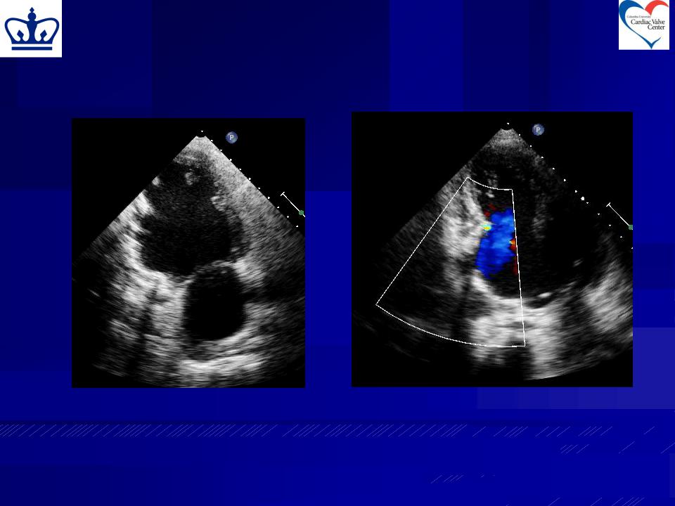
Apical 2Ch View |
Apical 3Ch View |
|
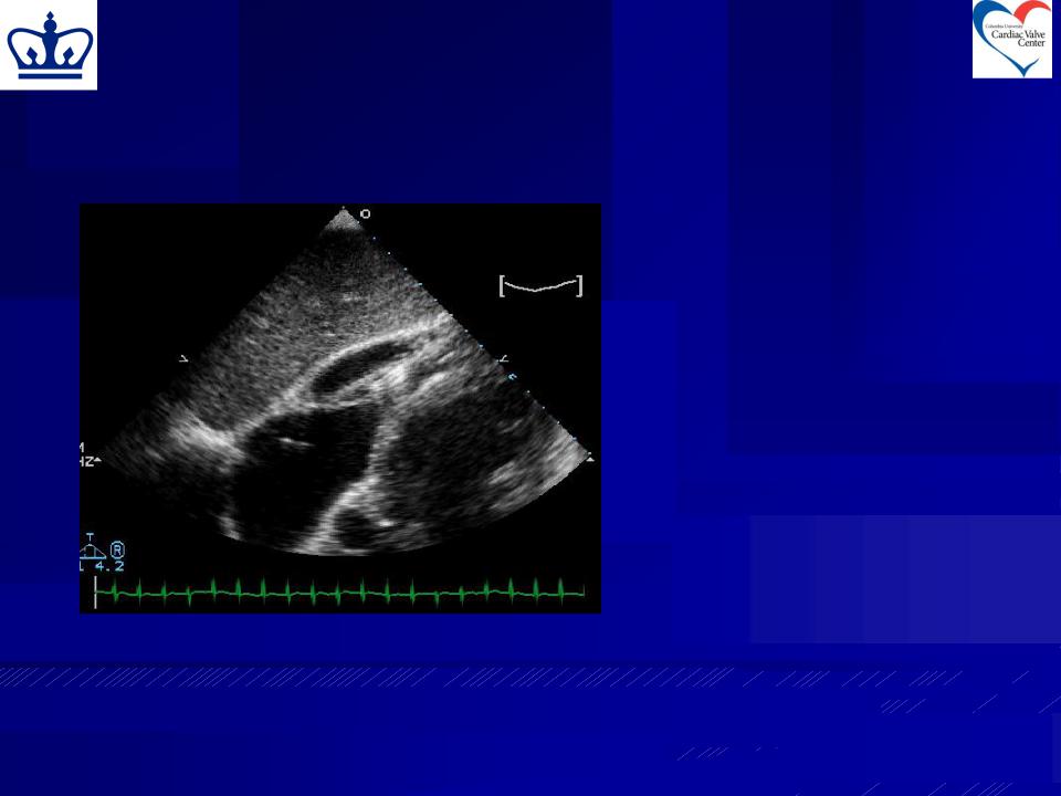
Diagnosis:
1. RV infarction with aneurysm of the inferior wall
2. Pericardial effusion
3. Pseudoaneurysm
4. Pericardial cyst
Saline Contrast Injection
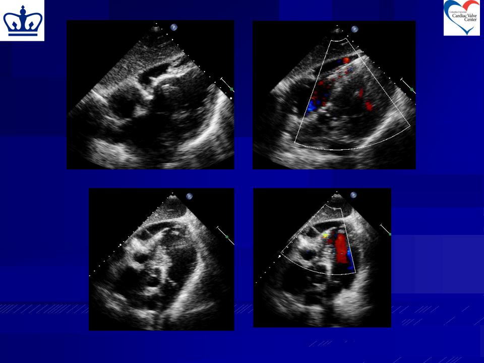
Rotating to Apical 2Ch View
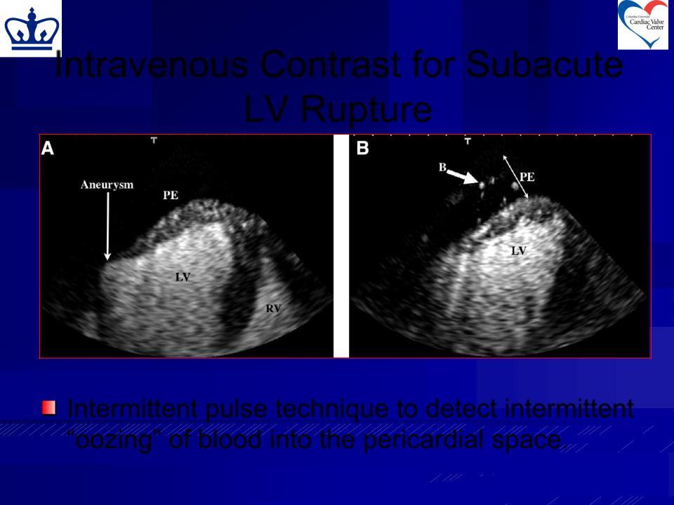
Intravenous Contrast for Subacute
LV Rupture |
Intermittent pulse technique to detect intermittent
“oozing” of blood into the pericardial space.
Uno K et al. J Am Soc Echocardiogr 2006;19:1401.e9-e11

Other Diagnostic Tests
MRI
–Loss of epicardial fat at the orifice of pseudoaneurysm
–Absence of myocardium (difficult if myocardial layer very thin)
May distinguish thrombus from myocardium from pericardium
Frances C et al. JACC 1998;32:557-61
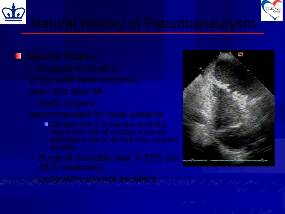
Natural History of Pseudoaneurysm
Natural history
– Rupture in 30-45%
of pts (with fatal outcome) even late after MI
– Early surgery
recommended for most patients
Chronic PA (> 3 months post MI) has lower risk of rupture however persistent risk of arrhythmia, embolic events.
–Surgical mortality rate: 7-23% (vs 48% medically)
– Long-term survival excellent
Harpaz D et al JASE 2001;14:219-27.
Frances C et al. JACC 1998;32:557-61
February |
March |
May |
79 year old woman with NIDDM, asthma, rheumatoid arthritis (S/P right shoulder and right knee replacements), Colon Ca (S/P resection >2 yr ago with no CTX) and AMI in February
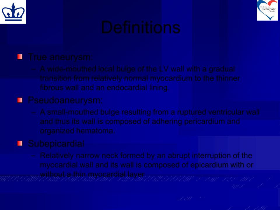
Definitions
True aneurysm:
–A wide-mouthed local bulge of the LV wall with a gradual transition from relatively normal myocardium to the thinner fibrous wall and an endocardial lining.
Pseudoaneurysm:
– A small-mouthed bulge resulting from a ruptured ventricular wall and thus its wall is composed of adhering pericardium and organized hematoma.
Subepicardial
–Relatively narrow neck formed by an abrupt interruption of the myocardial wall and its wall is composed of epicardium with or
without a thin myocardial layer
Hironaka, E et al. J Am Soc Echocardiogr 1997;10:192-96.