

ECHO 2013 / Artifacts of Imaging and Doppler Know Your Physics
.pdfState-of-the-Art Echo 2013
Artifacts of Imaging and Doppler:
Know Your Physics
Rebecca T. Hahn, MD
Columbia University
College of Physicians and
Surgeons
Nothing to Disclose

Sound
Then A
Miracle
Occurs
Images
How to Explain Echocardiographic Imaging

Physical Properties of Sound
Sound wave = series of compressions and rarefactions
Cycle Length = length of one compression + one rarefaction
Wavelength ( ) =
length from the peak of one compression to the peak of next compression (range 0.1-0.8 mm)
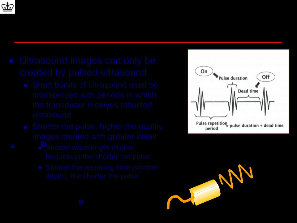
Pulsed Ultrasound
Ultrasound images can only be created by pulsed ultrasound
Short bursts of ultrasound must be interspersed with periods in which the transducer receives reflected ultrasound
Shorter the pulse, higher the quality images created with greater detail
Shorter wavelength (higher |
(mm) = 1.54 mm/µsec f (MHz) |
|
frequency) the shorter the pulse |
||
|
Shorter the receiving time (shorter depth), the shorter the pulse
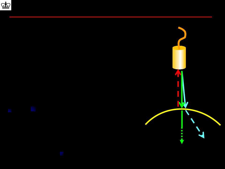
Interaction of Ultrasound with Tissue
•Impedence: The acoustic resistance to sound traveling through a medium
•Reflection
– High impedance mismatch between two media
’d REFLECTION
–Allows creation of an ultrasound image
•Refraction
–occurs at the interface of media of different impedances when the incident beam is OBLIQUE
–Property which allows use of lenses for focusing the beam
•Attenuation
–The weakening of sound waves as it propagates through tissue
–Due to reflection, absorption and scatter

Physical Properties of Sound
•Image resolution is
•The shorter the wavelength (higher the frequency), the better the longitudinal resolution (ie: smaller the distance between two distinguishable objects)
•Depth of penetration is
•The shorter the wavelength (higher the frequency), the worse the penetration (i.e: the shorter the depth that the ultrasound travels)
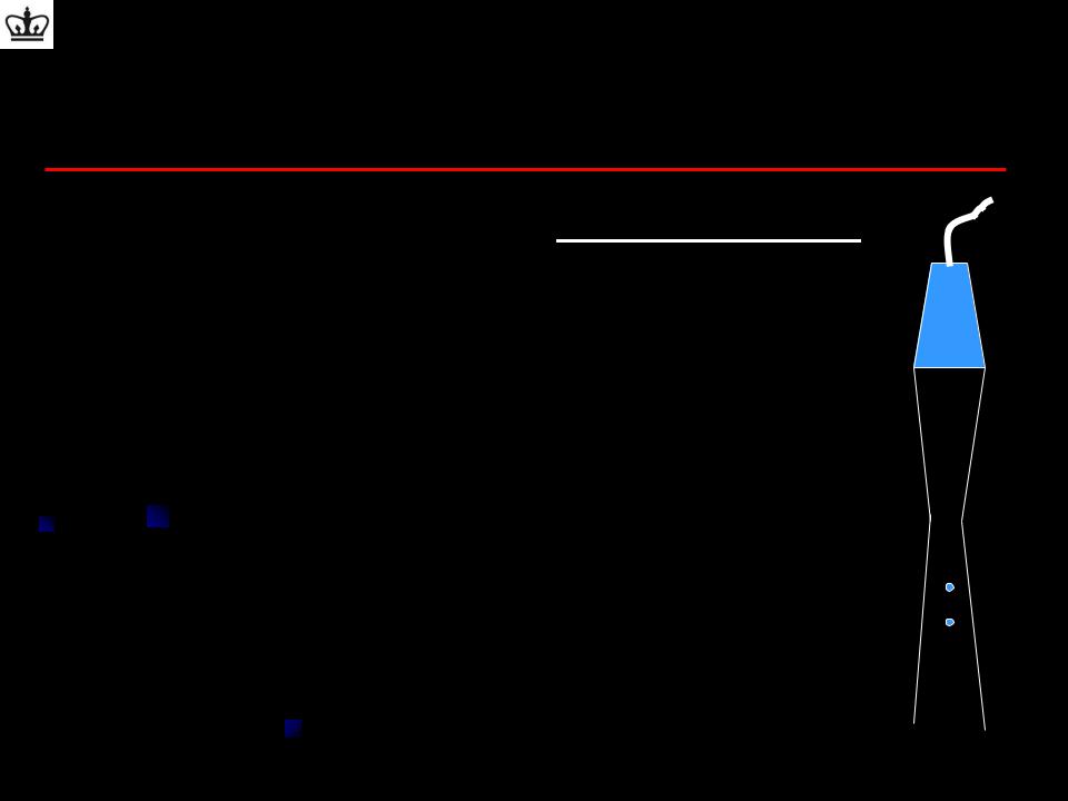
Longitudinal (Axial) Resolution
Longitudinal resolution (mm) ~ spatial pulse length 2
–Ability to distinguish two objects, one in front of the other
–The minimal required separation distance between two
reflectors is one-half of the spatial pulse length (SPL)
–SPL/2 avoids the overlap of returning echoes since the distance traveled between two reflectors is twice the
separation distance.
– Improve longitudinal resolution with
•Shorter pulse length (shorter wavelength)
•Higher frequency transducer
•Wider bandwidth transducer

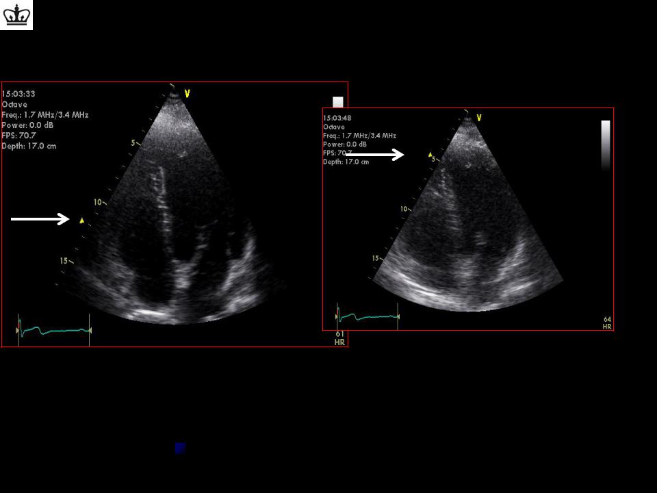
Ultrasound Transducers:
Resolution
Lateral Resolution:
Ability to distinguish two objects next to each other
Best at one focal length from transducer
 Dependent on DEPTH
Depths greater than or less than one focal length have a greater beam width and thus worse lateral resolution
Improves with focusing
Effect of Focusing
Note:
1.Each focal zone requires separate pulse echo sequences to acquire data.
2.Increasing the number of focal zones improves overall lateral resolution, but the amount of time required to produce an image increases and reduces the frame rate and/or number of scan lines per image
Elevational Resolution
The elevational or slice-thickness dimension of the ultrasound beam is perpendicular to the image plane.
Elevational resolution is dependent on the transducer element height in much the same way that the lateral resolution is
dependent on the transducer element width.