

ECHO 2013 / Cardiac Tumors and Masses
.pdfCARDIAC TUMORS AND
MASSES
Jennifer E. Liu, MD
Director of Cardiovascular Laboratories
Memorial Sloan Kettering Cancer Center

Disclosure Statement of Financial Interest
I, Jennifer Liu, DO NOT have a financial interest/arrangement or affiliation with one or more organizations that could be perceived as a real or apparent conflict of interest in the context of the subject of this presentation.

Differential Diagnosis
•True Intracardiac Mass
–Cardiac tumors (primary and secondary)
–Thrombus
–Vegetations
•Psuedo-masses
–Variants of anatomy
–Embryonic remnants
–Fat in AV groove, transverse sinus
–Q-tip on TEE
–Hiatal hernia
Cardiac Tumors and Masses
Echocardiographic Assessment
•TTE
•Best for endocardial lesions
•Size, shape, location, attachment, motion
•Hemodynamic consequences
•TEE
•Higher sensitivity and specificity
•Tissue characteristics
•Ability to assess invasion/extension
•Contrast Echo and 3D
•Vascularity
•Anatomy and spatial relationship to contiguous structures
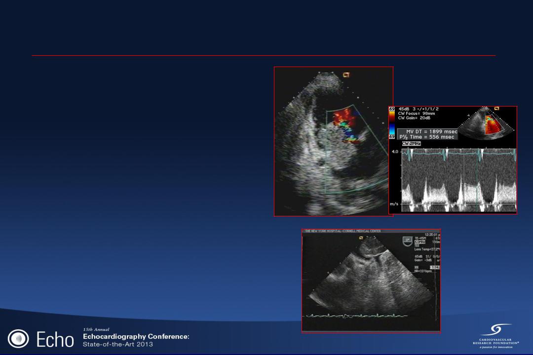
MVA = 0.4 cm2

Cardiac Tumors and Masses
Clinical Presentation
•CV – embolism, arrhythmia, heart failure
•Systemic – fever, fatigue, chest pain, weight loss
•Obstruction – chamber, valve or venous
•Cardiac compression
•Incidental

CARDIAC TUMORS
•Primary cardiac tumors are rare.
–incidence: <0.1% in autopsy series
•Secondary tumors are far more common
–occur 20-40x more frequent than primary tumor
–found in up to 20% of patients dying of cancer in autopsy series
•Cardiac tumor:
–endocardium, myocardium, epicardium

Primary Cardiac Tumors
Miscellaneous
17%
Fibroma
7%
Papillary
Fibroelastoma
9%
Rhabdomyoma 11%
Other sarcoma 6%
Benign: 75%
Malignant: 25%
Myxoma
42%
Rhabdomyosarcoma
5%
Angiosarcoma
3%
Miralles et al. Ann Thorac Surg 52:886-895, 1991

CARDIAC MYXOMA
•Most common primary tumor
–42% of all benign cardiac tumors; 25% of all primary cardiac tumors
•More common in the 3rd and 6th decade
•Female predominance with 3:1 ratio
•Usually sporadic as an isolated tumor

PATHOLOGY
•Gross morphology
–Soft with gelatinous consistency, often with areas of hemorrhage, thrombus or calcification
–Polypoid, pedunculated, friable
–size vary from 1 to 15 cm
–rapid growth rate: 1.2 gm/month
papillary |
smooth |

MYXOMA: Clinical Signs and Symptoms
•Constitutional
–fever, malaise, weight loss, arthralgia, elevated ESR, hypergammaglobulinemia
•Cardiac
–obstruction of atrioventricular blood flow; often mimic mitral stenosis; postural syncope
•Emboli
–tumor fragments or thrombi
–occur in 40-50% cases of myxoma
•Treatment
–prompt surgical excision