ECHO 2013 / Cardiac Tumors and Masses
.pdf
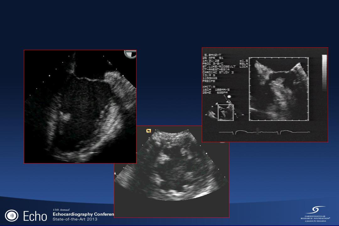
CPF: Left Ventricular Chambers

CPF: Clinical Signs and Symptoms
•Asymptomatic in vast majority
–incidental finding on surgically excised valves or necrospy
•CVA due to embolization of the papillary fronds of the tumor itself or from a superimposed thrombus can occur.
–Risk factors: size (≥ 1 cm), mobility, location (left sided lesions)
•Rare reports of angina or sudden cardiac death from coronary ostial occlusion

CPF: Management
Symptoms
yes |
no |
surgical candidate |
R side |
L side |
|
|
|
||
|
recommendations NOT based on |
|
|
no |
randomized controlled data |
high risk features |
|
yes |
Clinical f/u |
||
Clinical f/u |
|
Consider a/c |
|
|
surgery |
no |
yes |
||
Consider a/c |
||||
|
|
|
Sun et al, Circulation 2001; 103:2687-2693 Gowda et al, AHJ 2003; 146:404-410
low surgical risk |
|
yes |
no |
surgery |
Clinical f/u |
|
|
|
Consider a/c |

•Majority of cases (83%) can be safely resected by valve sparing, simple shave excision. Valve repair or replacement was rarely necessary.
•Low surgical mortality (<2%)
•No tumor recurrence and no tumor-related late mortality or morbidity during a follow-up period lasting up to 8.3 years.

Lambl’s Excrescences
•Fine thread like strands arising on the line of closure (contact surface) of heart valves
•Found in 70-80% of older adults; often multiple
•Pathogenesis
–Degenerative wear and tear of the valvular endocardial surfaces where the valve margins contact
•Association with embolization - controversial

RHABDOMYOMA
•Most common primary cardiac tumor in the pediatric age group
–approximately 3/4 occur in < 1 year of age
•Strongly associated with tuberous sclerosis (familial syndrome of systemic hemartoma)
•Derived from cardiac muscle; may actually be cardiac hemartoma or malformation rather than true neoplasm

RHABDOMYOMA: Gross Pathology
•Yellow-gray; circumscribed
•Range from 1mm to several cm in diameter
•Locations
–90% are multiple
–equal frequency in LV/RV/septum
– 1/3 also involve atria |
LV |
|
–mostly intramural with intracavitary extension in 50% of cases

Rhabdomyoma
Fetal Ultrasound: In-Utero

Rhabdomyoma
One month after birth

RHABDOMYOMA: Clinical Aspects
•Obstruction: inflow or outflow
•Arrythmia
–AV block or incessant VT
–sudden death
•Presence of multiple nodular masses in several chambers on 2D echo is diagnostic
•Limited ability to grow and tend to regress
•Surgery only indicated for symptomatic patients
