

ECHO 2013 / Artifacts of Imaging and Doppler Know Your Physics
.pdfPropagation Path Artifact: Side
Lobes
Dominant Side Lobes require
large, relatively echo-free left atrium
artifactual echoes must originate from strong reflecting surfaces
 Correction
Reduce gain or increase reject level
Change acoustic window
Harmonic imaging

Artifact
Pacing wire perforating the septum?
Side Lobe artifact

Side-lobe Color Artifact
•Ultrasound Property
–Extraneous beams of ultrasound are generated from the edges of individual elements
–Artifactual echoes must originate from strong reflecting surfaces
Propagation Path Artifact: Range
Ambiguity
Range Ambiguity due to high frame rate
Places an object closer to the transducer
Dependent on Frame Rate (thus depth and Frequency)
ASSUMPTIONS:
•Each pulse transmitted is received before the next pulse is delivered
•Distance determines the time to receive a pulse
Pulsed Doppler Limitations: Nyquist Limit
High Pulsed Repetition Frequency
Increase in pulsed repetition frequency in order to increase the Nyquist limit and increase detectable velocity
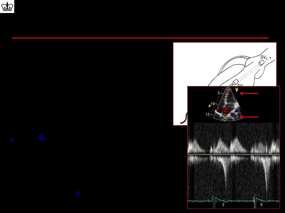
Introduces range ambiguity since signals that return to the transducer at twice (or half) the focal length are received during the receiving time of the transducer
Multiple sample volumes appear on sector scan

Range Ambiguity
Range Ambiguity due to high frame rate
Places an object closer to the transducer
Moving objects will appear to move in the same direction
Dependent on Frame Rate (thus depth and Frequency)
110sec
20 |
110 |
sec |
sec |
40 |
110 |
sec |
sec |
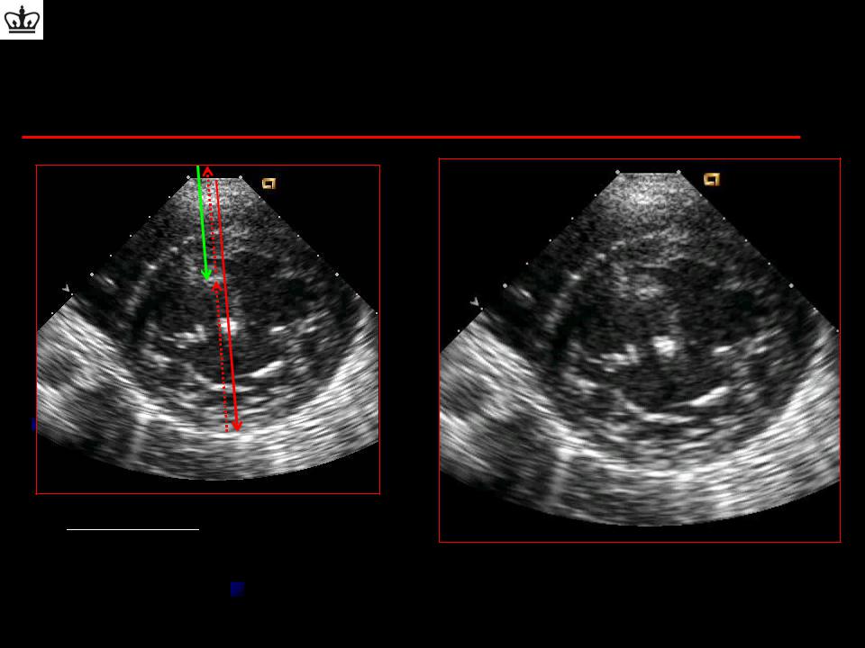
Return time = 110 sec
Frame rate = 110 sec
Frame rate = 110 sec
Propagation Path Artifact: Range
Ambiguity
Clues to Artifact:
•Indistinct edges
•Moves through real structure
Artifact moves in the same direction as the real object

Real Time 3D (RT3D)
RT3D Systems
Full volume (90 x 90 )
Larger cardiac volume
Requires ECG gating to merge 4-7 narrower pyramidal scans obtained over 4-7 consecutive heart beats
Lower resolution compared to real time
Houck R et al. AJR 2006; 187:1092–1106

Summary
Artifacts of echocardiography can be explained by the physical properties of ultrasound
It is important to recognize these artifact to prevent incorrect interpretation of the ultrasound or Doppler image
Characteristics suggestive of artifacts:
Not anatomically correct
Crosses borders
No attachment
Not consistent with cardiac cycle
Not seen in multiple views
Color or contrast goes through image No pathologic or anatomic correlate

Thank You