

Mapstone_2014
.pdfzooids. New material, much obtained by SCUBA diving, has allowed a reassessment [16] that retains four older species (F. edwardsi, F. contorta, F. formosa and F. tholoides), adds two new species (Table 4) and reduces F. leuckarti to a junior synonym of F. contorta [1]. The recent molecular analysis supports monophyly of this family (Figure 9), which uniquely possesses (for most species) four bract types [10], with one type on the stem (stem bracts) and three types on the elongate pedicels of the gastrozooids (bolster and two types of knee-shaped bracts) [16]. A single gonodendron also occurs on the stem, between two gastrozooids, and carries bunches of both male and female gonophores which can be attached in a species-specific pattern [16].
Resomiidae. Live colonies of this new monoecious family are mostly transparent with a short rigid siphosome that never relaxes, as also in species of Erenna and the agalmatid Agalma okeni. Three of the five species referred to the family are new (Table 4), and all are characterized by tentilla on the same tentacle which undergo transformation from a spirally coiled cnidoband to a zig-zagged cnidoband [17], a process superbly illustrated in colour for the three new species by Pugh and Haddock [91]. In the new species Resomia ornicephala, the involucrum floats above the cnidoband and fluoresces under incident blue light, attracting krill prey, as described further below.
Agalmatidae. A sensu stricto clade of this family has been identified from the molecular phylogeny of Dunn et al. [10] and includes three long-stemmed and two short-stemmed genera (Figures 9, 10, Table 4); all have tricornuate tentilla and tightly coiled cnidobands. A new species Halistemma transliratum from the Bahamas has nectophores with a single vertical-lateral ridge and three types of bract [92], whilst nectophores from another giant Halistemma species (H. foliacea) have been described for the first time [93] from Indonesian waters (Table 4); the latter species has nectophores with two vertical-lateral ridges and three types of thick foliaceous bracts. Both species have unicornuate tentilla with a vestigial involucrum and a long terminal filament terminating in a small cupulate process or sinker (see below). Cormidial development has been elucidated for Agalma elegans and Nanomia bijuga, using a SEM, and zooids found to develop differently from pro-buds in each species [8]. Tissue samples from very young nectophores and gastrozooids of N. bijuga have also been analysed (using next generation sequencing [96]) for gene expression in wild specimens, and a gene expressed only in the basigaster of the gastrozooid that encodes for a protein used in the formation of the nematocyst wall further characterized [96].
Unascribed monoecious physonects. The three monoecious genera with a ventral nectosome noted in Figure 10 and Table 4 have unique tentilla, and two of them (Frillagalma and Lychnagalma) are monotypic [1]. Two new species await description in the genus Cordagalma [17], and a re-description of F. vityazi from new submersible material shows that frilling of the ridges in the nectophores and bracts of the original net-caught specimens is a preservation artefact [97]. Sequencing of the 16S gene of L. utricularia shows its closest relations to be members of the family Physophoridae [17]; L. utricularia was also found to be the only non-bioluminescent physonect in the Alboran Sea [98]. Fresh specimens of a fourth unassigned monoecious physonect, Rudjakovia plicata, taken recently off California indicate that their much pleated nectosacs are also preservation artefacts. The nectophores of this species attach to the dorsal side of the nectosome, indicating that it may be referable to the Agalmatidae sensu stricto, but further material is needed to confirm this hypothesis.
Prayidae. Absolute axes applied to the colony, stem and zooids of two new prayine species in this family facilitate consistent future species descriptions [7], and are extrapolated to a further
Diversity and Phylogeny of Siphonophores
nine prayid species in another publication [6]. The prayid somatocyst is also redefined [6] to bring the terms applied to prayid proximal nectophore canals into line with those used for the homologous canals and diverticula in both physonects (which lack a somatocyst) and diphyomorph calycophorans (which have a somatocyst that penetrates into the mesogloea and develops from only one diverticulum of the pedicular canal). Bracts, larval nectophores and young definitive nectophores of Praya dubia and P. reticulata have been reliably distinguished for the first time since 1987 [103] and their mature nectophores also fully described from new specimens collected in the NE Pacific [6]. The recent siphonophore molecular phylogeny of Dunn et al. [10] suggests that Prayidae are paraphyletic, with Praya dubia and two nectopyramidines forming one clade and three other prayines forming a second (Figure 9).
Clausophyidae. New information on this diphyomorph family is given in Table 5, and its position intermediate between the Prayidae and Diphyidae is well shown in a figure by Mapstone (fig. 4 [6]). A useful time line is also given by Pugh [106] for descriptions of three widespread clausophyid species (Clausophyes galeata, C. moserae, Kephyes ovata). New deep-water records from various locations worldwide contribute further to our understanding of the ecology of this deep-water family [6,42,47,87, 105,106,109,116,117,118,119,120], and two further new clausophyid species await description [10,106].
Sphaeronectidae. A recent and thorough review of this diphyomorph family is given by Pugh [18], together with an updated systematic treatment of all valid species [1]. Beautiful images are available for six of the ten small species now comprising this family [18,107], and new siphonophore axes are extrapolated for sphaeronectids by Mapstone [6]. These axes are incorporated into descriptions of the two most recently introduced species [107,108]. For a useful schematic summary of the sphaeronectid life cycle see fig. 15 in [18].
Diphyidae. The first new Lensia species introduced for 36 years is L. quadriculata (Table 5 and [109]), and another, L. asymmetrica, is re-described with its posterior nectophore, bract and gonophore identified for the first time [110]. New bracts of a third small species L. reticulata indicate a close affinity to the family Clausophyidae for which it is transferred to a new subfamily (Table 5 and [111]), and a previously unassigned eudoxid referred to the large diphyid Lensia cossack (Table 5 and [112]). Seven diphyid species are recorded for the first time in Japanese waters [116].
Nematocysts and Lures
Nematocysts and tentilla were only briefly covered in the 1987 review of siphonophore biology [2], and are therefore described here in more detail.
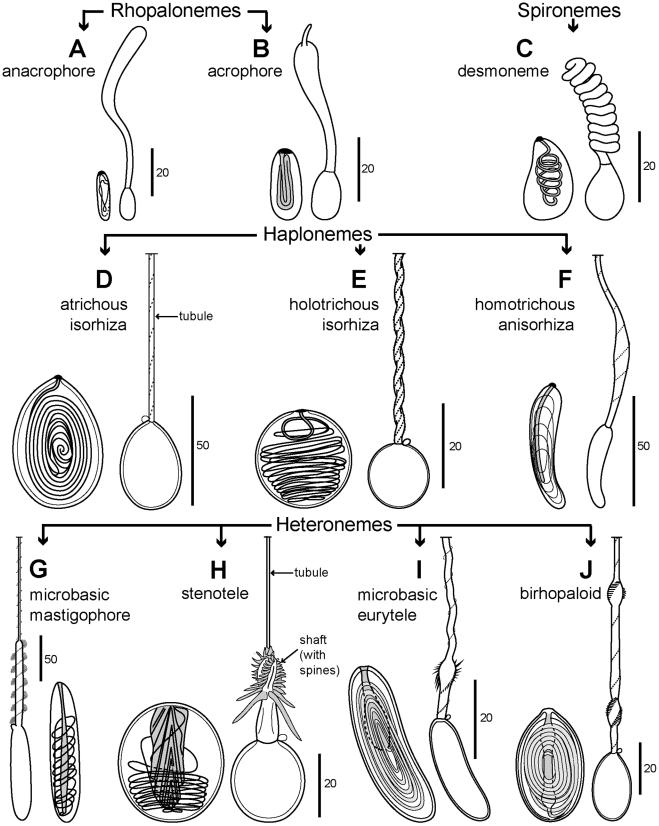
Nematocysts. Nematocysts are an apomorphy of the Cnidaria, and one of three types of cnidae which characterize the phylum; the others are ptychocysts and spirocysts (absent from Hydrozoa). More than 30 types of nematocyst are recognized and their classification is typically based on characters of the tubule (open or closed tip, diameter, presence or absence of a swollen shaft at the proximal end, pattern, distribution and size of spines on the tubule). Diversity among nematocysts, different methods of classifying them and the possible importance of cnidae in cnidarian evolution are reviewed by Fautin [121]. The total complement of cnidae in a species is termed the cnidome (p. 68 [6]). A summary of nematocyst characteristics of most siphonophore families and some genera and species is given in Figure 11 and Table 6. Five types are autapomorphic (exclusive) to Siphonophora, including two categories of rhopalonemes (acro-
PLOS ONE | www.plosone.org |
21 |
February 2014 | Volume 9 | Issue 2 | e87737 |

Diversity and Phylogeny of Siphonophores
Table 5. New systematics for calycophoran siphonophore families.
Family |
Comments |
|
|
13. Prayidae |
Probably paraphyletic, and includes nested family Hippopodiidae [10] (see below); Praya dubia (Subfamily Prayinae) and sub-family |
|
Nectopyramidinae maybe one lineage, with prayines Craseoa, Gymnopraia and Rosacea another [10], but broader taxa sampling is needed |
|
[6]. Prayine name Lilyopsis medusa has precedence over Lilyopsis rosea [1]; new prayine species Desmophyes haematogaster, Gymnopraia |
|
lapislazula, Lilyopsis fluoracantha, Rosacea repanda, R. limbata, R. arabiana introduced (see [1]); subfamily Nectopyramidinae revised [13] |
|
with Nectopyramis thetis and N. natans re-described and new genus Nectadamas introduced (for N. diomedeae and a new species N. |
|
richardi [13]). Prayine species R. cymbiformis also re-described [99] and nomenclature problems concerning R. plicata sensu Bigelow and |
|
Desmophyes annectens resolved [100,101]. Eudoxids are released in amphicaryonines and nectopyramidines, but not in prayines [6]. |
|
Rosacea villafrancae transferred to genus Desmophyes [102], and Prayoides intermedia found to be a junior synonym of Praya species |
|
[1,103]. Unique bio-optical properties identified in G. lapislazula and L. fluoracantha, though their function is still unknown [7]. |
|
|
14. Hippopodiidae |
Found nested within prayines in first siphonophore phylogeny, and Hippopodius nested within Vogtia [10]; hippopodiid distribution |
|
correlated with feeding on various species of ostracods, unlike other calycophorans [104]. Family characters recently summarized and the |
|
new axes applied, together with re-descriptions given and synonomies listed for V. serrata, V. spinosa and V. pentacantha [6]; V. |
|
microsticella considered a junior synonym of V. glabra, and V. kuruae a junior synonym of V. serrata [1,6]. |
|
|
15. Clausophyidae |
The 3 diphyomorph families below may have arisen from the Clausophyidae [10]. New species include Clausophyes laetmata [42] and Cl. |
|
tropica [105] and 2 others re-described include Cl. galeata and Cl. moserae [105]; a unique fuseudoxid life stage found in Crystallophyes |
|
amygdalina [47] and a new genus Kephyes introduced for Moser’s Cl. ovata, which, unlike Clausophyes species, has bracts with a pair of |
|
hydroecial canals [106]. 4 clausophyids re-described from NE Pacific and the new axes applied [6]. |
|
|
16. Sphaeronectidae |
Ten species now considered valid in this family with single retained larval nectophore. Family reviewed and history summarized [18]; 5 |
|
new species introduced: Sphaeronectes christiansonae, S. haddocki, S. tiburonae [18], S. pagesi [107] and S. pughi [108]. An old species S. |
|
brevitruncata reinstated [18] and S. bougisi concluded to likely be a calyconula of Lilyopsis medusa [1]. S. gracilis relegated to a junior |
|
synonym of S. koellikeri and probably restricted to the tropics [1,18]; specimens reported from Jervis Inlet, British Columbia [6] probably |
|
another species. |
|
|
17. Diphyidae |
Probably paraphyletic [10], vindicating earlier conclusions [9], but based on only 5 of 43 likely valid species [1]. Two main clades identified |
|
in the molecular study of Dunn et al. [10], within one of which is nested the Family Abylidae. New axes applied to all life stages of |
|
diphyids, muscular lamellae, median gastrovascular canals and pedicular canal arrangements also schematically shown for two basic |
|
types of diphyids [6]. A new small species added to genus Lensia (L. quadriculata [109]), another re-described in detail (L. asymmetrica |
|
[110]) and a third (L. reticulata) transferred to a new genus Gilia within a new subfamily Giliinae, for the two clausophyid-like canals in the |
|
bract (G. reticulata [111]). An enigmatic species Eudoxia macra shown, using the mitochondrial 16S gene, to be sexual stage of a larger |
|
species L. cossack [112]. A number of previously described Lensia species, several Sulculeolaria species and one Muggiaea species all |
|
reduced to junior synonyms of various better known species [1]. |
|
|
18. Abylidae |
Family nested with Diphyes dispar in one of two Diphyidae clades, based on 16S and 18S [10], but only Abylopsis tetragona tested and |
|
more taxa sampling needed. 10 valid species [1], all present in the S Atlantic and summarized in a recent report [113]; several species also |
|
re-described from around South Africa [82,114]. Junior synonyms (including those in a confusing abylid review by Sears [115]) given in the |
|
Worms World List [1]. |
|
|
doi:10.1371/journal.pone.0087737.t005
phores and anacrophores), haploneme homotrichous anisorhizas and two categories of heteroneme rhopaloids (shaft of unequal diameter with either two swellings along its length (birhopaloids) or one (rhopaloids)) (Figure 11).
Identification of nematocysts can be difficult, requiring examination of discharged tubules, although some larger types can be recognized in situ undischarged [124]. Successful discharge is best achieved with live material, though the procedure requires practice; discharging nematocysts from preserved specimens is not usually possible [121]. Smaller nematocysts are also more difficult to identify than larger examples, with the result that it has not been possible to identify specific rhopalonemes and smaller isorhizas in some siphonophore species (Table 6).
Although homotrichous anisorhizas occur in most siphonophore groups, they and the other three types of autapomorphic nematocysts are absent from cystonects (Physaliidae and Rhizophysidae in Table 6), which have simple tentacles. Cystonect cnidomes are composed almost exclusively of isorhizas, the most primitive type of cnidarian nematocyst [135]; these nematocysts can be present in enormous quantities, particularly in the tentacles of the Portuguese Man O’War Physalia physalis. Ultrastructure of the smaller isorhiza of Physalia was studied for the first time by Hessinger & Ford [136], enlarging upon an earlier light microscope study by Will [137]. The nematocyst capsule is held in position by a complex fibrillar basket anchored to the underlying mesogloea with hemidesmosomes and apically by enveloping processes from neighbouring epithelial cells [136].
Such basal anchoring fibrils, often termed a cnidopod (p. 114 [122]), also occur in the nematocysts of other Hydrozoa (p. 29 [138]). In Physalia, nematocysts are formed in basigasters (ampullae of Totton [39]) separated from their gastrozooids during development; each nematocyst migrates down either a tentacle to the nematocyst battery region (isorhizas) for prey capture or to gonopalpons in a cormidium (stenoteles), probably for defence of the spherical gonodendron after release from the colony (Table 6 and [81]). Rhizophysid tentacles have side branches with either a strip of isorhizas along one side (e.g. R. eysenhardti) or isorhizas in pads on swellings at the distal ends of the branches (e.g. R. filiformis fig. 5F [67]). Cystonects consume only soft-bodied prey, mainly fish and fish larvae, and when present in large numbers can deplete fish stocks [67,139,140].
The cnidome of apolemiids also reflects a diet of soft-bodied prey [67] and was studied in detail in Apolemia uvaria from the Mediterranean [127], in another apolemiid from off California [124] and recently in A. lanosa and A. rubriversa from Monterey Bay [11]. These physonects, sister to all other codonophorans (Figure 10), also lack complex nematocyst batteries and have simple unbranched gastrozooid tentacles, and palpons with elongate palpacles indistinguishable from the tentacles. Nematocysts include birhopaloids (Figure 11) of two sizes (fig. 1 [127] and fig. 3a–d [124]), and in other species rhopaloids with a single swelling on the shaft [11]. These rhopaloid types are unique to the Apolemiidae (amongst Siphonophora) and in A. uvaria birhopaloids occur in pairs down the lengths of relaxed tentacles [9]. There are
PLOS ONE | www.plosone.org |
22 |
February 2014 | Volume 9 | Issue 2 | e87737 |
Diversity and Phylogeny of Siphonophores
Figure 11. Schematic representation of ten nematocyst types found in Siphonophora. Undischarged and discharged nematocysts included. A: anacrophore rhopaloneme (after fig. 22a–b [122]); B: acrophore rhopaloneme (after fig. 23a–b [122]); C: desmoneme spironeme (after fig. 26a–b [122]); D: atrichous isorhiza haploneme (after fig. 4a–b [123]); E: holotrichous isorhiza haploneme (after figs. 1a, 1b [124] and fig. 7b [123]); F: homotrichous anisorhiza haploneme (after fig. 41a–b [122]); G: microbasic mastigophore heteroneme (derived from fig. 29 [127] and fig. 2a [124]); H:
PLOS ONE | www.plosone.org |
23 |
February 2014 | Volume 9 | Issue 2 | e87737 |

Diversity and Phylogeny of Siphonophores
stenotele heteroneme (derived from fig. 17 [127] and fig. 1d [124]); I: microbasic eurytele heteroneme (after pl. 1, figs. 6–7 [132]); J: birhopaloid heteroneme (after fig. 83 [122] and fig. 3d [124]).
doi:10.1371/journal.pone.0087737.g011
also two types of heteronemes in most apolemiids, including stenoteles (two size classes) and microbasic mastigophores (one size class) around the mouths of gastrozooids and palpons (Table 6), and haploneme isorhizas of two size classes on the surfaces of bracts and palpons, and probably also on the tentacles of some apolemiids from the NE Pacific [9,11].
The cnidomes of monoecious physonects include nematocysts on zooids other than the tentilla (see below). These likely include acrophores on the body and stenoteles around the mouth of the gonopalpons in most forskaliid species [16], white clusters of stenoteles or orange clusters of microbasic mastigophores on the tips of the enlarged palpons of Physophora species (Table 6, Figures 4C, 12F) and large microbasic mastigophores on the bracts of Resomia ornicephala, with similar nematocysts also on the palpacles and gonophores of this species, and two patches on the lateral surfaces of the nectophores [91].
Table 6. Nematocysts of siphonophores.
Tentilla. In all siphonophores other than cystonects and apolemiids, the nematocysts used for feeding are contained within complex nematocyst batteries on side branches of the tentacles, here termed tentilla (for definition, see p. 74 [6]). A few other authors refer to them as nematocyst batteries or tentillar batteries [67,141]. The appearance of these batteries during evolution coincides with the loss of large polyps from the nectosome (present in apolemiids) and a change in diet from soft-bodied prey to hardbodied crustacean prey [67]. The batteries represent a transition during the phylogeny of Siphonophora which might perhaps have occurred after the origin of the nectosome by pro-bud subdivision and before a change in sexual state from dioecy to monoecy (see fig. 7 in [8]).
The nematocysts of such tentilla are contained within a cnidosac [6], or saccus [9], which can be simple or complex. Complex batteries are better known than simple examples, because they are
|
|
|
|
|
|
|
Holotrich |
Homo |
|
Micro |
|
|
|
|
Family |
Des |
Acro |
Anacro |
Atrich Iso |
Iso |
Aniso |
Steno |
Mastig |
|
Micro Eury |
Birhop |
References |
||
|
|
|
|
|
|
|
|
|
|
|
|
|
|
|
|
Physaliidae |
– |
– |
– |
isorhizas |
|
– |
yes |
– |
|
– |
– |
[81,125] |
|
|
|
|
|
|
|
|
|
|
|
|
||||
Rhizophysidae |
– |
– |
– |
isorhizas |
|
– |
– |
– |
|
– |
– |
[67,126] |
||
|
|
|
|
|
|
|
|
|
|
|
|
|
||
|
Apolemiidae |
– |
– |
– |
isorhizas (2 sizes) |
– |
yes |
yes (or |
|
one sp. |
in one+sp. |
[11,124,127] |
||
|
|
|
|
|
|
|
|
|
(2 sizes) |
unknown |
|
probably |
(2 sizes) |
|
|
|
|
|
|
|
|
|
|
|
type) |
|
|
|
|
|
|
|
|
|
|
|
|
|
|
|
|
|
|
|
Pyrostephidae |
yes |
rhopalonemes |
– |
– |
– |
yes |
– |
|
– |
– |
[14] |
|||
|
|
|
|
|
|
|
|
|
|
|
|
|
|
|
|
Erennidae |
– |
– |
– |
|
isorhizas |
|
yes |
– |
– |
|
– |
– |
[15] |
|
|
|
|
|
|
|
|
|
|
|
|
|||
Rhodaliidae |
yes |
rhopalonemes |
– |
– |
yes |
– |
yes |
yes |
– |
[27,45,89] |
||||
|
|
|
|
|
|
|
|
|
|
|
|
|
|
|
|
Marrus spp. |
yes |
yes |
– |
|
– |
– |
yes |
– |
yes? |
|
yes |
– |
[90] |
|
|
|
|
|
|
|
|
|
|
|||||
Forskaliidae |
yes |
yes |
– |
? |
– |
yes |
yes |
– |
|
– |
– |
[122] |
||
|
|
|
|
|
|
|
|
|
|
|
|
|
|
|
|
Physophoridae |
– |
– |
– |
– |
– |
yes |
yes |
yes |
|
– |
– |
[77,128,129] |
|
|
|
|
|
|
|
|
|
|
|
|||||
Resomiidae |
yes |
yes |
– |
– |
– |
yes |
– |
yes |
|
– |
– |
[17,91] |
||
|
|
|
|
|
|
|
|
|
|
|
|
|
|
|
|
Agalma spp. |
yes |
yes |
– |
|
– |
– |
yes |
– |
yes |
|
yes |
– |
[67,73] |
|
|
|
|
|
|
|
|
|
|
|||||
Athorybia rosacea |
yes |
rhopalonemes |
– |
– |
yes |
yes |
– |
|
– |
– |
[67] |
|||
|
|
|
|
|
|
|
|
|
|
|
|
|
|
|
|
Halistemma spp. |
yes |
yes |
– |
|
– |
– |
yes |
yes |
– |
|
yes |
– |
[92,73] |
|
|
|
|
|
|
|
|
|
|
|||||
Nanomia spp. |
yes |
rhopalonemes |
– |
– |
yes |
yes |
– |
|
– |
– |
[67] |
|||
|
|
|
|
|
|
|
|
|
|
|
|
|
|
|
|
Cordagalma |
– |
– |
– |
|
yes |
– |
yes |
yes |
– |
|
– |
– |
[130] |
|
|
|
|
|
|
|
|
|
|
|||||
|
Frillagalma vityazi |
– |
– |
– |
|
– |
– |
yes |
yes |
– |
|
– |
– |
[97] |
|
|
|
|
|
|
|
|
|
|
|
|
|
|
|
|
Lychnagalma utricularia |
? |
rhopalonemes |
|
– |
– |
yes |
yes |
– |
|
– |
– |
[131] |
|
|
|
|
|
|
|
|
|
|||||||
|
Calycophorae |
|
|
|
|
|
|
|
|
|
|
|
|
|
|
|
|
|
|
|
|
|
|
|
|
|
|
|
|
|
Rosacea spp. |
yes |
– |
yes |
|
– |
yes |
yes |
– |
– |
|
yes |
– |
[46,67] |
|
|
|
|
|
|
|
|
|
|
|||||
|
Desmophyes villafrancae |
yes |
– |
yes |
|
– |
– |
yes |
– |
– |
|
yes |
– |
[46,132] |
|
|
|
|
|
|
|
|
|
|
|
|
|
|
|
|
Prayola tottoni |
yes |
– |
yes |
|
– |
– |
– |
yes |
– |
|
– |
– |
[133] |
|
|
|
|
|
|
|
|
|
|
|||||
|
Lilyopsis medusa |
yes |
– |
yes |
|
– |
– |
yes |
– |
yes |
|
– |
– |
[134] |
|
|
|
|
|
|
|
|
|
|
|
|
|
|
|
|
Nectadamas diomedeae |
yes |
– |
– |
|
– |
– |
probably |
probably |
? |
|
– |
– |
[13,38] |
|
|
|
|
|
|
|
|
|
|
|||||
|
Hippopodiidae |
yes |
rhopalonemes |
– |
– |
yes |
– |
yes |
– |
– |
[67] |
|||
|
|
|
|
|
|
|
|
|
|
|
|
|
|
|
|
Sphaeronectidae |
yes |
– |
yes |
– |
– |
yes |
– |
yes |
|
– |
– |
[18] |
|
|
|
|
|
|
|
|
|
|
||||||
|
Diphyidae |
yes |
– |
yes |
– |
yes |
yes |
– |
yes |
|
– |
– |
[67] |
|
|
|
|
|
|
|
|
|
|
|
|
|
|
|
|
|
Abylidae |
yes |
– |
yes |
– |
yes* |
yes* |
– |
yes |
|
– |
– |
[67]*except |
|
|
|
|
|
|
|
|
|
|
|
|
|
|
|
E.hyalinum |
|
|
|
|
|
|
|
|
|
|
|
|
|
|
|
|
|
|
|
|
|
|
|
|
|
|
|
|
|
|
Key: Des - desmoneme; Acro - acrophore; Anacro - anacrophore; Atrich Iso - Atrichous Isorhiza; Holotrich Iso – holotrichous isorhiza; Homo Aniso - homotrichous anisorhiza; Steno – stenotele; Micro Mastig – microbasic mastigophore; Micro Eury – microbasic eurytele; Birhop – birhopaloides (two swellings on tubule). doi:10.1371/journal.pone.0087737.t006
PLOS ONE | www.plosone.org |
24 |
February 2014 | Volume 9 | Issue 2 | e87737 |
characteristic of the better studied and more abundant species of physonects, for which they can be diagnostic. Each comprises a cnidoband, terminal filament(s) and elastic strands [70,142], which together function to rapidly entangle the prey and simultaneously release the cnidoband, by a mechanism explained in more detail below. In addition, the cnidome of non-apolemiid physonect codonophorans includes nematocysts on other zooids, including bracts, nectophores, palpons or palpacles. Such nematocysts are probably for defence (as in apolemiids). It is also important to remember that during collection the tentacles and their side branches are easily torn off or shed, due to the delicate nature and sensitivity of siphonophores [43]. The cnidome is, therefore, rarely completely known for less common siphonophores, or, indeed, for many common species, although details of the cnidomes of species in the monoecious physonect family Resomiidae are given by Pugh and Haddock [91].
Dioecious physonect tentilla: The tentilla of pyrostephids and erennids differ from those of other dioecious physonects and all monoecious codonophorans in having a cnidoband of very small nematocysts and an axial gastrovascular canal which penetrates the length of the terminal filament to the tip (Figure 12A–C, Table 7). These tentilla are probably held out straight in life, and their terminal filaments have either many small nematocysts similar to those in the centre of the cnidoband (pyrostephids), or none (erennids); in the latter there is a pair of pigmented photophores (ocelli) which are held out stiffly during feeding and vibrate to act as a lure (see below).
In rhodaliids and species in the genus Marrus the mature cnidoband, where known, is often, although not always, loosely coiled, and typically comprises a suite of larger nematocysts which include many small central haplonemes (probably homotrichous anisorhizas) flanked by some large heteronemes (Table 7, Figure 12D–E). The latter may be microbasic mastigophores, or in Thermopalia taraxacum, stenoteles [27]. No figures have yet been published showing the arrangement of nematocysts in rhodaliid tentilla.
Monoecious physonect tentilla: Monoecious species of the physonect families Forskaliidae, Physophoridae, Resomiidae and Agalmatidae sensu stricto typically have tightly coiled cnidobands and a single terminal filament, while tentilla of the unassigned genera Cordagalma, Frillagalma and Lychnagalma are more varied (Figure 12F–H, Figure 13A–E, Table 7). Cnidobands typically comprise many small homotrichous anisorhizas flanked proximally by large microbasic mastigophores or stenoteles, with the terminal filament composed of smaller desmonemes and rhopaloneme acrophores (Table 7). A thin and transparent protective involucrum partially or completely covers the cnidoband in many mature tentilla (Table 7).
Forskaliid tentilla have particularly long pedicels, a loosely coiled cnidoband without an involucrum (Figure 12H) and nematocysts as noted in Table 7. A larval-type tentillum has also been identified in one species [16]. Prey consumed is typically copepods and sometimes decapod larvae, shrimp and chaetognaths, but no ostracods or gelatinous zooplankton [140]. In physophorids, the tentilla are unusual and carried on tentacles which, when relaxed, are extremely elongate (pl. 1, fig. 1 [128]). Tentilla are similar in both Physophora species and of unique construction (Figure 12Fa–c) with the cnidoband becoming enclosed and inverted inside a layered capsule during maturation (Table 7). Resomiid tentacles bear two types of tentilla on each tentacle, and the cnidoband changes configuration from coiled to zigzag as it matures (Figure 12Ga–b). The transformation process is particularly well illustrated in the series of published images quoted in Table 7, and in all species but one the involucrum of the
Diversity and Phylogeny of Siphonophores
tentillum forms a transparent tube enclosing the cnidoband throughout the transformation process. Simplified larval tentilla with a short straight cnidoband have been identified on one tentacle of Resomia ornicephala [91].
Tentilla of Agalmatidae sensu stricto (Agalma, Athorybia, Melophysa, Halistemma and Nanomia) are tightly coiled in life; details of their cnidobands and terminal filaments are given in Table 7 and shown in Figure 13Aa, B–C. Larval tentilla occur only on the first tentacle [145], as in other monoecious species (see above); these tentilla are small and simple with some large heteronemes proximally, followed by small and large anisorhizas, and distally some isorhizas bearing elongate cnidocils for prey capture (Figure 13Ab). There are also microbasic euryteles (Table 6) on the larval bract of A. elegans [73], at the distal ends of each tentacle, and in two spots on each side of the ostium of the nectophores [146]. In Agalma species the tentilla are tricornuate because they have three distal structures: an ampulla and two terminal filaments (Fig. 13Aa). The terminal filament of Halistemma tentilla has a ‘sinker’ [142] (Figure 13B) or ‘cupulate process’ at the distal end which is specifically variable (Table 7) and similar to that found in many calycophorans (see Table 8 below). The larval bracts of H. rubrum, like those of A. elegans, contain euryteles [73].
Of the three monoecious genera with a ventral nectosome (named in Figure 10) only Lychnagalma has a tentillum similar to that of the Agalmatidae sensu stricto (Table 7), but it includes more terminal filaments and probably acts as a lure (see below). The other two genera have a much smaller cnidosac (except perhaps
Cordagalma tottoni), with that of Cordagalma ordinata (Figure 13D) resembling the larval tentillum of Agalma elegans (Figure 13Ab), and that of Frillagalma vityazi bears two enormous sequential distal ampullae (Figure 13Ea–b). Details of these three tentilla are given in Table 7, but their affinities with other monoecious physonects are unclear.
Calycophoran tentilla: Calycophorans are monoecious (see Figure 9) with tentilla mostly of uniform design, arising from more numerous and closely spaced tentacles than those of physonects. Calycophoran tentilla are laterally compressed with U-shaped, folded or relatively straight cnidobands, and a long terminal filament (Figure 14A–C, Figure 15D). Often, there is a swelling, or sinker [142], at the distal end of the terminal filament, which bears a ring of large desmonemes, and acts as a weight to hold down the fine terminal filament during feeding (Figure 14B, 15F). Cnidobands typically comprise many small anisorhizas flanked proximally by some large microbasic mastigophores (exceptionally stenoteles or euryteles), and with one or more tufts of desmonemes at the distal end (Table 6 and Figure 14A, B, D). The terminal filament contains alternating small desmonemes and rhopaloneme anacrophores, and the desmonemes of most calycophoran tentilla bear conspicuous cnidocils for prey capture (Figure 14E).
Amongst the prayids, only a single amphicaryonine tentillum has so far been figured in the literature (Table 8), although there are numerous published illustrations available of prayine tentilla from a range of species (figs. 5B, 8C, 12D [19]; fig. 3E [67]; pl. 3, fig. 5 [46]; pl. 2, figs. 3–4 [133]; pl. 1, fig. 5 [132]; pl. 3, fig. 1 [134]), all similar to that shown for Rosacea cymbiformis in Figure 14A. These tentilla probably all have a sinker at the distal end of the terminal filament, as described in R. cymbiformis (p. 157 [38]), Stephanophyes superba (Figure 15D; [151]) and other prayines [132,133]), although not always evident in published figures due to contraction. Nectopyramidine prayids have either a relatively conventional tentillum (Nectopyramis) or a unique club-shaped type (Nectadamas), as noted in Table 8. Tentilla of the two genera of prayomorphs in the family Hippopodiidae may reflect their
PLOS ONE | www.plosone.org |
25 |
February 2014 | Volume 9 | Issue 2 | e87737 |

Diversity and Phylogeny of Siphonophores
Figure 12. Schematic representations of tentilla of dioecious and monoecious physonect siphonophores. A: Pyrostephos vanhoeffeni
(after fig. 44 [9]); B: Bargmannia elongata (after fig. 14F [6]); C: Erenna richardi (after fig. 7D [15]); D: Steleophysema sulawensis (derived from fig. 4 [89]); E: Marrus orthocanna (after fig. 14D [6] and partly derived from fig. 5c [143]); F: Physophora hydrostatica a: (after pl. 6, fig. 8 [144]); b: (after pl. 5, fig. 8 [144]); c: (after pl. 5, fig. 10 [128]); G: Resomia convoluta a: zigzag tentillum (derived from pl. 32, fig. 4 [33] and fig. 11L [17]); b: spiral tentillum (derived from fig. 11G [17]); H: Forskalia edwardsi, derived from pl. 14, fig. 4 [128]). Labels: ca – capsule; cb – cnidoband; div – diverticulum; inv – involucrum; pe – pedicel; po – pore; rl – red lure (photophore); st – stenotele; t – tentacle (with tentilla); tf – terminal filament. doi:10.1371/journal.pone.0087737.g012
separate clades as shown in Figure 9, since the cnidoband of Hippopodius is short and U-shaped (Figure 14C) while that of Vogtia is much longer and folded (see Table 8 and fig. 86 [152]; pl. 4,
fig. 7 [153]). Clausophyid, sulculeolariine, diphyine and abylid tentilla are all of similar design with details and references to published figures included for a range of species in Table 8. The
PLOS ONE | www.plosone.org |
26 |
February 2014 | Volume 9 | Issue 2 | e87737 |

Diversity and Phylogeny of Siphonophores
Table 7. Physonect tentilla.
Family/genus |
Length, shape and cnidoband details |
Terminal filament(s) |
|
References |
|
|
|
|
|
Dioecious: |
|
|
|
|
Pyrostephidae |
,50 mm with straight cnidoband of many small rhopalonemes, |
Flexible with central axial canal and |
[14] |
|
|
likely acrophores and desmonemes, flanked proximally by a few |
comprising many of the same small |
|
|
|
large heteroneme stenoteles (Figure 12A–B); no involucrum |
nematocysts as in the cnidoband |
|
|
|
|
|
|
|
Erennidae |
,30 mm with straight cnidoband of many small haplonemes of |
Stiff and with central axial canal but no |
|
[15] |
|
two shapes flanked by slightly larger anisorhizas (Figure 12C); |
nematocysts; pair of pigmented |
|
|
|
no involucrum |
photophores near distal end |
|
|
|
|
|
|
|
Rhodaliidae |
,1.5 mm with loosely coiled or straight cnidoband of, where |
Flexible and without central axial |
[4,27,45,89] |
|
|
known, numerous anisorhizas flanked by larger heteronemes |
canal; many small |
|
|
|
(Figure 12D); no involucrum; tentilla carried only on the |
rhopaloneme nematocysts (Table 6) |
|
|
|
tentacles of type II gastrozooids in rhodaliids |
|
|
|
|
|
|
|
|
Marrus |
,5 mm with straight or loosely coiled cnidoband of many small |
Flexible with a string of desmonemes |
|
[90] |
|
central haplonemes flanked by two rows of larger heteronemes |
and acrophores (Table 6) |
|
|
|
(Figure 12E; Table 6); no involucrum |
and no central canal |
|
|
|
|
|
|
|
Monoecious: |
|
|
|
|
|
|
|
|
|
Forskalia |
,2 mm with pedicel contracted; coiled orange-red cnidoband |
Flexible with repeating pattern of one |
|
[16,142] |
|
of anisorhizas and possibly some isorhizas, flanked by two rows |
pair of desmonemes and two pairs |
|
|
|
of large stenoteles (Figure 12H); no involucrum |
of acrophores in F. edwardsi and |
|
|
|
|
F. contorta |
|
|
|
|
|
|
|
Physophora |
,5 mm long with distal capsule enclosing inverted coiled |
Absent in mature tentilla |
[9,25,128,142,144] |
|
|
cnidoband of many small anisorhizas flanked by a few large |
|
|
|
|
yellow microbasic mastigophores at its attached distal end; |
|
|
|
|
cnidoband discharge via a pore at proximal end of capsule |
|
|
|
|
(Figure 12F, a–c). |
|
|
|
|
|
|
|
|
Resomiidae |
,9 mm with cnidoband of many anisorhizas flanked by several |
Flexible string of desmonemes |
|
[17,91] |
|
microbasic mastigophores; tentilla from proximal end of |
and acrophores in R. ornicephala |
|
|
|
tentacle with coiled cnidoband, and from distal end with |
|
|
|
|
zigzagged cnidoband (Figure 12G, a–b); involucrum complete, |
|
|
|
|
with extra swelling from pedicel floating above cnidoband and |
|
|
|
|
forming a lure in R. ornicephala |
|
|
|
|
|
|
|
|
Agalma |
,4 mm with tightly coiled red cnidoband of many anisorhizas |
|
flanked proximally by microbasic mastigophores; complete |
|
involucrum (figure 13Aa). Larval tentilla on first tentacle only, |
|
small, with few nematocysts, long cnidocils for prey capture |
|
and no cnidoband or terminal filaments (Figure 13Ab) |
Two flexible terminal filaments of |
[68,145,147] |
desmonemes and acrophores separated by nematocyst-free ampulla in definitive tentillum
Athorybia and |
Similar to Agalma, except that in Athorybia there is a second |
As above except that in Athorybia |
[34,148] |
Melophysa |
tentillum type with uncoiled cnidoband, nematocyst-free |
lucida there is no ampulla and the two |
|
|
dendritic processes arising from the pedicel, with the |
terminal filaments are loosely fused |
|
|
heteronemes of Athorybia rosacea being stenoteles |
along their lengths |
|
|
|
|
|
Halistemma |
,6 mm with tightly coiled red cnidoband of many anisorhizas |
Flexible string of desmonemes and |
[32,92,93,128,142] |
|
flanked proximally by stenoteles; very reduced involucrum |
acrophores with specifically variable |
|
|
(Figure 13B). |
distal swollen sinker (cupulate process) |
|
|
|
comprising ring of nematocysts with inert |
|
cap (H. cupulifera), smaller swelling (H. foliacea) or small spiral
(H. rubrum)
Nanomia |
,9 mm with tightly coiled cnidoband; comprising 4500 |
|
anisorhizas flanked proximally by 15–35 large stenoteles |
|
in N. bijuga, 14000 anisorhizas flanked by 70–80 stenoteles |
|
in N. cara; partial involucrum (Figure 13C) |
Flexible string of one or two types |
[67,149] |
of smaller desmonemes and rhopalonemes (probably acrophores)
Lychnagalma |
,7.5 mm with large complexly coiled red cnidoband of many |
|
likely anisorhizas, flanked by two rows of larger heteronemes, |
|
probably stenoteles; complete involucrum (Figure 16C) |
Cordagalma |
,0.14 mm long with retained larval tentillum in |
|
C. ordinata 4–7 heteronemes, 15 haplonemes (Figure 13D); |
|
definitive tentillum in C. tottoni |
Eight terminal filaments surrounding |
[131] |
|
a large nematocyst-free ampulla |
|
|
which acts as a lure |
|
|
|
|
|
– |
|
[67,94,130] |
|
|
|
Frillagalma |
,2 mm, unique tentillum with no cnidoband; |
|
instead a simple capsule with 3 proximal |
|
stenoteles and 30–35 distal |
|
anisorhizas (Figure 13E) |
Absent; tentillum with 2 sequential |
[97] |
ampullae only beyond the cnidosac |
|
Note: tentillum lengths given here include cnidoband and any terminal structures and are derived from photographic images of tentilla, where available, most preserved (and therefore contracted).
doi:10.1371/journal.pone.0087737.t007
PLOS ONE | www.plosone.org |
27 |
February 2014 | Volume 9 | Issue 2 | e87737 |

Diversity and Phylogeny of Siphonophores
Figure 13. Schematic representations of tentilla from more monoecious physonect siphonophores. A: a: Agalma elegans (derived from pl. 7, fig. 17 [68]); b: Agalma elegans larval tentillum (derived from pl. 9, fig. 9 [147]); B: Halistemma transliratum (derived from fig. 7B [92]); C: Nanomia bijuga (derived from pl. 19, fig. 10 [34]); D: Cordagalma ordinatum (derived from pl. 3, fig. 7 [130] and pl. 15, fig. 12 [26]); E: a: Frillagalma vityazi (derived from fig. 6A [97]); b: cnidosac of F. vityazi tentillum (12a) enlarged (from fig. 7 [97]). Labels: am – ampulla; an – anisorhiza; cb – cnidoband; cn
– cnidocil; cs – cnidosac; el – elastic strand; he – heteroneme; inv – involucrum; is – isorhiza (some questionable are labelled ?is); mm – microbasic mastigophore; pe – pedicel; sk – sinker; st – stenotele; tf – terminal filament.
doi:10.1371/journal.pone.0087737.g013
PLOS ONE | www.plosone.org |
28 |
February 2014 | Volume 9 | Issue 2 | e87737 |

Diversity and Phylogeny of Siphonophores
Table 8. Calycophoran tentilla.
Family/species |
Length, shape and cnidoband |
Terminal filament |
References |
|
|
|
|
Prayomorphs: |
|
|
|
Amphicaryon peltifera |
0.1 mm with short curved cnidoband of presumed |
Unknown |
[26] as Mitrophyes |
|
anisorhizas flanked by two pairs of larger presumed |
|
peltifera |
|
microbasic mastigophores |
|
|
|
|
|
|
Rosacea cymbiformis |
1.25 mm with J-shaped cnidoband of 400 anisorhizas |
Two nematocyst types: |
[46,67], and as Praya [150] |
|
and 25–30 microbasic mastigophores proximally |
rhopalonemes alternating |
|
|
and some large desmonemes distally (Figure 14A) |
with small desmonemes |
|
|
|
|
|
Stephanophyes superba |
0.7–0.9 mm with long folded over cnidoband of |
Two nematocyst types: |
[67,151] |
|
2000 likely anisorhizas flanked by 32–50 larger |
rhopalonemes alternating |
|
|
heteronemes with group of large desmoneme |
with small desmonemes |
|
|
distally (Figure 15D) |
|
|
|
|
|
|
Nectadamas diomedeae |
2.5 mm long, straight and with unique bulb-shaped |
Absent |
[14,38,152] |
|
distal end of cnidoband comprising proximal |
|
|
|
ring of possible heteronemes and narrower distal |
|
|
|
rings of possible anisorhizas, with distal cap |
|
|
|
of 50–70 nematocysts, maybe |
|
|
|
stenoteles, with long cnidocils |
|
|
|
|
|
|
Hippopodius hippopus |
0.3 mm long with U-shaped cnidoband of |
One nematocyst type: |
[26] as Polyphyes |
|
200 anisorhizas and 7–10 microbasic |
either anacrophores or |
ungulata, [34,67] |
|
mastigophores |
small desmonemes |
|
|
|
|
|
Vogtia spinosa |
1.1 mm long with twice folded red cnidoband, |
Probably as in Hippopodius, |
[34] |
|
probably of anisorhizas and microbasic |
but needs confirmation |
|
|
mastigophores but needs confirmation |
|
|
|
|
|
|
Diphyomorphs: |
|
|
|
|
|
|
|
Kephyes ovata |
0.47 mm with L-shaped cnidoband of 5+ large |
Probably of anacrophores |
[25] |
|
heteronemes proximally and group of smaller |
and/or small desmonemes, |
|
|
haplonemes (probably large |
with larger desmonemes |
|
|
desmonemes) distally |
in sinker at distal end, |
|
|
|
but needs confirmation |
|
|
|
|
|
Sulculeolaria |
0.6 mm mm long with slightly curved cnidoband |
|
(S. turgida) of 200 anisorhizas and 8 heteronemes, |
|
probably mms (S. quadrivalvis) |
Very adhesive, with two |
[25,67,128] |
nematocyst types and sinker at distal end
Diphyes dispar |
0.5 mm long with long and slightly curved cnidoband of 250 |
One nematocyst type |
[26,67], and as |
|
anisorhizas, 12 microbasic mastigophores and group of large |
known; sinker at distal end |
Doramasia picta [154] |
|
desmonemes distally (Figure 14D) |
|
|
|
|
|
|
Dimophyes arctica |
0.8 mm long with slightly curved |
Unknown |
[31] |
|
cnidoband of many likely anisorhizas |
|
|
|
and c. 18 large heteronemes (more than most |
|
|
|
other diphyine diphyids) plus distal group |
|
|
|
of large desmonemes; all need |
|
|
|
confirmation |
|
|
|
|
|
|
Abylopsis tetragona |
2.2 mm long (longest known diphyomorph |
Two nematocyst types: |
[67,152] |
|
tentillum) with 800 haplonemes, 20–21 heteronemes |
anacrophores and small |
|
|
and probably a distal group of large |
desmonemes; sinker not |
|
|
desmonemes, though these |
yet identified, but may be |
|
|
need confirmation |
present |
|
|
|
|
|
Sphaeronectes |
0.1 mm, short, with short slightly curved |
Two nematocyst types: |
[18,67] |
|
cnidoband of 50 haplonemes (anisorhizas) |
probably anacrophores |
|
|
and 1–4 large proximal heteronemes (microbasic |
and small desmonemes, with |
|
|
mastigophores in S. haddocki) and a group of |
sinker distally in at least one |
|
|
prominent large desmonemes distally (with |
species (S. koellikeri) |
|
|
long cnidocils) |
|
|
|
|
|
|
Note: tentillum lengths are derived from published images, excluding the pedicel and including the terminal filament contracted. doi:10.1371/journal.pone.0087737.t008
cnidoband of sphaeronectid diphyomorphs, however, is relatively short, although overall tentillum structure is the same, and a sinker is figured on the terminal filament of one species (Table 8; [18]).
Tentillum discharge. Eruption of siphonophore tentilla is an explosive process studied only once in recent times, by Mackie and Marx [155] in the small physonect Nanomia bijuga (then thought to be N. cara). A looped elastic strand of mesogloeal origin extends distally inside the tentillum from the pedicel to the origin
of the terminal filament (Figure 15A–C), and plays an important role in tentillum discharge; it allows the cnidoband to slap onto the prey whilst still remaining attached to the pedicel. A descending portion of the elastic strand spirals around the axial endodermal gastrovascular canal, while an ascending portion passes back up on the inside surface of the cnidoband (Figure 15Ca). A transverse section through the tentillum (Figure 15B) shows how the prominent ectodermal cnidoband composed of haploneme and
PLOS ONE | www.plosone.org |
29 |
February 2014 | Volume 9 | Issue 2 | e87737 |

Diversity and Phylogeny of Siphonophores
Figure 14. Schematic representations of tentilla from calycophoran siphonophores. A: typical prayid tentillum, Rosacea cymbiformis (redrawn compilation from fig. 3E [67] and fig. 189 [150]); B: Sinker of Prayola tottoni (re-drawn from pl. 1, fig. 3 [133]); C: typical hippopodiid tentillum, Hippopodius hippopus (re-drawn from fig. 3C [67]); D: typical diphyomorph tentillum, Diphyes dispar (re-drawn from fig. 3I [67]); E: Detail of extended terminal filament of Eudoxoides spiralis (re-drawn from fig. 112 [122]). Labels: an – anisorhiza, cn – cnidocil; dl – large desmoneme; ds – small desmoneme; mm – microbasic mastigophore; nb – nematoblast; pe – pedicel; rh – rhopaloneme; sk – sinker; tf – terminal filament. doi:10.1371/journal.pone.0087737.g014
PLOS ONE | www.plosone.org |
30 |
February 2014 | Volume 9 | Issue 2 | e87737 |