

Mapstone_2014
.pdfDiversity and Phylogeny of Siphonophores
PLOS ONE | www.plosone.org |
11 |
February 2014 | Volume 9 | Issue 2 | e87737 |
Diversity and Phylogeny of Siphonophores
Figure 5. Calycophorans. A. Typical prayomorph Praya sp., with two rounded bells and a very long siphosome bearing over 100 cormidia; tentacles are extended for feeding, each bearing 80–90 nematocyst batteries, giving ,9000+ batteries in all (Steven Haddock MBARI); B. Atypical prayomorph Hippopodius hippopus with several facetted nectophores enclosing central chamber; latter contains short stem with cormidia which lack bracts to facilitate complete stem withdrawal (Russ Hopcroft, UAF); C. Typical diphyid diphyomorph Lensia conoidea with two angular linearly aligned bells; stem extended for feeding and with many closely spaced cormidia; each has an elongate tentacle with 15+ tentilla (better shown in Figure 3C) for feeding (Rob Sherlock, MBARI); D. Typical clausophyid diphyomorph Kephyes ovata with two staggered bells and a partly contracted stem bearing cormidia with bracts (MBA); E. Another typical diphyid diphyomorph Chelophyes appendiculata, with stem partly withdrawn into hydroecium of posterior (smaller) nectophore (P. Schuchert, MHNG); F. Typical abylid diphyomorph Abyla trigona, with two linearly aligned facetted bells and stem withdrawn into hydroecium of posterior bell (P.R. Pugh, with permission) G. Typical sphaeronectid diphyomorph Sphaeronectes pagesi, with a single bell (representing larval nectophore retained) and stem with tentacles (with tentilla) extended for feeding (D. Lindsay, R. Minemizu, JAMSTEC). doi:10.1371/journal.pone.0087737.g005
buoyancy and protection (bracts are absent in cystonects). Tentacles have side branches in most siphonophores, bearing either ‘pads’ of nematocysts (cystonects, Figure 3A inset) or complex nematocyst batteries (physonects and calycophorans, Figure 3C inset) here termed ‘tentilla’. Physonect cormidia also contain one or more reduced gastrozooids called palpons, which have a chemosensory or excretory function (Figure 3B); each palpon bears a reduced tentacle, the palpacle.
Cormidia can be pedunculate (attached at one point on the siphosome), as in calycophorans (Figure 3C) or dispersed along the length of the siphosome, as in long-stemmed cystonects and physonects (Figure 3A–B). In many calycophorans, mature cormidia detach as they reach the end of the stem to become free-living eudoxids, (the sexual stage in the life cycle) in the plankton. In other calycophorans cormidia are retained on the stem throughout life. Free-living eudoxids comprise a single bract (conical buoyant zooid) covering a gastrozooid with tentacle and a gonophore (see below). More gonophores form after the first is released and production may continue for several months.
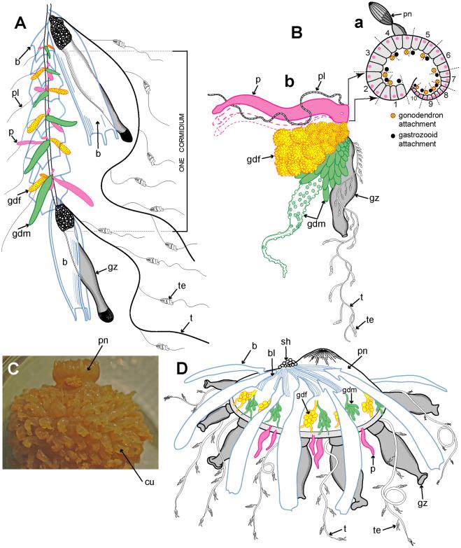
Example cormidia from a range of physonects are shown in Figure 6, covering typical long-stemmed as well as short-stemmed types. A cormidium from the typical long-stemmed physonect Nanomia bijuga comprises several palpon-gonodendra-bract complexes and large posterior gastrozooid with an associated elongate bract (Figure 6A). The palpon complexes become progressively older and larger posteriorly, and all elements of each cormidium originally arose from a pro-bud (as noted earlier) on the siphosomal horn at the anterior end of the siphosome [8] (Figure 3B). One of 10 cormidia from the physonect Physophora hydrostatica occupies a compact segment of the siphosomal corm, and includes three enlarged lateral palpons, an associated hermaphrodite gonodendron of male and female gonophores, with a gastrozooid and tentacle on the posterior surface, but no bracts (Figure 6B a–b). In the rhodaliid Dromalia alexandri (Figure 6C) cormidia are borne on branched cormidial units away from the corm surface, and these units spiral around the inflated corm to the posterior under-surface [4]. Cormidial units originate continuously on a siphosomal horn between the nectophores (swimming bells), on the ventral surface just below the pneumatophore, and each mature unit typically carries three cormidia. A single cormidium includes a gastrozooid, several palpons and many gonophores in a gonodendron [4]. Dendritic growth of the cormidial units enables a large number of cormidia to be carried on a single rotund D. alexandri individual. Cormidia on the enlarged float of Athorybia rosacea (Figure 6D) originate from a siphosomal horn adjacent to the float apex, and each includes a group of typically four large larval bracts, an associated branched hermaphrodite gonodendron with small palpons below, and a larger gastrozooid on the posterior corm surface.
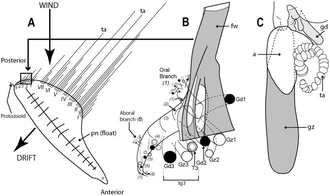
Figure 7A illustrates the complexity of a mature Portuguese Man O’War Physalia physalis viewed from above and ‘sailing’ with the wind, with many long tentacles extending from the cormidia and streaming out from the windward side. The cormidia of P.
physalis are shown diagrammatically in Figure 7A, and numbered 1–5 and I –VII; they originate directly from the underside of the float (pneumatophore) in this species and develop in a particularly complex pattern, as described and illustrated in a seminal paper by A.K. Totton [39]. Cormidia bud one from another in a series, and each such series is termed a cormidial complex. There are twelve cormidial complexes in a mature P. physalia, which are attached in two groups separated by a small gap; the oldest complex in each group, (which forms first) lies closest to the anterior (or aboral) end of the animal (Figure 7A). The smaller oral group of complexes (1– 5) lies just posterior of the first gastrozooid to form in the larva, the protozooid, and one cormidial complex from this region is shown in Figure 7B. It bears c. 13 cormidia, on two branches: a smaller oral branch above which is directed towards the oral end of the float, and a larger aboral branch below which is directed towards the aboral, or posterior, end of the animal. Almost all the cormidia of P. physalis comprise three zooids: a gastrozooid, gonodendron and a separate tentacle with ampulla (where the nematocysts are formed), which together form a tripartite group (Figure 7C). As growth proceeds more tripartite groups develop on lateral branches from the cormidial complex, filling every available space (Figure 7B). Indeed, no other siphonophore buds so prolifically as P. physalis [39]. As sexual maturity is reached, the gonodendra of each cormidial complex sub-branch many times, and detach. The largest such gonodendral ‘sphere’ found by Totton (from a female) measured ,5 cm across, and bore 2400 gonophores on seven main branches, plus 224 very small medusoid bells, an extra zooid present in the cormidial complexes of mature P. physalis.
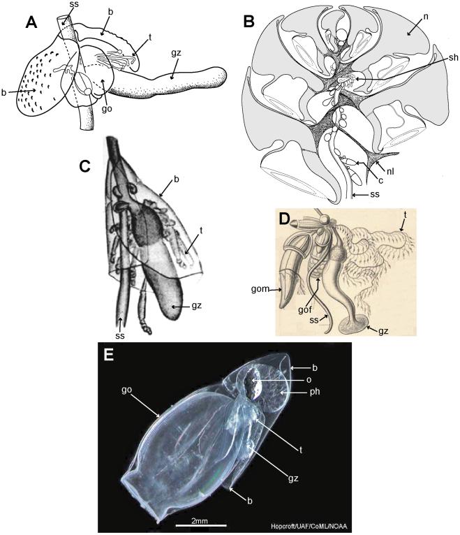
Cormidia are discrete in calycophorans, and, with one exception, lack the palpons present in physonect cormidia. In many calycophoran cormidia, the bract wraps around the stem in a cloak-like manner and gives protection to the underlying gastrozooid and gonophores (Figure 8A, C). As already noted above, when the cormidium of most diphyomorphs reaches maturity, it detaches and becomes a free-living eudoxid (Figure 8E). In some calycophorans, however, cormidia remain attached to the stem throughout life (prayine prayids and sulculeolariine diphyids). A few groups lack bracts, including members of the prayomorph family Hippopodiidae (see above), and Clausophyes species of the diphyomorph family Clausophyidae, both of which also probably retain their cormidia on the stem. In hippopodiids, a number of bells remain joined together when mature, forming a hollow cylinder from which the siphosomal stem emerges at the posterior end (Figure 8B). This stem originates between the two youngest nectophores but only the bottom two bells are functional in hippopodiids; their mesogloea, together with that of the other smaller bells, give buoyancy to compensate for the absence of bracts in the cormidia (Figure 8D). Cormidia arise from a siphosomal horn and are small, allowing the stem to be completely withdrawn into the cylindrical chamber when not feeding, as already noted above (Figure 8B).
PLOS ONE | www.plosone.org |
12 |
February 2014 | Volume 9 | Issue 2 | e87737 |
Diversity and Phylogeny of Siphonophores
Figure 6. Physonect cormidia. A: Nanomia bijuga cormidium (derived from [68] pl. 7, fig. 10); B: Physophora hydrostatica a. diagram of posterior view of corm surface bearing 10 cormidia (derived from [77] figs. 12a, 16a); b. one cormidium exploded (derived from [26] pl. 20, fig. 18 with two additional palpons added); C: Dromalia alexandri dorsal view of corm with many spirally arranged cormidial units, dorsal view (GMM); D: Athorybia rosacea lateral view of float with siphosomal horn and attached cormidia (derived from [50] txt fig. 45). Labels: b – bract; bl – bracteal lamella; cu – cormidial unit; gdf – female gonodendron; gdm – male gonodendron; gz – gastrozooid; p – palpon; pl – palpacle; pn – pneumatophore (float); sh – siphosomal horn; t – tentacle with tentilla; te - tentillum.
doi:10.1371/journal.pone.0087737.g006
PLOS ONE | www.plosone.org |
13 |
February 2014 | Volume 9 | Issue 2 | e87737 |
Diversity and Phylogeny of Siphonophores
Figure 7. Cystonect cormidia as exhibited by Physalia physalis. A: Left-handed drifting specimen viewed from above (derived with minor modification from [39] fig. 5) – added numbers 1–5 identify oral cormidial groups while numbers I–VI identify main cormidial groups – note how Physalia’s surface float drifts to starboard with the wind on a broad reach; B: Oral cormidial complex number 2 viewed from inside the float – note groups 3 to 8 are tripartite, with more tripartite groups on oral and aboral side branches (adapted from [39] txt fig. 12D) – numbers in brackets added to identify tripartite groups; C: A developing tripartite group from main cormidial complex number VI (derived from [39] txt fig. 14B, in part only). Labels: a – ampulla (basigaster); fw – float wall; gd – gonodendron; gz – gastrozooid; pn – pneumatophore (float); ta – tentacle with ampulla (basigaster); T – tentacle; tg – tripartite group.
doi:10.1371/journal.pone.0087737.g007
Old and New Phylogenies
The first detailed molecular study of a large range of Siphonophora [10] identified important morphological characters associated with their evolution not previously considered significant; it is reproduced here as Figure 9. A more recent study [79] used the barcoding gene mtCOI to generate a phylogeny for 95 medusozoan species (including 61 siphonophores), though this gene is more appropriate for phylogenetic characters at family level or below. Analysis of a third gene 28S is unresolved for the clade Codonophora [55], and further siphonophore taxa analyses and application of whole genome sequencing to the group are awaited for more clarification of this clade. The study of Dunn et al. [10] led to further changes in physonect systematics by Pugh [17] as discussed below (Figure 10). The old and new phylogenies are compared in Table 3, from 15 families recognized in 1987 and 16 different families and 67 genera recognized today.
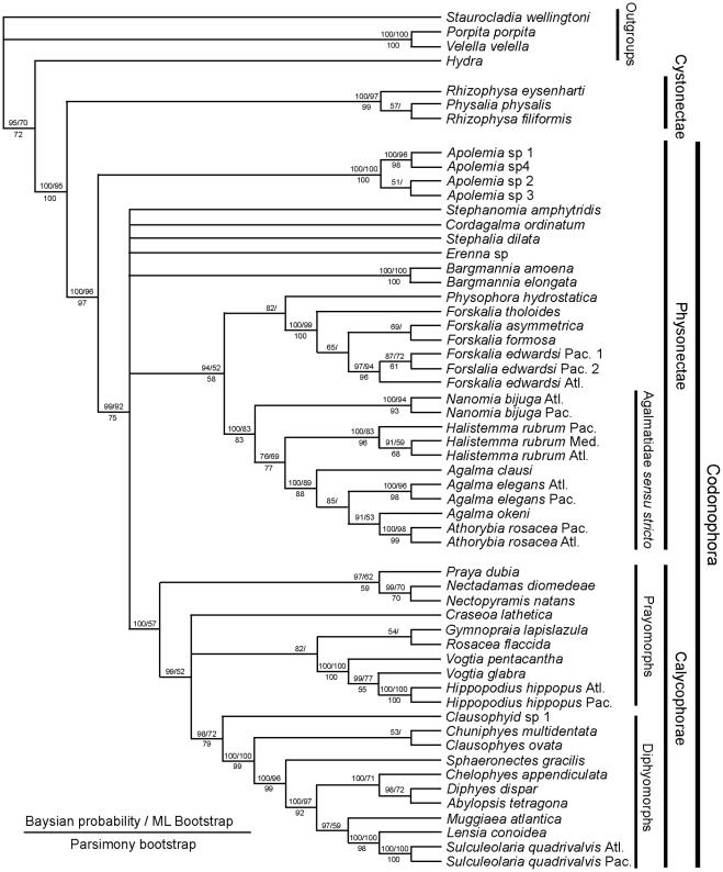
The consensus tree from the molecular study of Dunn et al. (see fig. 6 [10]) is based on data from two genes: the nuclear gene 18S and mitochondrial gene 16S, and is figured here as Figure 9. It concludes that cystonects are sister to all other siphonophores, with the remainder ranked together in a new clade Codonophora, meaning ‘bell bearers’. Within the Codonophora clade, the traditional grouping ‘Physonectae’ are paraphyletic, with the ‘physonect’ family Apolemiidae sister to all other taxa. Clades for the physonect families Forskaliidae and Agalmatidae sensu stricto are well supported, although resolution for taxa representing rhodaliids, erennids, pyrostephids and physophorids is poor. The
traditional group Calycophorae are nested within the nonapolemiid Codonophora and form a monophyletic clade. Within the Calycophorae, prayomorphs are paraphyletic, based on taxa and genes sampled in 2005. Hippopodiid prayomorphs form a distinct clade, and diphyomorphs, together with Sphaeronectes (ignoring one undescribed clausophyid species) form another distinct clade. Intraspecific variation is also demonstrated in multiple individuals of Hippopodius hippopus and Sulculeolaria quadrivalvis collected in the Atlantic and Pacific Oceans. The final important finding of Dunn et al. [10] places abylids within a clade containing the five diphyids tested. Five cryptic species pairs are also identified amongst the Atlantic and Pacific ‘physonects’ analysed (Figure 9).
The new phylogeny shows that character evolution within the Siphonophora is related to reproductive state (figs. 7–8 [10]). Separately sexed individuals are dioecious, whereas monoecious siphonophores bear differentially maturing male and female gonophores on the same individual. Zooid types scored by Dunn et al. [10] include nectosomal nectophores, siphosomal bracts, gastrozooids and palpons, as well as the number of types of each zooid present in each taxon. They found that cystonects, apolemiids, pyrostephids, erennids and rhodaliids, are all dioecious, and, surprisingly, all lack a descending ‘pallial canal’ (‘descending surface diverticulum’ of Mapstone [6]) on the proximal surface of the nectophore. In contrast, all remaining codonophorans are monoecious, and in taxa tested from the families Agalmatidae sensu stricto, Forskaliidae and Physophoridae
PLOS ONE | www.plosone.org |
14 |
February 2014 | Volume 9 | Issue 2 | e87737 |
Diversity and Phylogeny of Siphonophores
Figure 8. Calycophoran cormidia. A: Rosacea cymbiformis cormidium (after [6] fig. 2D); B. Hippopodius hippopus section through colony (adapted from [31] fig. 11, [78] txt fig. 13 and [27] fig. 44b); C: Chelophyes appendiculata cormidium (from [34] pl. 11, fig. 1); D. Hippopodius hippopus cormidium; note, no bracts (from [26] pl. 29, fig. 1 in part); E. Dimophyes arctica eudoxid (Russ Hopcroft, UAF). Labels: b – bract, c – cormidium; go – gonophore; gof – female gonophore; gom – male gonophore; gz – gastrozooid; n – nectophore; nl – nectophoral lamella; o – oil globule (in phyllocyst); ph – phyllocyst; sh – siphosomal horn; ss – siphosomal stem; t – tentacle with tentilla.
doi:10.1371/journal.pone.0087737.g008
PLOS ONE | www.plosone.org |
15 |
February 2014 | Volume 9 | Issue 2 | e87737 |
Diversity and Phylogeny of Siphonophores
Figure 9. Molecular phylogeny of siphonophores from Dunn et al. (fig. 6 [10]). Consensus tree of all trees for the Bayesian analysis of the combined data set (from an initial 20 million trees). The left score above the branch is the Bayesian posterior probability (%), the right score above the branch is the ML bootstrap support value (%), and the score below the branch is the MP bootstrap support value (%). The bars to the right of the species names indicate clades and grade taxa. Abbreviations: Atl – Atlantic; Med – Mediterranean; Pac – Pacific. For full details of analyses and consensus tree computations refer to Dunn et al. [10].
doi:10.1371/journal.pone.0087737.g009
PLOS ONE | www.plosone.org |
16 |
February 2014 | Volume 9 | Issue 2 | e87737 |
Diversity and Phylogeny of Siphonophores
Figure 10. Possible phylogeny of the Siphonophora (derived from [17], fig. 21, and [11]). MFZ – muscle-free zone on nectophore; * - dorsal nectosome; ** - one species monoecious.
doi:10.1371/journal.pone.0087737.g010
PLOS ONE | www.plosone.org |
17 |
February 2014 | Volume 9 | Issue 2 | e87737 |
(except Athorybia rosacea which lacks nectophores) this condition is coincident with the presence of a descending ‘pallial canal’ on the proximal surface of the nectophore.
Nectosomal nectophores are an apomorphy of the Codonophora and may have been derived from retained reproductive medusae [10]. Their presence together with the presence or absence of a descending pallial canal, suggests these two characters might have pleiotropic links [10]. Many of one type of nectophore (homomorphic) were found in all the ‘physonects’ tested except Athorybia rosacea, which lacks nectophores. Amongst the Calycophorae, nectophores are reduced to two of one type in most prayomorphs tested, except for the two nectopyramidines which had only one of one type, and hippopodiids which, as Dunn et al. [10] conclude, have developed several nectophores of one type from an ancestor which probably had only two of one type (see their fig. 8a). Most diphyomorph calycophorans, in contrast, have two nectophores of two types (an anterior and a posterior: heteromorphic), with one nectophore lost in Muggiaea atlantica, and only a single larval nectophore retained in Sphaeronectes gracilis (fig. 8a [10]).
Palpons are another character found in almost all ‘physonects’, but absent from all the calycophorans tested by Dunn et al. (fig. 8b [10]). Parsimony indicates that palpons were probably present in the ancestral siphonophore and have been lost one or two times, while bracts appeared first in the Codonophora, and might have developed into two or more types several times and at several different specific locations during siphonophore evolution [10]. Bracts, however, which are also characteristic of the Codonophora, are all of one type in apolemiids and also in all calycophorans which possess them, as well as in some Agalmatidae sensu stricto (Agalma and Athorybia). In erennids and other Agalmatidae sensu stricto (Nanomia and Halistemma species) two types of bracts develop, and four types are found in Forskalia species (see fig. 8b in [10]). Thus, as Dunn et al. [10] conclude, there has been both gain and loss of zooids during siphonophore evolution.
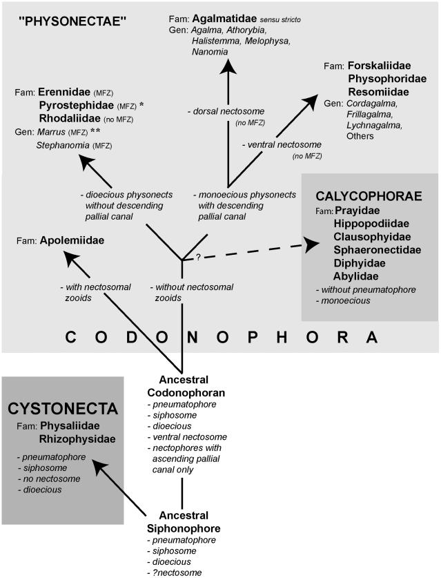
The importance of these characters in shaping siphonophore evolution is reflected in the higher rankings given in Table 3B. A new hypothesis for character evolution given by Pugh [17], which is shown here in Figure 10, proposes a dioecious ancestral siphonophore with pneumatophore and siphosome, but maybe not a nectosome. Such an ancestor may have given rise to two clades: the dioecious cystonects with a pneumatophore and siphosome but no nectosome, and a dioecious ancestral codonophoran with a pneumatophore, nectosome and siphosome. Nectophores of the latter have only an ascending ‘pallial canal’ on their proximal surfaces. The first nectosome to evolve is thought to have had all nectophores attached on the same side of the stem as the siphosomal zooids, which is, by convention, the ventral surface of the stem (p. 931 [10]). A similar condition is found in most families and genera of physonects today.
Apolemiids are also dioecious, with nectosomal palpons between the nectophores [11,80], and were the first group to split from the other Codonophora, with both lineages evolving simultaneously and independently thereafter. The ancestral sister group to the Apolemiidae could have been another clade that lacked nectosomal palpons (Figure 10) and from which, perhaps later in time, a monoecious ancestor emerged. Dioecy could have persisted in a group of physonects which lacked a descending ‘pallial canal’ on the proximal surface of their nectophores, including three extant families and two unascribed dioecious genera (see Table 3B). In one of these families, the Pyrostephidae, a twist may have occurred at the junction between the nectosome and siphosome resulting in nectophores arising on the dorsal
Diversity and Phylogeny of Siphonophores
surface (‘dorsal nectosome’) and siphosomal cormidia on the ventral surface. The first monoecious siphonophores could have been physonects with a descending ‘pallial canal’ on the proximal surface of their nectophores, a new diagnostic character. From this clade Pugh [17] proposes a split into the Family Agalmatidae sensu stricto with a dorsal nectosome, and a non-agalmatid clade including the families Forskaliidae, Physophoridae and Resomiidae together with the unascribed monoecious genera Cordagalma, Frillagalma and Lychnagalma (Table 3B) which all exhibit a ventral nectosome (Figure 10). Pugh [17] also suggests that a further monoecious group of siphonophores, the Calycophorae, appeared at some point during the evolution of these various physonect families, (Figure 10). In calycophorans the pneumatophore is lost and the nectosome typically reduced to just two nectophores.
Systematics 1987 to Present
This section summarizes the changes in siphonophore systematics since the last review in 1987 and is based on the new phylogenies as outlined above [10,17], together with details of families that have been revised or newly introduced, and new genera and species added, moved or now considered invalid. Most of this information for cystonects and physonects is given in Table 4, and for calycophorans in Table 5. Ongoing debates about the validity of certain species, and other systematic information too extensive for inclusion in the tables, is briefly summarized below.
Apolemiidae. Unique nectosomal palpons (previously nectosomal tentacles or polyps) are probably a synapomorphy of the Codonophora, being greatly reduced or absent in other codonophorans [10]. These zooids arise on the nectosome from the posterior ends of the nectophoral muscular lamellae, either singly or in bunches [85], and are identified as small buds on the nectosome of some other long-stemmed physonects [8]. Other important specific characters include the presence or absence of diverticula penetrating into the mesogloea from the lateral radial canals of the nectophores, the relative size of the siphosomal horn, the type of siphosomal cormidia present (pedunculate or dispersed), and the number of palpon types on the siphosome (one or two) [11]. In older cormidia, secondary gastrozooids may form independent of the growth zone, directly on the siphosome [11], as also shown in the agalmatid Nanomia bijuga [8] (see above). Pedunculate cormidia may be either ancestral or derived for the Codonophora [11], but further work and denser sampling of siphonophore phylogeny is needed to resolve this question [11].
Currently, the family is monotypic for Apolemia, and includes A. uvaria (Lesueur, 1815), A. vitiazi (Stepanjants, 1967) and A. contorta (Margulis, 1976) [1], together with two newly described species A. lanosa and A. rubriversa [11] and a third species not yet described (A. trinegra [84]). Two types of siphosomal palpons are exhibited by A. uvaria (shorter red/brown type and longer opaque type [85,95]), but may also be characteristic of other species, together with pigment distribution in the siphosomal palpons [84]. Apolemiids can reach more than 30 m in length, and the recent paper by Siebert et al. forms the foundation for descriptions of up to 15 further new species [11]. Apolemiids frequently undergo autotomy [6,95], releasing many lengths of siphosome which float freely in the water without nectophores, while the latter swim off or drifted away in a different direction.
Erennidae. Collection of several excellent quality specimens by submersible from the Dry Tortugas and Bahamas in the tropical Atlantic has enabled introduction of this new family, with three new species and an older re-described species (Table 4 and [15]). These deep-sea physonects have much enlarged tentilla that are held close to the body and in most species vibrate to attract prey (deep-sea fish); these lures are described in a later section.
PLOS ONE | www.plosone.org |
18 |
February 2014 | Volume 9 | Issue 2 | e87737 |

Diversity and Phylogeny of Siphonophores
Table 3. Old and new classification of the Siphonophora.
A. OLD TAXONOMY |
B. NEW PHYLOGENY |
|
|
|
|
|
|
High Rank |
High Rank |
Family & Sub-family |
Genera |
|
|
||
Sub-order Cystonectae |
|
|
|
|
|
|
|
Families: Physaliidae, Rhizophysidae |
|
|
|
|
I - CYSTONECTA |
01. Physaliidae |
Physalia |
|
|
|
|
|
|
02. Rhizophysidae |
Bathyphysa, Rhizophysa |
|
|
|
|
|
II - CODONOPHORA |
|
|
|
|
|
|
Sub-order Physonectae |
Physonectae |
|
|
|
|
|
|
Families: Apolemiidae, Agalmatidae, |
|
|
|
Pyrostephidae,Physophoridae, |
|
|
|
Athorybiidae, Rhodaliidae, |
|
|
|
Forskaliidae |
|
|
|
|
|
|
|
|
Dioecious families |
03. Apolemiidae |
Apolemia |
|
|
|
|
|
|
04. Erennidae |
Erenna, Parerenna |
|
|
|
|
|
|
05. Pyrostephidae |
Bargmannia, Pyrostephos |
|
|
|
|
|
|
06. Rhodaliidae |
Angelopsis, Aranciala, Dromalia, |
|
|
|
|
|
|
|
Archangelopsis, Steleophysema, |
|
|
|
|
|
|
|
Stephalia, Thermopalia |
|
|
|
|
|
|
07. Unascribed dioecious genera |
Marrus, Stephanomia |
|
|
|
|
|
Monoecious families |
09. Forskaliidae |
Forskalia |
|
|
|
|
|
|
10. Physophoridae |
Physophora |
|
|
|
|
|
|
11. Resomiidae |
Resomia |
|
|
|
|
|
|
08. Agalmatidae sensu stricto |
Agalma, Athorybia, Melophysa, |
|
|
|
|
|
|
|
Halistemma, Nanomia |
|
|
|
|
|
|
12. Unascribed monoecious genera |
Cordagalma, Frillagalma, |
|
|
|
|
|
|
|
Lychnagalma, Rudjakovia |
|
|
|
|
Sub-order Calycophorae |
|
|
|
Families: Prayidae, Diphyidae, |
|
|
|
Hippopodiidae, Clausophyidae, |
|
|
|
Sphaeronectidae, Abylidae |
|
|
|
|
|
|
|
|
Calycophorae |
|
|
|
|
|
|
|
Prayomorphs |
13. Prayidae |
|
|
|
|
|
|
|
S-f Amphyicaryoninae |
Amphicaryon, Maresearsia |
|
|
|
|
|
|
S-f Prayinae |
Craseoa, Desmophyes, Rosacea, |
|
|
|
|
|
|
|
Gymnopraia, Lilyopsis, |
|
|
|
|
|
|
|
Mistoprayina, Praya, Prayola, |
|
|
|
|
|
|
|
Stephanophyes |
|
|
|
|
|
|
S-f Nectopyramidinae |
Nectadamas, Nectopyramis |
|
|
|
|
|
|
14. Hippopodiidae |
Hippopodius, Vogtia |
|
|
|
|
|
Diphyomorphs |
15. Clausophyidae |
Chuniphyes, Clausophyes, |
|
|
|
|
|
|
|
Crystallophyes, Kephyes, |
|
|
|
|
|
|
|
Heteropyramis |
|
|
|
|
|
|
16. Sphaeronectidae |
Sphaeronectes |
|
|
|
|
|
|
17. Diphyidae |
|
|
|
|
|
|
|
S-f Sulculeolariinae |
Sulculeolaria |
|
|
|
|
|
|
S-f Diphyinae |
Chelophyes, Dimophyes, |
|
|
|
|
|
|
|
Diphyes, Eudoxoides, Lensia, |
|
|
|
|
|
|
|
Muggiaea |
|
|
|
|
|
|
S-f Giliinae |
Gilia |
|
|
|
|
|
|
18. Abylidae |
|
|
|
|
|
|
|
S-f Abylinae |
Abyla, Ceratocymba |
|
|
|
|
|
|
S-f Abylopsinae |
Abylopsis, Bassia, Enneagonum |
|
|
|
|
doi:10.1371/journal.pone.0087737.t003 |
|
|
|
PLOS ONE | www.plosone.org |
19 |
February 2014 | Volume 9 | Issue 2 | e87737 |

Diversity and Phylogeny of Siphonophores
Table 4. New systematics for cystonect and physonect siphonophore families.
|
Family |
Comments |
|
|
|
|
|
01. |
|
Physaliidae |
Monotypic for Physalia physalis (P. utriculus considered a junior synonym [81] |
|
|
||
02. |
|
Rhizophysidae |
Long-stemmed; Bathyphysa japonica a junior synonym of B. conifera; SEM studies of budding sequences described for B. sibogae, |
|
|
|
Rhizophysa filiformis and R. eysenhardti [8] |
|
|
|
|
03. |
|
Apolemiidae |
Long stemmed; monophyletic and sister to all other Codonophora, with unique nectophoral palpons on the nectosome. |
|
|
|
Nectophores distinctive and ridge-less, cormidia dispersed or discrete; gastrozooids with simple tentacles (no tentilla) resembling |
|
|
|
palpacles of palpons. Monogeneric for Apolemia. Two new species include A. lanosa and A. rubriversa [11] and three older species |
|
|
|
A. contorta, A. uvaria and A. vitiazi (Tottonia contorta sensu Mapstone 2003 now referable to A. lanosa). A number of other species |
|
|
|
are known to exist [2,10,11,52,82,83,84,85], and await full description. |
|
|
|
|
04. |
Erennidae |
Long-stemmed family erected for 4 species with large prominent straight tentilla, no involucrum and a rigid terminal process |
|
|
|
|
lacking nematocysts [15]. Two genera: Erenna (3 species) and Parerenna (1 species). E. richardi Bedot, 1904, and a new species E. |
|
|
|
laciniata have large flattened nectophores and large tentilla held close to body and vibrate to attract prey; two further new species |
|
|
|
E. cornuta and Parerenna emilyae have different and also unique tentilla and gastrozooids [15]. |
|
|
|
|
05. |
Pyrostephidae |
Long-stemmed family reviewed and revised [14], with 3 new species of Bargmannia: B. amoena, B. gigas, B. lata [14,86]; also Mica |
|
|
|
|
micula, the putative post-larva of a pyrostephid [87,88]. Nectophores with unique lower-lateral wings and much enlarged triangular |
|
|
|
thrust block; in B. elongata two growth zones on stem and composition of the cormidia studied using SEM [80]; pyrostephid |
|
|
|
cormidia either have oleocysts (modified tentacle-less palpons) (in Pyrostephos) or none (in Bargmannia) [14]. |
|
|
|
|
06. |
Rhodaliidae |
Short-stemmed family of 8 genera, with 4 new species including Archangelopsis jagoa, Arancialia captonia [45,89], and two others |
|
|
|
|
herein referred to Steleophysema Moser, 1924, including S. sulawensis and S. rotunda. Sagamalia hinomaru reduced to a junior |
|
|
|
synonym of Steleophysema aurophora [1,89]. First in situ feeding observations on four species [89]. Dromalia alexandri re-described |
|
|
|
[4]. |
|
|
|
|
07. |
Unascribed dioecious |
Long-stemmed genera Marrus Totton, 1954 [90] and Stephanomia Lesueur & Petit, 1807 [10] both with muscle-free zones on |
|
|
genera |
nectosac and other characters (Fig. 10). A new species M. claudanielis described [90] and new specimens of an old species S. |
|
|
|
|
amphytridis [10] await re-description. |
|
|
|
|
08. |
Forskaliidae |
Long stemmed and delicate monotypic family, probably sister to the Physophoridae [10]. Recently revised [16] with two new |
|
|
|
|
species added (Forskalia asymmetrica, F. saccula) and one reduced to a Species Inquirenda [1]. |
|
|
|
|
09. |
Physophoridae |
Family with long nectosome but short corm-like siphosome; previously monotypic for Physophora hydrostatica bract present only |
|
|
|
|
in larva; now a new smaller and less colourful second species P. gilmeri, is added, with bracts retained on adult colony [77]; unique |
|
|
|
tentilla in both species. |
|
|
|
|
10. |
Resomiidae |
Long-stemmed family newly introduced for two species previously referred to the Agalmatidae (Moseria convoluta, M. similis) and |
|
|
|
|
now transferred to a new monotypic genus Resomia [17]; two tentilla types uniquely present on each tentacle. Three new species |
|
|
|
R. dunni, R. ornicephala, R. persica described in 2010 [91]. |
|
|
|
|
11. |
Agalmatidae sensu stricto |
Mostly long-stemmed and recently restricted to genera with dorsal nectosome (see above) and involucrate tricornuate tentilla with |
|
|
|
|
tightly coiled cnidoband (see below). Now includes two short-stemmed genera (Athorybia, Melophysa) [17]. New species added |
|
|
|
(Halistemma transliratum) [92] and another re-described (H. foliacea, as H. amphytridis) [17,93]. |
12. |
Unascribed monoecious |
Long-stemmed monotypic genera Cordagalma, Frillagalma and Lychnagalma with ventral nectosomes have been removed from |
|
|
genera |
the Agalmatidae [17] and a new species C. tottoni described [94]. Rudjakovia plicata considered a valid species [1] and may be |
|
|
|
|
transferred to Agalmatidae when more characters are elucidated [17]. |
|
|
|
|
|
|
|
|
For fundamental characters of the physonect families listed above (sex, proximal surface canals etc), see Figure 10. doi:10.1371/journal.pone.0087737.t004
Pyrostephidae. This family has been properly diagnosed for the first time and three new species introduced [14]. A likely pyrostephid post-larva has also been described (Table 4), and a comprehensive study of the organisation of siphosomal zooids in Bargmannia elongata shows that new cormidia are formed on a protrusion from the stem termed the ‘‘horn’’ [80]. Here a series of ‘‘probuds’’ form, which each subdivides a number of times to form eight zooids and together these form a single cormidium (see above).
Rhodaliidae. Four new species have been added to this epibenthic family in recent years (Table 4 and [45,89]). Dromalia alexandri has been re-described including the first figures of a rhodaliid siphosomal horn, mature cormidial units and a mature bract, together with a more comprehensive distribution map including both range and density [4]. Herein the doubtful species Steleophysema aurophora Moser, 1924 [27], is re-validated from observations made by Dhugal Lindsay (pers. comm.) of new specimens collected off Japan, and as a result Sagamalia hinomaru is reduced to a junior synonym [1]. The genus Tridensa Hissmann, 2005, is also reduced to a junior synonym of Steleophysema, based on the shape and attachment point of its bracts (at base of each
cormidial unit), attachment of the gonophores (with egg pouch) directly to the thin polygastric cormidia just distal of each cormidial gastrozooid, and attachment of the gonopalpons just distal of the gonophores. The two species T. sulawensis and T. rotunda become junior synonyms of S. sulawensis and S. rotunda [1]. A full re-description of S. aurophora is underway (D. Lindsay, pers. comm.).
Unascribed dioecious physonects. The genera Marrus and
Stephanomia perhaps diverged early from other codonophorans (Figure 10). Marrus orthocannoides may not belong to the genus Marrus, because it has a fully muscular nectosac, whereas those of other Marrus species have a proximal muscle-free zone [90]. The genus name Stephanomia has been applied to many species in the past (p. 102 [6]), but is herein restricted to the large species Stephanomia amphytridis of Lesueur and Petit, 1807 [1] as applied by Dunn et al. [10] and mentioned on p. 103 of Mapstone [6]. This species has been collected recently in both the Atlantic and Pacific [17], sequenced for 16S and 18S genes [10] and a morphological description is underway.
Forskaliidae. The fragile and often snake-like colonies of this monoecious family have a spiral stem with diffusely attached
PLOS ONE | www.plosone.org |
20 |
February 2014 | Volume 9 | Issue 2 | e87737 |