Добавил:
Опубликованный материал нарушает ваши авторские права? Сообщите нам.
Вуз: Предмет: Файл:

Garrett R.H., Grisham C.M. - Biochemistry (1999)(2nd ed.)(en)

.pdf
Скачиваний:
182
Добавлен:
15.08.2013
Размер:
24.81 Mб
Скачать
FIGURE 19.30

19.6 Anaerobic Pathways for Pyruvate

631

19.5 The Metabolic Fates of NADH and Pyruvate—

The Products of Glycolysis

In addition to ATP, the products of glycolysis are NADH and pyruvate. Their processing depends upon other cellular pathways. NADH must be recycled to NAD , lest NAD become limiting in glycolysis. NADH can be recycled by both aerobic and anaerobic paths, either of which results in further metabolism of pyruvate. What a given cell does with the pyruvate produced in glycolysis depends in part on the availability of oxygen. Under aerobic conditions, pyruvate can be sent into the citric acid cycle (also known as the tricarboxylic acid cycle; see Chapter 20), where it is oxidized to CO2 with the production of additional NADH (and FADH2). Under aerobic conditions, the NADH produced in glycolysis and the citric acid cycle is reoxidized to NAD in the mitochondrial electron transport chain (Chapter 21).

19.6 Anaerobic Pathways for Pyruvate

Under anaerobic conditions, the pyruvate produced in glycolysis is processed differently. In yeast, it is reduced to ethanol; in other microorganisms and in animals, it is reduced to lactate. These processes are examples of fermentation —the production of ATP energy by reaction pathways in which organic molecules function as donors and acceptors of electrons. In either case, reduction of pyruvate provides a means of reoxidizing the NADH produced in the glyc- eraldehyde-3-phosphate dehydrogenase reaction of glycolysis (Figure 19.30). In yeast, alcoholic fermentation is a two-step process. Pyruvate is decarboxylated to acetaldehyde by pyruvate decarboxylase in an essentially irreversible reaction. Thiamine pyrophosphate is a required cofactor for this enzyme. The second step, the reduction of acetaldehyde to ethanol by NADH, is catalyzed

(a) Alcoholic fermentation


O


(b) Lactic acid fermentation


O


CHO


HPO24


C OPO23


CHO

HPO24


C OPO23


H


C


OH


H


C


OH


H


C


OH


H


C


OH


G3PDH


G3PDH


CH2OPO23


CH2OPO23


CH2OPO23


CH2OPO23

D-Glyceraldehyde-


1,3-BPG


D-Glyceraldehyde-


1,3-BPG

3-phosphate


3-phosphate


NAD+

NADH + H+


NAD+

NADH + H+


O


COO


CH3C


Pyruvate


OH


O


COO


COO


CH

CH

OH


CH

CHO

CO


CH

3


C


CH

C


3

2


Alcohol


3


2


Lactate

3


Ethanol


Acetaldehyde


Pyruvate


dehydrogenase


H


dehydrogenase


Lactate

(a) Pyruvate reduction to ethanol in yeast provides a means for regenerating NAD consumed in the glyceraldehyde-3-P dehydrogenase reaction. (b) In oxy- gen-depleted muscle, NAD is regenerated in the lactate dehydrogenase reaction.
632 Chapter 19 Glycolysis

by alcohol dehydrogenase (Figure 19.30). At pH 7, the reaction equilibrium strongly favors ethanol. The end products of alcoholic fermentation are thus ethanol and carbon dioxide. Alcoholic fermentations are the basis for the brewing of beers and the fermentation of grape sugar in wine making. Lactate produced by anaerobic microorganisms during lactic acid fermentation is responsible for the taste of sour milk and for the characteristic taste and fragrance of sauerkraut, which in reality is fermented cabbage.

Lactate Accumulates Under Anaerobic Conditions in Animal Tissues

In animal tissues experiencing anaerobic conditions, pyruvate is reduced to lactate. Pyruvate reduction occurs in tissues that normally experience minimal access to blood flow (e.g., the cornea of the eye) and also in rapidly contracting skeletal muscle. When skeletal muscles are exercised strenuously, the available tissue oxygen is consumed, and the pyruvate generated by glycolysis can no longer be oxidized in the TCA cycle. Instead, excess pyruvate is reduced to lactate by lactate dehydrogenase (Figure 19.30). In anaerobic muscle tissue, lactate represents the end of glycolysis. Anyone who exercises to the point of consuming all available muscle oxygen stores knows the cramps and muscle fatigue associated with the buildup of lactic acid in the muscle. Most of this lactate must be carried out of the muscle by the blood and transported to the liver, where it can be resynthesized into glucose in gluconeogenesis. Moreover, because glycolysis generates only a fraction of the total energy available from the breakdown of glucose (the rest is generated by the TCA cycle and oxidative phosphorylation), the onset of anaerobic conditions in skeletal muscle also means a reduction in the energy available from the breakdown of glucose.

19.7 The Energetic Elegance of Glycolysis

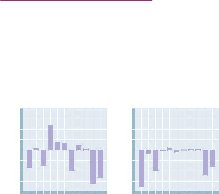
FIGURE 19.31 A comparison of free energy changes for the reactions of glycolysis (step 1 hexokinase) under (a) standard-state conditions and (b) actual intracellular conditions in erythrocytes. The values of G° provide little insight into the actual free energy changes that occur in glycolysis. On the other hand, under intracellular conditions, seven of the glycolytic reactions operate near equilibrium (with G near zero). The driving force for glycolysis lies in the hexokinase (1), phosphofructokinase (3), and pyruvate kinase (10) reactions. The lactate dehydrogenase (step 11) reaction also exhibits a large negative G under cellular conditions.

The elegance of nature’s design for the glycolytic pathway may be appreciated through an examination of Figure 19.31. The standard-state free energy changes for the 10 reactions of glycolysis and the lactate dehydrogenase reaction (Figure 19.31a) are variously positive and negative and, taken together, offer little insight into the coupling that occurs in the cellular milieu. On the other hand, the values of G under cellular conditions (Figure 19.31b) fall into two distinct classes. For reactions 2 and 4 through 9, G is very close to zero, so that these reactions operate essentially at equilibrium. Small changes

(a)

G at standard state (∆ G°')

(b) ∆ G in erythrocytes (∆ G)


40


40


30


30

kJ/mol

20

kJ/mol

20

10

10

energy,

energy,

0

0


Free

–10

Free

–10

–20

–20


–30


–30


–40


–40

0

1

2

3

4

5

6

7

8

9

10 11

0

1

2

3

4

5

6

7

8

9

10 11


Steps of glycolysis


Steps of glycolysis


19.8 Utilization of Other Substrates in Glycolysis

633

in the concentrations of reactants and products could “push” any of these reactions either forward or backward. By contrast, the hexokinase, phosphofructokinase, and pyruvate kinase reactions all exhibit large negative G values under cellular conditions. These reactions are thus the sites of glycolytic regulation. When these three enzymes are active, glycolysis proceeds and glucose is readily metabolized to pyruvate or lactate. Inhibition of the three key enzymes by allosteric effectors brings glycolysis to a halt. When we consider gluconeo- genesis—the biosynthesis of glucose—in Chapter 23, we will see that different enzymes are used to carry out reactions 1, 3, and 10 in reverse, effecting the net synthesis of glucose. The maintenance of reactions 2 and 4 through 9 at or near equilibrium permits these reactions (and their respective enzymes!) to operate effectively in either the forward or reverse direction.

19.8 Utilization of Other Substrates in Glycolysis

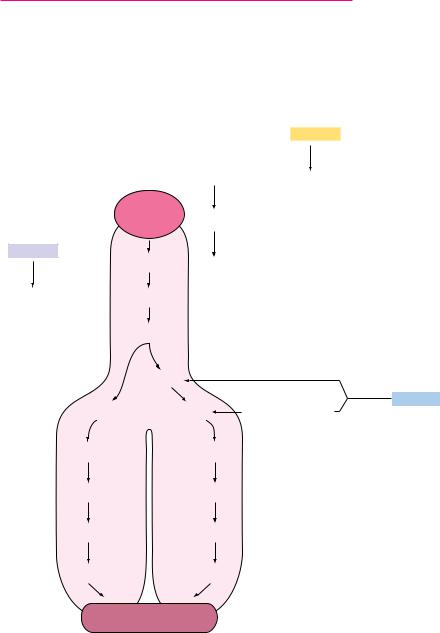
The glycolytic pathway described in this chapter begins with the breakdown of glucose, but other sugars, both simple and complex, can enter the cycle if they can be converted by appropriate enzymes to one of the intermediates of glycolysis. Figure 19.32 shows the mechanisms by which several simple metabolites can enter the glycolytic pathway. Fructose, for example, which is pro-

Galactose

UDP–Gal Galactose–1–P

Glucose UDP–Glucose

Mannose

G6P Glucose–1–P

Mannose–6–P F6P

FBP


DHAP

Aldolase


Triose


kinase

Fructose

G3P

D- Glyceraldehyde

G3P

FIGURE 19.32 Mannose, galactose, fructose, and other simple metabolites can enter the glycolytic pathway.

BPG

BPG

3PG

3PG

2PG

2PG

PEP

PEP

2 Pyruvate

634 Chapter 19 Glycolysis

duced by breakdown of sucrose, may participate in glycolysis by at least two different routes. In the liver, fructose is phosphorylated at C-1 by the enzyme fructokinase:

D-Fructose ATP4 88n D-fructose-1-phosphate2 ADP3 H (19.10)

Subsequent action by fructose-1-phosphate aldolase cleaves fructose-1-P in a manner like the fructose bisphosphate aldolase reaction to produce dihydroxyacetone phosphate and D-glyceraldehyde:

D-Fructose-1-P2 88n D-glyceraldehyde dihydroxyacetone phosphate2

(19.11)

Dihydroxyacetone phosphate is of course an intermediate in glycolysis. D-Gly- ceraldehyde can be phosphorylated by triose kinase in the presence of ATP to form D-glyceraldehyde-3-phosphate, another glycolytic intermediate.

In the kidney and in muscle tissues, fructose is readily phosphorylated by hexokinase, which, as pointed out above, can utilize several different hexose substrates. The free energy of hydrolysis of ATP drives the reaction forward:

D-Fructose ATP4 88n D-fructose-6-phosphate2 ADP3 H (19.12)

Fructose-6-phosphate generated in this way enters the glycolytic pathway directly in step 3, the second priming reaction. This is the principal means for channeling fructose into glycolysis in adipose tissue, which contains high levels of fructose.

The Entry of Mannose into Glycolysis

Another simple sugar that enters glycolysis at the same point as fructose is mannose, which occurs in many glycoproteins, glycolipids, and polysaccharides (Chapter 7). Mannose is also phosphorylated from ATP by hexokinase, and the mannose-6-phosphate thus produced is converted to fructose-6-phosphate by phosphomannoisomerase.

D-Mannose ATP4 88n D-mannose-6-phosphate2 ADP3 H

(19.13)

D-Mannose-6-phosphate2 88n D-fructose-6-phosphate2

(19.14)

The Special Case of Galactose

A somewhat more complicated route into glycolysis is followed by galactose, another simple hexose sugar. The process, called the Leloir pathway after Luis Leloir, its discoverer, begins with phosphorylation from ATP at the C-1 position by galactokinase:

D-Galactose ATP4 88n D-galactose-1-phosphate2 ADP3 H (19.15)

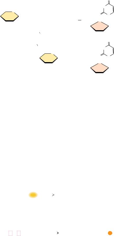
Galactose-1-phosphate is then converted into UDP-galactose (a sugar nucleotide) by galactose-1-phosphate uridylyltransferase (Figure 19.33), with concurrent production of glucose-1-phosphate and consumption of a molecule of UDPglucose. The uridylyltransferase reaction proceeds via a “ping-pong” mechanism (Figure 19.34) with a covalent enzyme-UMP intermediate. The glucose- 1-phosphate produced by the transferase reaction is a substrate for the phosphoglucomutase reaction (Figure 19.33), which produces glucose-6-phos- phate, a glycolytic substrate. The other transferase product, UDP-galactose, is converted to UDP-glucose by UDP-glucose-4-epimerase. The combined action of the uridylyltransferase and epimerase thus produces glucose-1-P from galac- tose-1-P, with regeneration of UDP-glucose.

A rare hereditary condition known as galactosemia involves defects in galac- tose-1-P uridylyltransferase that render the enzyme inactive. Toxic levels of

19.8 Utilization of Other Substrates in Glycolysis

635

Galactose


ATP


Galactokinase


ADP


Galactose-1- P


Galactose-1- P


UDP-Glucose

UDP-Galactose-


uridylyltransferase


4-epimerase


UDP-Galactose


Glucose-1- P


Phosphoglucomutase


Glucose-6- P

FIGURE 19.33 Galactose metabolism via the Leloir pathway.

galactose accumulate in afflicted individuals, causing cataracts and permanent neurological disorders. These problems can be prevented by removing galactose and lactose from the diet. In adults, the toxicity of galactose appears to be less severe, due in part to the metabolism of galactose-1-P by UDP-glucose pyrophosphorylase, which apparently can accept galactose-1-P in place of glucose-1-P (Figure 19.35). The levels of this enzyme may increase in galactosemic individuals, in order to accommodate the metabolism of galactose.


CH2OH


CH2OH


O


O


O


HO


O


O


OH


+


OH


O


P


HO


O


P


O


P


O


Uridine


H


O


H

OH


H


OH


O


O


O


CH2OH

UDP-glucose


CH2OH


α -D-Galactose-1-P


O


O


HO


O


O


O


HO


OH


O

+


OH


O


P


O


P


O


P


O


Uridine


H

OH


H

OH


O


O


O


α -D-Glucose-1-P


UDP-galactose


UDPGlc


Glc–1–P

Gal–1–P


UDPGal

k1


k1


k3


k3

k4


k4


k6


k6


k2


k5


E

E • UDPGlc


E–UMP • Glc–1–P

E–UMP

E–UMP • Gal–1–P


E–UDPGal

E


k2


k5


FIGURE 19.34 The galactose-1-phosphate uridylyltransferase reaction involves a “pingpong” kinetic mechanism.
636 Chapter 19 Glycolysis

FIGURE 19.35 The UDP–glucose pyrophosphorylase reaction.

FIGURE 19.36


O


CH2OH


HN


HO


O


O


O


O


O

O

N


OH


O


H


O


P


O+


P


O


P


O


P O


CH

O


H

OH


O


O


O


O


α -D-Galactose-1-P


UTP


H


HO

OH


O


CH2OH


HN


HO


O


O


O


O


O

O

N

O


O


+


OH


P


O


P


O


P


O


P


O


CH

O


H OH


O


O


O


O


H


Pyrophosphate


UDP-galactose


HO

OH


(UDPGal)

Lactose Intolerance

A much more common metabolic disorder, lactose intolerance, occurs commonly in most parts of the world (notable exceptions being some parts of Africa and northern Europe). Lactose intolerance is an inability to digest lactose because of the absence of the enzyme lactase in the intestines of adults. The symptoms of this disorder, which include diarrhea and general discomfort, can be relieved by eliminating milk from the diet.

Glycerol Can Also Enter Glycolysis

Glycerol is the last important simple substance whose ability to enter the glycolytic pathway must be considered. This metabolite, which is produced in substantial amounts by the decomposition of triacylglycerols (see Chapter 24) can be converted to glycerol-3-phosphate by the action of glycerol kinase and then oxidized to dihydroxyacetone phosphate by the action of glycerol phosphate dehydrogenase, with NAD as the required coenzyme (Figure 19.36). The dihydroxyacetone phosphate thereby produced enters the glycolytic pathway as a substrate for triose phosphate isomerase.

The glycerol kinase reaction


CH2OH


CH2OH


+


Mg2+


+


HOCH

ATP


HOCH

ADP


CH2OH


CH2

OPO32–


Glycerol


sn-Glycerol-3-phosphate


The glycerol phosphate dehydrogenase reaction


CH2OH


CH2OH


+ NAD+


+ NADH + H+


H

OC

H


C


O


CH2OPO32–


CH2OPO32–


sn-Glycerol-3-phosphate


Dihydroxyacetone


phosphate


PROBLEMS

1. List the reactions of glycolysis that

a.are energy-consuming (under standard-state conditions).

b.are energy-yielding (under standard-state conditions).

c.consume ATP.

d.yield ATP.

e.are strongly influenced by changes in concentration of substrate and product because of their molecularity.

f.are at or near equilibrium in the erythrocyte (see Table 19.2).

2.Determine the anticipated location in pyruvate of labeled car-

bons if glucose molecules labeled (in separate experiments) with 14C at each position of the carbon skeleton proceed through the

glycolytic pathway.

3. In an erythrocyte undergoing glycolysis, what would be the effect of a sudden increase in the concentration of

a. ATP?

b. AMP?

c. fructose-1,6-bisphosphate?

d. fructose-2,6-bisphosphate?

e. citrate?

f. glucose-6-phosphate?


4.Discuss the cycling of NADH and NAD in glycolysis and the related fermentation reactions.

5.For each of the following reactions, name the enzyme that carries out this reaction in glycolysis and write a suitable mechanism for the reaction.


CH2OPO32


C


O


CH2OPO32


CHO


HOCH


O


C

HCOH


HCOH


CH2OPO32


CH2OH


HCOH


CH2OPO32


O

OPO32


CHO


C


HCOH


HCOH


CH

OPO 2


CH OPO 2


2

3


2

3

6.Write the reactions that permit galactose to be utilized in glycolysis. Write a suitable mechanism for one of these reactions.

7.How might iodoacetic acid affect the glyceraldehyde-3- phosphate dehydrogenase reaction in glycolysis? Justify your answer.

8.If 32P-labeled inorganic phosphate were introduced to erythrocytes undergoing glycolysis, would you expect to detect 32P in

glycolytic intermediates? If so, describe the relevant reactions and the 32P incorporation you would observe.

9.Sucrose can enter glycolysis by either of two routes:

Sucrose phosphorylase:

Sucrose Pi 34 fructose glucose-1-phosphate

Invertase:

Sucrose H2O 34 fructose glucose

Problems 637

Would either of these reactions offer an advantage over the other in the preparation of hexoses for entry into glycolysis?

10.What would be the consequences of a Mg2 ion deficiency for the reactions of glycolysis?

11.Triose phosphate isomerase catalyzes the conversion of dihy- droxyacetone-P to glyceraldehyde-3-P. The standard free energy change, G° , for this reaction is 7.6 kJ/mol. However, the observed free energy change ( G) for this reaction in erythrocytes is 2.4 kJ/mol.

a.Calculate the ratio of [dihydroxyacetone-P]/[glyceraldehyde- 3-P] in erythrocytes from G.

b.If [dihydroxyacetone-P] 0.2 mM, what is [glyceraldehyde- 3-P]?

12. Enolase catalyzes the conversion

of 2-phosphoglycerate

to phosphoenolpyruvate H2O. The

standard free energy

change, G° , for this reaction is 1.8 kJ/mol. If the concentration of 2-phosphoglycerate is 0.045 mM and the concentration of phosphoenolpyruvate is 0.034 mM, what is G, the free energy change for the enolase reaction, under these conditions?

13. The standard free energy change ( G° ) for hydrolysis of phosphoenolpyruvate (PEP) is 61.9 kJ/mol. The standard free energy change ( G° ) for ATP hydrolysis is 30.5 kJ/mol.

a. What is the standard free energy change for the pyruvate kinase reaction:

ADP phosphoenolpyruvate 88n ATP pyruvate

b.What is the equilibrium constant for this reaction?

c.Assuming the intracellular concentrations of [ATP] and [ADP] remain fixed at 8 mM and 1 mM, respectively, what will be the ratio of [pyruvate]/[phosphoenolpyruvate] when the pyruvate kinase reaction reaches equilibrium?

14. The standard free energy change ( G° ) for hydrolysis of fruc- tose-1,6-bisphosphate (FBP) to fructose-6-phosphate (F-6-P) and Pi is 16.7 kJ/mol:

FBP H2O 88n fructose-6-P Pi

The standard free energy change ( G° ) for ATP hydrolysis is30.5 kJ/mol:

ATP H2O 88n ADP Pi

a. What is the standard free energy change for the phosphofructokinase reaction:

ATP fructose-6-P 88n ADP FBP

b.What is the equilibrium constant for this reaction?

c.Assuming the intracellular concentrations of [ATP] and [ADP] are maintained constant at 4 mM and 1.6 mM, respectively, in a rat liver cell, what will be the ratio of [FBP]/[fructose-6-P] when the phosphofructokinase reaction reaches equilibrium?

15. The standard free energy change ( G° ) for hydrolysis of 1,3- bisphosphoglycerate (1,3-BPG) to 3-phosphoglycerate (3-PG) and Pi is 49.6 kJ/mol:

1,3-BPG H2O 88n 3-PG Pi

638 Chapter 19 Glycolysis

The standard free energy change ( G° ) for ATP hydrolysis is30.5 kJ/mol:

ATP H2O 88n ADP Pi

a. What is the standard free energy change for the phosphoglycerate kinase reaction:

ADP 1,3-BPG 88n ATP 3-PG

FURTHER READING

Arkin, A., Shen, P., and Ross, J., 1997. A test case of correlation metric construction of a reaction pathway from measurements. Science 277:1275– 1279.

Beitner, R., 1985. Regulation of Carbohydrate Metabolism. Boca Raton, FL: CRC Press.

Bioteux, A., and Hess, A., 1981. Design of glycolysis. Philosophical Transactions, Royal Society of London B 293:5–22.

Bodner, G. M., 1986. Metabolism: Part I, Glycolysis. Journal of Chemical Education 63:566–570.

Bosca, L., and Corredor, C., 1984. Is phosphofructokinase the rate-limit- ing step of glycolysis? Trends in Biochemical Sciences 9:372–373.

Boyer, P. D., 1972. The Enzymes, 3rd ed., vols. 5–9. New York: Academic Press.

Braun, L., Puskas, F., Csala, M., et al., 1997. Ascorbate as a substrate for glycolysis or gluconeogenesis: Evidence for an interorgan ascorbate cycle.

Free Radical Biology and Medicine 23:804–808.

Conley, K. E., Blei, M. L., Richards, T. L., et al., 1997. Activation of glycolysis in human muscle in vivo. American Journal of Physiology 273:C306– C315.

Fothergill-Gilmore, L., 1986. The evolution of the glycolytic pathway. Trends in Biochemical Sciences 11:47–51.

Goncalves, P. M., Giffioen, G., Bebelman, J. P., and Planta, R. J., 1997. Signalling pathways leading to transcriptional regulation of genes involved in the activation of glycolysis in yeast. Molecular Microbiology 25:483–493.

Green, H. J., 1997. Mechanisms of muscle fatigue in intense exercise.

Journal of Sports Sciences 15:247–256.

Jucker, B. M., Rennings, A. J., Cline, G. W., et al., 1997. In vivo NMR investigation of intramuscular glucose metabolism in conscious rats. American Journal of Physiology 273:E139–E148.

b.What is the equilibrium constant for this reaction?

c.If the steady-state concentrations of [1,3-BPG] and [3-PG] in an erythrocyte are 1 M and 120 M, respectively, what will be the ratio of [ATP]/[ADP], assuming the phosphoglycerate kinase reaction is at equilibrium?

Knowles, J., and Albery, W., 1977. Perfection in enzyme catalysis: The energetics of triose phosphate isomerase. Accounts of Chemical Research 10:105– 111.

Luczak-Szczurek, A., and Flisinska-Bojanowska, A., 1977. Effect of highprotein diet on glycolytic processes in skeletal muscles of exercising rats.

Journal of Physiology and Pharmacology 48:119–126.

Newsholme, E., Challiss, R., and Crabtree, B., 1984. Substrate cycles: Their role in improving sensitivity in metabolic control. Trends in Biochemical Sciences 9:277–280.

Pilkus, S., and El-Maghrabi, M., 1988. Hormonal regulation of hepatic gluconeogenesis and glycolysis. Annual Review of Biochemistry 57:755–783.

Saier, M., Jr., 1987. Enzymes in Metabolic Pathways. New York: Harper and Row.

Sparks, S., 1997. The purpose of glycolysis. Science 277:459–460.

Vertessy, B. G., Orosz, F., Kovacs, J., and Ovadi, J., 1997. Alternative binding of two sequential glycolytic enzymes to microtubules. Molecular studies in the phosphofructokinase/aldolase/microtubule system. Journal of Biological Chemistry 272:25542–25546.

Wackerhage, H., Mueller, K., Hoffmann, U., et al., 1996. Glycolytic ATP production estimated from 31P magnetic resonance spectroscopy measurements during ischemic exercise in vivo. Magma 4:151–155.

Waddell, T. G., et al., 1997. Optimization of glycolysis: A new look at the efficiency of energy coupling. Biochemical Education 25:204–205.

Walsh, C. T., 1979. Enzymatic Reaction Mechanisms. San Francisco: W. H. Freeman.

Chapter 20

The Tricarboxylic

Acid Cycle

A time-lapse photograph of a ferris wheel at night. Aerobic cells use a metabolic wheel—the tricarboxylic acid cycle—to generate energy by acetyl-CoA oxidation.

(Ferns Wheel, DelMar Fair © Corbis/Richard Cummins)

The glycolytic pathway converts glucose to pyruvate and produces two molecules of ATP per glucose—only a small fraction of the potential energy available from glucose. Under anaerobic conditions, pyruvate is reduced to lactate in animals and to ethanol in yeast, and much of the potential energy of the glucose molecule remains untapped. In the presence of oxygen, however, a much more interesting and thermodynamically complete story unfolds. Under aerobic conditions, NADH is oxidized in the electron transport chain, rather than becoming oxidized through reduction of pyruvate to lactate or acetaldehyde to ethanol, for example. Further, pyruvate is converted to acetyl-coenzyme A and oxidized to CO2 in the tricarboxylic acid (TCA) cycle (also called the citric acid

Thus times do shift, each thing his turn does hold;

New things succeed, as former things grow old.

ROBERT HERRICK (Hesperides [1648], “Ceremonies for Christmas Eve”)

OUTLINE


20.1

Hans Krebs and the Discovery of the


TCA Cycle

20.2

The TCA Cycle—A Brief Summary

20.3

The Bridging Step: Oxidative


Decarboxylation of Pyruvate

20.4

Entry into the Cycle: The Citrate


Synthase Reaction

20.5

The Isomerization of Citrate by


Aconitase

20.6

Isocitrate Dehydrogenase—The First


Oxidation in the Cycle

20.7

-Ketoglutarate Dehydrogenase—A


Second Decarboxylation

20.8

Succinyl-CoA Synthetase—A Substrate-


Level Phosphorylation

20.9

Succinate Dehydrogenase—An


Oxidation Involving FAD

20.10

Fumarase Catalyzes Trans-Hydration of


Fumarate

20.11

Malate Dehydrogenase—Completing


the Cycle

20.12

A Summary of the Cycle

20.13

The TCA Cycle Provides Intermediates


for Biosynthetic Pathways

20.14

The Anaplerotic, or “Filling Up,”


Reactions

20.15

Regulation of the TCA Cycle

20.16

The Glyoxylate Cycle of Plants and


Bacteria

639

P

NAD+

NADH

ADP

ATP


Glycolysis


Glucose

ATP

first


ADP

priming

reaction


Glucose-6-phosphate


(G6P)

Fructose-6-phosphate

(F6P)

ATP

second ADP priming reaction

Fructose-1,6-bisphosphate

(FBP)


Dihydroxyacetone phosphate


(DHAP)

Glyceraldehyde-3-phosphate

Glyceraldehyde-3-phosphate

(G3P)

(G3P)

1,3-bisphosphoglycerate

1,3-bisphosphoglycerate

(BPG)

(BPG)

first

first

ATP-forming

ATP-forming

reaction

reaction

3-phosphoglycerate (3PG)

3-phosphoglycerate (3PG)

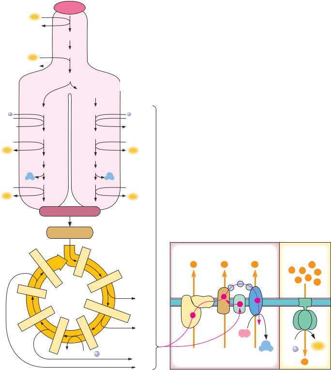
FIGURE 20.1 Pyruvate produced in glycolysis is oxidized in the tricarboxylic acid (TCA) cycle. Electrons liberated in this oxidation flow through the electron transport chain and drive the synthesis of ATP in oxidative phosphorylation. In eukaryotic cells, this overall process occurs in mitochondria.

P

NAD+

NADH

ADP

ATP

ADP

ATP

2-phosphoglycerate (2PG)

2-phosphoglycerate (2PG)

H2O

H2O

Phosphoenolpyruvate (PEP)

Phosphoenolpyruvate (PEP)

second

second

ATP-forming

ATP-forming

reaction

reaction

ADP

ATP

2 Pyruvate


Acetyl–CoA


Oxidative


Electron transport


phosphorylation


Citrate


Intermembrane space


Proton


Oxaloacetate


H+

H+

H+

gradient H+


H+ H+

H+


Isocitrate


H+ H+

+


Citric


H

Malate


e


acid


NADH


e


α


e


cycle

-


Ketoglutarate


arate


e


Fum

Succinate

Succinyl


NADH


O2


-


ADP +


CoA


H2O

ATP


P


GTP

GDP + P


[FADH2]

Mitochondrial matrix


H+


NADH


cycle). The electrons liberated by this oxidative process are then passed through an elaborate, membrane-associated electron transport pathway to O2, the final electron acceptor. Electron transfer is coupled to creation of a proton gradient across the membrane. Such a gradient represents an energized state, and the energy stored in this gradient is used to drive the synthesis of many equivalents of ATP.

640

Соседние файлы в предмете Химия