16.7 ● General Acid–Base Catalysis
Nearly all enzyme reactions involve some degree of acid or base catalysis. There are two types of acid–base catalysis: (1) specific acid–base catalysis, in which H or OH accelerates the reaction, and (2) general acid–base catalysis, in which an acid or base other than H or OH accelerates the reaction. For ordinary solution reactions, these two cases can be distinguished on the basis of simple experiments. As shown in Figure 16.11, in specific acid or base catalysis, the buffer concentration has no effect. In general acid or base catalysis, however, the buffer may donate or accept a proton in the transition state and thus affect the rate. By definition, general acid–base catalysis is catalysis in which a proton is transferred in the transition state. Consider the hydrolysis of p-nitro- phenylacetate with imidazole acting as a general base (Figure 16.12). Proton transfer apparently stabilizes the transition state here. The water has been made more nucleophilic without generation of a high concentration of OH or without the formation of unstable, high-energy species. General acid or general base catalysis may increase reaction rates 10to 100-fold. In an enzyme, ionizable groups on the protein provide the H transferred in the transition state. Clearly, an ionizable group will be most effective as a H transferring agent at or near its pKa. Because the pKa of the histidine side chain is near 7, histidine is often the most effective general acid or base. Descriptions of several cases of general acid–base catalysis in typical enzymes follow.
16.8 ● Metal Ion Catalysis
Many enzymes require metal ions for maximal activity. If the enzyme binds the metal very tightly or requires the metal ion to maintain its stable, native state, it is referred to as a metalloenzyme. Enzymes that bind metal ions more weakly, perhaps only during the catalytic cycle, are referred to as metal activated. One role for metals in metal-activated enzymes and metalloenzymes is to act as electrophilic catalysts, stabilizing the increased electron density or negative charge that can develop during reactions. Among the enzymes that function in this
16.8 |
● Metal Ion Catalysis |
511 |
(a) |
|
|
|
|
|
|
|
|
|
pH 8 |
|
|
|
|
|
|
|
|
|
|
|
|
|
kobs |
|
|
|
|
pH 7 |
|
|
|
|
|
|
|
|
|
|
pH 6 |
|
|
|
|
|
|
|
|
|
|
|
|
|
|
|
|
|
|
|
|
|
Buffer concentration
(b)
pH 8
pH 7
pH 6
Buffer concentration
FIGURE 16.11 ● Specific and general acid– base catalysis of simple reactions in solution may be distinguished by determining the dependence of observed reaction rate constants (kobs) on pH and buffer concentration.
(a) In specific acid–base catalysis, H or OH concentration affects the reaction rate, kobs is pH-dependent, but buffers (which accept or donate H /OH ) have no effect. (b) In general acid–base catalysis, in which an ionizable buffer may donate or accept a proton in the transition state, kobs is dependent on buffer concentration.
Reaction |
|
|
|
|
|
|
|
|
|
|
|
|
|
|
|
|
|
|
|
|
O |
|
|
|
|
|
|
|
|
O |
|
|
|
|
|
|
|
|
|
|
|
|
|
|
|
|
|
|
|
NO2 + H2O |
|
|
|
|
|
|
|
|
O– + HO |
|
NO2 + H+ |
|
CH3C |
|
O |
|
|
|
|
|
|
|
|
|
CH3C |
|
|
|
|
|
|
|
|
|
|
|
|
|
|
|
|
|
Mechanism |
|
|
|
|
|
|
|
|
|
|
|
|
|
|
|
|
|
|
|
|
|
|
|
|
|
|
O |
|
|
|
|
|
|
|
|
O– H+ |
|
|
O |
|
|
|
|
|
|
|
|
|
|
|
|
|
|
|
|
|
|
|
|
|
|
|
|
|
|
|
|
|
|
O– + HO |
NO2 + H+ |
|
|
|
|
CH3C |
|
O |
|
NO2 |
|
|
|
CH3 |
|
C O |
NO2 |
CH3C |
|
|
|
|
|
|
|
|
|
|
|
|
|
|
|
|
|
|
|
|
|
|
|
|
|
|
|
|
|
|
|
|
|
|
|
|
|
|
|
|
|
|
|
|
H |
|
O |
|
|
|
|
|
|
|
|
O |
|
|
|
|
|
|
|
|
|
|
|
|
|
|
|
H |
|
|
|
|
|
|
H |
|
|
|
|
|
|
|
|
HN N |
|
|
|
|
|
|
|
|
|
|
|
|
|
|
|
|
|
|
|
|
|
|
|
|
|
|
|
|
|
|
FIGURE 16.12 ● Catalysis of p-nitrophenylacetate hydrolysis by imidazole—an example of general base catalysis. Proton transfer to imidazole in the transition state facilitates hydroxyl attack on the substrate carbonyl carbon.
FIGURE 16.13
512 Chapter 16 ● Mechanisms of Enzyme Action
Zn2+
● Liver alcohol dehydrogenase catalyzes the transfer of a hydride ion (H: ) from NADH to acetaldehyde (CH3CHO), forming ethanol (CH3CH2OH). An active-site zinc ion stabilizes negative charge development on the oxygen atom of acetaldehyde, leading to an induced partial positive charge on the carbonyl C atom. Transfer of the negatively charged hydride ion to this carbon forms ethanol.
manner (Figure 16.13) is liver alcohol dehydrogenase. Another potential function of metal ions is to provide a powerful nucleophile at neutral pH. Coordination to a metal ion can increase the acidity of a nucleophile with an ionizable proton:
M2 NucH 34 M2 (NucH) 34 M2 (Nuc ) H
The reactivity of the coordinated, deprotonated nucleophile is typically intermediate between that of the un-ionized and ionized forms of the nucleophile. Carboxypeptidase (Chapter 5) contains an active site Zn2 , which facilitates deprotonation of a water molecule in this manner.
16.9 ● Proximity
Chemical reactions go faster when the reactants are in proximity, that is, near each other. In solution or in the gas phase, this means that increasing the concentrations of reacting molecules, which raises the number of collisions, causes higher rates of reaction. Enzymes, which have specific binding sites for particular reacting molecules, essentially take the reactants out of dilute solution and hold them close to each other. This proximity of reactants is said to raise the “effective” concentration over that of the substrates in solution, and leads to an increased reaction rate. In order to measure proximity effects in enzyme reactions, enzymologists have turned to model studies comparing intermolecular reaction rates with corresponding or similar intramolecular reaction rates. A typical case is the imidazole-catalyzed hydrolysis of p-nitrophenylacetate (Figure 16.14a). Under certain conditions the rate constant for this bimolecular reaction is 35 M 1 min 1. By comparison, the first-order rate constant for the analogous but intramolecular reaction shown in Figure 16.14b is 839 min 1. The ratio of these two rate constants
(839 min 1)/(35 M 1 min 1) 23.97 M
has the units of concentration and can be thought of as an effective concentration of imidazole in the intramolecular reaction. Put another way, a concentration of imidazole of 23.9 M would be required in the intermolecular reaction to make it proceed as fast as the intramolecular reaction.
There is more to this story, however. Enzymes not only bring substrates and catalytic groups close together, they orient them in a manner suitable for catalysis as well. Comparison of the rates of reaction of the molecules shown
|
O |
|
|
H2O |
O |
|
(a) |
|
|
|
|
|
N + H3C C O |
|
|
HN N + H3C C O– + HO |
NO2 + H+ |
HN |
|
NO2 |
|
|
|
|
kobs = 35 M –1min–1 |
|
|
(b) |
|
|
H2O |
HN N C O– + HO |
NO2 + H+ |
|
|
|
|
|
HN |
N C O |
NO2 |
|
|
|
O |
|
|
O |
|
|
|
|
|
|
kobs = 839 min–1 |
|
|
FIGURE 16.14 ● An example of proximity effects in catalysis. (a) The imidazolecatalyzed hydrolysis of p-nitrophenylacetate is slow, but the corresponding intramolecular reaction is 24-fold faster (assuming [imidazole] 1 M in [a]).
(Adapted from Milstien, S., and Cohen, L. A., 1972.
FIGURE 16.15
16.10 ● Typical Enzyme Mechanisms |
513 |
|
|
Reaction |
|
Rate const. |
Ratio |
|
|
|
|
|
|
(M –1sec–1) |
|
|
|
|
|
|
|
O |
|
HOOC |
|
|
|
|
|
|
|
OH |
|
H2O |
O |
|
|
|
|
|
|
|
|
|
|
|
|
|
5.9x10⁻⁶ |
|
|
|
|
|
|
|
O |
|
HOOC |
|
|
|
|
|
|
|
OH |
CH3 |
H |
|
O |
O |
CH3 |
|
H3C |
2 |
H3C |
|
|
|
|
|
|
|
|
|
|
1.5x10⁶ |
2.5x10¹¹ |
|
CH3 |
|
|
|
|
|
|
|
|
|
CH3 |
|
|
CH3 |
|
|
|
|
CH3 |
|
● Orientation effects in intramolecular reactions can be dramatic. Steric crowding by methyl groups provides a rate acceleration of 2.5 1011 for the lower reaction compared to the upper reaction.
Stereopopulation control I. Rate enhancements in the lactonization of o-hydroxyhydrocinnamic acid. Journal of the American Chemical Society 94:9158–9165.)
in Figure 16.15 makes it clear that the bulky methyl groups force an orientation on the alkyl carboxylate and the aromatic hydroxyl groups that makes them approximately 250 billion times more likely to react. Enzymes function similarly by placing catalytically functional groups (from the protein side chains or from another substrate) in the proper position for reaction.
Clearly, proximity and orientation play a role in enzyme catalysis, but there is a problem with each of the above comparisons. In both cases, it is impossible to separate true proximity and orientation effects from the effects of entropy loss when molecules are brought together (described the Section 16.4). The actual rate accelerations afforded by proximity and orientation effects in Figures 16.14 and 16.15, respectively, are much smaller than the values given in these figures. Simple theories based on probability and nearest-neighbor models, for example, predict that proximity effects may actually provide rate increases of only 5- to 10-fold. For any real case of enzymatic catalysis, it is nonetheless important to remember that proximity and orientation effects are significant.
16.10 ● Typical Enzyme Mechanisms
The balance of this chapter will be devoted to several classic and representative enzyme mechanisms. These particular cases are well understood, because the three-dimensional structures of the enzymes and the bound substrates are known at atomic resolution, and because great efforts have been devoted to kinetic and mechanistic studies. They are important because they represent reaction types that appear again and again in living systems, and because they demonstrate many of the catalytic principles cited above. Enzymes are the catalytic machines that sustain life, and what follows is an intimate look at the inner workings of the machinery.
FIGURE 16.16
514Chapter 16 ● Mechanisms of Enzyme Action
16.11● Serine Proteases
● Comparison of the amino acid sequences of chymotrypsinogen, trypsinogen, and elastase. Each circle represents one amino acid. Numbering is based on the sequence of chymotrypsinogen. Filled circles indicate residues that are identical in all three proteins. Disulfide bonds are indicated in yellow. The positions of the three catalytically
important active-site residues (His57, Asp102, and Ser195) are indicated.
Serine proteases are a class of proteolytic enzymes whose catalytic mechanism is based on an active-site serine residue. Serine proteases are one of the bestcharacterized families of enzymes. This family includes trypsin, chymotrypsin, elastase, thrombin, subtilisin, plasmin, tissue plasminogen activator, and other related enzymes. The first three of these are digestive enzymes and are synthesized in the pancreas and secreted into the digestive tract as inactive proenzymes, or zymogens. Within the digestive tract, the zymogen is converted into the active enzyme form by cleaving off a portion of the peptide chain. Thrombin is a crucial enzyme in the blood-clotting cascade, subtilisin is a bacterial protease, and plasmin breaks down the fibrin polymers of blood clots. Tissue plasminogen activator (TPA) specifically cleaves the proenzyme plasminogen, yielding plasmin. Owing to its ability to stimulate breakdown of blood clots, TPA can minimize the harmful consequences of a heart attack, if administered to a patient within 30 minutes of onset. Finally, although not itself a protease, acetylcholinesterase is a serine esterase and is related mechanistically to the serine proteases. It degrades the neurotransmitter acetylcholine in the synaptic cleft between neurons.
The Digestive Serine Proteases
Trypsin, chymotrypsin, and elastase all carry out the same reaction—the cleavage of a peptide chain—and although their structures and mechanisms are quite similar, they display very different specificities. Trypsin cleaves peptides
Chymotrypsinogen |
Trypsinogen |
C |
Elastase |
|
|
|
C |
|
|
|
C |
|
|
|
|
|
|
|
|
|
|
|
|
|
|
|
|
|
|
|
|
|
|
|
245 |
|
|
|
245 |
|
|
|
|
245 |
|
|
|
80 |
240 |
|
|
80 |
240 |
|
|
80 |
|
240 |
|
70 |
|
90 |
230 |
70 |
|
90 |
S |
|
70 |
90 |
|
230 |
|
|
|
|
|
230 |
|
|
|
|
60 |
|
|
|
60 |
|
|
|
|
60 |
|
|
220 |
|
|
100 |
220 S |
|
100 |
220 |
S |
|
S 100 |
|
S |
|
S |
|
S |
His |
|
His |
His |
|
|
|
Asp |
|
|
Asp |
|
|
Asp |
|
|
|
50 |
|
|
|
50 |
|
|
|
|
50 |
|
|
|
|
|
110 |
|
|
|
|
|
|
110 |
|
210 |
|
|
|
210 |
|
|
110 |
210 |
|
|
|
|
|
S |
|
|
|
S |
|
|
|
|
S |
|
|
|
|
|
|
|
|
|
|
40 |
|
|
|
|
40 |
|
|
|
40 |
|
|
|
|
|
|
|
|
|
|
S 200 |
|
|
S 200 |
|
|
120 |
S |
200 |
|
|
|
120 |
|
|
120 |
|
|
|
|
|
|
|
|
|
|
|
|
|
S |
|
|
|
|
|
|
|
|
|
|
|
|
|
|
Ser |
|
|
|
Ser |
|
|
|
|
Ser |
|
|
|
|
S |
|
|
|
S |
S |
30 |
|
|
190 |
S |
|
|
130 |
|
|
130 |
130 |
|
|
30 |
|
190 |
30 |
|
190 |
|
|
|
|
|
|
|
|
|
|
|
|
|
|
|
|
|
|
S |
|
|
|
S |
|
|
|
S |
|
|
|
|
140 |
S |
|
|
S |
|
S |
20 |
140 |
|
|
S |
20 |
|
20 |
|
140 |
180 |
|
180 |
|
|
180 |
|
|
|
|
|
|
|
|
|
|
|
|
|
|
|
|
|
|
|
|
|
|
|
|
|
|
|
N |
|
|
|
|
|
|
|
|
|
|
|
|
|
|
|
|
|
|
|
|
|
|
|
|
|
|
|
|
|
|
|
|
|
|
|
|
150 |
|
|
|
|
|
150 |
|
|
150 |
|
|
|
|
|
|
10 |
|
|
10 |
|
|
|
|
|
|
|
|
|
|
|
|
|
|
|
|
|
|
|
|
|
|
170 |
|
N |
|
170 |
|
|
|
|
170 |
S |
|
|
|
|
|
S |
|
|
|
|
|
|
|
S |
|
|
S |
160 |
|
|
160 |
|
|
|
|
160 |
|
|
|
|
|
|
S
N
FIGURE 16.21
Lag
516 Chapter 16 ● Mechanisms of Enzyme Action
|
|
|
|
|
|
H3C |
|
C |
|
O |
|
|
|
NO2 |
|
|
|
|
|
|
O |
|
H3C |
|
p-Nitrophenylacetate |
|
|
O CH2
H O
Acetylphenylalanine
methyl ester
FIGURE 16.20 ●
O CH2 CH3
|
|
|
|
|
|
C |
|
|
|
C |
C |
O |
|
CH3 |
|
C |
O |
|
CH3 |
|
|
|
|
H |
N |
H |
C |
O |
N |
H |
C |
|
|
|
|
|
|
|
|
|
|
|
|
|
|
|
H |
|
O |
|
H |
|
O |
|
Formylphenylalanine |
|
Benzoylalanine |
|
methyl ester |
|
methyl ester |
Artificial substrates used in studies of the mechanism of chymotrypsin.
– 2 |
|
|
–Nitrophenolate |
|
|
Steady-state |
pNOrelease |
|
release |
|
|
|
Acetateor |
phenolate |
|
p |
|
Acetate |
|
|
|
|
|
|
Burst |
|
|
|
|
|
|
|
|
Time
● Burst kinetics observed in the chymotrypsin reaction. A burst of nitrophenolate production is followed by a slower, steady-state release. After an initial lag period, acetate release is also observed. This kinetic pattern is consistent with rapid formation of an acyl-enzyme intermediate (and the burst of nitrophenolate). The slower, steady-state release of products corresponds to rate-limiting breakdown of the acyl-enzyme intermediate.
formylphenylalanine methyl ester and acetylphenylalanine methyl ester (Figure 16.20). p-Nitrophenylacetate is an especially useful model substrate, because the nitrophenolate product is easily observed, owing to its strong absorbance at 400 nm. When large amounts of chymotrypsin are used in kinetic studies with this substrate, a rapid initial burst of p-nitrophenolate is observed (in an amount approximately equal to the enzyme concentration), followed by a much slower, linear rate of nitrophenolate release (Figure 16.21). Observation of a burst, followed by slower, steady-state product release, is strong evidence for a multistep mechanism, with a fast first step and a slower second step.
In the chymotrypsin mechanism, the nitrophenylacetate combines with the enzyme to form an ES complex. This is followed by a rapid second step in which an acyl-enzyme intermediate is formed, with the acetyl group covalently bound to the very reactive Ser195. The nitrophenyl moiety is released as nitrophenolate (Figure 16.22), accounting for the burst of nitrophenolate product. Attack of a water molecule on the acyl-enzyme intermediate yields acetate as the second product in a subsequent, slower step. The enzyme is now free to bind another molecule of p-nitrophenylacetate, and the p-nitrophenolate product produced at this point corresponds to the slower, steady-state formation of product in the upper right portion of Figure 16.21. In this mechanism, the release of acetate is the rate-limiting step, and accounts for the observation of burst kinetics—the pattern shown in Figure 16.21.
Serine proteases like chymotrypsin are susceptible to inhibition by organic fluorophosphates, such as diisopropylfluorophosphate (DIFP, Figure 16.23). DIFP
NO2
+H+
NO2 |
|
|
|
– |
|
|
|
|
|
|
|
|
|
|
|
|
|
|
|
O |
|
|
|
|
|
H2O |
H+ |
|
|
|
|
|
|
|
|
|
E OH + |
|
|
|
|
E O C |
|
|
CH3 |
|
|
–O |
|
C |
|
CH3 |
|
|
|
|
|
|
|
|
|
|
|
|
|
|
|
Ser195 |
|
Fast step |
Ser195 |
|
|
|
|
Slow step |
|
|
|
|
|
|
|
|
|
|
|
|
|
|
|
|
|
|
|
|
|
|
|
|
|
|
|
O |
|
|
|
|
O |
O C |
|
CH3 |
|
|
|
|
|
|
|
|
|
|
|
|
|
|
|
|
|
|
|
|
|
|
|
|
|
|
|
|
|
|
|
O
FIGURE 16.22 ● Rapid formation of the acyl-enzyme intermediate is followed by slower product release.
16.11 ● Serine Proteases |
517 |
|
|
|
|
|
|
|
|
|
|
|
|
|
|
|
|
|
|
|
|
|
|
|
|
E |
|
|
|
|
|
|
|
E |
|
|
CH3 |
|
|
F |
|
|
|
CH3 |
F |
– |
|
|
CH3 |
|
|
O |
|
|
|
CH3 |
|
|
|
|
|
|
|
|
|
|
|
|
|
|
|
|
OH + H |
|
|
|
|
|
|
|
|
|
O |
|
|
|
|
|
|
|
|
|
|
|
|
|
|
|
|
O |
|
|
|
|
|
|
|
|
|
|
|
|
|
|
|
|
|
|
|
|
|
|
|
|
|
|
|
|
|
|
|
|
|
|
|
|
C |
|
O |
|
P |
|
|
C |
|
H |
|
|
H |
|
C |
|
O |
|
P |
|
|
C |
|
H |
|
|
|
|
|
|
|
|
|
|
|
|
|
|
|
|
|
|
|
|
|
|
|
|
|
|
|
|
|
|
|
|
|
|
|
|
|
|
|
|
|
|
|
|
|
|
|
|
|
|
|
|
CH3 |
|
|
O |
|
|
|
CH3 |
|
|
|
|
CH3 |
|
|
O |
|
|
|
CH3 |
|
Diisopropylfluorophosphate |
|
|
|
|
|
Diisopropylphosphoryl |
|
|
|
|
|
|
|
|
|
|
|
|
|
|
|
|
|
|
|
|
derivative of chymotrypsin |
FIGURE 16.23 ● Diisopropylfluorophosphate (DIFP) reacts with active-site serine residues of serine proteases (and esterases), causing permanent inactivation.
reacts rapidly with active-site serine residues, such as Ser195 of chymotrypsin and the other serine proteases (but not with any of the other serines in these proteins), to form a DIP-enzyme. This covalent enzyme-inhibitor complex is extremely stable, and chymotrypsin is thus permanently inactivated by DIFP.
The Serine Protease Mechanism in Detail: Events at the Active Site
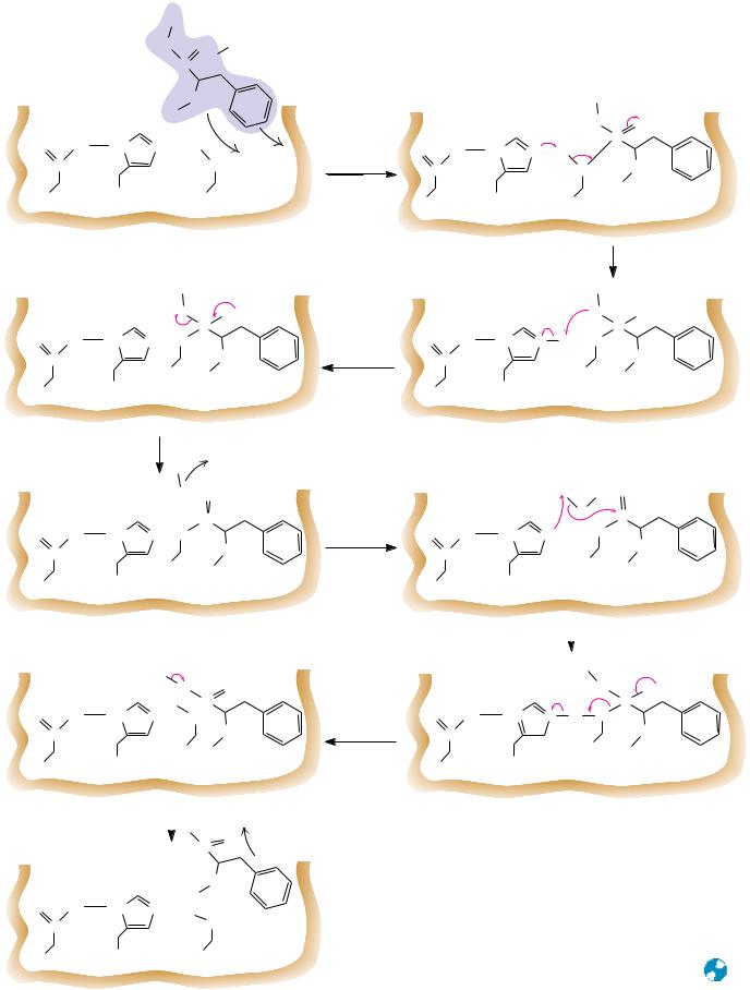
A likely mechanism for peptide hydrolysis is shown in Figure 16.24. As the backbone of the substrate peptide binds adjacent to the catalytic triad, the specific side chain fits into its pocket. Asp102 of the catalytic triad positions His57 and immobilizes it through a hydrogen bond as shown. In the first step of the reaction, His57 acts as a general base to withdraw a proton from Ser195, facilitating nucleophilic attack by Ser195 on the carbonyl carbon of the peptide bond to be cleaved. This is probably a concerted step, because proton transfer prior to Ser195 attack on the acyl carbon would leave a relatively unstable negative charge on the serine oxygen. In the next step, donation of a proton from His57 to the peptide’s amide nitrogen creates a protonated amine on the covalent, tetrahedral intermediate, facilitating the subsequent bond breaking and dissociation of the amine product. The negative charge on the peptide oxygen is unstable; the tetrahedral intermediate is short-lived and rapidly breaks down to expel the amine product. The acyl-enzyme intermediate that results is reasonably stable; it can even be isolated using substrate analogs for which further reaction cannot occur. With normal peptide substrates, however, subsequent nucleophilic attack at the carbonyl carbon by water generates another transient tetrahedral intermediate (Figure 16.24). His57 acts as a general base in this step, accepting a proton from the attacking water molecule. The subsequent collapse of the tetrahedral intermediate is assisted by proton donation from His57 to the serine oxygen in a concerted manner. Deprotonation of the carboxyl group and its departure from the active site complete the reaction as shown.
Until recently, the catalytic role of Asp102 in trypsin and the other serine proteases had been surmised on the basis of its proximity to His57 in structures obtained from X-ray diffraction studies, but it had never been demonstrated with certainty in physical or chemical studies. As can be seen in Figure 16.17, Asp102 is buried at the active site and is normally inaccessible to chemical modifying reagents. In 1987, however, Charles Craik, William Rutter, and their colleagues used site-directed mutagenesis (see Chapter 13) to prepare a mutant trypsin with an asparagine in place of Asp102. This mutant trypsin possessed a hydrolytic activity with ester substrates only 1/10,000 that of native trypsin, demonstrating that Asp102 is indeed essential for catalysis and that its ability to immobilize and orient His57 is crucial to the function of the catalytic triad.
|
|
R |
|
|
|
|
HN |
O |
Substrate |
|
|
|
C |
|
|
|
R' |
NH |
|
|
|
|
|
O |
O– |
HN N |
H |
Binding of |
C |
|
|
O |
substrate |
Asp 102 |
|
His 57 |
Ser 195 |
|
(a) |
|
|
|
(b) |
|
|
|
R |
|
|
|
|
|
Proton |
|
|
|
|
donation |
|
|
|
|
by His 57 |
(d) |
(c) |
|
C—N bond cleavage |
|
R |
|
Release of |
|
amino product |
R
Formation of covalent ES complex
R
(e) |
(f) |
|
Nucleophilic |
|
|
attack by water |
|
|
|
|
|
|
H |
Collapse of tetrahedral intermediate
(h) |
Carboxyl product release |
(g) |
|
– |
|
|
|
|
|
|
|
O |
O |
|
|
|
|
|
|
C |
|
|
|
|
|
(i) |
FIGURE 16.24 ● A detailed mechanism for the chymotrypsin reaction. |
518
16.12 ● The Aspartic Proteases |
519 |
C R I T I C A L D E V E L O P M E N T S I N B I O C H E M I S T R Y
Transition-State Stabilization in the Serine Proteases
X-ray crystallographic studies of serine protease complexes with transition-state analogs have shown how chymotrypsin stabilizes the tetrahedral oxyanion transition states (structures (c) and (g)
in Figure 16.24) of the protease reaction. The amide nitrogens of Ser195 and Gly193 form an “oxyanion hole” in which the sub-
strate carbonyl oxygen is hydrogen-bonded to the amide N-H groups.
Formation of the tetrahedral transition state increases the interaction of the carbonyl oxygen with the amide N-H groups in two ways. Conversion of the carbonyl double bond to the longer tetrahedral single bond brings the oxygen atom closer to the amide hydrogens. Also, the hydrogen bonds between the charged oxygen and the amide hydrogens are significantly stronger than the hydrogen bonds with the uncharged carbonyl oxygen.
Transition-state stabilization in chymotrypsin also involves the side chains of the substrate. The side chain of the departing amine product forms stronger interactions with the enzyme upon formation of the tetrahedral intermediate. When the tetrahedral intermediate breaks down (Figure 16.24d and e), steric repulsion between the product amine group and the carbonyl group of the acyl-enzyme intermediate leads to departure of the amine product.
The “oxyanion hole” of chymotrypsin stabilizes the tetrahedral oxyanion transition states of the mechanism in Figure 16.24.
The oxyanion hole
Gly193
 Ser195
The oxyanion hole
Gly193
....
–
 ....
....
 Ser195
16.12 ● The Aspartic Proteases
Mammals, fungi, and higher plants produce a family of proteolytic enzymes known as aspartic proteases. These enzymes are active at acidic (or sometimes neutral) pH, and each possesses two aspartic acid residues at the active site. Aspartic proteases carry out a variety of functions (Table 16.3), including digestion (pepsin and chymosin), lysosomal protein degradation (cathepsin D and E), and regulation of blood pressure (renin is an aspartic protease involved in the production of angiotensin, a hormone that stimulates smooth muscle contraction and reduces excretion of salts and fluid). The aspartic proteases display a variety of substrate specificities, but normally they are most active in the cleavage of peptide bonds between two hydrophobic amino acid residues. The preferred substrates of pepsin, for example, contain aromatic residues on both sides of the peptide bond to be cleaved.
Most aspartic proteases are composed of 323 to 340 amino acid residues, with molecular weights near 35,000. Aspartic protease polypeptides consist of
520 Chapter 16 ● Mechanisms of Enzyme Action
Table 16.3
Some Representative Aspartic Proteases
Name |
Source |
Function |
|
|
|
Pepsin* |
Animal stomach |
Digestion of dietary protein |
Chymosin† |
Animal stomach |
Digestion of dietary protein |
Cathepsin D |
Spleen, liver, and many |
Lysosomal digestion of |
|
other animal tissues |
proteins |
Renin‡ |
Kidney |
Conversion of angiotensinogen |
|
|
to angiotensin I; regulation |
|
|
of blood pressure |
HIV-protease§ |
AIDS virus |
Processing of AIDS virus |
|
|
proteins |
*The second enzyme to be crystallized (by John Northrup in 1930). Even more than urease before it, pepsin study by Northrup established that enzyme activity comes from proteins. †Also known as rennin, it is the major pepsinlike enzyme in gastric juice of fetal and newborn animals.
‡A drop in blood pressure causes release of renin from the kidneys, which converts more angiotensinogen to angiotensin.
§A dimer of identical monomers, homologous to pepsin.
(a)
(b)
FIGURE 16.25 ● Structures of (a) HIV-1 protease, a dimer, and (b) pepsin (a monomer). Pepsin’s N-terminal half is shown in red; C-ter- minal half is shown in blue.
two homologous domains that fold to produce a tertiary structure composed of two similar lobes, with approximate twofold symmetry (Figure 16.25). Each of these lobes or domains consists of two -sheets and two short -helices. The two domains are bridged and connected by a six-stranded, antiparallel -sheet. The active site is a deep and extended cleft, formed by the two juxtaposed domains and large enough to accommodate about seven amino acid residues. The two catalytic aspartate residues, residues 32 and 215 in porcine pepsin, for example, are located deep in the center of the active site cleft. The N-termi- nal domain forms a “flap” that extends over the active site, which may help to immobilize the substrate in the active site.
On the basis, in part, of comparisons with chymotrypsin, trypsin, and the other serine proteases, it was hypothesized that aspartic proteases might function by formation of covalent enzyme-substrate intermediates involving the active-site aspartate residues. Two possibilities were proposed: an acyl-enzyme intermediate involving an acid anhydride bond and an amino-enzyme intermediate involving an amide (peptide) bond (Figure 16.26). All attempts to trap or isolate a covalent intermediate failed, and a mechanism (see following paragraph) favoring noncovalent enzyme-substrate intermediates and general acid–general base catalysis is now favored for aspartic proteases.
The Mechanism of Action of Aspartic Proteases
A crucial datum supporting the general acid–general base model is the pH dependence of protease activity (see Critical Developments in Biochemistry:
The pH Dependence of Aspartic Proteases and HIV-1 Protease, page 525). Enzymologists hypothesize that the aspartate carboxyl groups function alternately as general acid and general base. This model requires that one of the aspartate carboxyls be protonated and one be deprotonated when substrate binds. X-ray diffraction data on aspartic proteases show that the active-site structure in the vicinity of the two aspartates is highly symmetric. The two aspartates appear to act as a “catalytic dyad” (analogous to the catalytic triad of the serine proteases). The dyad proton may thus be covalently bound to either of