Добавил:
Опубликованный материал нарушает ваши авторские права? Сообщите нам.
Вуз: Предмет: Файл:

Garrett R.H., Grisham C.M. - Biochemistry (1999)(2nd ed.)(en)

.pdf
Скачиваний:
183
Добавлен:
15.08.2013
Размер:
24.81 Mб
Скачать

Further Reading

531

5. Active -chymotrypsin is produced from chymotrypsinogen, an inactive precursor, as shown in the color figure on page 530.

The first intermediate— -chymotrypsin—displays chymotrypsin activity. Suggest proteolytic enzymes that might carry out these cleavage reactions effectively.

6. Based on the reaction scheme shown below, derive an expression for ke/ku, the ratio of the rate constants for the catalyzed and uncatalyzed reactions, respectively, in terms of the free energies of activation for the catalyzed ( Ge) and the uncatalyzed ( Gu) reactions.


K u


X


k u


E

S


P


K S


E


K e


EX

ke


CH3H ECH3


CH3H ECH3


ES


EP


OH


CH3


OH

CH


CH


A


A

A


A


A

CH2OCONHOCHOCONHOCHOCONHOCHOCHOCH2OCONHOCHOCONHOCHOCHOCH2OCOOH


A


A


A


CH

HCH3

CH2


CH2


CH3E

A


A


ECHH


ECHH


CH3

CH3


CH3

CH3

Iva

Val

Val

Sta


Ala


Sta


Pepstatin

FURTHER READING

General

Cannon, W. R., Singleton, S. F., and Benkovic, S. J., 1997. A perspective on biological catalysis. Nature Structural Biology 3:821–833.

Eigen, M., 1964. Proton transfer, acid–base catalysis, and enzymatic hydrolysis. Angewandte Chemie, Int. Ed. 3:1–72.

Fersht, A., 1985. Enzyme Structure and Mechanism, 2nd ed. New York: W. H. Freeman and Company.

Gerlt, J. A., Kreevoy, M. M., Cleland, W. W., and Frey, P. A., 1997. Understanding enzymic catalysis: The importance of short, strong hydrogen bonds. Chemistry and Biology 4:259–267.

Jencks, W. P., 1969. Catalysis in Chemistry and Enzymology. New York: McGrawHill.

Jencks, W., 1997. From chemistry to biochemistry to catalysis to movement.

Annual Review of Biochemistry 66:1–18.

Page, M. I., and Williams, A., eds., 1987. Enzyme Mechanisms. London, England: Royal Society of London.

Radzicka, A., and Wolfenden, R., 1995. A proficient enzyme. Science 267:90–93.

Simopoulos, T. T., and Jencks, W. P., 1994. Alkaline phosphatase is an almost perfect enzyme. Biochemistry 33:10375–10380.

Smithrud, D. B., and Benkovic, S. J., 1997. The state of antibody catalysis.

Current Opinions in Biotechnology 8:459–466.

Walsh, C., 1979. Enzymatic Reaction Mechanisms. San Francisco: W. H. Freeman and Company.

Transition-State Stabilization and Transition-State Analogs

Bearne, S. L., and Wolfenden, R., 1997. Mandelate racemase in pieces: Effective concentrations of enzyme functional groups in the transition state. Biochemistry 36:1646–1656.

Kraut, J., 1988. How do enzymes work? Science 242:533–540.

Kreevoy, M., and Truhlar, D. G., 1986. Transition-state theory. Chapter 1 in Investigations of Rates and Mechanisms of Reactions, Vol. 6, Part 1, edited by C. F. Bernasconi. New York: John Wiley & Sons.

Lolis, E., and Petsko, G., 1990. Transition-state analogues in protein crystallography: Probes of the structural source of enzyme catalysis. Annual Review of Biochemistry 59:597–630.

Radzicka, A., and Wolfenden, R., 1995. Transition state and multisubstrate analog inhibitors. Methods in Enzymology 249:284–312.

Wolfenden, R., 1972. Analogue approaches to the structure of the transition state in enzyme reactions. Accounts of Chemical Research 5:10–18.

Wolfenden, R., and Frick, L., 1987. Transition state affinity and the design of enzyme inhibitors. Chapter 7 in Enzyme Mechanisms, edited by M. I. Page and A. Williams. London, England: Royal Society of London.

Wolfenden, R., and Kati, W. M., 1991. Testing the limits of protein-ligand binding discrimination with transition-state analogue inhibitors. Accounts of Chemical Research 24:209–215.

Serine Pro t e a s e s

Cassidy, C. S., Lin, J., and Frey, P. A., 1997. A new concept for the mechanism of action of chymotrypsin: The role of the low-barrier hydrogen bond.

Biochemistry 36:4576–4584.

Craik, C. S., et al., 1987. The catalytic role of the active site aspartic acid in serine proteases. Science 237:909–919.

Lesk, A. M., and Fordham, W. D., 1996. Conservation and variability in the structures of serine proteinases of the chymotrypsin family. Journal of Molecular Biology 258:501–537.

Plotnick, M. I., Mayne, L., Schechter, N. M., and Rubin, H., 1996. Distortion of the active site of chymotrypsin complexed with a serpin. Biochemistry 35:7586–7590.

Renatus, M., Engh, R. A., Stubbs, M. T., et al., 1997. Lysine-156 promotes the anomalous proenzyme activity of tPA: X-ray crystal structure of singlechain human tPA. EMBO Journal 16:4797–4805.

532 Chapter 16 Mechanisms of Enzyme Action

Sprang, S., et al., 1987. The three-dimensional structure of Asn102 mutant of trypsin: Role of Asp102 in serine protease catalysis. Science 237:905–909.

Stavridi, E. S., O’Malley, K., Lukacs, C. M., et al., 1996. Structural change in -chymotrypsin induced by complexation with 1-antitrypsin as seen by enhanced sensitivity to proteolysis. Biochemistry 35:10608–10615.

Steitz, T., and Shulman, R., 1982. Crystallographic and NMR studies of the serine proteases. Annual Review of Biophysics and Bioengineering 11:419–444.

Tsukada, H., and Blow, D., 1985. Structure of -chymotrypsin refined at 1.68 Å resolution. Journal of Molecular Biology 184:703–711.

Aspartic Pro t e a s e s

Fruton, J., 1976. The mechanism of the catalytic action of pepsin and related acid proteinases. Advances in Enzymology 44:1–36.

Oldziej, S., and Ciarkowski, J., 1996. Mechanism of action of aspartic proteinases: Application of transition-state analogue theory. Journal of Computer-Aided Molecular Design 10:583–588.

Polgar, L., 1987. The mechanism of action of aspartic proteases involves “push-pull” catalysis. FEBS Letters 219:1–4.

HIV-1 Protease

Bardi, J. S., Luque, I., and Friere, E., 1997. Structure-based thermodynamic analysis of HIV-1 protease inhibitors. Biochemistry 36:6588–6596.

Beaulieu, P. L., Wernic, D., Abraham, A., et al., 1997. Potent HIV protease inhibitors containing a novel (hydroxyethyl)amide isostere. Journal of Medicinal Chemistry 40:2164–2176.

Blundell, T., et al., 1990. The 3-D structure of HIV-1 proteinase and the design of antiviral agents for the treatment of AIDS. Trends in Biochemical Sciences 15:425–430.

Carr, A., and Cooper, D. A., 1996. HIV protease inhibitors. AIDS 10:S151–S157.

Chen, Z., Li, Y., Chen, E., et al., 1994. Crystal structure at 1.9-Å resolution of human immunodeficiency virus (HIV) II protease complexed with L-735,524, an orally bioavailable inhibitor of the HIV proteases. Journal of Biological Chemistry 269:26344–26348.

Chen, Z., Li, Y., Schock, H. B., et al., 1995. Three-dimensional structure of a mutant HIV-1 protease displaying cross-resistance to all protease inhibitors in clinical trials. Journal of Biological Chemistry 270:21433–21436.

Hyland, L., et al., 1991. Human immunodeficiency virus-1 protease 1: Initial velocity studies and kinetic characterization of reaction intermediates by 18O isotope exchange. Biochemistry 30:8441–8453.

Hyland, L., Tomaszek, T., and Meek, T., 1991. Human immunodeficiency virus-1 protease 2: Use of pH rate studies and solvent isotope effects to elucidate details of chemical mechanism. Biochemistry 30:8454–8463.

Korant, B., Lu, Z., Strack, P., and Rizzo, C., 1996. HIV protease mutations leading to reduced inhibitor susceptibility. Advances in Experimental Medicine and Biology 389:241–245.

Rose, R. B., Craik, C. S., Douglas, N. L., and Stroud, R. M., 1996. Threedimensional structures of HIV-1 and SIV protease product complexes.

Biochemistry 35:12933–12944.

Vondrasek, J., and Wlodawer, A., 1997. Database of HIV proteinase structures. Trends in Biochemical Sciences 22:183.

Wang, Y. X., Freedberg, D. I., Yamazaki, T., et al., 1997. Solution NMR evidence that the HIV-1 protease catalytic aspartyl groups have different ionization states in the complex formed with the asymmetric drug KNI-272.

Biochemistry 35:9945–9950.

Ly s o z y m e

Chipman, D., and Sharon, N., 1969. Mechanism of lysozyme action. Science 165:454–465.

Ford, L., et al., 1974. Crystal structure of a lysozyme-tetrasaccharide lactone complex. Journal of Molecular Biology 88:349–371.

Kirby, A., 1987. Mechanism and stereoelectronic effects in the lysozyme reaction. CRC Critical Reviews in Biochemistry 22:283–315.

Phillips, D., 1966. The three-dimensional structure of an enzyme molecule. Scientific American 215:75–80.

Chapter 17

Molecular Motors

Michaelangelo’s “David” epitomizes the musculature of the human form. (The Firenze Academia/photo by Stephanie

Colasanti/Corbis)

Movement is an intrinsic property associated with all living things. Within cells, molecules undergo coordinated and organized movements, and cells themselves may move across a surface. At the tissue level, muscle contraction allows higher organisms to carry out and control crucial internal functions, such as peristalsis in the gut and the beating of the heart. Muscle contraction also enables the organism to carry out organized and sophisticated movements, such as walking, running, flying, and swimming.

17.1 Molecular Motors

Motor proteins, also known as molecular motors, use chemical energy (ATP) to orchestrate all these movements, transforming ATP energy into the mechanical energy of motion. In all cases, ATP hydrolysis is presumed to drive and

Under the spreading chestnut tree The village smithy stands;

The smith a mighty man is he With large and sinewy hands.

And the muscles of his brawny arms Are strong as iron bands.

HENRY WADSWORTH LONGFELLOW, “The Village

Blacksmith”

OUTLINE

17.1 Molecular Motors

17.2 Microtubules and Their Motors

17.3 Skeletal Muscle Myosin and Muscle

Contraction

17.4 A Proton Gradient Drives the Rotation of Bacterial Flagella

533

FIGURE 17.1

534 Chapter 17 Molecular Motors

(a)

(b)

(c)

(d)

control protein conformational changes that result in sliding or walking movements of one molecule relative to another. To carry out directed movements, molecular motors must be able to associate and dissociate reversibly with a polymeric protein array, a surface or substructure in the cell. ATP hydrolysis drives the process by which the motor protein ratchets along the protein array or surface. As fundamental and straightforward as all this sounds, elucidation of these basically simple processes has been extremely challenging for biochemists, involving the application of many sophisticated chemical and physical methods in many different laboratories. This chapter describes the structures and chemical functions of molecular motor proteins and some of the experiments by which we have come to understand them.

17.2 Microtubules and Their Motors

One of the simplest self-assembling structures found in biological systems is the microtubule, one of the fundamental components of the eukaryotic cytoskeleton and the primary structural element of cilia and flagella (Figure 17.1). Microtubules are hollow, cylindrical structures, approximately 30 nm in diameter, formed from tubulin, a dimeric protein composed of two similar 55-kD subunits known as -tubulin and -tubulin. Eva Nogales, Sharon Wolf, and Kenneth Downing have determined the structure of the bovine tubulin dimer to 3.7 Å resolution (Figure 17.2a). Tubulin dimers polymerize as shown in Figure 17.2b to form microtubules, which are essentially helical structures, with 13 tubulin monomer “residues” per turn. Microtubules grown in vitro are dynamic structures that are constantly being assembled and disassembled.

(f)

(e)

Micrographs and electron micrographs of cytoskeletal elements, cilia, and flagella: (a) microtubules, (b) rat sperm tail microtubules (cross-section),

(c) Stylonychia, a ciliated protozoan (undergoing division), (d) cytoskeleton of a eukaryotic cell, (e) Pseudomonas fluorescens (aerobic soil bacterium), showing flagella, (f) nasal cilia.

(a, K. G. Murti/Visuals Unlimited; b, David Phillips/Visuals Unlimited; c, Eric Grave/Phototake; d, Fawcett and Heuser/Photo Researchers, Inc.; e, Dr. Tony Brain/Custom Medical Stock; f, Veronika Burmeister, Visuals Unlimited)

FIGURE 17.2

24 nm

ubulin

Tubulin heterodimer

β

α

(8 nm)


ubulin

17.2 Microtubules and Their Motors

535

(a) The structure of the tubulin heterodimer. (b) Microtubules may be viewed as consisting of 13 parallel, staggered protofilaments of alternating -tubulin and-tubulin subunits. The sequences of the andsubunits of tubulin are homologous, and thetubulin dimers are quite stable if Ca2 is present. The dimer is dissociated only by strong denaturing agents.

Protofilament

(a)

(b)


Because all tubulin dimers in a microtubule are oriented similarly, microtubules are polar structures. The end of the microtubule at which growth occurs is the plus end, and the other is the minus end. Microtubules in vitro carry out a GTPdependent process called treadmilling, in which tubulin dimers are added to the plus end at about the same rate at which dimers are removed from the minus end (Figure 17.3).

Dimers on

β

α

Plus end

(Growing end)

Microtubules Are Constituents of the Cytoskeleton

Although composed only of 55-kD tubulin subunits, microtubules can grow sufficiently large to span a eukaryotic cell or to form large structures such as cilia and flagella. Inside cells, networks of microtubules play many functions, including formation of the mitotic spindle that segregates chromosomes during cell division, the movement of organelles and various vesicular structures through the cell, and the variation and maintenance of cell shape. Microtubules are, in fact, a significant part of the cytoskeleton, a sort of intracellular scaffold formed of microtubules, intermediate filaments, and microfilaments (Figure 17.4). In most cells, microtubules are oriented with their minus ends toward the centrosome and their plus ends toward the cell periphery. This consistent orientation is important for mechanisms of intracellular transport.

Microtubules Are the Fundamental Structural

Units of Cilia and Flagella

As already noted, microtubules are also the fundamental building blocks of cilia and flagella. Cilia are short, cylindrical, hairlike projections on the surfaces of the cells of many animals and lower plants. The beating motion of cilia functions either to move cells from place to place or to facilitate the movement of extracellular fluid over the cell surface. Flagella are much longer structures found singly or a few at a time on certain cells (such as sperm cells). They pro-

Minus end

Dimers off

FIGURE 17.3 A model of the GTP-depen- dent treadmilling process. Both - and-tubulin possess two different binding sites for GTP. The polymerization of tubulin to form microtubules is driven by GTP hydrolysis in a process that is only beginning to be understood in detail.

FIGURE 17.5
(a, b, M. Schliwa/Visuals Unlimited)
FIGURE 17.4

536 Chapter 17 Molecular Motors

(a)


8

9 10


Protofilaments


7

9


9


6

B-


10

8


10

8

tubule 11


7

11


7

5


12


A-


6

12


6

4


13

tubule

13


5


5

3


1


1


2

1

2


4


2


4


3


3

Inner dynein arm

Outer dynein arm

Radial

Nexin

spoke

Spoke

Central singlet

head


microtubules with

Plasma

connecting bridge

membrane


The structure of an axoneme. Note the manner in which two microtubules are joined in the nine outer pairs. The smaller-diameter tubule of each pair, which is a true cylinder, is called the A-tubule and is joined to the center sheath of the axoneme by a spoke structure. Each outer pair of tubules is joined to adjacent pairs by a nexin bridge. The A-tubule of each outer pair possesses an outer dynein arm and an inner dynein arm. The larger-diameter tubule is known as the B-tubule.

(b)

Intermediate filaments have diameters of approximately 7 to 12 nm, whereas microfilaments, which are made from actin, have diameters of approximately 7 nm. The intermediate filaments appear to play only a structural role (maintaining cell shape), but the microfilaments and microtubules play more dynamic roles. Microfilaments are involved in cell motility, whereas microtubules act as long filamentous tracks, along which cellular components may be rapidly transported by specific mechanisms. (a) Cytoskeleton, double-labeled with actin in red and tubulin in green. (b) Cytoskeletal elements in a eukaryotic cell, including microtubules (thickest strands), intermediate filaments, and actin microfilaments (smallest strands).

pel cells through fluids. Cilia and flagella share a common design (Figure 17.5). The axoneme is a complex bundle of microtubule fibers that includes two central, separated microtubules surrounded by nine pairs of joined microtubules. The axoneme is surrounded by a plasma membrane that is continuous with the plasma membrane of the cell. Removal of the plasma membrane by detergent and subsequent treatment of the exposed axonemes with high concentrations of salt releases the dynein molecules (Figure 17.6), which form the dynein arms.

The Mechanism of Ciliary Motion

The motion of cilia results from the ATP-driven sliding or walking of dyneins along one microtubule while they remain firmly attached to an adjacent microtubule. The flexible stems of the dyneins remain permanently attached to A-tubules (Figure 17.6). However, the projections on the globular heads form transient attachments to adjacent B-tubules. Binding of ATP to the dynein heavy chain causes dissociation of the projections from the B-tubules. These projections then reattach to the B-tubules at a position closer to the minus end. Repetition of this process causes the sliding of A-tubules relative to B-tubules. The cross-linked structure of the axoneme dictates that this sliding motion will occur in an asymmetric fashion, resulting in a bending motion of the axoneme, as shown in Figure 17.7

Microtubules Also Mediate Intracellular

Motion of Organelles and Vesicles

The ability of dyneins to effect mechano-chemical coupling—i.e., motion coupled with a chemical reaction—is also vitally important inside eukaryotic cells, which, as already noted, contain microtubule networks as part of the cytoskeleton. The mechanisms of intracellular, microtubule-based transport of organelles and vesicles were first elucidated in studies of axons, the long pro-

FIGURE 17.7

17.2 Microtubules and Their Motors

537

(a)

(b)

Intermediateand


A-tubule

low-molecular- B-tubule


weight chains

Fixed attachment to A-tubule

ATP

cycling

Outer dynein arm heavy
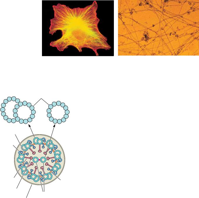
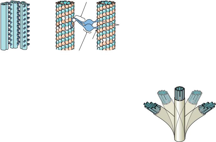
chain

Transient attachments to B-tubule

FIGURE 17.6 (a) Diagram showing dynein interactions between adjacent microtubule pairs. (b) Detailed views of dynein crosslinks between the A-tubule of one microtubule pair and the B-tubule of a neighboring pair. (The B-tubule of the first pair and the A-tubule of the neighboring pair are omitted for clarity.) Isolated axonemal dyneins, which possess ATPase activity, consist of two or three “heavy chains” with molecular masses of 400 to 500 kD, referred to as and (and when present), as well as several chains with intermediate (40 to 120 kD) and low (15 to 25 kD) molecular masses. Each outerarm heavy chain consists of a globular domain with a flexible stem on one end and a shorter projection extending at an angle with respect to the flexible stem. In a dynein arm, the flexible stems of several heavy chains are joined in a common base, where the intermediateand low-molecular-weight proteins are located.

jections of neurons that extend great distances away from the body of the cell. In these cells, it was found that subcellular organelles and vesicles could travel at surprisingly fast rates—as great as 2 to 5 m/sec—in either direction. Unraveling the molecular mechanism for this rapid transport turned out to be a challenging biochemical problem. The early evidence that these movements occur by association with specialized proteins on the microtubules was met with some resistance, for two reasons. First, the notion that a network of microtubules could mediate transport was novel and, like all novel ideas, difficult to accept. Second, many early attempts to isolate dyneins from neural tissue were unsuccessful, and the dynein-like proteins that were first isolated from cytosolic fractions were thought to represent contaminations from axoneme structures. However, things changed dramatically in 1985 with a report by Michael Sheetz and his coworkers of a new ATP-driven, force-generating protein, different from myosin and dynein, which they called kinesin. Then, in 1987, Richard McIntosh and Mary Porter described the isolation of cytosolic dynein proteins from Caenorhabditis elegans, a nematode worm that never makes motile axonemes at any stage of its life cycle. Kinesins have now been found in many eukaryotic cell types, and similar cytosolic dyneins have been found in fruit flies, amoebae, and slime molds; in vertebrate brain and testes; and in HeLa cells (a unique human tumor cell line).

Dyneins Move Organelles in a Plus-to-Minus Direction; Kinesins, in a Minus-to-Plus Direction

The cytosolic dyneins bear many similarities to axonemal dynein. The protein isolated from C. elegans includes a “heavy chain” with a molecular mass of approximately 400 kD, as well as smaller peptides with molecular mass ranging from 53 kD to 74 kD. The protein possesses a microtubule-activated ATPase

A mechanism for ciliary motion. The sliding motion of dyneins along one microtubule while attached to an adjacent microtubule results in a bending motion of the axoneme.

538 Chapter 17 Molecular Motors

C R I T I C A L D E V E L O P M E N T S I N B I O C H E M I S T R Y

Effectors of Microtubule Polymerization as Therapeutic Agents

Microtubules in eukaryotic cells are important for the maintenance and modulation of cell shape and the disposition of intracellular elements during the growth cycle and mitosis. It may thus come as no surprise that the inhibition of microtubule polymerization can block many normal cellular processes. The alkaloid colchicine (see figure), a constituent of the swollen, underground stems of the autumn crocus (Colchicum autumnale) and meadow saffron, inhibits the polymerization of tubulin into microtubules. This effect blocks the mitotic cycle of plants and animals. Colchicine also inhibits cell motility and intracellular transport of vesicles and organelles (which in turn blocks secretory processes of cells). Colchicine has been used for hundreds of years to alleviate some of the acute pain of gout and rheumatism. In gout, white cell lysosomes surround and engulf small crystals of uric acid. The subsequent rupture of the lysosomes and the attendant lysis of the white cells initiate an inflammatory response that causes intense pain. The mechanism of pain alleviation by colchicine is not known for certain, but appears to involve inhibition of white cell movement in tissues. Interestingly, colchicine’s ability to inhibit mitosis has given it an important role in the commercial development of new varieties of agricultural and ornamental plants. When mitosis is blocked by colchicine, the treated cells may be left with an extra set of chromosomes. Plants with extra sets of chromosomes are typically larger and more vigorous than normal plants. Flowers developed in this way may grow with double the normal number of petals, and fruits may produce much larger amounts of sugar.

Another class of alkaloids, the vinca alkaloids from Vinca rosea, the Madagascar periwinkle, can also bind to tubulin and inhibit microtubule polymerization. Vinblastine and vincristine are used as potent agents for cancer chemotherapy, owing to their ability to inhibit the growth of fast-growing tumor cells. For reasons that are not well understood, colchicine is not an effective chemotherapeutic agent, though it appears to act similarly to the vinca alkaloids in inhibiting tubulin polymerization.

A new antitumor drug, taxol, has been isolated from the bark of Taxus brevifolia, the Pacific yew tree. Like vinblastine and colchicine, taxol inhibits cell replication by acting on microtubules. Unlike these other antimitotic drugs, however, taxol stimulates microtubule polymerization and stabilizes microtubules. The remarkable success of taxol in treatment of breast and ovarian cancers stimulated research efforts to synthesize taxol directly and to identify new antimitotic agents that, like taxol, stimulate microtubule polymerization.


N


CH2

CH3


OH


N


N


H3CO C


CH2

CH3


O


N


O C

CH3


H3CO


HO


O


R


C

OCH3


Vinblastine: R = CH3


O


Vincristine: R = CHO


H3C

O


O


H3C

O


NH

C

CH3


CH3

O


O


Colchicine


O

CH3


O


O


H3C

O


O OH


NH

O


O

H


O


H

CH3


OH


OH

O


O

O


Taxol

O


The structures of vinblastine, vincristine, colchicine, and taxol.

activity, and, when anchored to a glass surface in vitro, these proteins, in the presence of ATP, can bind microtubules and move them through the solution. In the cell, cytosolic dyneins specifically move organelles and vesicles from the plus end of a microtubule to the minus end. Thus, as shown in Figure 17.8, dyneins move vesicles and organelles from the cell periphery toward the centrosome (or, in an axon, from the synaptic termini toward the cell body). The

17.2 Microtubules and Their Motors

539

(a)

Rough endoplasmic reticulum

Cell body


Multivesicular

Lysosome

body


Microtubule

Nucleus

Vesicles

Synaptic


Golgi apparatus

terminal


Mitochondrion


(b)

Kinesin


Organelle

Vesicle


Minus end

Plus end

FIGURE 17.8 (a) Rapid axonal transport

along microtubules permits the exchange of


material between the synaptic terminal and the


body of the nerve cell. (b) Vesicles, multivesicu-


lar bodies, and mitochondria are carried


through the axon by this mechanism.


(Adapted from a drawing by Ronald Vale)

kinesins, on the other hand, assist the movement of organelles and vesicles from the minus end to the plus end of microtubules, resulting in outward movement of organelles and vesicles. Kinesin is similar to cytosolic dyneins but smaller in size (360 kD), and contains subunits of 110 kD and 65 to 70 kD. Its length is 100 nm. Like dyneins, kinesins possess ATPase activity in their globular heads, and it is the free energy of ATP hydrolysis that drives the movement of vesicles along the microtubules.

The N-terminal domain of the kinesin heavy chain (38 kD, approximately 340 residues) contains the ATPand microtubule-binding sites and is the domain responsible for movement. Electron microscopy and image analysis of tubulin–kinesin complexes reveals (Figure 17.9) that the kinesin head domain is compact and primarily contacts a single tubulin subunit on a microtubule surface, inducing a conformational change in the tubulin subunit. Optical trapping experiments (see page 554) demonstrate that kinesin heads move in 8-nm (80-Å) steps along the long axis of a microtubule. Kenneth Johnson and his coworkers have shown that the ability of a single kinesin tetramer to move unidirectionally for long distances on a microtubule depends upon cooperative interactions between the two mechanochemical head domains of the protein.

(Taken from Kikkawa et al., 1995. Nature

540 Chapter 17 Molecular Motors

FIGURE 17.9 The structure of the tubulin–kinesin complex, as revealed by image analysis of cryoelectron microscopy data. (a) The computed, three-dimensional map of a microtubule, (b) the kinesin globular head domain–microtubule complex, (c) a contour plot of a horizontal section of the kinesin–microtubule complex, and (d) a contour plot of a vertical section of the same complex.

376:274–277. Photo courtesy of Nobutaka Hirokawa.)

(a)

(b)

(c)

?

'

P1

(d)

#

P2

17.3 Skeletal Muscle Myosin and Muscle Contraction

The Morphology of Muscle

Four different kinds of muscle are found in animals (Figure 17.10). They are skeletal muscle, cardiac (heart) muscle, smooth muscle, and myoepithelial cells.

The cells of the latter three types contain only a single nucleus and are called myocytes. The cells of skeletal muscle are long and multinucleate and are referred to as muscle fibers. At the microscopic level, skeletal muscle and cardiac muscle display alternating light and dark bands, and for this reason are often referred to as striated muscles. The different types of muscle cells vary widely in structure, size, and function. In addition, the times required for contractions and relaxations by various muscle types vary considerably. The fastest responses (on the order of milliseconds) are observed for fast-twitch skeletal

diac muscle are striated. Cardiac muscle, smooth muscle, and myoepithelial cells are mononucleate, whereas skeletal muscle is multinucleate.

Соседние файлы в предмете Химия