

Biomedical Nanotechnology - Neelina H. Malsch
.pdfIMPLANTS AND PROSTHESES |
59 |
A major opportunity in using peptides instead of complete proteins is to target specific cellular interactions to a given peptide, while eliminating possible undesired responses of an intact protein. Furthermore, displaying short peptides appeared to enhance the availability and activity of receptor-binding domains as compared with displaying the entire native protein.75 Presumably, the use of entire proteins is associated with many possible orientations and occasional sterical hindrance, resulting in a less effective display of the receptor-binding domains as compared to short peptides. Although several domains are known to be beneficial in the enhancement of cell binding to biomaterial surfaces,76 peptides containing the arginine–gly- cine–aspartic acid (RGD) amino acid sequence are mostly used. This tripeptide is the cell-binding domain of fibronectin, and known to serve as a ligand for an integrin receptor (α5β1) expressed on the surfaces of many cells and involved in many cellular processes, including adhesion, migration, assembly of ECM products, and signal transduction.77
The previously mentioned modifications of biomaterials involving elements of native ECM can be useful for enhancing tissue integration of implants in both soft and hard tissues.78–80 However, since natural hard tissues comprise precipitated minerals, they are also used for creating biomimetic biomaterial surfaces. The most important inorganic constituents of biological hard tissues such as bones and teeth are calcium phosphates, and they are widely used as biomaterial surface coatings for bone implants. Furthermore, calcium phosphates are bioactive, which means that they allow dynamic interactions favoring bone formation with implant surroundings.6,81 Many techniques have been developed to deposit calcium phosphates on biomaterial surfaces, including magnetron sputtering techniques,82,83 plasma spraying techniques,84 and the novel electrostatic spray deposition technique.85 These techniques allow the generation of nanostructured calcium phosphate coatings with several potential phases of calcium phosphate.
6. DNA Coatings
Another example of nanoscale modifications on biomaterial surfaces deals with the generation of DNA-containing coatings for biomaterial purposes. The hypothesis is that DNA can have several advantages when used as a structural element, regardless of its genetic information. Vertebrate DNA, a natural polymeric material, is regarded as nonimmunogenic or slightly immunogenic,86 unlike bacterial DNA, a potent stimulator of immune reactions.87,88 This difference in immunostimulatory reaction is due to an abundance of unmethylated cysteine–phosphate–guanine (CpG) dinucleotides in bacterial DNA.89 Additionally, DNA can be used as a drug delivery vehicle.
The structure of DNA allows its interaction with other molecules via mechanisms including groove binding and intercalation.90–92 In view of this, the loading of DNA with molecules that elicit specific cellular responses (cytokines, growth factors, antibiotics, etc.) can deliver these signal molecules at an implantation site. A third application of DNA may be its use as a suitable bone deposition material. Since phosphate groups favor the deposition of calcium phosphate,93,94 the high content of phosphate groups in DNA may also favor the deposition of calcium phosphates.
\
60 |
BIOMEDICAL NANOTECHNOLOGY |
DNA |
amphiphilic |
DNA-lipid complex |
|
lipid |
|
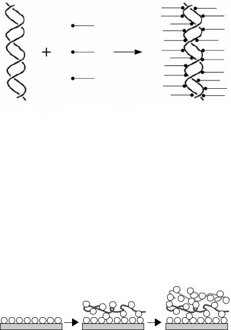
Figure 3.7 Formation of DNA–lipid complexes. Aqueous solutions of DNA and amphiphilic lipids are mixed in an appropriate phosphate anion-to-amphiphilic lipid cation ratio. Formation of DNA–lipid complexes is accompanied by their precipitation in the aqueous (mixed) solution. Via subsequent wash steps and lyophilization, dry DNA–lipid complexes that are soluble in organic solvents are produced.
Finally, DNA–lipid complexes, depending on composition, may exert antibacterial activities.95 Since infections are common problems associated with implantation procedures, a coating that possesses antibacterial activity may diminish the incidence of implantation-related infections.
The use of DNA as a nanocoating on a biomaterial surface, however, implies the necessity to circumvent certain properties of DNA, including its water solubility and easy degradation by nucleases. DNA can be complexed with amphiphilic lipids96,97 (Figure 3.7) or cationic polyelectrolytes98 (Figure 3.8). The structures generated by this process are stable through electrostatic interactions between anionic phosphate groups in the DNA and cationic groups in the amphiphilic lipid or polymer. The application of DNA coatings in implantology may lead ultimately to multifunctional coatings that can be applied at various sites in the body, evoke minimal immunologic reactions, and deliver biologically active substances to modulate cellular behavior.
|
|
|
|
– |
– |
– – |
– |
|
|
|
|
– |
– |
||
|
|
+ + |
|
– |
|||
|
+ |
+ |
+ |
+ – + |
|
+ |
|
|
+ |
+ |
+ + |
+ |
+ |
+ |
+ |
– – – – – – – – |
|
|
|||||
– – – – – – – – |
– – – – – – – – |
||||||
Figure 3.8 Formation of multilayered polyelectrolyte coatings. Polyanionic (e.g., DNA) and polycationic polymers can be used to generate multilayers based on electrostatic interactions between alternate layers. This technique allows a wide variation in the number of polyelectrolyte layers that form a multilayered coating and the types of polyelectrolytes.
IMPLANTS AND PROSTHESES |
61 |
C. Influence of Biomaterials with Nanostructures on Cell Behavior
This section describes the influence of nanostructured biomaterials on cell behavior based on a selection of recently published research work. Due to their recent development, nanotechnologically modified implants have not achieved clinical applications yet. Before clinical application is possible, in vitro and in vivo test models must demonstrate the benefits of nanotechnologically modified implants.
In our view, nanostructured biomaterials contain features that possess at least one dimension (x, y, or z) in the submicron (<1 μm) range. Although classification of nanotechnological methodologies is difficult, we have tried to generate an overview of cell behavior in relation to biomaterials with topographical, protein–peptide, and calcium phosphate nanostructures.
1. Topographical Nanostructures
The topography of biomaterial surfaces has been a major topic in biomaterials science in the past decade. Surface topography can be of great importance with respect to area enlargement. An increase in surface area may provide greater potential for tissue integration (mechanical interlocking). Excellent reviews on this topic56,57,99–101 evidence a general consensus that topography indeed influences cell behavior. In the first studies that explored the effect of topography on cell behavior, microscale topographical cues were usually used. An enormous diversity of topographical cues was used: grooves, pits, ridges, cliffs, tunnels, steps, waves, wells, tubes, nodes, pillars, pores, spheres, and cylinders. Researchers used many different cell types in studies to examine the effects of microscale topographical cues on the behavior of primary isolated cells or immortalized cell lines including fibroblasts, macrophages, epithelial cells, leukocytes, neuronal cells, endothelial cells, and osteoblasts.
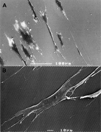
Although the reaction is dependent on cell type, cells react on contacting microscale topographies in a wide variety of manners including orientation, extension, movement, and activation [phosphorylation, actin polymerization, messenger ribonucleic acid (mRNA) expression, and phagocytic activity]. A phenomenon called contact guidance is observed when cells are cultured on microgrooved substrata (Figure 3.9); the cells align along the axes of the grooves. Control over cellular alignment (including the alignment of cell extensions) may be a pivotal factor in orchestrating cell morphology and orientation for the generation of nerve and other well-organized tissues.
Unfortunately, the precise biological effects of microscale topographies remain unclear — various research groups have obtained contradictory results. Parker et al.102 found no favorable effects of surface texturing on capsule formation around subcutaneous implants, but in vivo studies by others indicated that grooved implant surfaces produced beneficial effects on tissues surrounding the implant.99 In the latter studies, grooved topographies appeared to encourage tissue organization and showed a reduction in fibrous capsule formation as compared to smooth implant surfaces.
\
62 |
BIOMEDICAL NANOTECHNOLOGY |
Figure 3.9 Contact guidance phenomena of rat dermal fibroblasts on microgrooved substrates. (A) Cells align themselves along the axes of the microgrooves (groove width = 1 μm, ridge width = 1 μm, and groove depth = 1 μm). (B) Higher magnification showing cell extensions along the axes of the microgrooves.
Although the technology for creating topographical cues on biomaterial surfaces with nanoscale dimensions is a novelty, several studies have demonstrated the substantial effect the cues can exert on cell behavior. The effect on cell behavior of grooves with nanoscale dimensions has been a subject of interest in many in vitro studies.103–106 Regarding the effects of grooves with nanoscale dimensions, the findings generally indicate that cells become oriented and elongate along the surface grooves. Furthermore, the activity of cells on nanotopographically modified substrates is increased compared to activity on smooth control substrates. This is demonstrated by higher proliferation rates and enhanced spreading of fibroblasts,107
IMPLANTS AND PROSTHESES |
63 |
increased phagocytic activity of macrophages,106 and a notable up-regulation of gene expression related to cell signaling, proliferation, and the production of cytoskeleton and ECM proteins.108
A specific role for biomaterial surface nanotopography has been demonstrated for growing nerve tissue. Control over the outgrowth of neurites from the cell bodies of neurons, both the sites at which they emerge from cell bodies and directions, was achieved in vitro using biomaterials with nanoscale groove dimensions.104 The general conclusion of this study was that nanoscale substratum topography can be a potent morphogenetic factor for developing neurons and can assist in establishing neuronal polarity.
In addition to nanoscale texture, nanoscale roughness has been shown to affect cellular behavior. Using polymeric inverse replicas of native tissue, bladder smooth muscle cells109 and cardiovascular endothelial cells110 showed higher proliferation rates, more rapid spreading, and a more native-like appearance, respectively. Cells of osteogenic lineage also appear to be influenced by nanoscale topographies. In a series of studies using nanophase ceramics,111–114 osteoblast (bone-forming) cells and osteoclast (bone-degrading) cells showed different behaviors dependent on nanophase ceramics. Proliferation, expression of differentiation markers, and calcium deposition were increased for osteoblasts. Similarly, the functions of osteoclastlike cells (including formation of resorption pits) were significantly enhanced.
2. Protein and Peptide Nanostructures
While synthetic surfaces lack specific signals cells can recognize, naturally derived materials may possess numerous signals involved in a wide variety of biological processes. In view of this characteristic, modulation of a biomaterial surface with nanoscale-sized, naturally occurring components of ECM proteins containing such signals has become a common modality. These nano approaches attempt to integrate living and nonliving systems. The effect of the immobilization of specific peptide sequences including RGD onto the surfaces of materials has been studied for biomaterials intended for implantation in both soft and hard tissues. Excellent reviews dealing with this topic115,116 outline proven applications and promising potential for the future.
Biomaterial surfaces and matrices endowed with peptides for tissue engineering have been shown to enhance cellular behavior substantially (Table 3.3). Beneficial effects of such surfaces have been demonstrated for cell types with different functions and originating from both soft and hard tissues, i.e., connective tissues (fibroblasts), muscle tissues (myoblasts and smooth muscle cells), vascular tissues (endothelial cells), nerve tissues (neuronal cells), bone (osteoblasts), and cartilage (chondrocytes).
A pivotal factor determining the capacities of peptides to modulate cellular behavior is spatial distribution. In order to evoke functional intracellular signaling, receptors on a cell surface (e.g., integrins) must be clustered. This clustering can be achieved by increasing peptide density or flexibility using spacers. However, exaggerating the density of peptides on a biomaterial surface can also dramatically affect cell motility, which may be beneficial in immobilizing endothelial cells for vascular
\

64 |
|
BIOMEDICAL NANOTECHNOLOGY |
||
|
Table 3.3 Application of Immobilized Peptides onto Biomaterials |
|||
|
|
|
|
|
|
Peptide |
ECM Molecule Source |
Application |
|
|
|
|
|
|
|
RGD |
Multiple ECM molecules, |
Enhance bone and cartilage tissue |
|
|
|
e.g., fibronectin, |
formation in vitro and in vivo; |
|
|
|
vitronectin, laminin, |
regulate neurite outgrowth in vitro |
|
|
|
collagen, and |
and in vivo; promote myoblast |
|
|
|
thrombospondin |
adhesion, proliferation, and |
|
|
|
|
differentiation; enhance |
|
|
|
|
endothelial cell adhesion and |
|
|
|
|
proliferation |
|
|
IKVAV YIGSR |
Laminin |
Regulate neurite outgrowth in vitro |
|
|
RNIAEIIKDI |
|
and in vivo |
|
|
Recombinant fibronectin |
Fibronectin |
Promote formation of focal contacts |
|
|
fragment (FNIII7–10) |
|
in preosteoblasts |
|
|
Ac-GCRDGPQ- |
Common MMP |
Encourage cell-mediated |
|
|
GIWGQDRCG |
substrates, e.g., |
proteolytic degradation, |
|
|
|
collagen, fibronectin, |
remodeling, and bone |
|
|
|
and laminin |
regeneration in vivo |
|
Source: Adapted from Boontheekul, T. and Mooney, D.J. Curr Opin Biotechnol, 14, 559–565, 2003.137
grafts, but is undesirable for the application of tissue ingrowth into tissue engineering matrices.
In vitro and in vivo studies investigating peptide-modulated biomaterials are numerous and focus primarily on RGD peptides. A comprehensive overview of some of these for use with both soft and hard tissue cells is presented below.
In vitro, RGD peptides significantly enhance attachment and spreading of cells, for example, endothelial cells.117 Fibroblast proliferation rates were demonstrated to be significantly increased on RGD-modified polymeric material.118 In addition to these cellular characteristics, cell migration and ECM deposition appear to be controllable using adequate density and distribution of RGD peptides.119,120 In an attempt to enhance the attachment of endothelial cells to artificial blood vessels, polymeric materials were functionalized with a peptide domain selective for endothelial cells (and not for other cell types such as fibroblasts, vascular smooth muscle cells, and platelets).121,122 Endothelial monolayers cultured on these functionalized polymeric materials proved to be nonthrombogenic, resulting in increased patency of such vascular grafts.
In vitro experiments have also clearly demonstrated the beneficial effects RGD immobilization can exert on cells of osteogenic lineage. Depending on the density and type of peptide immobilized on a material surface, both osteoblast-like cell adhesion and mineralization of synthesized ECM can be increased substantially.72,123 Tissue responses to peptide-treated polymeric material in vivo were assessed after intraperitoneal or subcutaneous implantation for 12 weeks.124 Although blood sample analysis revealed no adverse responses, histological evaluation after 12 weeks demonstrated the presence of thicker fibrous capsules around RGD-treated implants compared to controls. In contrast, RGD-coated porous poly(methyl-methacrylate) implants in a rabbit model demonstrated enhanced and accelerated cancellous bone ingrowth compared to noncoated controls.125 Moreover, apposition of newly formed
IMPLANTS AND PROSTHESES |
65 |
bone directly toward the implant surface was observed, whereas noncoated control implants were surrounded by fibrous tissue layers that prevented direct bonding of bone to the implant surface.
3. Calcium Phosphate Nanostructures
A number of studies have described the effectiveness of calcium phosphatecoated implants and materials for bones. It is evident that calcium phosphate coatings improve the biological performance of endosseous implants.6,126,127 Further expansion of calcium phosphate deposition techniques to generate coatings that resemble the nano-sized dimensions of native bone tissue have led to only a limited number of scientific publications to date.
Hydroxyapatite (HA) with nanometer-scale crystal size was used to modify commercially available collagen sheets.128 Using an organ culture technique in which bone fragments were used to provide osteogenic cells, the composite scaffolds were demonstrated to be suitable for culturing osteogenic cells. The cells migrated from the bone fragments into the porous composite scaffold and eventually acquired a three-dimensional polygonal appearance. The three-dimensional osteogenic cells (nanoHA–collagen complexes) are suggested as promising candidates for the engineering of bone tissue. In a subsequent study using nanoHA–collagen composites, composite implants without bone cells (or fragments) were implanted into rabbit femurs.129 Upon implantation, both new bone formation and implant degradation reminiscent of the bone remodeling process were observed. However, the lack of organization of bone constituents in the composite (compared to natural bone) resulted in a decreased mechanical strength that only reached the lower limit of that of natural bone.
V. CONSIDERATIONS
Although nanotechnologically modulated biomaterials have potential effects on cellular behavior, several issues must be considered. These issues involve determination of what actually causes specific cellular behavior and whether man-made nanoscale cues will be as powerful as natural nanoscale entities.
A. Topographical versus Chemical Cues
Roughly, all earlier described methods can be divided into techniques that (1) create isotropic nanotechnological structures that do not differ chemically from the intrinsic substratum or (2) create anisotropic nanotechnological structures using patterns of molecules chemically different from those of the intrinsic substratum.
Although both types of nanofabrication allow production of nanoscale cues on a biomaterial surface, the question of what causes a potential distinct cell behavior is justified since the former technique uses topographical cues to control cell behavior and the latter also uses chemical cues. Although chemical means to analyze possible slight differences in chemical properties of nanostructures on isotropic modified
\
66 |
BIOMEDICAL NANOTECHNOLOGY |
biomaterial surfaces are not yet available, topographical cues in the nanoscale range appear to affect the behavior of cells directly. The evidence for this is that the reactions of cells to similar topographies on chemically different biomaterial surfaces are comparable.130
Although we noted that the adsorption of proteins to biomaterial surfaces is responsible for cell attachment and subsequent cell reaction to biomaterial surfaces, the protein adsorption characteristics of materials still determine the constitution of the layer of proteins. Thus, if reactions of cells to topographical cues are similar regardless of the biomaterial used, it seems plausible to conclude that the adsorbed layer of proteins whose constitution is governed by the type of biomaterial131 has a minor effect on cell reaction compared to the effect of the biomaterial surface topography.
Further evidence for this hierarchy of topographical over chemical cues in control of cell reactions has emerged from a study in which topography and chemistry competed in controlling alignments of neurites.132 The biomaterial surfaces used in this experiment contained grooves or protein (laminin) patterns or both. Patterns were made either parallel or orthogonal. The conclusion drawn after culturing nerve cells on these substrates was that such morphogenetic guidance cues preferentially act synergistically. However, at sites where the depth of the grooves exceeded 500 nm, the topographical cues appeared to be higher in hierarchy than the chemical ones. It is important to emphasize that reactions of cells to either isotropic or anisotropic cues are dependent on cell type. Moreover, the presence of chemical cues is inevitably accompanied by minor parallel topographical cues since patterns of chemicals have certain thicknesses to which the cells may also react.
An in vivo model in which titanium implants were used showed that topography indeed is a major factor in modulating cell responses.133 Polished, grit-blasted, HAcoated, and titanium film-covered HA-coated titanium implants were used to determine the relative contributions of surface topography and chemistry to the osseointegration of hard tissue implants. Using thin section histological evaluation and subsequent scanning electron microscopy, the authors showed that, although osseointegration was significantly greater on HA-coated titanium implants, 80% of the maximal bone forming response was observed on HA-coated titanium implants covered by a thin titanium film. The conclusion was that topography is a suprahierarchical factor compared to chemistry in bone apposition.
B. Natural versus Synthetic Nanostructures
The creation of nanostructures on biomaterial surfaces aimed to enhance implant success rates remains a man-made modification method. Therefore, even nanotechnological approaches to generating biomimetic biomaterial surfaces result in surfaces considerably dissimilar from those found in nature. The main reason is the lack of bilateral dynamic interactions and responses between the biomaterial surface and its biological surroundings. For example, the natural interaction of cell surface receptors (e.g., integrins) with their respective ligands is a highly dynamic process in which receptors and ligands continuously associate and disassociate, resulting in signaling
IMPLANTS AND PROSTHESES |
67 |
into the cell via consecutive events of intracellular domains and accessory complexes and cascades.134,135
The clustering of receptor–ligand complexes is important for several cellular processes including motility136 and the spatial distribution of receptor–ligand complexes is an important issue. Since the immobilization of ECM components on biomaterial surfaces does not allow spatial changes in ligand distribution upon cell binding, receptor clustering at the surfaces of cells adhering to the biomaterial surface is restricted to sites at which ligand density is appropriate after immobilization. Studies by Massia and Hubbell75 aimed at elucidating the minimal distribution of RGD peptides required for interactions with the αvβ3–integrin receptor demonstrated that for human foreskin fibroblasts, a 440-nm peptide-to-peptide (RGD) spacing is required for cellular spreading, whereas the formation of focal contacts and stress fiber organization require an approximately three-fold lower RGD spacing.
VI. CONCLUSIONS
The generation of implants that will succeed in their intended functions requires a multidisciplinary approach that involves comprehension of divergent processes. For that reason, collaboration of researchers is recommended to develop clinically safe and reliable implants. When bulk properties of biomaterials meet the criteria required for a specific intended function, surface properties become important for minimizing potential undesirable responses by the surrounding biological environment. In view of this, we hope that nanotechnology may provide a significant approach by which a biomaterial surface can be modulated to decrease common host-versus-biomaterial responses.
Nanotechnology can provide strategies that can help create features on biomaterial surfaces in a dimensional range that may be adequate for cells. In its natural habitat, a cell is surrounded by other cells and by ECM proteins that provide a diverse range of signals (via cell–cell or cell–ECM contact) influencing cellular behavior. The majority of these signals are transmitted via receptor–ligand interactions, and their dimensions lie within the nanometer range. Therefore, several approaches using modulations of biomaterial surfaces with nanoscale features have been undertaken to study their effects on the responses of tissues in the direct vicinity of the implant. A wide variety of approaches include nanoscale topographical and chemical alterations at the biomaterial surface. Combinations of approaches (e.g., using both nanotopography and peptide functionalization) could offer additional power over cellular behavior.
Although research in this area is still in its infancy, several published studies indicate the beneficial effects nanotechnologically modified surfaces can have for implantology.138 Because many aspects of cellular responses to materials are still unknown, further expansion of our understanding of nanotechnology and biological responses to nanoscale features will eventually result in clinically applicable designs for biomaterial surfaces that will be able to adjust to the required functionality of an implant.
\
68 |
BIOMEDICAL NANOTECHNOLOGY |
The techniques described in this chapter are limited to experimental and laboratory settings. The clinical use of nanotechnologically modulated implants and prostheses awaits unambiguous proof of beneficial effects for given applications. However, improvements in exploiting currently available and future techniques combined with a better understanding of the influence of nanoscale features on cells and tissues surrounding implants and a multidisciplinary approach in implantology will pave the road for the use of nanostructures in the design of implants and prostheses.
REFERENCES
1.Anderson, J.M. et al. Implants and devices, in Ratner, B.D., Hoffman, A.S., Schoen, F.J., and Lemons, J.E., Eds., Biomaterials Science: Introduction to Materials in Medicine. Academic Press, London, 1996.
2.Ratner, B.D. Replacing and renewing: synthetic materials, biomimetics, and tissue engineering in implant dentistry. J Dent Educ 65, 1340–1347, 2001.
3.Crubezy, E., Murail, P., Girard, L., and Bernadou, J.P. False teeth of the Roman world. Nature 391, 29, 1998.
4.Cooke, F.W. Bulk properties of materials, in Ratner, B.D., Hoffman, A.S., Schoen, F.J., and Lemons, J.E., Eds., Biomaterials Science: Introduction to Materials in Medicine. Academic Press, London, 1996.
5.Black, J. Biological Performance of Materials: Fundamentals of Biocompatibility. Marcel Dekker, New York, 1992.
6.Lacefield, W.R. Materials characteristics of uncoated/ceramic-coated implant materials. Adv Dent Res 13, 21–26, 1999.
7.Schakenraad, J.M. Cells: their surfaces and interactions with materials, in Ratner, B.D., Hoffman, A.S., Schoen, F.J., and Lemons, J.E., Eds., Biomaterials Sciences: Introduction to Materials in Medicine. Academic Press, London, 1996.
8.Cordero, J., Munuera, L., and Folgueira, M.D. The influence of the chemical composition and surface of the implant on infection. Injury 27, Suppl. 3, SC34–SC37, 1996.
9.Bauer, T.W. and Schils, J. The pathology of total joint arthroplasty. II. Mechanisms of implant failure. Skeletal Radiol 28, 483–497, 1999.
10.Allara, D.L. Critical issues in applications of self-assembled monolayers. Biosensors Bioelectron 10, 771–783, 1995.
11.Williams, D.F. and Williams, R.L. Degradative effects of the biological environment on metals and ceramics, in Ratner, B.D., Hoffman, A.S., Schoen, F.J., and Lemons, J.E., Eds., Biomaterials Science: Introduction to Materials in Medicine. Academic Press, London, 1996.
12.Liu, W., Cao, Y., and Longaker, M.T. Gene therapy of scarring: a lesson learned from fetal scarless wound healing. Yonsei Med J 42, 634–645, 2001.
13.Webb, J.C.J. and Tricker, J. Bone biology: a review of fracture healing. Curr Orthop 14, 457–463, 2000.
14.Glowacki, J. Angiogenesis in fracture repair. Clin Orthop 355 (Suppl) S82–S89, 1998.
15.Harding, K.G., Morris, H.L., and Patel, G.K. Science, medicine and the future: healing chronic wounds. Br Med J 324, 160–163, 2002.
16.Singer, A.J. and Clark, R.A. Cutaneous wound healing. New Engl J Med 341, 738–746, 1999.