

Biomedical Nanotechnology - Neelina H. Malsch
.pdfIMPLANTS AND PROSTHESES |
69 |
17.Bello, Y.M. and Phillips, T.J. Recent advances in wound healing. JAMA 283, 716–718, 2000.
18.Clark, R.A. Fibrin and wound healing. Ann NY Acad Sci 936, 355–367, 2001.
19.Hart, J. Inflammation. 1: Its role in the healing of acute wounds. J Wound Care 11, 205–209, 2002.
20.Hart, J. Inflammation. 2: Its role in the healing of chronic wounds. J Wound Care 11, 245–249, 2002.
21.Hollinger, J. and Wong, M.E. The integrated processes of hard tissue regeneration with special emphasis on fracture healing. Oral Surg Oral Med Oral Pathol Oral Radiol Endod 82, 594–606, 1996.
22.Wong, M.E., Hollinger, J.O., and Pinero, G.J. Integrated processes responsible for soft tissue healing. Oral Surg Oral Med Oral Pathol Oral Radiol Endod 82, 475–492, 1996.
23.Werner, S. and Grose, R. Regulation of wound healing by growth factors and cytokines. Physiol Rev 83, 835–870, 2003.
24.Gerstenfeld, L.C., Cullinane, D.M., Barnes, G.L., Graves, D.T., and Einhorn, T.A. Fracture healing as a post-natal developmental process: molecular, spatial, and temporal aspects of its regulation. J Cell Biochem 88, 873–884, 2003.
25.Baj-Krzyworzeka, M. et al. Platelet-derived microparticles stimulate proliferation, survival, adhesion, and chemotaxis of hematopoietic cells. Exp Hematol 30, 450–459, 2002.
26.Mackay, C.R. Chemokines: immunology’s high impact factors. Nat Immunol 2, 95–101, 2001.
27.Janeway, C.A., Travers, P., Walport, M., and Capra, J. Immunobiology: the immune system in health and disease. Elsevier Science Ltd. (Garland Publishing), 1999.
28.Einhorn, T.A. The cell and molecular biology of fracture healing. Clin Orthop S7–S21, 1998.
29.Mutsaers, S.E., Bishop, J.E., McGrouther, G., and Laurent, G.J. Mechanisms of tissue repair: from wound healing to fibrosis. Int J Biochem Cell Biol 29, 5–17, 1997.
30.Visse, R. and Nagase, H. Matrix metalloproteinases and tissue inhibitors of metalloproteinases: structure, function, and biochemistry. Circ Res 92, 827–839, 2003.
31.Chamay, A. and Tschantz, P. Mechanical influences in bone remodeling: experimental research on Wolff’s law. J Biomech 5, 173–180, 1972.
32.Thomsen, P. and Gretzer, C. Macrophage interactions with modified material surfaces.
Curr Opin Sol State Mater Sci 5, 163–176, 2001.
33.Vogler, E.A. Structure and reactivity of water at biomaterial surfaces. Adv Colloid Interface Sci 74, 69–117, 1998.
34.Hallab, N.J., Bundy, K.J., O’Connor, K., Moses, R.L., and Jacobs, J.J. Evaluation of metallic and polymeric biomaterial surface energy and surface roughness characteristics for directed cell adhesion. Tissue Eng 7, 55–71, 2001.
35.Walboomers, X.F., Croes, H.J., Ginsel, L.A., and Jansen, J.A. Contact guidance of rat fibroblasts on various implant materials. J Biomed Mater Res 47, 204–212, 1999.
36.Jansen, J.A., van der Waerden, J.P., and de Groot, K. Effect of surface treatments on attachment and growth of epithelial cells. Biomaterials 10, 604–608, 1989.
37.Horbett, T.A. and Schay, M.B. Correlations between mouse 3T3 cell spreading and serum fibronectin adsorption on glass and hydroxyethylmethacrylate–ethylmethacry- late copolymers. J Biomed Mater Res 22, 763–793, 1988.
38a. Kasemo, B. and Gold, J. Implant surfaces and interface processes. Adv Dent Res 13, 8–20, 1999.
\
70 |
BIOMEDICAL NANOTECHNOLOGY |
38b. Hanarp, P., Sutherland, D., Gold, J., and Kasemo, B. Nanostructured model biomaterial surfaces prepared by colloidal lithography. Nanostructured Mater. 12(1–4), 429–432, 1999.
39.Horbett, T.A. The role of adsorbed adhesion proteins in cellular recognition of biomaterials. BMES Bull 23, 5–9, 1999.
40.Horbett, T.A. and Klumb, L.A. Cell culturing: surface aspects and considerations, in Brash, W.P., Ed., Interfacial Phenomena and Bioproducts. Marcel Dekker, New York, 1996.
41.Steele, J.G., Dalton, B.A., Johnson, G., and Underwood, P.A. Adsorption of fibronectin and vitronectin onto Primaria and tissue culture polystyrene and relationship to the mechanism of initial attachment of human vein endothelial cells and BHK-21 fibroblasts. Biomaterials 16, 1057–1067, 1995.
42.Steele, J.G. et al. Roles of serum vitronectin and fibronectin in initial attachment of human vein endothelial cells and dermal fibroblasts on oxygenand nitrogen-con- taining surfaces made by radiofrequency plasmas. J Biomater Sci Polym Ed 6, 511–532, 1994.
43.Ratner, B.D. Reducing capsular thickness and enhancing angiogenesis around implant drug release systems. J Control Release 78, 211–218, 2002.
44.Seare, W.J., Jr. Alloplasts and biointegration. J Endourol 14, 9–17, 2000.
45.Ducheyne, P. et al. Effect of hydroxyapatite impregnation on skeletal bonding of porous coated implants. J Biomed Mater Res 14, 225–237, 1980.
46.Geesink, R.G., de Groot, K., and Klein, C.P. Bonding of bone to apatite-coated implants. J Bone Joint Surg Br 70, 17–22, 1988.
47.Anderson, J. M. Biological responses to materials. Annu Rev Mater Res 31, 81–110, 2001.
48.van Diest, P.J., Beekman, W.H., and Hage, J.J. Pathology of silicone leakage from breast implants. J Clin Pathol 51, 493–497, 1998.
49.Anderson, J.M. Multinucleated giant cells. Curr Opin Hematol 7, 40–47, 2000.
50.Zhao, Q. et al. Foreign body giant cells and polyurethane biostability: in vivo correlation of cell adhesion and surface cracking. J Biomed Mater Res 25, 177–183, 1991.
51.Whitesides, G.M. and Love, J.C. Nanofabrication: the art of building small. Sci Am 39–47, September 2001.
52.Sorribas, H., Padeste, C., and Tiefenauer, L. Photolithographic generation of protein micropatterns for neuron culture applications. Biomaterials 23, 893–900, 2002.
53.Kane, R.S., Takayama, S., Ostuni, E., Ingber, D.E., and Whitesides, G.M. Patterning proteins and cells using soft lithography. Biomaterials 20, 2362–2376, 1999.
54.Wilkinson, J.M. Nanotechnology: applications in medicine. Med Dev Tech 29–31, June 2003.
55.ten Wolde, A. Nanotechnology: Toward a Molecular Construction Kit. Netherlands Study Centre for Technology Trends, The Hague, 1998.
56.Curtis, A. and Wilkinson, C. Topographical control of cells. Biomaterials 18, 1573–1583, 1997.
57.Ito,Y. Surface micropatterning to regulate cell functions. Biomaterials 20, 2333–2342, 1999.
58.Desai, T.A. Microand nano-scale structures for tissue engineering constructs. Med Eng Phys 22, 595–606, 2000.
59.Walboomers, X.F. Engineered Implant Surfaces: Modification of Cell and Tissue Response by Microgrooves. University of Nijmegen, Netherlands, 2000.
60.Prokop, A. Bioartificial organs in the 21st century: nanobiological devices. Ann NY Acad Sci 944, 472–490, 2001.
IMPLANTS AND PROSTHESES |
71 |
61.Yoshinari, M., Matsuzaka, K., Inoue, T., Oda, Y., and Shimono, M. Effects of multigrooved surfaces on fibroblast behavior. J Biomed Mater Res 65A, 359–368, 2003.
62.von Recum, A.F., Shannon, C.E., Cannon, E.C., Long, K.J., van Kooten, T.G., and Meyle, J. Surface roughness, porosity, and texture as modifiers of cellular adhesion. Tissue Eng 2, 241–253, 1996.
63.Zhang, S. Emerging biological materials through molecular self-assembly. Biotechnol Adv 20, 321–339, 2002.
64.Whitesides, G.M., Mathias, J.P., and Seto, C.T. Molecular self-assembly and nanochemistry: a chemical strategy for the synthesis of nanostructures. Science 254, 1312–1319, 1991.
65.Mrksich, M., Dike, L.E., Tien, J., Ingber, D.E., and Whitesides, G.M. Using microcontact printing to pattern the attachment of mammalian cells to self-assembled monolayers of alkanethiolates on transparent films of gold and silver. Exp Cell Res 235, 305–313, 1997.
66.Pierschbacher, M.D. and Ruoslahti, E. Cell attachment activity of fibronectin can be duplicated by small synthetic fragments of the molecule. Nature 309, 30–33, 1984.
67.Hynes, R.O. Integrins: versatility, modulation, and signaling in cell adhesion. Cell 69, 11–25, 1992.
68.Danen, E.H. and Sonnenberg, A. Integrins in regulation of tissue development and function. J Pathol 200, 471–480, 2003.
69.Kim, B.S. and Mooney, D.J. Development of biocompatible synthetic extracellular matrices for tissue engineering. Trends Biotechnol 16, 224–230, 1998.
70.Sakakura, S., Saito, S., and Morikawa, H. Stimulation of DNA synthesis in trophoblasts and human umbilical vein endothelial cells by hepatocyte growth factor bound to extracellular matrix. Placenta 20, 683–693, 1999.
71.Barber, T.A., Golledge, S.L., Castner, D.G., and Healy, K.E. Peptide-modified pAAm- co-EG/AAc IPNs grafted to bulk titanium modulate osteoblast behavior in vitro. J Biomed Mater Res 64, 38–47, 2003.
72.Rezania, A. and Healy, K.E. Biomimetic peptide surfaces that regulate adhesion, spreading, cytoskeletal organization, and mineralization of the matrix deposited by osteoblast-like cells. Biotechnol Prog 15, 19–32, 1999.
73.Hubbell, J.A. Bioactive biomaterials. Curr Opin Biotechnol 10, 123–129, 1999.
74.Hubbell, J.A. Biomaterials in tissue engineering. Biotechnology 13, 565–576, 1995.
75.Massia, S.P. and Hubbell, J.A. An RGD spacing of 440 nm is sufficient for integrin alpha V beta 3-mediated fibroblast spreading and 140 nm for focal contact and stress fiber formation. J Cell Biol 114, 1089–1100, 1991.
76.Healy, K.E., Rezania, A., and Stile, R.A. Designing biomaterials to direct biological responses. Ann NY Acad Sci 875, 24–35, 1999.
77.Hill, I.R., Garnett, M.C., Bignotti, F., and Davis, S.S. Determination of protection from serum nuclease activity by DNA–polyelectrolyte complexes using an electrophoretic method. Anal Biochem 291, 62–68, 2001.
78.Reyes, C.D. and Garcia, A.J. Engineering integrin-specific surfaces with a triplehelical collagen mimetic peptide. J Biomed Mater Res 65A, 511–523, 2003.
79.Cutler, S.M. and Garcia, A.J. Engineering cell adhesive surfaces that direct integrin alpha 5–beta 1 binding using a recombinant fragment of fibronectin. Biomaterials 24, 1759–1770, 2003.
80.Garcia, A.J. and Keselowsky, B.G. Biomimetic surfaces for control of cell adhesion to facilitate bone formation. Crit Rev Eukaryot Gene Expr 12, 151–162, 2002.
\
72 |
BIOMEDICAL NANOTECHNOLOGY |
81.Barrere, F. et al. In vitro and in vivo degradation of biomimetic octacalcium phosphate and carbonate apatite coatings on titanium implants. J Biomed Mater Res 64A, 378–387, 2003.
82.Jansen, J.A., Wolke, J.G., Swann, S., Van der Waerden, J.P., and de Groot, K. Application of magnetron sputtering for producing ceramic coatings on implant materials. Clin Oral Implants Res 4, 28–34, 1993.
83.Wolke, J.G., van Dijk, K., Schaeken, H.G., de Groot, K., and Jansen, J.A. Study of the surface characteristics of magnetron-sputter calcium phosphate coatings. J Biomed Mater Res 28, 1477–1484, 1994.
84.Rivero, D.P., Fox, J., Skipor, A.K., Urban, R.M., and Galante, J.O. Calcium phos- phate-coated porous titanium implants for enhanced skeletal fixation. J Biomed Mater Res 22, 191–201, 1988.
85.Leeuwenburgh, S., Wolke, J., Schoonman, J., and Jansen, J. Electrostatic spray deposition ESD of calcium phosphate coatings. J Biomed Mater Res 66A, 330–334, 2003.
86.McMichael, A.J. Antigens and MHC systems, in McGee, J.O.D., Isaacson, P.G., and Wright, N.A., Eds. Oxford Textbook of Pathology. Oxford University Press, Oxford, 1992.
87.Yamamoto, S. et al. DNA from bacteria, but not from vertebrates, induces interferons, activates natural killer cells and inhibits tumor growth. Microbiol Immunol 36, 983–997 1992.
88.Krieg, A.M. Immune effects and mechanisms of action of CpG motifs. Vaccine 19, 618–622, 2000.
89.Krieg, A.M. et al. CpG motifs in bacterial DNA trigger direct B-cell activation. Nature 374, 546–549, 1995.
90.Werner, M.H., Gronenborn, A.M., and Clore, G.M. Intercalation, DNA kinking, and the control of transcription. Science 271, 778–784 1996.
91.Wilson, W.D. Reversible interactions of nucleic acids with small molecules, in Blackburn, G.M. and Gait, M.J., Eds. Nucleic Acids in Chemistry and Biology. Oxford University Press, Oxford, 1996.
92.Goldman, A. and Glumoff, T. Interaction of proteins with nucleic acids, in Blackburn, G.M. and Gait, M.J., Eds. Nucleic Acids in Chemistry and Biology. Oxford University Press, Oxford, 1996.
93.Kamei, S., Tomita, N., Tamai, S., Kato, K., and Ikada, Y. Histologic and mechanical evaluation for bone bonding of polymer surfaces grafted with a phosphate-containing polymer. J Biomed Mater Res 37, 384–393, 1997.
94.Tretinnikov, O.N., Kato, K., and Ikada, Y. In vitro hydroxyapatite deposition onto a film surface-grated with organophosphate polymer. J Biomed Mater Res 28, 1365–1373, 1994.
95.Inoue, Y. et al. Antibacterial characteristics of newly developed amphiphilic lipids and DNA-lipid complexes against bacteria. J Biomed Mater Res 65A, 203–208, 2003.
96.Tanaka, K. and Okahata, Y. A DNA-lipid complex in organic media and formation of an aligned cast film. J Am Chem Soc 118, 10679–10683, 1996.
97.Okahata, Y. and Tanaka, K. Oriented thin films of a DNA–lipid complex. Thin Solid Films 284–285, 6–8, 1996.
98.Decher, G. Fuzzy nanoassemblies: toward layered polymeric multicomposites. Science 277, 1232–1237, 1997.
99.Brunette, D.M. and Chehroudi, B. The effects of the surface topography of micromachined titanium substrata on cell behavior in vitro and in vivo. J Biomech Eng 121, 49–57, 1999.
100.Singhvi, R. et al. Engineering cell shape and function. Science 264, 696–698, 1994.
IMPLANTS AND PROSTHESES |
73 |
101.Walboomers, X.F. and Jansen, J.A. Cell and tissue behavior on micro-grooved surfaces. Odontology 89, 2–11, 2001.
102.Parker, J.A., Walboomers, X.F., Von den Hoff, J.W., Maltha, J.C., and Jansen, J.A. Soft-tissue response to silicone and poly-L-lactic acid implants with a periodic or random surface micropattern. J Biomed Mater Res 61, 91–98, 2002.
103.den Braber, E.T., de Ruijter, J.E., Ginsel, L.A., von Recum, A.F., and Jansen, J.A. Quantitative analysis of fibroblast morphology on microgrooved surfaces with various groove and ridge dimensions. Biomaterials 17, 2037–2044, 1996.
104.Rajnicek, A., Britland, S., and McCaig, C. Contact guidance of CNS neurites on grooved quartz: influence of groove dimensions, neuronal age and cell type. J Cell Sci 110, Pt. 23, 2905–2913, 1997.
105.Clark, P., Connolly, P., Curtis, A.S., Dow, J.A., and Wilkinson, C.D. Cell guidance by ultrafine topography in vitro. J Cell Sci 99, Pt. 1, 73–77, 1991.
106.Wojciak-Stothard, B., Curtis, A., Monaghan, W., MacDonald, K., and Wilkinson, C. Guidance and activation of murine macrophages by nanometric scale topography. Exp Cell Res 223, 426–435, 1996.
107.Dalby, M.J., Riehle, M.O., Johnstone, H.J., Affrossman, S., and Curtis, A.S. Polymerdemixed nanotopography: control of fibroblast spreading and proliferation. Tissue Eng 8, 1099–1108, 2002.
108.Dalby, M.J. et al. Increasing fibroblast response to materials using nanotopography: morphological and genetic measurements of cell response to 13-nm-high polymer demixed islands. Exp Cell Res 276, 1–9, 2002.
109.Thapa, A., Miller, D.C., Webster, T.J., and Haberstroh, K.M. Nano-structured polymers enhance bladder smooth muscle cell function. Biomaterials 24, 2915–2926, 2003.
110.Goodman, S.L., Sims, P.A., and Albrecht, R.M. Three-dimensional extracellular matrix textured biomaterials. Biomaterials 17, 2087–2095, 1996.
111.Webster, T.J., Ergun, C., Doremus, R.H., Siegel, R.W., and Bizios, R. Enhanced osteoclast-like cell functions on nanophase ceramics. Biomaterials 22, 1327–1333, 2001.
112.Webster, T.J., Ergun, C., Doremus, R.H., Siegel, R.W., and Bizios, R. Enhanced functions of osteoblasts on nanophase ceramics. Biomaterials 21, 1803–1810, 2000.
113.Webster, T.J., Ergun, C., Doremus, R.H., Siegel, R.W., and Bizios, R. Specific proteins mediate enhanced osteoblast adhesion on nanophase ceramics. J Biomed Mater Res 51, 475–483, 2000.
114.Webster, T.J., Siegel, R.W., and Bizios, R. Osteoblast adhesion on nanophase ceramics. Biomaterials 20, 1221–1227, 1999.
115.Hersel, U., Dahmen, C., and Kessler, H. RGD-modified polymers: biomaterials for stimulated cell adhesion and beyond. Biomaterials 24, 4385–4415, 2003.
116.LeBaron, R.G. and Athanasiou, K.A. Extracellular matrix cell adhesion peptides: functional applications in orthopedic materials. Tissue Eng 6, 85–103, 2000.
117.Porte-Durrieu, M.C. et al. Development of RGD peptides grafted onto silica surfaces: XPS characterization and human endothelial cell interactions. J Biomed Mater Res 46, 368–375, 1999.
118.Davis, D.H., Giannoulis, C.S., Johnson, R.W., and Desai, T.A. Immobilization of RGD to <111> silicon surfaces for enhanced cell adhesion and proliferation. Biomaterials 23, 4019–4027, 2002.
119.Maheshwari, G., Brown, G., Lauffenburger, D.A., Wells, A., and Griffith, L.G. Cell adhesion and motility depend on nanoscale RGD clustering. J Cell Sci 113, Pt. 10, 1677–1686, 2000.
\
74 |
BIOMEDICAL NANOTECHNOLOGY |
120.Mann, B.K. and West, J.L. Cell adhesion peptides alter smooth muscle cell adhesion, proliferation, migration, and matrix protein synthesis on modified surfaces and in polymer scaffolds. J Biomed Mater Res 60, 86–93, 2002.
121.Hubbell, J.A., Massia, S.P., Desai, N.P., and Drumheller, P.D. Endothelial cell-selec- tive materials for tissue engineering in the vascular graft via a new receptor. Biotechnology 9, 568–572, 1991.
122.Massia, S.P. and Hubbell, J.A. Tissue engineering in the vascular graft. Cytotechnology 10, 189–204, 1992.
123.Rezania, A. and Healy, K.E. The effect of peptide surface density on mineralization of a matrix deposited by osteogenic cells. J Biomed Mater Res 52, 595–600, 2000.
124.Johnson, R. et al. Fibrous capsule formation in response to ultrahigh molecular weight polyethylene treated with peptides that influence adhesion. Biomed Sci Instrum 34, 47–52, 1997.
125.Kantlehner, M. et al. Surface coating with cyclic RGD peptides stimulates osteoblast adhesion and proliferation as well as bone formation. Chem Biochem 1, 107–114, 2000.
126.Dorozhkin, S.V. and Epple, M. Biological and medical significance of calcium phosphates. Angew Chem Int Ed 41, 3130–3146, 2002.
127.Kokubo, T., Kim, H.M., and Kawashita, M. Novel bioactive materials with different mechanical properties. Biomaterials 24, 2161–2175, 2003.
128.Du, C., Cui, F.Z., Zhu, X.D., and de Groot, K. Three-dimensional nano-HAp/collagen matrix loading with osteogenic cells in organ culture. J Biomed Mater Res 44, 407–415, 1999.
129.Du, C., Cui, F.Z., Feng, Q.L., Zhu, X.D., and de Groot, K. Tissue response to nanohydroxyapatite/collagen composite implants in marrow cavity. J Biomed Mater Res 42, 540–548, 1998.
130.Curtis, A. and Wilkinson, C. Nantotechniques and approaches in biotechnology.
Trends Biotechnol 19, 97–101, 2001.
131.Hlady, V. V. and Buijs, J. Protein adsorption on solid surfaces. Curr Opin Biotechnol 7, 72–77, 1996.
132.Britland, S. et al. Morphogenetic guidance cues can interact synergistically and hierarchically in steering nerve cell growth. Exp Biol Online 1, 2, 1996.
133.Hacking, S.A., Tanzer, M., Harvey, E.J., Krygier, J.J., and Bobyn, J.D. Relative contributions of chemistry and topography to the osseointegration of hydroxyapatite coatings. Clin Orthop 133, 24–38, 2002.
134.Zamir, E. and Geiger, B. Molecular complexity and dynamics of cell-matrix adhesions. J Cell Sci 114, 3583–3590, 2001.
135.Fernandez, C., Clark, K., Burrows, L., Schofield, N.R., and Humphries, M.J. Regulation of the extracellular ligand binding activity of integrins. Front Biosci 3, D684–D700, 1998.
136.Maheshwari, G., Brown, G., Lauffenburger, D.A., Wells, A., and Griffith, L.G. Cell adhesion and motility depend on nanoscale RGD clustering. J Cell Sci 113, Pt. 10, 1677–1686, 2000.
137.Boontheekul, T. and Mooney, D.J. Protein-based signaling systems in tissue engineering. Curr Opin Biotechnol 14, 559–565, 2003.
138.Castner, D.G. and Ratner, B.D. Biomedical surface science: foundations to frontiers. Surf Sci 500, 28–60, 2002.
CHAPTER 4
Diagnostics and High Throughput Screening
Aránzazu del Campo and Ian J. Bruce
CONTENTS |
|
||
I. |
High Throughput Screening and Nanotechnology Tools |
|
|
|
for Biomedicine.............................................................................................. |
76 |
|
|
A. |
Definition of High Throughput Screening.......................................... |
76 |
|
B. |
HTPS Architectures............................................................................. |
77 |
|
C. |
Nanotechnology and HTPS................................................................. |
78 |
|
D. |
Principal Applications of HTPS in Biomedicine................................ |
79 |
II. |
Flat Surface Microarrays ............................................................................. |
80 |
|
|
A. |
DNA Microarrays................................................................................ |
80 |
|
B. |
Protein Arrays...................................................................................... |
83 |
|
C. |
Affinity Capture Arrays....................................................................... |
86 |
|
D. |
Carbohydrate Arrays ........................................................................... |
88 |
|
E. |
Cell Arrays........................................................................................... |
90 |
|
F. |
Tissue Microarrays .............................................................................. |
91 |
III. |
Nonpositional HTPS Platforms ................................................................... |
93 |
|
|
A. |
Automated Ligand Identification System ........................................... |
93 |
|
B. |
Fiberoptic Arrays................................................................................. |
93 |
|
C. |
Suspension Arrays ............................................................................... |
95 |
IV. |
Microfluidics, Microelectromechanical Systems, and Micro Total |
|
|
|
Analysis Systems ........................................................................................... |
95 |
|
V. |
New Trends in Detection Systems .............................................................. |
97 |
|
|
A. |
New Labeling Systems: Nanoparticles and Quantum Dots ............... |
97 |
|
B. |
Label-Free Detection Systems ............................................................ |
99 |
VI. |
Bioinformatics............................................................................................ |
100 |
|
VII. |
Applications of HTPS in Biomedicine ..................................................... |
101 |
|
|
A. |
Genetic Diseases ............................................................................... |
101 |
75
76 |
|
BIOMEDICAL NANOTECHNOLOGY |
|
B. |
Cancer................................................................................................ |
|
102 |
C. |
Genetic Epidemiology....................................................................... |
|
104 |
D. |
Tissue Typing .................................................................................... |
|
104 |
E. |
Infectious Diseases............................................................................ |
|
104 |
F. |
Therapeutics: Drug Discovery and Validation.................................. |
105 |
|
VIII. Future of Nanotechnology and HTPS....................................................... |
|
105 |
|
References |
.............................................................................................................. |
|
106 |
I. HIGH THROUGHPUT SCREENING AND NANOTECHNOLOGY
TOOLS FOR BIOMEDICINE
A. Definition of High Throughput Screening
Molecular biology has become a common tool for research in medicine, particularly where investigations are undertaken to associate cellular dysfunction, disease, and therapeutic methods with specific perturbations at the molecular level. The vast range and complexity of biomolecules and the uniqueness of each individual’s molecular (genetic or biochemical) profile make such investigations extremely complicated. Global screening of the entire molecular species of an organism or cell is an impossible task using traditional biological methods in which only analysis of selected components can be performed in parallel. In fact, considering disease and its underlying causes, selective screening may lead to false conclusions since changes in more than a single biomolecule are usually involved — simultaneously or consecutively — in the development of most illnesses.
This situation has encouraged researchers from many different disciplines to cooperate in the development of “global” analytical tools that allow the investigation of large sample collections in the context of biological targets in order to accurately identify active chemotypes. High throughput screening (HTPS) as we know it today at the start of the so-called “molecular age” of biomedicine is a consequence of this development.
HTPS approaches must fulfill certain criteria in order to be useful in a research or diagnostic laboratory. They must be able to perform large numbers of assays rapidly and simultaneously in a user-friendly manner and be small in format. They must be configured to provide robust and reproducible results that allow standardization and comparison of experiments performed in different laboratories. Because biological samples and reagents are usually small and costly to generate, HTPS methods should be capable of handling small volumes and detecting low concentrations of analytes in order to reduce cost. Finally, they should be capable of many reuses without significant reductions in accuracy or sensitivity.
Taking all these requirements into account, the goal for HTPS remains the fabrication of miniaturized laboratory reactors that can work in parallel and be compatible with high sensitivity detection systems to monitor their outputs. The emerging discipline of nanotechnology comes into play in this context by facilitating the creation of such systems through improved understanding and control of matter on a nanometer-length scale and its consequent exploitation.1,2
DIAGNOSTICS AND HIGH THROUGHPUT SCREENING |
77 |
B. HTPS Architectures
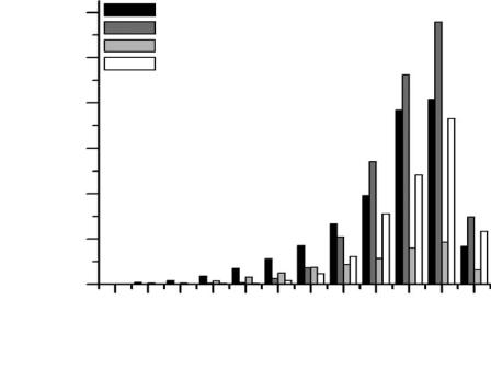
Until now, microarray technology has represented the most widespread platform for HTPS in biomedical experimentation and diagnostics. Its impact is reflected in the increasing volume of scientific literature related to microarrays and their applications (Figure 4.1) and in the growth of the microarray market from $232 million in 1999 to $2.6 billion in 2004.3
Classic solid phase substrates such as microtiter plates, membrane filters, and microscopic slides used in biotesting inspired the development of microarrays. These media effectively represent flat substrates that can be modified so as to possess multiple (often hundreds or thousands) probe sites. Each site bears a ligand or probe whose molecular recognition of a complementary molecule can produce a signal that, when detected by an imaging technology, most often fluorescence, can indicate the interaction both quantitatively and qualitatively. These probe spots are microto nanometer-sized.
Microarrays can be classified on the basis of the materials arrayed upon them (Figure 4.2). They have been constructed using DNA and nucleic acids (natural and synthetic), proteins, antibodies, carbohydrates, tissues, and cells. Numerous
Number of publications
3000
2500
2000
1500
1000
500
0
1980-1990 |
1991-1994 |
'HTPS' (Sci Finder)
'Microarrays' (Sci Finder)
'HTPS' (ISI Web of Science)
'Microarrays' (ISI Web of Science)
1995 |
1996 |
1997 |
1998 |
1999 |
2000 |
2001 |
2002 |
2003 |
2004 |
|
|
|
Year |
|
|
|
|
March |
|
|
|
|
|
|
|
|
|
||
Figure 4.1 Number of literature reports concerning microarrays and HTPS published from 1980 to 2004. Data extracted from ISI Web of Science and SciFinder on-line search engines.
\

78 |
BIOMEDICAL NANOTECHNOLOGY |
Protein array
DNA array
Cell membrane protein array
Microarray
Anitobdy array
Tissue array
Aptamer array
Cell array
Carbohydrate array
Figure 4.2 Types of flat surface microarrays by arrayed material.
examples of these arrays are commercially available, and descriptions of the most relevant formats will appear later in this chapter.
Newly emerging HTPS strategies are moving away from the classical formats. The new approaches may introduce significant benefits including diminished cost of fabrication and application and improvement in throughput. In this context, suspension arrays based on combinatorial libraries of encoded beads promise to enable ultra-HTPS analysis.4
Finally it is worth mentioning that microarrays would not be as effective as they are in HTPS without the help of microfluidics — a new term that defines any process or hardware involved in microvolume liquid management. In fact, HTPS and microfluidics overlap as commercial biosensors (biochips) often represent complicated networks of microsize channels, chambers, valves, and pumps.5
C. Nanotechnology and HTPS
Nanotechnology comes into play in the manufacture of microarrays and biochips because they benefit from the use of a great number of nanofabrication tools. Examples of nanotechnological contributions include (1) spatial positioning (microprinting and ink jetting) necessary for gridding arrayed materials in microto nanometer-scale spots; (2) patterning (photolithography) and microconstruction (micromachining,