

Biomedical Nanotechnology - Neelina H. Malsch
.pdfIMPLANTS AND PROSTHESES |
49 |
remodeling phase may last for several years. The general process of soft tissue remodeling involves rapid synthesis and degradation of connective tissue proteins.29 The degradation of these extracellular matrix (ECM) proteins is accomplished through the actions of matrix metalloproteinases (MMPs).30 The common outcome of soft tissue remodeling is scar formation, which results mainly from an imbalance between the stimulation of collagen synthesis and degradation of extracellular collagen.
Remodeling in hard tissues involves bone resorption by osteoclasts, followed by the synthesis of new bone matrix and its mineralization by osteoblasts. The remodeling process in hard tissues is subject to mechanical forces acting upon it (Wolff’s law31). In contrast with soft tissue remodeling, hard tissue remodeling is devoid of scarring. Furthermore, healed hard tissue is able to resume its original configuration.
B. Macrophages
Several cell types are involved in the biological processes that occur after the implantation of a biomaterial. The interplay among these cells is extremely important because inadequate cellular responses could directly or indirectly impede the functionality of the implanted device. Cells respond to stimuli mostly via receptors on their surfaces. Via these receptors, cells can recognize a large variety of ligands including soluble mediators secreted by other cells (cytokines), molecules present on the surfaces of adjacent cells, and distinct patterns in molecules of ECM proteins.
Due to their early appearance at an implantation site, their longevity, and the large number of cytokines they can produce and secrete, macrophages are generally considered the most important cell type in the vicinity of a newly implanted device.32 Macrophages perform multiple functions at a site of implantation ranging from phagocytosis of cell debris and potential pathogens via initiation of an inflammatory reaction to orchestration of the processes necessary to heal the damaged tissue resulting from the surgical procedure. In summary, the macrophages at an implantation site govern the magnitude and duration of all phases and subphases of the wound healing process by means of the versatility in the mediators they secrete that control the responses and functions of many other cell types.
C. Biomaterial Interface Processes
Although an implant is subject to cellular biological processes upon introduction, as described above, the initial contact of implant and host relies on noncellular interactions. A newly introduced implant is surrounded by an aqueous liquid. The water molecules in the direct vicinity of an implant can substantially alter the appearance of the biomaterial surface for the biological environment.33 The abundance of water molecules within this liquid means that water is the primary molecule involved in the first series of interactions of a biomaterial surface interface with its in vivo surroundings.
An important parameter is the free energy of a biomaterial surface reflected by its water wettability. Biomaterial surfaces are often categorized as hydrophobic or hydrophilic. A related parameter of biomaterial surfaces is cell adhesion. Although some authors assert the existence of a correlation between surface free energy and
\
50 |
BIOMEDICAL NANOTECHNOLOGY |
cell adhesion,34,35 others impugn this correlation36 or even postulate the inverse.37 The water molecules in the direct vicinity of the biomaterial surface will form a water monolayer or bilayer in which the arrangement of the water molecules depends on the surface properties at the atomic scale and completely differs from that of liquid water.33
Subsequent to interface interactions with water molecules, a biomaterial surface will first encounter ions and then the proteins present within the surrounding liquid. In the monolayer or bilayer of water molecules, natural ions (e.g., Na+ and Cl–) are incorporated as hydrated ions.38 The surface properties of the biomaterial determine the type, amount, and conformational state of the adsorbed proteins.39,40 Thus, the spectrum of adsorbed proteins will not necessarily reflect the amounts and ratios of the proteins within the surrounding liquid.41,42 Additionally, denaturation of the adsorbed proteins may occur. As a result, biologically important sites may become inaccessible or nonfunctional, limiting interactions with counter-receptors present on cellular membranes.
Finally, living cells will become involved. The presence of a wide variety of membrane-bound receptors on the surfaces of cells enables them to adhere to adsorbed proteins on the biomaterial surface. Because the interaction of cells with the biomaterial surface does not rely on direct contact between cells and biomaterial, but merely on an indirect interaction mediated by adsorbed proteins, it has been suggested that the biomaterial is not what causes unwanted responses.43 The nonspecific layer of proteins adsorbed on the biomaterial surface immediately after implantation is recognized by the host as a foreign or unnatural material. This assumption seems plausible because such an adsorbed mixture of proteins with random orientations and conformational states presents a divergence from natural, intentionally arranged protein layers.
D. Foreign Body Reaction
The cumulative effects of all separate contributive processes that occur at the biomaterial interface result in one of the following outcomes of implantation: (1) integration, (2) extrusion, (3) resorption, or (4) encapsulation. Although integration of the biomaterial device is the most favorable outcome, the number of cases in which true biointegration is achieved is limited.44 Most frequently, true biointegration occurs after implantation of compatible biomaterials such as titanium coated with hydroxyapatite (HA) into bone tissue.45,46 Implantation of biomaterials into soft tissues usually results in one of the other three outcomes.
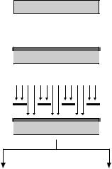
Extrusion occurs when an implanted device is in direct contact with epithelial tissue. The epithelium will form a pocket continuous with the adjacent epithelial membrane that subsequently dissipates the implant. In the case of external epithelium, the implant will be externalized from the host. Resorption of the implant can occur when an implant is made of degradable material. After complete resorption, only a collapsed scar will remain at the implantation site. In most cases, implanted biomaterials in soft tissues become encapsulated by a process known as the foreign body reaction2,47 (Figure 3.3). The capsule commonly consists of a relatively hypocellular membrane with a high collagen content.48 Adjacent to this collagenous

IMPLANTS AND PROSTHESES |
51 |
Figure 3.3 Foreign body reaction. The introduction of an implant (1) into a receptor leads to the adsorption of proteins in all possible configurations on the surface (2). Subsequently, cells (including macrophages) will attach to the implant surface via cell surface receptors that recognize corresponding ligands in the adsorbed protein layer (3). Attached cells secrete a wide variety of signal molecules that influence the behavior of perceptive cells (4) that become activated and start to produce extracellular matrix (5). Finally, the implant becomes enclosed in a fibrous capsule that isolates the implant from the body (6).
membrane, a layer of myofibroblasts is occasionally observed. Furthermore, foreign body giant cells (FBGCs; fused macrophages) are frequently observed in the space between implant and capsule.49,50
In general, the organization of cells and matrix surrounding an implant is built up in such a way that a barrier between the foreign material and the body is created, and this structure more or less isolates the implant from the body. The capsule,
\
52 |
BIOMEDICAL NANOTECHNOLOGY |
including FBGCs, surrounding the implant may persist for the lifetime of the implant. However, it is not yet clear whether FBGCs present at the biomaterial surface remain activated during the lifetime of the implant or become quiescent.47 Since encapsulated implants can perform their functions for many years, the isolation of an implant in a collagenous capsule is not necessarily an unwanted phenomenon. It may even help the body live in symbiosis with a synthetic device, although the presence of genuine symbiosis in this respect may be arguable. Unfortunately, the presence of myofibroblasts within the capsule may lead to contraction and thus cause pain and/or implant failure. Furthermore, the formation of a capsule associated or not associated with wearing of the biomaterial may result in loosening.
IV. NANOTECHNOLOGY IN IMPLANTOLOGY
A. Introduction
From the previous descriptions of biomaterial properties and interfacial biological processes, it is evident that the placement of an implant into a living organism causes specific reactions of the biological environment. The biomolecules and cells on the one hand and the intrinsic properties of the biomaterials on the other determine the biocompatibility and longevity of synthetic devices. Since the interaction of biomolecules and cells with the biomaterial surface is a vital element in evaluating the suitability of a biomaterial for its intended function, it is not necessary to note that every attempt to avoid undesired responses and/or enhance desired responses to implants is of utmost interest.
In many disciplines including biomaterial science, miniaturization has been a topic of interest for several years51 and led to the evolution of microtechnology techniques52,53 that allow the creation of features with microscale dimensions on biomaterial surfaces. Further expansion of many of these techniques, development of novel techniques, and focusing on medical applications resulted in expansion of the field of biomedical nanotechnology54 dealing with dimensions 1000-fold smaller than previously possible. In general, the emerging field of nanotechnology aims to increase control over material structures of nanoscale size in at least one dimension (x, y, or z).55 As already shown, microscale features can exert control over cellular behavior,56–59 and recent improvements in the field of nanotechnology may yield powerful additional tools to increase control over reactions of the biological environment to submicron cues in the direct vicinity of a biomaterial device.60
The general difference between microtechnology and nanotechnology is the size of the created microscale or nanoscale structures. Generation of nanoscale structures can be based on the miniaturization of higher scale structures (top-down) or on the assembly of nanoscale structures from ultimately small structures (bottom-up). The convergence of top-down and bottom-up strategies to create features with nanoscale dimensions via the collaboration of many scientific disciplines, for example, chemistry, physics, biology, and medicine, makes it possible to produce materials that resemble natural surroundings for biological entities.

IMPLANTS AND PROSTHESES |
53 |
The three-dimensional organizations of structures surrounding cells in vivo influence most cellular processes, e.g., adhesion, migration, growth, differentiation, secretion, and gene expression. The majority of such structures such as ECM components and membrane-bound receptors on cells encompass dimensions down to nanoscale size. The organization of cells and ECM proteins has been hypothesized to be of importance in controlling cellular behavior, and this was shown in an elegant experiment using multigrooves (a combination of microgrooves and macrogrooves).61 The experiment demonstrated that control over both cellular orientation and ECM orientation is feasible. Consequently, it was suggested that multigrooves may allow the production of three-dimensional ECM in vivo.
The introduction of nanodimensional structures on the surface of a biomaterial is possible by means of present nanotechnology, and such structures may influence biological reactions to implants and prostheses. Although distinct from natural nanostructures, synthetic nanostructures may be able to influence cellular responses to biomaterial implantation. Because nanotechnology is still in its infancy, future developments could expand the efficacy and thus the importance of creating nanostructures on biomaterial surfaces. A number of nanotechnology-based methods to modify biomaterial surfaces are described below and the effects of such nanotechnologically modified biomaterial surfaces on cell behavior will be discussed.
B. Current Nanofabrication Methods
The production of nanostructures on biomaterial surfaces is an emerging field of technology that may involve utilization of many techniques. Several, but certainly not all, methods for the fabrication of biomaterial surfaces with nanoscale topological or chemical cues are listed in Table 3.2. Their principles are described below. In general, nanotechnological modifications of biomaterial surfaces can be categorized into those that alter a surface topographically and those that introduce nanoscale chemical molecules (or groups) on a surface. The techniques described below, however, do not necessarily restrain themselves to one of these types of modifications. Many techniques can serve multiple purposes, for example, using some kind
Table 3.2 Available Methods for Nanofabrication of Biomaterial Surfaces
Type of System |
Materials |
Resolution |
Lithography |
Silica, silicon, silicon nitride, |
x, y, and z to 10 nm |
|
silicon carbide |
|
Colloidal resist |
Silica, silicon, silicon nitride, silicon carbide |
x, y, and z to 5 nm |
Self-organizing or |
Polymer demixing, self-assembling particles |
In 10-nm range |
self-assembling |
and monolayers, other self-assembling |
|
|
systems |
|
Soft lithography |
Any fairly large molecule |
x and y to 200 nm, z |
|
|
to one monolayer |
Biomimicry |
Many |
Actual native |
|
|
dimensions |
Source: Partly adapted from Curtis, A. and Wilkinson, C. Trends Biotechnol, 19, 97–101, 2001.130
\
54 |
BIOMEDICAL NANOTECHNOLOGY |
of mask may involve either topography or chemistry. Topography can be further specified as having either texture or roughness. The difference between texture and roughness is determined by the regularity of the topographical cues. While texture is characterized by an organized regularity in topography, roughness encompasses a random topography.62
1. Lithography
Figure 3.4 depicts the basic principles of photolithography. Lithography is a technique by which a material is coated with a film prior to the creation of desired features. The film is usually a polymer that is sensitive to a particular type of energy applied. Polymers sensitive to light or to electrons can be used. Depending on the sensitivity of the polymer (also called the resist), lithographical techniques are categorized as photolithography (light-sensitive resist) or electron beam lithography (electron-sensitive resist).
The irradiation of a specific pattern in a sensitive polymer modifies the polymer properties in that area. A subsequent dissolution step removes the affected sensitive polymer, leaving a specific pattern of sensitive polymer at the surface of a biomaterial. Photolithography commonly employs a mask to allow control over the irradiation of the resist, whereas in electron beam lithography, the beams of electrons can be focused at and maneuvered to the desired positions to gain control over the
Material substrate
Resist
1. Coating
Material substrate
Light source
Mask
2. Irradiation
Resist
Material substrate
Resist |
|
|
|
|
|
|
|
|
|
|
|
|
|
|
|
|
|
|
|
|
|
|
|
|
|
|
|
3. Development |
Material substrate |
|
|
|
|
|
|
|
|
|
|
|
|
|
|
|
|
|
|
||||||||||
|
|
Negative tone |
|
|
|
|
|
Positive tone |
||||||||||||||||||||
|
|
|
|
|
|
|
|
|||||||||||||||||||||
Figure 3.4 Photolithography techniques. In conventional lithography, a resist is coated on a material substrate and the resist is subsequently irradiated through a mask, creating a pattern corresponding to the mask in the resist. Development of the resist will result in a positive or negative tone on the material surface that can be used for coating or etching techniques.
IMPLANTS AND PROSTHESES |
55 |
irradiated zone. Two types of further modification of the surface from which the polymer has been removed can be applied: (1) etching and (2) film deposition. Etching allows pits, grooves, and other topographies of controlled shape and size to be created. On the other hand, the deposition of a thin film basically relies on coating the exposed area with a desired solution, from which the solvent evaporates or in which the particles (molecules) organize themselves in a specific conformation (selfassembly). The selectivity of and the precision by which the energy used to irradiate the sensitive polymer is applied determine the range of the dimensions of patterns that can be created. Generally, the resolution of conventional photolithography is 300 nm, whereas lithography features down to 10 nm in size can be created with electron beam lithography.38a
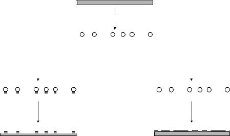
2. Colloidal Resists
In addition to masks (as in photolithography) or precision maneuvers of an electron beam (as in electron beam lithography) the application of colloidal particles is possible (Figure 3.5). Colloidal particles of different materials and sizes down to 5 nm can be produced and subsequently dispersed over a biomaterial surface. The distribution (e.g., density) of the particles on a surface can be controlled by the salinity38b and acidity (pH) of the solution. Subsequently, the adsorbed particles can be used as a template for patterning the underlying surface. In a technique similar to photolithography and electron beam lithography, the space not covered by colloidal particles can be etched or a thin film can be deposited. After removal of the colloidal particles, a patterned surface remains. Using colloidal resist techniques followed by etching or thin film
Colloidal dispersion
|
|
|
|
|
|
|
|
|
|
|
|
|
|
|
|
|
|
|
|
|
|
|
|
|
|
|
|
|
|
|
|
|
|
|
|
|
|
|
|
|
|
|
|
|
|
|
|
|
|
|
|
|
|
|
|
|
|
|
|
|
|
|
|
|
|
|
|
|
|
|
|
|
|
|
|
|
|
|
|
|
Etching |
|
|
|
Thin film deposition |
|||||||||
|
|
|
|
|
|
|
|
|
|
|
|
|
|
|
|
|
|
|
|
|
|
|
|
|
|
|
|
|
|
|
|
|
|
|
|
|
|
|
|
|
|
|
|
|
|
|
|
|
|
|
|
|
|
|
|
|
|
|
|
|
|
|
|
|
|
|
|
|
|
|
|
|
|
|
|
|
|
|
|
|
|
|
|
|
|
|
|
|
|
|
|
|
|
|
Removal of colloidal particles
Figure 3.5 Colloidal resist techniques. A colloidal suspension is dispersed on the surface of a material. Subsequent etching or coating, followed by removal of the colloidal particles, results in a pattern on the material surface.
\
56 |
BIOMEDICAL NANOTECHNOLOGY |
deposition and subsequent removal of the colloidal particles, the variations in the pattern are related to particle size and spatial distribution.
3. Self-Assembly Systems
Self-assembly is a common phenomenon in nature. It is described as a “spontaneous association of numerous individual entities into a coherent organization and well-defined structures to maximize the benefit of the individual without external instruction.”63 If this phenomenon is downscaled to smaller entities, molecular self-assembly results. Molecules organize spontaneously into structurally well defined and rather stable arrangements via noncovalent interactions under equilibrium conditions.64
The formation of a cell membrane from single phospholipid moieties is a good example of naturally occurring self-assembly at the molecular scale. It becomes apparent from this example that self-assembly allows the formation of stable structures, whereas single noncovalent interactions may be somewhat weak and collective interactions can be more than sufficient to create very stable structures and materials. The establishment of a self-assembling system relies on chemical complementarity and structural compatibility. Therefore, a vital prerequisite for self-assembly is the use of molecules of correct size and orientation (chirality). Monolayers of molecules with distinct properties exposed at the “new” surface can be generated and are designated self-assembling monolayers, or SAMs.
One common application of SAM technology is protein patterning. The generation of a self-assembled monolayer of molecules (e.g., alkylsilanes or alkane thiol molecules) into an organized layer10 results in the possibility of effectively modulating the properties of the layer of free end groups. Via variation of unique reactive end groups in the SAM, homogeneous interactions (hydrophilic end group with hydrophilic protein and v.v.) with proteins can provide a mechanism of protein patterning.
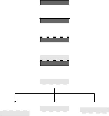
4. Soft Lithography
Soft lithography (Figure 3.6) is a term collectively used for a group of lithographic techniques in which a patterned elastomer, usually poly(dimethylsiloxane) or PDMS, is used to generate or transfer this specific pattern via molding, stamping, or masking onto a biomaterial surface. Additionally, the PDMS inverse replica can be used as a “master” to generate positive replicas of the original template.
Microcontact printing is a soft lithographic technique using the contact of the relief pattern of the PDMS stamp with the biomaterial surface to generate a pattern on the latter. Prior to the moment of contact between the PDMS stamp and the biomaterial surface, the stamp is “inked” to create the pattern of the stamp at the biomaterial surface. Most commonly, microcontact printing is used together with SAMs on gold substrates. Patterns of specific SAMs can be created, after which the intrapattern space can be filled using another SAM. SAMs have, respective to their chemical properties, selective adsorption profiles for proteins. Selecting appropriate SAMs and designing them into a pattern can control protein adhesion. Such
IMPLANTS AND PROSTHESES |
57 |
Silicon wafer
Resist
'master'
PDMS casting
PDMS inverse replica
|
|
|
|
|
|
|
|
|
|
|
|
|
|
|
|
|
|
|
|
|
|
|
|
|
|
|
|
|
|
|
|
|
|
|
|
|
|
|
|
|
|
|
|
|
|
|
|
|
|
|
|
|
|
|
|
|
|
|
|
|
|
|
|
|
|
|
|
|
|
|
|
|
|
|
|
Positive replica |
|
|
Microcontact printing |
|
|
Microfluidic patterning |
||||||||||||
Figure 3.6 Soft lithography techniques. Using conventional lithography techniques, a master is prepared, onto which PDMS is cast.The PDMS inverse replica can subsequently be used to create patterns (via etching, coating, etc.) on material surfaces via techniques like casting, microcontact printing, and microfluidic patterning.
patterned, protein-containing surfaces can serve as ligands for cell receptors, thus providing the opportunity for directed cell attachment.65 In addition to indirect protein immobilization through SAMs, direct patterning of proteins using microcontact printing is also possible.
Microfluidic patterning is a technique using the network of microchannels created during contact of the PDMS stamp for the generation of patterns on a biomaterial surface. Via these microchannels, fluids can be delivered to selected areas of a substrate. In microcontact printing, the pattern is created at the sites of contact between stamp and biomaterial. In contrast, in microfluidic patterning, the areas where the stamp is not in contact with the biomaterial are responsible for the
\
58 |
BIOMEDICAL NANOTECHNOLOGY |
patterning. Depending on the type of fluid used, several possibilities for creation of a pattern are feasible: (1) solidization of the fluid, (2) deposition of soluble constituents, or (3) removal of underlying material.
5. Biomimetic Approaches
A completely different approach regarding the modulation of implant surfaces is the use of biomimicry. Biomimetic approaches attempt to create an implant surface, which is not, or to a lesser extent, recognized as foreign by the host. Constituents of the natural cellular environment (i.e., ECM proteins) that often have nanoscale dimensions can be of help in creating biomimetic surfaces.
Under natural conditions, cellular functions are regulated via interactions of cells with their direct surroundings, and cells recognize specific components of their surroundings, including ECM components. For that reason, research has focused on mimicking such surroundings on biomaterial surfaces, both topographically and biologically. Much effort has been devoted to creating biomaterial surfaces that contain elements of native ECM proteins. Such proteins have been demonstrated to contain domains that can influence cell behavior. Receptors located on the surface of a cell can recognize such domains that can function as their counterparts (ligands; key–lock principle).66 The interactions of the receptor family of integrins with such domains are particularly known for their impacts on cellular processes.67,68 For example, adhesion of cells to specific domains of ECM proteins can be achieved via receptor-mediated interactions.
Additionally, receptor-mediated interactions can influence other cellular processes including proliferation, migration, morphological change, gene expression, and cell survival by intracellular signaling. The introduction of native ECM components onto the surfaces of biomaterials is an interesting modification method that can generate a biomaterial interface akin to a natural one (biomimicry) onto which cellular behavior can be influenced. An additional prospect of using ECM components for the generation of biomimetic surfaces involves the capacity of ECM components to strongly bind growth factors69,70 that can further modulate cellular behavior, depending on the type of growth factor applied.
In general, three major methods exist for the immobilization of biomolecules such as proteins and peptides onto surfaces: (1) physical adsorption (e.g., via van der Waals or electrostatic interactions); (2) physical entrapment (use of a barrier); and (3) covalent attachment. In addition to these methods, more sophisticated techniques such as covalent linking to polymeric networks can be used to generate biomimetic surfaces containing elements of native ECM components.71,72
Although adsorption of entire proteins (e.g., fibronectin) is demonstrated to be effective in enhancing cellular attachment,42 research has focused on the design of materials representing only parts of ECM proteins. Generally, these parts (or peptides) are based on the primary structure of the receptor-binding domain of an entire protein such as fibronectin or laminin. These peptides, whether linear or cyclized, can possess similar functionalities, for example, receptor specificity, binding affinity, and signaling of cell responses, compared to their native proteins.73,74