

Emergency Echocardiography
.pdf106 ECHOCARDIOGRAPHY IN ACUTE AORTIC SYNDROMES
intramural echolucent zone6 (Fig. 7.4). The diagnosis of IMH as opposed to severe atherosclerotic plaque is supported by demonstrating the intimal layer overlying the abnormal area rather than being underneath but the differentiation from a severely diseased atherosclerotic aortic wall may be difficult.7 An even greater challenge may be the differentiation from the thrombosed false lumen of a dissection without a detectable intimal tear, though in this case a misdiagnosis is less critical since the treatment is dictated by the localization of the aortic process and not by differentiating between the two entities.
7.4 PENETRATING AORTIC ULCER
Penetrating aortic ulcer is the result of an atherosclerotic ulceration disrupting the internal elastic lamina and advancing through the media with possible involvement of the full aortic wall thickness. The clinical presentation may be similar to that of a typical dissection and the process can develop to full-pledged dissection, IMH, pseudoaneurysm formation, or rupture and, in fact, these pathologies can coexist in the same patient.8 Since PAU is strongly associated with aortic atherosclerosis, it involves mainly the descending aorta where severe atherosclerotic disease is more likely. The typical appearance of a penetrating ulcer is described as an outpouching of the aortic wall, generally associated with complex atherosclerotic wall changes.8 The optimal management of PAU is still to be defined but recent reports suggest a good outcome using endovascular stent grafting.9,10
References
1. Januzzi JL, Isselbacher EM, Cooper JV, et al. Characterizing the young patient with aortic dissection: results from the International Registry of Aortic Dissection (IRAD). J Am Coll Cardiol. 2004;43(4):665-669.
2. Erbel R, Alfonso F, Boileau C, et al. Diagnosis and management of aortic dissection. Eur Heart J. 2001;22(18):1642-1681.
3. Ince H, Nienaber CA. Diagnosis and management of patients with aortic dissection. Heart. 2007;93(2):266–270.
4. Nienaber CA, Eagle KA. Aortic dissection: new frontiers in diagnosis and management: part I: from etiology to diagnostic strategies. Circulation. 2003;108(5):628–635.
5. Nienaber CA, von Kodolitsch Y, Nicolas V, et al. The diagnosis of thoracic aortic dissection by noninvasive imaging procedures. N Engl J Med. 1993;328(1):1–9.
6. Evangelista A, Avegliano G, Elorz C, González-Alujas T, del Castillo HG, Soler-Soler J. Transesophageal echocardiography in the diagnosis of acute aortic syndrome. J Card Surg. 2002;17(2):95–106.
7. Flachskampf FA. Assessment of aortic dissection and hematoma.
Semin Cardiothorac Vasc Anesth. 2006;10(1):83–88.

7.4 PENETRATING AORTIC ULCER 107
8. Vilacosta I, San Roman JA, Aragoncillo P et al. Penetrating atherosclerotic aortic ulcer: documentation by transesophageal echocardiography. J Am Coll Cardiol. 1998;32(1):83–89.
9. Pauls S, Orend KH, Sunder-Plassmann L., Kick J, Schelzig, H et al. Endovascular repair of symptomatic penetrating atherosclerotic ulcer of the thoracic aorta. Eur J Vasc Endovasc Surg. 2007;34(1):66–73.
10. Botta L, Buttazzi K, Russo V et al. Endovascular repair for penetrating atherosclerotic ulcers of the descending thoracic aorta: early and midterm results. Ann Thorac Surg. 2008;85(3):987–992.

Chapter 8
Echocardiography in Acute
Pulmonary Embolism
High-risk pulmonary embolism (PE) is associated with significant right ventricular dilatation and hypocontractility. Major PE can be safely ruled out as a cause of hemodynamic deterioration if the right ventricle appears normal on a bedside echocardiographic study. This chapter details the role of emergency echocardiography in the diagnosis of patients with suspected PE, including the possible visualization of the thrombus with transesophageal echocardiography.
8.1 INTRODUCTION
PE is suspected whenever a patient presents with chest pain, dyspnea, hypoxemia, suggestive ECG changes, and an elevated D-dimer. Right ventricular failure and hemodynamic compromise define massive or high-risk PE while evidence of right ventricular (RV) impairment without hemodynamic instability define nonmassive or intermediate-risk PE.1 Echocardiography is potentially useful in all cases but it has a special role in highand intermediate-risk PE. These patients may be acutely ill with hypoxemia and cardiogenic shock and emergent echocardiography may strongly support the diagnosis of massive PE or make it unlikely. As a general rule, massive PE should cause significantly enough right ventricular enlargement and systolic dysfunction to be easily recognized on echocardiography (Fig. 8.1). Conversely, the absence of these findings practically rules out PE as the cause for the patient’s hemodynamic deterioration.
109
A. Chenzbraun, Emergency Echocardiography, DOI: 10.1007/978-1-84882-336-5_8, © Springer-Verlag London Limited 2009

110 ECHOCARDIOGRAPHY IN ACUTE PULMONARY EMBOLISM
FIG. 8.1. Apical four-chamber view imaging in a patient with acute onset of chest pain, hypoxemia, and hypotension. There is severe dilatation and hypokinesis of the right ventricle and the interventricular septum is displaced toward an underfilled left ventricle. Massive pulmonary embolism (PE) was subsequently confirmed by computed tomography (CT) angiography.
Positive findings to be actively sought and reported in suspected massive PE are:
•RV enlargement and hypocontractility*
•Pulmonary artery dilatation
•Tricuspid regurgitation
•Dilated interior vena cava (IVC) with reduced respiratory varia tions
•Evidence for pulmonary hypertension**
°As demonstrated by tricuspid regurgitation (TR) peak systolic velocity
°As inferred by systolic flattening of the interventricular septum
* The differential diagnosis between preexistent RV hypokinesis due to chronic lung disease and acutely developed RV failure due to PE may be occasionally challenging. Among others, preserved normal contractility of the RV apex in the presence of hypokinesis of the other RV free wall segments (McConell sign) has been reported as specific for the acute cor pulmonale, secondary to PE.1
8.1 INTRODUCTION 111
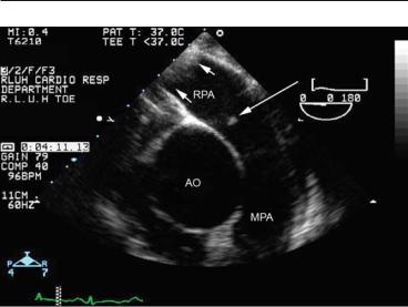
FIG. 8.2. Emergency transesophageal echocardiography (TEE) imaging of the pulmonary artery (PA) in a patient with evidence of a right atrial mass on transthoracic echocardiography (TTE), who became acutely hypoxemic. A small thrombus (long arrow) is seen at the origin of the right pulmonary artery, which more distally is almost totally obstructed by a large thrombus (short arrows). Computed tomography (CT) angiography confirmed multiple pulmonary emboli.
** The pulmonary artery (PA) pressure may be only moderately elevated with systemic hypotension and acute RV failure. PA pressure to systemic pressure ratio and IVS flattening may be more useful and demonstrate relative pulmonary hypertension even with relatively low TR velocities.
There is no practical role for echocardiography in the immediate diagnosis of suspected low-risk PE.
Though CT angiography is classically used to demonstrate PE, TEE is, again, a convenient alternative in critically ill patients who need a rapid, bedside diagnosis. The main PA, its bifurcation, and the right PA are easily visualized in most patients (Fig. 2.4c, 8.2). Since massive PE is frequently associated with either bilateral or main PA emboli, TEE should be able to visualize the obstructing embolus in these patients and indeed sensitivity and specificity of 80% and 100%, respectively, have been reported in severe PE.2 Accordingly, TEE may be considered as a valid imaging method in critically ill patients with suspected massive PE allowing a rapid diagnostic confirmation.3

112 ECHOCARDIOGRAPHY IN ACUTE PULMONARY EMBOLISM
References
1. Torbicki A, Perrier A, Konstantinides S, et al. Guidelines on the diagnosis and management of acute pulmonary embolism: the task force for the diagnosis and management of acute pulmonary embolism of the European Society of Cardiology (ESC). Eur Heart J. 2008;29(18):2276–2315.
2. Pruszczyk P, Torbicki A, Pacho R, et al.. Noninvasive diagnosis of suspected severe pulmonary embolism: transesophageal echocardiography vs spiral CT. Chest 1997;112(3):722–728.
3. Krivec B, Voga G, Zuran I, et al. Diagnosis and treatment of shock due to massive pulmonary embolism: approach with transesophageal echocardiography and intrapulmonary thrombolysis. Chest 1997;112(5):1310–1316.

Chapter 9
Echocardiography in Stroke
and Systemic Embolism
This chapter covers the main pathologies associated with stroke or a peripheral embolic event in acutely ill patients, with implications for an echocardiographic diagnosis.
Generally accepted algorithms are in place for the use of echocardiography in the work up of patients after an ischemic stroke or a peripheral embolism.12 Depending on the overall clinical picture, the occurrence of an embolic event in a critically ill patient raises some specific diagnostic questions as detailed below.
•Left ventricular (LV) thrombus in a patient with acute/recent myocardial infarction
°More likely with apical MI (Fig. 6.2)
•Left atrial thrombus in a patient with atrial fibrillation (Fig. 9.1)
°More likely with mitral valve disease or LV dysfunction
•Native or prosthetic valve endocarditis
•Prosthetic valve thrombosis
•Aortic dissection
°The tributary artery feeds from the false lumen
All these conditions are echocardiographic diagnoses so echocardiography should be used liberally in this clinical setup or if the presenting event is a systemic embolus, which, due to the size necessary to obstruct a peripheral or visceral artery is likely to be cardiac in origin.
113
A. Chenzbraun, Emergency Echocardiography, DOI: 10.1007/978-1-84882-336-5_9, © Springer-Verlag London Limited 2009
114 ECHOCARDIOGRAPHY IN STROKE AND SYSTEMIC EMBOLISM
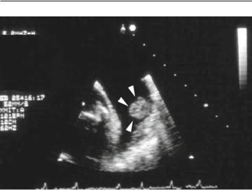
FIG. 9.1. Rounded, protruding thrombus (arrow heads) in a large left atrial appendage in a patient with atrial fibrillation.
References
1. Cheitlin MD, Alpert JS, Armstrong WF, et al. ACC/AHA guidelines for the clinical application of echocardiography: executive summary. A report of the American College of Cardiology/American Heart Association Task Force on Practice Guidelines (Committee on Clinical Application of Echocardiography). Developed in collaboration with the American Society of Echocardiography. J Am Coll Cardiol. 1997;29(4):862–879.
2. Douglas PS, Khandheria B, Stainback RF, et al. ACCF/ASE/ACEP/ ASNC/SCAI/SCCT/SCMR 2007 appropriateness criteria for transthoracic and transesophageal echocardiography: a report of the American College of Cardiology Foundation Quality Strategic Directions Committee Appropriateness Criteria Working Group, American Society of Echocardiography, American College of Emergency Physicians, American Society of Nuclear Cardiology, Society for Cardiovascular Angiography and Interventions, Society of Cardiovascular Computed Tomography, and the Society for Cardiovascular Magnetic Resonance endorsed by the American College of Chest Physicians and the Society of Critical Care Medicine. J Am Coll Cardiol. 2007;50(2):187–204.

Chapter 10
Echocardiography
in the Septic Patient
Maintenance of cardiac output may be compromised in sepsis due to a combination of myocardial depression and inappropriate vasodilatation. Echocardiographic information on both ventricular contractility and filling status is paramount for appropriate management. This chapter summarizes the role of echocardiography as an increasingly accepted alternative to invasive hemodynamic monitoring in septic patients.
Sepsis is diagnosed when there is evidence of both an infectious process and a systemic inflammatory response syndrome. The cardiovascular response to sepsis is complex and multifactorial, related to endothelial and coagulation system activation, microvascular dysfunction and release of numerous mediators, some of which, such as cytokines and prostanoids, act as myocardial depressors.1 The hemodynamic hallmarks of sepsis are myocardial depression, which can range from occult to frank pump failure and inappropriate peripheral vasodilatation, which induces a hyperdynamic state and thus can mask mild degrees of myocardial dysfunction. The majority of septic-shock patients will be hyperdynamic and vasodilated and benefit from fluid resuscitation and vasoconstrictors such as norepinephrine or dopamine. A sizable minority of about one-third of the cases2,3 can present nevertheless with decreased cardiac index and evidence of reduced ejection fraction and will require inotropic support with dobutamine as well.
Echocardiography in this setup can be performed as either a transthoracic or a transesophageal study depending on images quality and the existence of a specific indication for transesophageal
115
A. Chenzbraun, Emergency Echocardiography, DOI: 10.1007/978-1-84882-336-5_10, © Springer-Verlag London Limited 2009

116 ECHOCARDIOGRAPHY IN THE SEPTIC PATIENT
echocardiography (TEE). The main objectives of echocardiographic evaluation in the septic patient and the relevant echo findings to be specifically sought are:
•Confirmation of a hyperdynamic state and low filling pressures (see Sect. 3.3 and Fig. 3.2).
°Hyperdynamic contraction of both ventricles; this is a subjective assessment, which, however, should be within the competence of even less-experienced operators.
°E/A < 1 and/or E/E´ < 10 by Doppler interrogation of the mitral valve and annulus are:
■less useful with tachycardia when fusion of the earlyand late-diastolic waves can occur.
°Optional: actual measurement of cardiac index using the velocity time integral of the aortic flow and the aortic diameter (Appendix B)
•Prediction of response to fluid challenge:
°> 50% respiratory changes in inferior vena cava diameter
°> 60% respiratory changes in superior vena cava diameter (use TEE in ventilated patients)
•Exclusion/assessment of concomitant obvious systolic dysfunction.
•Unmasking of previously unrecognized left ventricular (LV) systolic dysfunction when the systemic vascular resistance is increased by using vasopressors. This requires a protocol of echocardiographic monitoring and may help to decide putting the patient on dobutamine as well.
•Confirmation or exclusion of possible endocarditis, when there are reasonable grounds to suspect this diagnosis.
The hemodynamic information input of echocardiography in these patients is of such quality and reliability that it should be seen as a viable alternative to invasive monitoring. In fact, some units report using repeat echo assessment as only mean of monitoring their septic patients.3
References
1. Merx MW, Weber C. Sepsis and the heart. Circulation 2007;116(7): 793–802.
2. Jardin F, Brun-Ney D, Auvert B, et al. Sepsis-related cardiogenic shock. Crit Care Med. 1990;18(10):1055–1060.
3. Vieillard-Baron A, Prin S, Chergui K, et al. Hemodynamic instability in sepsis: bedside assessment by Doppler echocardiography. Am J Respir Crit Care Med. 2003;168(11):1270–1276.