

Emergency Echocardiography
.pdf2.2TRANSESOPHAGEAL ECHOCARDIOGRAPHY 45
□Structures: MV, proximal interventricular septum (IVS), aortic root and AV
°90° with clockwise rotation (bicaval view)
□Structures: SVC, interior vena cava (IVC), right atrial appendage (RAA)
Position V: Transgastric (Fig. 2.7).
°At 0°
□Equivalent transthoracic view: PSSA
□Structures: LV and RV
°At 90°–120°
□Equivalent transthoracic views: parasternal long-axis (PSLA) and 2-chamber view
□Structures: LV, MV, subvalvular apparatus
Position VI (Fig. 2.8): Withdraw to a mid-esophageal level and then rotate shaft posteriorly in a counter-clockwise direction with transducer at 0° until the thoracic descending aorta is visualized in a short-axis cut. Advance the probe until the aorta is lost at diaphragmatic level and then withdraw slowly until it changes course and disappears again at distal arch level. Shaft rotation may be necessary to follow a tortuous aorta. When pathology is noted, the level should be recorded using the
FIG. 2.7. Transgastric views. These are essential if the main query is left ventricle contractility or mitral subvalvular pathology, but are not mandatory if this information is available from the transthoracic study. a Cross-sectional view allowing good visualization of ventricular walls at mid-ventricle level. b Long-axis view.
FIG. 2.6. (continued) level. b 120°: long-axis view of the left ventricular outflow tract, mitral and aortic valve and aortic root. c 90° with rightwards rotation of the transducer. The junctions of the two vennae cavae and the interatrial septum at fossa ovalis level (thin arrow) are visualized at this level. Thick arrow: crista terminalis.
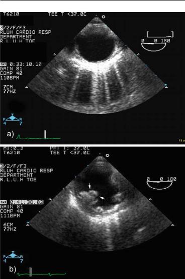
46 EMERGENCY ECHOCARDIOGRAPHY
FIG. 2.8. Cross-sectional view of the descending thoracic aorta. This view is mandatory for type B aortic dissection, intramural hematoma or aortic atherosclerosis. a Normal aorta. b Severe atherosclerosis of descending aorta with complex plaque and mobile atheroma (arrows), found as an incidental finding in a patient undergoing TEE before cardioversion; this underscores the importance of routine scanning of the aorta.

2.3HAND-HELD ECHOCARDIOGRAPHY 47
TABLE 2.2. Frequently encountered clinical scenario and recommended structure-targeted modular approach in unstable patients when a brief TEE examination is required.
Dg. module |
“Not to be missed” echo targets |
Essential views |
(a) Hemodynamic |
• LV/RV contractility |
I, IV, VI |
instability |
• LV/RV dimensions |
|
|
• LV Doppler filling pattern by |
|
|
PW Doppler of mitral inflow |
|
(b) Difficulty to wean |
• LV/RV contractility |
I, IV, VI |
from ventilator |
• MR presence |
|
|
• R L shunt (PFO) |
|
(c) Aortic dissection |
• Aortic root |
II, III, VII |
|
• Descending aorta |
|
(d) Massive pulmo- |
• RV size and function |
I, II, IV |
nary |
• Pulmonary artery size and |
|
embolism |
scanning for clot |
|
|
• TR |
|
PFO patent foramen ovale, TR tricuspid regurgitation, RV right ventricle, LV left ventricle, MR mitral regurgitation, PW pulsed wave
distance markers on the probe shaft. Long-axis cuts should also be used for detailed imaging of wall pathology.
If the patient is unstable use a diagnostic module approach (Table 2.2) and start with a “structure targeted” study to address the main clinical question. Complete the study if possible, after obtaining the critical information.
2.3 HAND-HELD ECHOCARDIOGRAPHY
IN THE CRITICALLY ILL PATIENT
Hand-held echocardiography, formally referred to as hand-carried ultrasound (HCU), is an ultrasound diagnostic modality whose role and indications are under continuous review reflecting evolving technological advances. A 2002 report of the American Society of Echocardiography emphasizes HCU as an extension of physical examination while acknowledging that revisions of this document may be required to reflect ongoing technologically driven developments. A wealth of data has accumulated to support the reliability of HCU echocardiography when compared with standard platform systems with best results for two-dimensional information and somewhat less for color Doppler.15

48 EMERGENCY ECHOCARDIOGRAPHY
2.3.1 What Is a HCU?
A HCU system will typically fulfil the following criteria:
•Light (usually < 6 lbs)
•Easily portable, without the need of a carrying trolley
•Battery powered
•Significantly cheaper than a conventional system
2.3.2 Particularities of HCU Imaging
Because of the basic capabilities of first generation devices and the initial scope of HCU use mainly as a convenient aid to bedside examination, some study limitations are accepted as compared with a standard examination:
•Study duration:
°brief (<5–10 min)
•Scope:
°targeted to confirm or rule out a major finding or clinical diagnosis:
□Pericardial effusion
□Severe systolic dysfunction
□Severe hypertrophy
□Severe valvular pathology
•Imaging modalities:
°possibly limited (i.e., spectral Doppler may not be available)
•Image quality:
°possibly limited:
□small screen size
□restricted color capabilities
•Report:
°not standardized, limited, findings may be only briefly mentioned in the patient’s notes
•Images storage:
°optional, thus without the possibility of later review
•Accepted minimal training level of the operator: Level 1
However, with continuous technological advances, some of these limitations may become irrelevant and the capabilities of new models tend to blur the differences between a high-end HCU and a portable conventional system (Figs. 2.9 and 2.10).
2.3.3 HCU Echocardiography in the Critically Ill
HCU devices should be used in critically ill patients mainly for screening purposes and assessment at the time of the first evaluation. Because of their portability and good two-dimensional images, they may be the preferred tool for an instant, emergency assessment as outlined in Fig. 2.1. The results of the scan may
2.3HAND-HELD ECHOCARDIOGRAPHY 49
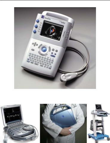
FIG. 2.9. Light, hand-held SonoHeart Elite echocardiographic unit.
FIG. 2.10. Newer generation M-Turbo unit, still light and portable but suitable for trolley-mounting and carrying as well. (Images used in Figs. 2.9 and 2.10 are courtesy of SonoSite, Inc., 21919 30th Drive SE, Bothell, WA 98021-3904).
immediately point to the underlying pathology, for example, empty ventricles, tamponade or severe systolic dysfunction, making a comprehensive standard examination not necessary at that moment though it may still be required at a later stage. Despite the expected quality limitations in difficult to image ICU patients, HCU scanning was found to have a diagnostic accuracy and therapeutic impact similar to those of conventional systems, mainly for two-dimensional imaging.16,17

50 EMERGENCY ECHOCARDIOGRAPHY
2.4 CONTRAST ECHOCARDIOGRAPHY
IN THE CRITICALLY ILL PATIENT
Contrast echocardiography can be performed either as a “conventional study” with agitated saline, for right heart visualization, or using transpulmonary agents for left heart imaging.
2.4.1 “Conventional” Contrast Echocardiography for Right-to-Left Shunts
Right-to-left shunts occur mainly at interatrial septal level through a PFO or previously unrecognized ASD and, occasionally, at pulmonary level through arteriovenous fistulae, either isolated or in association with liver disease (hepatopulmonary syndrome). A PFO is present in about 25–30% of the general population; the magnitude and occurrence of the shunt may increase with pulmonary hypertension, which is frequently encountered in ICU patients due to either cardiac or primary lung conditions. A right- to-left shunt should be suspected in cases of persistent hypoxemia (see Sect. 2.4) or unexplained embolic stroke. Color Doppler is not sensitive enough, especially for transthoracic studies and small shunts, and a formal contrast study should be performed when a right-to left shunt is suspected. Rapid injection of agitated saline through an antecubital or central vein is the most practical way to check whether a right-to-left shunt is present. The solution is
prepared by vigorous mixing of 10 ml normal saline with a small amount ( 0.5 ml) of air using two syringes, preferably of Luer lock
type, connected by a three-way stopcock. Apical 4-chamber imaging by either TTE (Fig. 2.11) or TEE is the view of choice, though a bicaval view should be used as well with TEE. A first injection is generally performed without provocation and, in cooperative patients, is repeated with provocation using:
•A Valsalva manoeuvre with the patient instructed to build the pressure during injection and to abruptly release it when the contrast reaches the right heart
•A forceful cough at the time of right heart opacification
The efficacy of a provocation manoeuvre during TEE can be checked by visualizing a brief bowing of the IAS towards the LA due to the transient raise of the right atrium (RA) pressures. Some patients are not able to perform an effective Valsalva manoeuvre or to cough during a TEE study. A high-quality TTE with good provocation should, therefore, complete a negative TEE or even may obviate the need for it if presence or absence of the shunt was the only query.18 In ventilated patients, manual inflation of the lungs, followed by
2.4 CONTRAST ECHOCARDIOGRAPHY 51
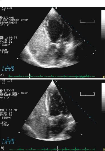
FIG. 2.11. Agitated saline injection in a young patient with unexplained transient ischemic attack (TIA). a Good contrast injection without provocative maneuver fully opacifies the right heart without any passage of bubbles to the left heart. b Repeat injection with a vigorous cough shows a moderate amount of bubbles in the left cavities, making this a positive test for right-to-left shunt.

52 EMERGENCY ECHOCARDIOGRAPHY
abrupt release, may be equivalent to a Valsalva manoeuvre.15 If left cavities opacification does occur, both its timing and magnitude should be noted. A shunt is diagnosed if 1 bubble is seen in the left heart, though some studies required 3 bubbles for clinical significance.19 The appearance of contrast later than five cycles after right heart opacification suggests intrapulmonary rather than intracardiac shunt. The number of bubbles observed in the left heart defines the shunt magnitude with some authors considering 10 and others 30 bubbles as the cut-off for a large shunt.19 Of note, the magnitude of the shunt as defined by the number of bubbles transiting to the left heart is a functional assessment different from the anatomical size of the PFO, which generally is assessed with TEE or during surgery.
Other uses for right-sided contrast echocardiography
Agitated saline injection has been also shown to enhance the Doppler signal of tricuspid regurgitation (TR). This technique may be used to improve the accuracy of pulmonary artery pressure measurement in patients with mild or poorly defined TR signal.
2.4.2 Contrast Echocardiography for Left Ventricular Opacification
The technical aspects, indications and contraindications of contrast studies using transpulmonary agents for better visualization of the LV have been discussed in Sect. 1.6. As mentioned, poor LV imaging may be an even more relevant factor in critically ill patients. Ventricular cavity opacification using contrast echocardiography has been shown to dramatically improve left ventricular function assessment by transthoracic studies in the intensive care unit setting20-23 and thus may obviate the need for a TEE.
Reference
1. Stewart WJ, Douglas PS, Sagar K, et al. Echocardiography in emergency medicine: a policy statement by the American Society of Echocardiography and the American College of Cardiology. The Task Force on Echocardiography in Emergency Medicine of the American Society of Echocardiography and the Echocardiography TPEC Committees of the American College of Cardiology. J Am Soc Echocardiogr. 1999;12(1):82–84.
2. Chenzbraun A, Pinto FJ, Schnittger I Transesophageal echocardiography in the intensive care unit: impact on diagnosis and decisionmaking. Clin Cardiol. 1994;17(8):438–444.

2.4 CONTRAST ECHOCARDIOGRAPHY 53
3. Denault AY, Couture P, McKenty S, et al. Perioperative use of transesophageal echocardiography by anesthesiologists: impact in noncardiac surgery and in the intensive care unit. Can J Anaesth. 2002;49(3):287–293.
4. Bruch C, Comber M, Schmermund A, et al. Diagnostic usefulness and impact on management of transesophageal echocardiography in surgical intensive care units. Am J Cardiol. 2003;91(4):510–513.
5. Stanko LK, Jacobsohn E, Tam JW, De Wet CJ, Avidan M. Transthoracic echocardiography: impact on diagnosis and management in tertiary care intensive care units. Anaesth Intensive Care. 2005;33(4):492–496.
6. Douglas PS, Hendel RC, Patel RM, et al. ACCF/ASE/ACEP/ASNC/ SCAI/SCCT/SCMR 2007 appropriateness criteria for transthoracic and transesophageal echocardiography: a report of the American College of Cardiology Foundation Quality Strategic Directions Committee Appropriateness Criteria Working Group, American Society of Echocardiography, American College of Emergency Physicians, American Society of Nuclear Cardiology, Society for Cardiovascular Angiography and Interventions, Society of Cardiovascular Computed Tomography, and the Society for Cardiovascular Magnetic Resonance endorsed by the American College of Chest Physicians and the Society of Critical Care Medicine. J Am Coll Cardiol. 2007;50(2):187–204.
7. Flachskampf FA, Decoodt P, Fraser AG, Daniel WG, Roelandt JR Guidelines from the Working Group. Recommendations for performing transesophageal echocardiography. Eur J Echocardiogr. 2001;2(1):8–21.
8. Cook CH, Praba AC, Beery PR, Martin LC. Transthoracic echocardiography is not cost-effective in critically ill surgical patients. J Trauma. 2002;52(2):280–284.
9. Beaulieu Y, Marik PE. Bedside ultrasonography in the ICU: part 1. Chest. 2005;128(2):881–895.
10. Slama MA, Novara A, Van de Putte P, et al. Diagnostic and therapeutic implications of transesophageal echocardiography in medical ICU patients with unexplained shock, hypoxemia, or suspected endocarditis. Intensive Care Med. 1996;22(9):916–922.
11. Vieillard-Baron A, Slama M, Cholley B, Janvier G, Vignon P. Echocardiography in the intensive care unit: from evolution to revolution? Intensive Care Med. 2008;34(2):243–249.
12. Vignon P, Mentec H, Terré S, Gastinne H, Guéret P, Lemaire F. Diagnostic accuracy and therapeutic impact of transthoracic and transesophageal echocardiography in mechanically ventilated patients in the ICU. Chest. 1994;106(6):1829–1834.
13. Hwang JJ, Shyu KG, Chen JJ, et al. Usefulness of transesophageal echocardiography in the treatment of critically ill patients. Chest. 1993;104(3):861–866.
14. Shanewise JS, Cheung AT, Aronson S, et al. ASE/SCA guidelines for performing a comprehensive intraoperative multiplane transesophageal echocardiography examination: recommendations of the American Society of Echocard-iography Council for Intraoperative Echocardiography and the Society of Cardiovascular Anesthesiologists Task Force for

54 EMERGENCY ECHOCARDIOGRAPHY
Certification in Perioperative Transesophageal Echocardiography. J Am
Soc Echocardiogr. 1999;12(10):884–900.
15.Beaulieu Y, Marik PE. Bedside ultrasonography in the ICU: part 2. Chest. 2005;128(3):1766–1781.
16. Goodkin GM, Spevack DM, Tunick PA, Kronzon, I. How useful is hand-carried bedside echocardiography in critically ill patients? J Am Coll Cardiol. 2001;37(8):2019–2022.
17. Vignon P, Chastagner C, Françoiset B, et al. Diagnostic ability of handheld echocardiography in ventilated critically ill patients. Crit Care. 2003;7(5):R84–R91.
18.Clarke NR, Timperley J, Kelion AD, Banning AP. Transthoracic echocardiography using second harmonic imaging with Valsalva manoeuvre for the detection of right to left shunts. Eur J Echocardiogr. 2004;5(3):176–181.
19. Pinto FJ. When and how to diagnose patent foramen ovale. Heart. 2005;91(4):438–440.
20. Makaryus AN, Zubrow ME, Gillam LDet al. Contrast echocardiography improves the diagnostic yield of transthoracic studies performed in the intensive care setting by novice sonographers. J Am Soc Echocardiogr. 2005;18(5):475–480.
21.Nash PJ, Kassimatis KC, Borowski AG, et al. Salvage of nondiagnostic transthoracic echocardiograms on patients in intensive care units with intravenous ultrasound contrast. Am J Cardiol. 2004;94(3):409–411.
22. Yong Y, Wu D, Fernandes V, et al. Diagnostic accuracy and costeffectiveness of contrast echocardiography on evaluation of cardiac function in technically very difficult patients in the intensive care unit. Am J Cardiol. 2002;89(6):711–718.
23.Reilly JP, Tunick PA, Timmermans RJ, Stein B, Rosenzweig BP, Kronzon I. Contrast echocardiography clarifies uninterpretable wall motion in intensive care unit patients. J Am Coll Cardiol. 2000;35(2):485–490.