

Emergency Echocardiography
.pdfChapter 4
Echocardiography in the
Hypoxemic Patient
Unexplained hypoxemia can occasionally be related to right-to-left shunting through an unsuspected interatrial communication. This chapter deals with the role of echocardiography in persistently hypoxemic intensive care units (ICU) patients, without a clear cardiac or pulmonary etiology.
Isolated hypoxemia without accompanying heart failure or obvious cardiac pathology is unlikely to be cardiac in origin. However, persistent hypoxemia in a patient with right ventricular infarction,1 or mechanically ventilated,2 should raise the suspicion of right-to-left shunt through a patent foramen ovale (PFO) (Fig. 4.1). Using color Doppler or contrast, a PFO can be diagnosed with either a good quality TTE or, if needed, a TEE (see Sect. 6.4). Selected cases may benefit from a transcatheter closure as the only therapeutic measure.3-5
75
A. Chenzbraun, Emergency Echocardiography, DOI: 10.1007/978-1-84882-336-5_4, © Springer-Verlag London Limited 2009

76 ECHOCARDIOGRAPHY IN THE HYPOXEMIC PATIENT
FIG. 4.1. Transesophageal echocardiography (TEE) demonstration of a patent foramen ovale (PFO) (short arrows) using color Doppler in this slightly modified bicaval view. The thin fossa ovalis area (long thin arrow) is clearly seen between the boundaries of septum secundum (short arrows).6
References
1. Silver MT, Lieberman EH, Thibault GE. Refractory hypoxemia in inferior myocardial infarction from right-to-left shunting through a patent foramen ovale: a case report and review of the literature. Clin Cardiol. 1994;17(11):627–630.
2. Cujec B, Polasek P, Mayers I, Johnson D. Positive end-expiratory pressure increases the right-to-left shunt in mechanically ventilated patients with patent foramen ovale. Ann Intern Med. 1993;119(9):887–894.
3. Cox D, Taylor J, Nanda NC. Refractory hypoxemia in right ventricular infarction from right-to-left shunting via a patent foramen ovale: efficacy of contrast transesophageal echocardiography. Am J Med. 1991;91(6):653–655.
4. Nguyen DQ, Das GS, Grubbs BC, Bolman RM III, Park SJ. Transcatheter closure of patent foramen ovale for hypoxemia during left ventricular assist device support. J Heart Lung Transplant. 1999;18(10):1021–1023.
5. Kuch B, Riehle M, von Scheidt W. Hypoxemia from right-to-left shunting through a patent foramen ovale in right ventricular infarction: treatment by revascularization, preload reduction, and, finally, interventional PFO closure. Clin Res Cardiol. 2006;95(12):680–684.
6. Chenzbraun A, Pinto FJ, Schnittger I. Biplane transesophageal echocardiography in the diagnosis of patent foramen ovale. J Am Soc Echocardiogr. 1993;6(4):417–421.

Chapter 5
Echocardiography in Valvular
Emergencies
Valvular emergencies present clinically as either primary hemodynamic instability or as part of another acute condition. This chapter summarizes the echocardiographic diagnosis of both native and prosthetic valves acute pathologies. Emphasis is given to both common and specific echocardiographic features as they relate to the underlying mechanism. Special situations such as acute myocardial infarction-related mitral regurgitation or prosthetic valve pathology are further addressed in different sections.
5.1 INTRODUCTION
In most cases chronic valvular lesions do not result in acute hemodynamic deterioration, although the patient with uncorrected end-stage aortic or mitral disease will eventually develop severe, irreducible heart failure. The overall clinical picture of acute severe mitral or aortic regurgitation is a variable combination of rapidly evolving shock and left ventricular (LV) failure, but occasional patients may tolerate them surprisingly well.
The acute valvular pathologies associated with possible new onset hemodynamic instability and their main common echocardiographic features are discussed below.
5.2 MAIN MECHANISMS FOR ACUTE VALVULAR REGURGITATION
•Native valves
°Acute mitral regurgitation (MR)
Spontaneous chordae tendinae rupture
Infectious endocarditis
77
A. Chenzbraun, Emergency Echocardiography, DOI: 10.1007/978-1-84882-336-5_5, © Springer-Verlag London Limited 2009

78ECHOCARDIOGRAPHY IN VALVULAR EMERGENCIES
–leaflet perforation or disruption
–chordal rupture
Complicating an acute myocardial infarction (see Sect. 6.4)
Traumatic (see Sect. 12.5)
°Acute aortic regurgitation (AR)
Infectious endocarditis
–leaflet perforation or disruption
Type A aortic dissection
Traumatic (see Sect. 12.5)
•Prosthetic valves (see Sect. 12.2 and Appendix G )
°Mechanical valves
Stuck valve
–Clot
–Pannus
–Vegetation
Paravalvular leak
°Bioprosthetic valves
Paravalvular leak
Vegetation
Degenerated valve
5.3 COMMON ECHOCARDIOGRAPHIC FINDINGS WITH ACUTE VALVULAR REGURGITATION
Acute valvular regurgitations result in acute ventricular volume overload, therefore my share some common features, irrespective of the specific valve involved:
•Severe regurgitant jet (eccentric jets may be underevaluated)
•Hyperdynamic LV* (in the absence of previous impairment)
•Pulmonary hypertension, possibly enlarged and hypokinetic right ventricular (RV)
•The specific pathology responsible**
*The finding of a hyperdynamic ventricle in a patient who clinically is in severe acute cardiac failure should prompt a search for acute regurgitation.
**Not always obvious from a transthoracic study.
5.4 SPECIFIC VALVULAR PATHOLOGIES
5.4.1 Native Valve Pathology: Acute MR
5.4.1.1 Acute MR Due to Spontaneous Chordal Rupture
Spontaneous chordal rupture is a possible complication of degenerative mitral valve disease, 1 that is, myxomatous degeneration

5.4 SPECIFIC VALVULAR PATHOLOGIES 79
or fibroelastic deficiency. Frequently, these patients are diagnosed because of either a murmur or progressive heart failure symptoms. However, a small minority of previously healthy patients will present with a dramatic picture of pulmonary edema and, possibly, shock. Auscultatory findings may be unreliable in this context but a possible clinical clue to suspect the diagnosis is the acute onset of pulmonary edema in absence of any identifiable etiology.
Urgent echocardiography should always be performed with unexplained pulmonary edema.
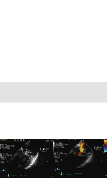
The overall echocardiographic picture will include (Fig. 5.1):
•Hyperdynamic ventricle
•Severe mitral regurgitant jet
FIG. 5.1. Emergency echocardiography in a patient with sudden onset of unexplained acute pulmonary edema in whom transthoracic echocardiography (TTE) showed a hyperdynamic heart and possibly a very eccentric mitral regurgitation (MR) jet. a Transthoracic apical 4-chamber view showing small end systolic ventricular volume consistent with a hyperdynamic ventricle. b Transesophageal echocardiography (TEE) imaging at 0° in mid-esophageal position with clockwise rotation, confirms the diagnostic of severe MR with eccentric, anteriorly directed jet. c TEE scanning at 116° in mid-high esophageal position demonstrates flail central scallop of the posterior mitral leaflet (thick arrow) with ruptured chorda (thin arrow).
80ECHOCARDIOGRAPHY IN VALVULAR EMERGENCIES
°eccentric and, thus, possibly underestimated or even missed altogether or
°extremely wide, completely filling the left atrium.
•The specific valvular pathology involved
°a torn chorda and/or a flail leaflet can be seen sometimes on a transthoracic echocardiography (TTE) but if in doubt or for better visualization, transesophageal echocardiography (TEE) is needed.
•Pulmonary hypertension and various degrees of right ventricular enlargement and failure
5.4.1.2 Acute MR Due to Infective Endocarditis
The mitral valve can be involved in infective endocarditis (IE) either primarily or due to extension from aortic valve endocarditis, so the importance of careful scanning of all valves in IE patients cannot be overemphasized. Acute MR can develop mainly as a result of:
•progressive leaflets destruction
•vegetation impeding the leaflets coaptation in systole
•localized leaflet perforation, and
•chordal rupture.
Especially with the last two mechanisms, the MR occurrence or worsening can be rapid and dramatic, translating clinically in pulmonary edema and shock. The echocardiographic findings will include the common findings of acute severe MR and specific changes as described above, though, occasionally, the degree of regurgitation will be unexpectedly milder than anticipated (Fig. 5.2).
FIG. 5.2. Transesophageal echocardiography (TEE) study in a patient with staphylococcal infective endocarditis (IE). a Large vegetation is present on the atrial side of the leaflets. b Color Doppler shows mild-moderate mitral regurgitation (MR) only.

5.4 SPECIFIC VALVULAR PATHOLOGIES 81
5.4.2 Native Valve Pathology: Acute AR
There are three instances of acute, hemodynamically compromising, aortic regurgitation (see below). Echocardiographically, they share the finding of a possibly hyperdynamic ventricle and the visualization of the wide and turbulent regurgitant jet by color Doppler. The specific findings will reflect the etiology.
5.4.2.1 Infective Endocarditis (Fig. 5.3)
The presence of vegetation and/or leaflet disruption/perforation should be seen on either TTE or TEE.
FIG. 5.3. Transesophageal echocardiography (TEE) long-axis view of aortic root in a patient with acute endocarditis and hemodynamic deterioration. a Thickened aortic non-coronary cusp with complex, elongated vegetation with a mobile part (thin arrows). A small area of valve disruption is seen at the base of the leaflet (thick arrow). b Diastolic frame in the same view with color Doppler demonstrates severe aortic regurgitation with eccentric jet.

82 ECHOCARDIOGRAPHY IN VALVULAR EMERGENCIES
5.4.2.2 Type A Aortic Dissection
Acute or worsening AR is frequently found in the setting of type A aortic dissection, due to:
•tethering of the leaflets by the dilated aortic root,
•change in the valvular geometry, and
•prolapse of intimal flap through the valve.
Though the presence of AR in these patients can generally be documented by TTE, TEE is necessary to clarify the mechanism involved.
5.4.2.3 Blunt Chest Trauma (See Sect. 12.5)
Aortic cusps disruption can rarely occur with severe chest trauma. This emphasizes the need for an echocardiographic scan in unstable patients after severe blunt chest trauma.
5.5 ACUTE PROSTHETIC VALVE PATHOLOGY (SEE ALSO SECT. 12.3 AND APPENDIX G)
There are five main pathological processes that may affect the functioning of prosthetic valves (Table 5.1). The resulting clinical entities and the echocardiographic diagnostic approach are discussed below.
5.5.1 Stuck Prosthetic Valve 2-4
Represents acute or rapidly progressive limitation of the motion of the valve occluder (ball, disk, leaflets). Although, it is generally associated with obstruction, a stuck valve may become regurgitant if the closing excursion of the occluder is impeded as well.
Can occur in:
•Mechanical valves, due to:
°Thrombus formation – more likely with
inadequate anticoagulation
shorter time interval from valve implantation.
°Pannus (fibrous tissue) growth - more likely with:
long-time interval from valve implantation.
°Vegetation
•Bioprosthetic valves, due to:
°Vegetation
Risk factors for stuck valve include
•Mitral position
•Low cardiac output
•Atrial fibrillation

TABLE 5.1. Main pathologies involving mechanical and biological prosthetic valves.
|
|
Prosthetic valve type |
|
|
Clinical and echocardiographical picture |
|
||
|
|
|
|
|
|
|
|
|
|
|
|
|
|
Valvular |
Valvular |
Paravalvular |
|
|
|
Mechanical |
Biological |
|
obstruction |
regurgitation |
regurgitation |
Embolism |
|
|
|
|
|
|
|
||
Thrombosisa |
+ |
− |
+ |
+ |
− |
+ |
||
Pannusa |
+ |
− |
+ |
+ |
− |
− |
||
Infection |
+ |
+ |
+ |
+ |
+ |
+ |
||
“Wear and tear”b |
− |
+ |
+ |
+ |
+ |
− |
||
|
|
|
|
|
|
|
|
|
aFor all practical purposes, restriction of valve motion due to thrombus or pannus is a mechanical valve pathology
b“Wear and tear” deterioration of mechanical valves is not a practical concern anymore but it is a limiting factor for durability of bioprosthetic valves
83 PATHOLOGY VALUE PROSTHETIC ACURE 5.5

84 ECHOCARDIOGRAPHY IN VALVULAR EMERGENCIES
The clinical picture will include:
•Rapidly progressing heart failure and/or hypotension up to shock and pulmonary edema
•Possible new murmur and/or muffling of prosthesis sounds
The echocardiographic workup of a suspected obstructed valve is detailed in Sect. 12.2.
5.5.2 Regurgitant Prosthetic Valve5
A prosthetic valve can become acutely regurgitant as part of a “stuck” condition, as detailed above, or due to a paravalvular leak. The progressive regurgitation due to “wear and tear” of a bioprosthetic valve is a more chronic process, so it will not be included in this discussion.
5.5.2.1 Paravalvular Leaks
Small (<5mm) dehiscences are not unusual in postmortem studies.5 Clinically significant paravalvular leaks may result from failure of the sutures in prone areas, especially in patients with heavily calcified or degenerated valvular annuli, but are mainly the result of prosthetic valve endocarditis. From a practical standpoint, the acute development of a paravalvular leak should always prompt a workup to rule out infective endocarditis. Besides the possible signs and symptoms of endocarditis, the acute emergence of a significant paravalvular leak may manifest itself as a combination of:
•New murmur
•New onset/rapidly progressive cardiac failure
•Hemolytic anemia
Possible echocardiographic findings with severe paravalvular leaks are (Fig. 5.4):
•De novo hyperdynamic contractility
•High peak gradient with a normal mean gradient
°for a prosthetic valve in mitral position, a peak velocity of ≥2m/s with a normal (<5mmHg) mean gradient and a normal
pressurehalf-time(P1/2T)ishighlysuggestiveofatleastmoderate regurgitation.
•Visualization of a high velocity, turbulent jet, different from the normal appearance for a given valve
•“Rocking” motion of the valve if the dehiscence involves a large portion of the suture circumference