

Emergency Echocardiography
.pdf2.2 TRANSESOPHAGEAL ECHOCARDIOGRAPHY 35
•LV
°Size
°Contractility Global Regional
°Filling pattern
□Suggestive of underfilled ventricle
□Suggestive of high filling pressures
•Right ventricular (RV)
°Size
°Contractility
•Pericardial fluid
°Amount
°Evidence for tamponade
•Significant valvular pathology
•Pulmonary hypertension
•Any major pathological finding
A typical study should begin with an attempt at trans-thoracic imaging. It is the responsibility of the operator to decide whether the information obtained is satisfactory or more advanced techniques such as contrast or transesophageal echocardiography are needed.
2.2 TRANSESOPHAGEAL ECHOCARDIOGRAPHY IN THE CRITICALLY ILL PATIENT
Transesophageal echocardiography rapidly entered into clinical use starting with the 1980s. Its specific diagnostic advantages as compared with transthoracic echocardiography (TTE) (see below) derive from the positioning of the transducer into the esophagus, in the proximity of the heart (Fig. 2.2), thus allowing unimpeded imaging from a posterior-to-anterior view and the use of high frequencies providing better resolution.
2.2.1 TEE Imaging Advantages
•Interrogation of posterior cardiac structures generally not well imaged with TTE:
°Pulmonary veins
°Atrial appendages
°Descending aorta
•Better imaging of difficult and/or small targets such as:
°Vegetations
°Prosthetic valves

FIG. 2.2. Imaging planes and typical views of cardiac structures at standard esophageal positions and transducer angles. Although the transducer is situated posterior to the heart, the image is displayed as seeing the crosssectional view from an anterior position. Left-sided structures are displayed on the right side of the screen and posterior structures are displayed in the upper part. a Mid-low esophageal positions, b mid-high esophageal
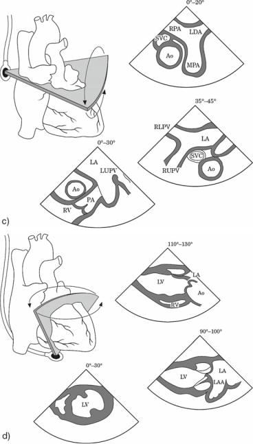
FIG. 2.2. (continued) positions, c high esophageal positions, and d transgastric positions. Adapted with permission from Flachskampf et al.7

38 EMERGENCY ECHOCARDIOGRAPHY
2.2.2 TEE “First Line” Indications Are Summarized Below and Are Detailed in Available Guidelines6,7
•Aortic pathology
°Acute aortic syndromes
°Aortic trauma
•Infective endocarditis (selected cases)
°Positive diagnostic
°Complications
•Prosthetic valves assessment
°Obstruction
°Regurgitation
°Prosthetic valve endocarditis
•Suspected high-risk pulmonary embolism
•Source of systemic/cerebral emboli
°Intracardiac masses/clots
°Intracardiac shunts
°Aortic atheromas
•Valvular pathology
°Equivocal TTE studies
°Pre-repair assessment
•Imaging guiding during procedures
°Atrial septal defect (ASD)/patent foramen ovale (PFO) closure
°Mitral valve balloon valvuloplasty
°Percutaneous aortic valve replacement
•Hemodynamic monitoring during surgery
•Poor quality TTE imaging when the echocardiographic information is considered essential for management.
2.2.3 TEE Versus TTE in the Critically Ill Patient
The tendency exists to consider transesophageal echocardiography (TEE) as the first step for echocardiographic imaging in intensive care units (ICU) patients. Indeed, due to limitating factors in these patients, TTE imaging failure rates of up to 40% have been previously quoted.8 However, following the continuous improvement in image quality with modern systems and the use of harmonic imaging, TTE can actually provide useful information in a surprisingly high percentage of patients with a failure rate possibly as low as 10–15%.9 Moreover, some areas such as LV apex or the aortic valve in the presence of a prosthetic mitral valve are usually better visualized by transthoracic echocardiography. Cavities size and ventricular contractility are also occasionally more accurate by TTE. Accordingly, it is our policy to always attempt a transthoracic study first and to continue with a TEE, if:

2.2TRANSESOPHAGEAL ECHOCARDIOGRAPHY 39
•Image quality is unsatisfactory for a confident diagnosis or
•The clinical question in itself is a “TEE-first line” indication (see above).
2.2.4 TEE Contraindications, Safety and Precautions
In critically ill patients, TEE has a special role due to its input in some of the encountered pathologies and to the higher likelihood of limited quality transthoracic studies in this population. A large body of literature has documented the excellent yield and safety profile of TEE in intensive care patients.2,10–13 Except for some esophageal pathologies and cervical spine instability, there are no absolute contraindications for TEE. Hemodynamic instability and acute conditions as such are not a contraindication, in fact, if appropriate, they may be an indication for TEE diagnostic work-up. The TEE technique in critically ill patients is similar to the one used in stable, elective ones, however, some precautions are to be taken:
•In hemodynamically unstable or hypoxemic patients, it may be preferable to have the sedation performed, or at least supervised, by an anesthetist. The “threshold”for requiring this assistance depends on the operator’s expertise and the severity of the case.
•In mechanically ventilated patients, achieving the appropriate level of sedation is generally the responsibility of the ICU team. Removing the naso-gastric tube before the procedure is recommended to facilitate probe advance and improve imaging. Also care should be exercised not to dislodge the endotracheal tube during intubation of the esophagus and when withdrawing the probe.
•As a rule, a TEE in an unstable patient should be performed by an experienced practitioner and not by a beginner or an occasional operator.
2.2.5 TEE Examination Protocol
•Individual routines vary but the concept of following a study protocol is crucial in order to avoid missing important information. Guidelines on how to perform TEE and how to assess specific pathologies exist7,14 and may be adapted to personal preferences.
•If no recent TTE study is available, the TEE should be complemented, if possible, by even a quick TTE scan.
•Though a complete and systematic study should be attempted in all cases, it is a good routine in patients who are unstable or tolerate the procedure poorly, to start with the “target” (i.e., prosthetic valve, aorta, LV function) and then continue with a full study so that if the procedure has to be prematurely terminated, the important information has been acquired.

40EMERGENCY ECHOCARDIOGRAPHY
•The examination can be either “view guided,” that is, the operator follows an orderly sequence of standard views as in a transthoracic study, or “structure guided” where each structure is fully interrogated in appropriate views, before the next structure is targeted. It is our opinion that a “view guided” approach is generally superior since it is less likely to omit certain views and miss important information, while a “structure guided” approach may be more appropriate in unstable patients.
A suggested sequence for a TEE “view guided” examination protocol is detailed below. Recommended probe positions and transducer angles are orientative and have to be adapted to the patient’s specific anatomical particularities. A standard view of a given structure is “expected” but not certain to be found at a certain angle and depth, so scanning has to be performed in a true “windscreen wiper” manner until the required view is achieved. As a general rule, try first to understand the two-dimensional anatomy and then use color Doppler.
Position I: Mid-esophageal at 0° (Fig. 2.3).
This is a good starting view to give an overall “feel of the heart” and detect pericardial effusion or extracardiac compressing masses.
FIG. 2.3. Systolic frame of an apical 4-chamber view obtained from a midesophageal position with the transducer at 0°.

2.2TRANSESOPHAGEAL ECHOCARDIOGRAPHY 41
–Equivalent transthoracic view: apical 4-chamber view
–Structures: atria, ventricles, mitral and tricuspid valves (TV)
–Suggested manoeuvres at this level are:
•Retroversion of the probe tip using the big wheel may help to avoid a short-axis cut with vertical hearts in this view.
•Rotation of the probe shaft counter-clockwise and clockwise to scan the left atrium (LA) leftwards and rightwards, respectively, and the mitral valve (MV) from the septal to the lateral aspect of the annulus.
•Clockwise rotation with slight advancement of the probe may optimize the TV and the IAS as well.
Position II: High esophageal at 0° (Fig. 2.4).
There are three levels here to be structure targeted through gradual withdrawing of the probe.
–Aortic valve level (roughly equivalent to a 5-chamber view) for left ventricular outflow tract (LVOT) and anterior segments of the MV
–Left atrial appendage (LAA) and left upper pulmonary vein (LUPV) level
–Superior vena cava (SVC), ascending aorta and main pulmonary artery (MPA) level
Position III (Fig. 2.5): Back to mid-esophageal level, slightly advancing the probe to “lose” the aortic valve with counterclockwise rotation to bring the MV in the centre of the image. Scan from 40° to 90° for MV interrogation.
–Equivalent transthoracic views: apical 2-chamber view
–Structures: MV, LV, LA, LAA
–Once at 90°, clockwise rotation will open the right ventricular outflow tract (RVOT) and the MPA.
Position IV (Fig. 2.6): Back to midto high-esophageal position until the aortic valve (AV) becomes visible again. There are three transducer angulations to be used here:
°0°
□Equivalent transthoracic view: basal parasternal shortaxis view
□Structures: TV, AV, RVOT, PV, left main coronary artery, LAA
°120°
□Equivalent transthoracic view: parasternal long-axis view

FIG. 2.4. Transesophageal echocardiography (TEE) imaging at 0° with gradual withdrawing of the probe from midto high- (a–c) esophageal positions. a Aortic valve level position for left ventricular outflow tract (LVOT) and anterior mitral valve (MV) segments. b Left atrial appendage position. The left upper pulmonary vein is visualized on its left (right on screen), separated by a ridge. c Proximal ascending aorta position with good imaging of the proximal aortic root and the superior vena cava in cross-sectional view and the main pulmonary artery in long-axis view.

FIG. 2.5. Transesophageal echocardiography (TEE) scanning from 40° to 90° at mitral valve level. a Bicommissural view to identify the mitral valve segments. The anterior leaflet central segment is seen between the posterior mitral leaflet posterior and anterior segments in this early-diastolic frame. b 2-Chamber view demonstrating the anterior and inferior left ventricular (LV) walls. c Rightwards (clockwise) rotation from position b brings in the right ventricular outflow tract, pulmonic valve, and proximal main pulmonary artery.

FIG. 2.6. Transesophageal echocardiography (TEE) scanning starting at aortic valve level in short-axis view and continuing to a bicaval view. a 20°–40°: short-axis view of the aortic valve, allowing clear identification of the cusps. The tricuspid and the pulmonic valves, the left atrial appendage and, occasionally, the proximal left coronary system are also seen at this