

Dale_Molecular Genetics of Bacteria 4th ed
.pdfGENETIC METHODS FOR INVESTIGATING BACTERIA |
257 |
|
|
Receptor |
|
|
FruA |
|
|
FruA - P |
|
csgA |
csgA |
|
Activation of developmental genes
C-signal
Figure 9.8 Simplified model of some elements of the C-signal pathway in Myxococcus xanthus
are resistant to drying and enable the bacteria to survive until conditions improve, when they germinate and form individual cells again.
Development from a swarm of individual cells to an aggregate and then on to sporulation is controlled by a series of signals. One of these, the A-signal is a mixture of amino acids that operates as a quorum-sensing mechanism (as described in Chapter 3) at an early stage of the process. Aggregation only occurs if the A-signal reaches a threshold level, thus ensuring that there are enough cells to form a functional fruiting body. Further development is controlled by another gene (csgA) coding for the C-signal, so that csgA mutants are also defective in sporulation. Unlike the A-signal, which is secreted, the C-signal requires cell contact, since it is a cell-surface protein which interacts with a surface receptor on another cell (Figure 9.8). This interaction has two main consequences. Firstly it stimulates transcription of csgA, so more of the signal is made, thus amplifying the effect. Secondly, it alters the regulation of more than 50 genes, including those needed for sporulation; this is, in part, mediated by phosphorylation of a regulatory protein called FruA. The overall effect is that sporulation will only occur in those cells within the mound which are in contact with other cells, thus contributing to a spatial difference in gene expression within the fruiting body.
9.3 Bacterial virulence
9. 3.1 Wide range mechanisms of bacterial pathogenesis
Bacteria exploit a number of common molecular mechanisms to achieve a range of different objectives during infection. This section describes how bacterial
258 |
MOLECULAR GENETICS OF BACTERIA |
molecular genetics has been used to uncover these key mechanisms of microbial pathogenicity.
From genome sequence information, it is clear that virulence genes often occur in clusters and that these regions are absent from closely related non-pathogenic bacteria. Furthermore, based on the %GC content of these regions compared to the rest of the genome, it became apparent that these large sections (e.g. 30–50 kb) of DNA had been acquired from other organisms. Together, these observations gave rise to the concept of pathogenicity islands which suggests that bacteria can acquire, en masse, complete systems that expand their ability to exploit different host environments. A previous example is Vibrio cholerae (Chapter 4), where the toxin genes are located on a pathogenicity island which was subsequently shown to be an integrated bacteriophage.
Although not expected at the time, a number of related studies on the plant pathogen Pseudomonas syringae were to have dramatic implications for our understanding of pathogenicity. In the mid-1980s transposon mutagenesis (see Chapter 10) was used to identify a cluster of genes which were essential for the generation of plant disease symptoms. The importance of this region was confirmed when the gene cluster was cloned into non-pathogenic bacteria using shuttle vectors and was shown to confer upon these avirulent cells the ability to generate specific disease symptoms. In the early 1990s it became clear that similar gene clusters existed in many other bacterial pathogens and that some of the proteins encoded by these gene clusters resembled parts of the machinery for exporting the bacterial flagellum. This information gave rise to the concept that a common secretion pathway existed in these bacterial pathogens.
It is now known that these regions encode a specialized secretory apparatus called a type III secretion system (for information on other types of secretion system see Chapter 1). These comprise at least 20 proteins and are the most complex transport systems known in bacteria. They are often referred to as ‘molecular syringes’ as they enable Gram-negative bacteria to secrete and inject effector proteins directly into the cytosol of eukaryotic host cells. The injected proteins can redirect the normal cellular signal transduction pathways and can result in disarmament of host immune responses or in cytoskeletal reorganization to establish pathways for bacterial colonization.
Much of the type III secretory apparatus is conserved between distantly related pathogens (although the effector proteins differ). Thus the same general bacterial pathogenicity mechanism is involved in a multitude of diseases from bubonic plague in humans to southern wilt in tomato plants. Yersinia spp., for example, inject at least three different effector molecules to destroy key functions of immune cells. The genes encoding type III secretion systems, including effector proteins and structural proteins, are clustered together as another example of pathogenicity islands that have been transferred between bacterial species.
GENETIC METHODS FOR INVESTIGATING BACTERIA |
259 |
9.3.2 Detection of virulence genes
Many bacterial pathogens have separate free-living and pathogenic life cycles, so they encounter very different environments and require very different functions for survival. As a consequence, pathogens must be able to recognize signals in the host that convey the need to express virulence genes. The expression of certain virulence gene functions in Shigella, for example, is triggered at body temperature (37 8C) but not at environmental temperatures (< 30 C). Therefore, if the genes which are only expressed during infection can be identified, then the key virulence traits can also be identified. Reporter genes have been especially useful for this purpose.
In Vivo Expression Technology (IVET)
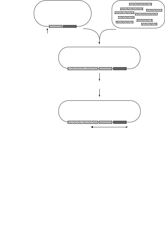
One of the most widely used methods of using reporters to identify virulence genes is known as In Vivo Expression Technology (IVET). This is a technique that selects bacterial promoters which are only expressed in the host and which thus drive the expression of virulence traits. In most examples of IVET, random fragments of DNA from the bacterial host are inserted adjacent to a promoterless reporter gene whose product confers a phenotype that can be positively selected for in the host. For example, a purA mutant of Salmonella typhimurium is unable to survive in an animal model because purine biosynthesis is essential for Salmonella in this environment. This defect can be complemented by a plasmid carrying a functional purA gene, but the purA gene on the plasmid will only be expressed if a promoter is inserted which is functional in vivo (Figure 9.9). (The construct shown also contains a lacZ gene whose expression is also controlled by the inserted promoter so that constitutively expressed promoters can be identified and excluded). When animals are infected with a pool of clones each carrying a different DNA fragment, only those clones containing an active promoter are able to survive and to be recovered from the animals, thus providing direct selection for those promoters which are active during infection. Identification of these promoter fragments then leads to identification of the genes that are normally regulated by them, which are likely to include genes which are essential for the virulence of Salmonella.
Signature tagged mutagenesis
As shown previously, the classical approach to identifying the genes responsible for a given phenotype would be to generate mutants that are defective in that phenotype. For virulence genes, the phenotype of the mutant would be the
260 |
MOLECULAR GENETICS OF BACTERIA |
Random fragments of Salmonella DNA
purA lacZ
Cloning site
Pool of recombinant plasmids
Insert |
purA |
lacZ |
Selection of PurA+ clones in mice
Active promoter |
purA |
lacZ |
Figure 9.9 In vivo expression technology (IVET). The plasmid vector contains promoterless purA and lacZ genes; expression of these genes is dependent on insertion of a DNA fragment with promoter activity. Infection of mice with a pool of Salmonella containing recombinant plasmids is selective for PurAþ clones which contain a promoter that is active under these conditions
inability to survive in an in vivo model for the disease. Isolation of the mutants cells will prove to be difficult as they are the very cells which do not survive. To circumvent this obstacle, a procedure called signature tagged mutagenesis (STM) has been invented. This is essentially a negative selection technique derived from transposon mutagenesis (see Chapter 10) in which each individual mutant is labelled with a unique DNA signature. By comparing the mutants present in the initial inoculum with those that are recovered after infection of the model, it is possible to identify those that did not survive and thus identify the genes that are required for virulence.
GENETIC METHODS FOR INVESTIGATING BACTERIA |
261 |
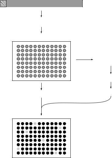
An overview of STM is given in Figure 9.10. A library of mutants is made for the bacterium of interest and each mutated gene tagged with a section of DNA containing a unique central region and two flanking arms which share their sequence with all of the other tags. The key to STM is that each individual mutant
Variable tag
Pool of uniquely tagged transposons
Transposon mutagenesis
Array of tagged transposon mutants
Infect mice with pooled mutants
Recover tagged bacteria
PCR amplify tags
Membrane
Probe the membrane
Negative clones show the absence of the specific tag in mice
Clones are recovered from the original array
Figure 9.10 Signature tagged mutagenesis. This procedure uses transposon mutagenesis to inactivate genes that are needed for infection of mice and provides a method of identifying those genes
262 |
MOLECULAR GENETICS OF BACTERIA |
can be distinguished from every other mutant based on the possession of its own unique tag. The mutants are then stored individually in ordered arrays and DNA from each one spotted onto a membrane in an grid-like manner. The mutants are then pooled and inoculated into a relevant animal model and the bacteria that are able to survive and establish infection are recovered. PCR is then used to amplify all the tags present in the recovered bacteria and the mixed product is used to probe the gridded membrane. Mutants that fail to survive in the infected animal (those which are defective in virulence) can be identified since their tags will not be present in the output pool. They can be recovered from the original stored arrays for further study. This system has been widely used to identify novel virulence factors that are involved in colonization, immune system evasion and attachment to human cells in a number of bacterial pathogens.
9.4 Specific mutagenesis
So far in this chapter, conventional mutational techniques have been considered – producing mutants with an altered phenotype and then identifying the genes affected and determining their functions. These techniques can be complemented by the use of recombinant DNA technology. In contrast to conventional genetics, the recombinant DNA approach starts with an hypothesis that a specific gene is involved. This gene is then modified or deleted and the resultant phenotype characterized. So, while conventional genetics starts with the phenotype and works towards identifying the nature and function of the genes involved, the molecular approach starts by altering the gene and works towards an analysis of the phenotype.
9.4.1 Gene replacement
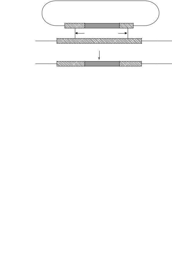
A key technique for determining the function of a specific gene is to inactivate it by a process known variously as gene replacement, allelic replacement or gene knock-out. Essentially, this uses homologous recombination to remove all or part of a specific gene or to replace it with an altered or inactivated gene. An example of how this can be done is shown in Figure 9.11. In this case, a plasmid has been constructed in which the central part of the cloned gene has been removed and replaced by an antibiotic resistance gene (aph, aminoglycoside phosphotransferase which confers resistance to kanamycin). The kanamycin resistance gene is flanked by regions of DNA that are the same as those in the host strain and it is within those regions that homologous recombination will occur. Note that two recombination events (a double crossover) are needed for gene replacement. Recombination at a single site (a single crossover) will merely integrate the plasmid into the chromosome. Additional techniques are needed to ensure that a double crossover is achieved.
GENETIC METHODS FOR INVESTIGATING BACTERIA |
263 |
Plasmid |
|
aph
Recombination sites |
Chromosome |
Target gene
Homologous recombination
Chromosome
Target gene disrupted
Figure 9.11 Gene disruption by allelic replacement. Homologous recombination between the disrupted cloned gene and the target gene on the chromosome leads to inactivation of the target gene. aph, aminoglycoside phosphotransferase (kanamycin resistance)
The plasmid that is used is one that cannot replicate in the chosen host cell. Thus after transformation, selection for kanamycin resistance will isolate cells in which the aph gene has been inserted into the chromosome by homologous recombination with the target gene, thereby inactivating that gene. Tests can be carried out to ascertain whether the expected phenotype is produced. For example, if it has been assumed that the target gene is necessary for survival and growth within macrophages, then testing the mutant for a deficiency in this respect will confirm or deny the assumption.
The results do not necessarily prove the case absolutely. Genes interact in many ways and knocking out one gene may have indirect effects on others. In particular, if the target gene is part of an operon, the gene knockout may affect the expression of other genes within the operon (in other words, the mutation may be polar). This possibility can be partly eliminated by the use of complementation. A plasmid carrying the wild-type gene (in this case using a plasmid that can replicate in this host) can be introduced into the mutant and if the original gene knock-out was responsible for the observed effect (e.g. loss of ability to grow within macrophages), then the introduced plasmid will restore the original, wildtype phenotype. Successful complementation therefore supports the contention that the product of this gene is necessary for survival in macrophages. But it needs to be said that it still does not finally establish a direct role for the gene product – for example, it could function by altering the expression of other genes.
264 |
MOLECULAR GENETICS OF BACTERIA |
Instead of knocking out a gene completely, it may be desirable to replace it with a specifically altered gene. For this gene replacement should be combined with the techniques of site-directed mutagenesis, as covered earlier in this chapter. In this way, the significance of a specific amino acid in the activity of an enzyme for example, can be tested by replacing it with a series of different residues and determining the activity of the product.
9.4.2 Antisense RNA
One problem with gene knock-outs is that if the gene is essential for the growth of the cell in the laboratory, then the complete loss of that gene would be lethal and therefore no recombinants would be obtained. It may therefore be useful to partially reduce the expression of the gene or the functionality of its product – thus leaving enough activity to cope with the comfortable conditions of normal laboratory growth but not enough to deal with the stress conditions that may be imposed on it subsequently. One alternative strategy is to use antisense RNA.
For this purpose, part of the gene would be cloned in an expression vector, so that it is transcribed from a promoter on the vector, but the insert would be deliberately put in the wrong orientation. The insert will therefore be transcribed in the opposite direction from normal – or in other words, the opposite strand will be transcribed. The RNA produced (the antisense RNA) will be complementary to the normal mRNA and will pair with it to produce a double-stranded RNA molecule. This may interfere with translation of the mRNA and thus reduce the level of the protein that is made. Using a stronger or a weaker promoter (or even better, using a promoter that can be switched on and off) will lead to different amounts of the antisense RNA being made and hence will alter the extent of the reduction in the amount of the protein product formed.
9.5 Taxonomy, evolution and epidemiology
9.5.1 Molecular taxonomy
It is possible to distinguish between even closely related organisms using a range of biochemical tests, but this approach does not necessarily give an accurate picture of the true taxonomic or evolutionary relationship between different organisms. Conventional taxonomy therefore resulted in some quite different organisms being erroneously grouped together in the same genus or family. Molecular approaches have played an important role in resolving these issues.
One simple molecular characteristic that is used to classify bacteria is the base composition of the DNA, defined as the number of guanine and cytosine residues as a percentage of the total number of bases (%GC). It is not necessary to
GENETIC METHODS FOR INVESTIGATING BACTERIA |
265 |
sequence the genome to determine this value (although of course that gives the most accurate and precise value). The %GC can be determined using physical techniques. The base composition of bacterial DNA varies widely from one species to another – over a range of 20–80 per cent – but closely related organisms tend to have similar DNA base compositions. There are good reasons for this. Replication, transcription and translation are all, in different ways and to varying extents, sensitive to the base composition of the nucleic acids and so have evolved together. Earlier in this chapter it became evident that this can be used as evidence that a portion of the genome has been acquired more recently in the evolution of the organism, as its base composition is different from the rest of the genome.
If this concept is applied to bacterial taxonomy, it is sometimes found that organisms which are otherwise quite similar have quite different DNA base composition. For example the genera Staphylococcus and Micrococcus are morphologically similar Gram-positive cocci (although they can be distinguished biochemically). However Micrococcus has a high GC content (about 70 per cent GC), while Staphylococcus DNA has a low proportion of GþC (30–39 per cent).
Ribosomal RNA sequencing is a much more powerful technique. The ribosomal RNA genes are very highly conserved, being remarkably similar in all bacteria, and yet there are small variations in the sequence from one species to another. These variations (most commonly in the 16S rRNA) not only distinguish between species but also indicate the degree of difference. In other words, by counting the number of bases that are different in two species a measure of the evolutionary distance that separates them can be calculated. If a number of such sequences is compared, a phylogenetic tree can be constructed which will show a possible route by which these species have diverged from a common ancestor. A simple example (with a much shorter sequence than would be used in practice) is shown in Figure 9.12. The sequence of organism A is more similar to B than it is to C or D for example and this is reflected in the arrangement of the tree. A word of caution: construction of a phylogenetic tree is much more complicated than this simple description and many trees can be drawn from a single set of data. The computer produces the best fit, but it is only a model and does not necessarily reflect the true evolution of the organisms involved.
Cloning the ribosomal RNA genes to do this is not necessary. The variation in the 16S (or 23S) rRNA gene is not evenly spread across the gene. Some regions are particularly highly conserved, so a pair of PCR primers can be used which recognize conserved sequences on either side of a variable region and amplify the region which contains differences. This amplified product can then be sequenced (see Chapter 10). The degree of conservation is such that the same pair of primers can be used for any organism, without knowing anything about it. The sequence obtained can then be compared with sequences of rRNA from known organisms and thus the identity of the unknown bacterium and its relationship to known species can be determined, at least provisionally.

266 |
MOLECULAR GENETICS OF BACTERIA |
|
||||||||||||||||
|
Organism |
|
|
|
|
|
|
|
Sequence |
|
||||||||
|
A |
C |
|
U A G A |
|
|
|
U G |
|
C G C C |
|
|||||||
|
A |
C |
C |
A |
|
|||||||||||||
|
|
|
|
|
|
|
|
|||||||||||
|
B |
C C U A G A G |
C |
U G G C G C |
G |
|
||||||||||||
|
|
|
|
|
|
|
|
|
|
|||||||||
|
C |
C |
G |
|
A |
A G A G |
G |
U G G C G C C |
|
|||||||||
|
D |
|
|
C U A G A |
|
|
|
G |
U G G C G |
|
|
C |
|
|||||
|
G |
|
A |
G |
|
|||||||||||||
|
|
|
|
|
|
|
|
|
|
|
|
|
|
|
|
|
|
|
Only a short sequence is shown as an example
In practice much longer sequences would be used
A
Possible phylogenetic tree
B
C
D
Figure 9.12 Construction of a phylogenetic tree from sequence data. This illustrates the principle only. In practice a much longer sequence would be used and extensive computer analysis is required to test the many possible trees that can be drawn
The power of PCR to amplify minute amounts of DNA means that the bacterium in question does not have to be cultured. This is significant as standard bacteriological techniques are designed to culture certain bacteria, especially medically important pathogens. The range of bacteria that can be isolated can be extended by using different media and different growth conditions. But however wide the range of conditions used, there will still be some bacteria – often a substantial majority – that are unable to grow. Applying PCR to such a sample, using primers directed at the 16S rRNA gene, will produce a very wide range of amplified products. Cloning this mixture of products, rather like constructing a gene library (Chapter 8), enables each one to be isolated and sequenced so that the bacteria present in the sample can be identified (within the limitations of the known sequences in the database). For example, the bacterial flora of the human colon has been extensively investigated using cultural techniques and its constituent bacteria were thought to have been thoroughly characterized. However, the genotypic approach described above showed that 76 per cent of the 16S rRNA