

Pediatrics
Treatment
No treatment is recommended for intrusions as spontaneous re-eruption may occur with healing. Follow-ups are recommended.
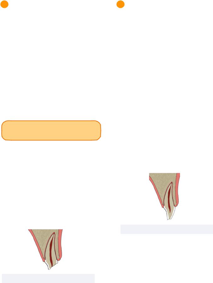
Extrusion
Extrusion injuries refer to the coronal displacement of teeth. The severity and luxation distance of the injury determines the possibility of severing the blood supply to the tooth and subsequent pulpal necrosis.
INBDE Pro Tip: Extraction is not required to be followed by space maintenance if the permanent successor is close to eruption.
Treatment
If the extrusion is more than 3mm, the tooth must be extracted and space maintained. However if the patient is seen before formation of a blood clot apically, the tooth may be carefully repositioned and splinted for 1-2 weeks prior to necessary endodontic treatment. Follow-ups are also recommended.
Figure 2.02 Extrusion Luxation
2021 Di Giorgio, G., et al. MDPI, Basel, Switzerland. CCBY 4.0
33
Avulsion
Avulsion occurs when a tooth is completely displaced from the socket.
Treatment
Previously, replantation of the avulsed primary tooth was recommended if the extra-alveolar dry time (time out of mouth and appropriate solution) was less than 30minutes. In this scenario, replantation and splinting for 1-2 weeks on a soft diet and antibiotics was indicated prior to endodontic treatment.
Currently, it is recommended to not replant the tooth as replanting can cause damage to the permanent successor.
Figure 2.03 Total Avulsion
Damdent, CC BY 3.0, via Wikimedia Commons
Alveolar and Crown-Root Fracture
Root fractures are rare in pediatric patients, as their alveolar bone is malleable. This makes apical root fractures less likely than those occurring in the coronal half.
Treatment
If the root fracture occurs in the apical half, no treatment is indicated and the root segment is left to undergo physiological resorption.
However if the fracture occurs in the coronal half, a rigid splint or extraction is required.
INBDE Booster | Booster PrepTM
Pediatrics |
34 |
Root Resorption - Review
Root resorption can be a pathological response to dental trauma which may occur after root fracture.
Internal root resorption occurs when the odontoblastic layer in the pulp is damaged, while external root resorption occurs when the cementoblastic layer in the periodontal ligament is damaged.
External root resorption can occur in various forms:
•Surface - occurs in small areas
•occurs when periodontal ligament is relatively normal
•Replacement - leads to ankylosis
•long-term splinting may increase risk
•Inflammatory - may be visualized as radiolucency
•replacement of root with granulation tissue
•Cervical Root Resorption - pink spot in biological width area
•Apical Root Resorption
•often caused by orthodontic forces
INBDE Booster | Booster PrepTM
Pediatrics
3 Moderate Injuries
Moderate injuries require more complex and higher-cost interventions which may require a multi-disciplinary approach. They have a moderate prognosis and require a longer term follow up.
Subluxation
Subluxation refers to direct damage of the periodontal ligament which may cause mobility.
Treatment
No treatment is recommended. However, patients may be recommended a soft diet and provided with oral hygiene instruction. Followups may be conducted.
INBDE Pro Tip: Teeth with open apices are more likely to remain vital after trauma.
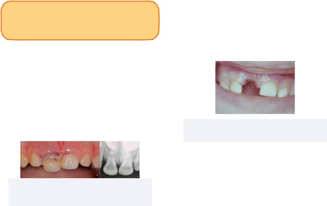
Extensive Tooth Structure Involvement
Crown fractures involving more tooth structure and pulp are more likely in primary teeth due to their large pulp spaces.
Treatment
For vital primary teeth, a pulpotomy is indicated.
For non-vital teeth, a pulpectomy is indicated. In the more severe case where pathologic root resorption is observed, the tooth may be indicated for extraction and space maintenance.
Figure 3.01 Extensive Crown Fracture
35
4 Minor Injuries
Minor injuries are require simpler, low-cost treatments and minor procedures. They have a good prognosis and short term follow up.
Concussion
Concussion refers to tenderness caused by inflammation of the periodontal ligament following a dental injury.
Treatment
Similar to subluxation injuries, no treatment is recommended for concussion injuries. Patients may be recommended a soft diet and continued oral hygiene.
Craze Lines and Enamel Fractures
Fractures limited to the enamel can be smoothed for patient comfort.
Enamel and Dentin Fractures
Fractures including enamel and dentin without pulpal involvement are indicated for minimal preparation and restoration to functional and esthetic demand.
Figure 4.01 Enamel and Dentin Fracture
INBDE Booster | Booster PrepTM

Pediatrics
4 Dental Trauma Management
Medical History
For all trauma patients, information regarding the medical history must be obtained to ensure the safety of the patient.
Coagulation Disorders
Information of coagulation disorders must be obtained as they can alter the course of intervention.
Tetanus Coverage
Tetanus coverage is important to ensure the patient is not at risk of active tetanus infection.
Active immunization through the tetanus, diphtheria, pertussis (Tdap) vaccine can be obtained by receiving:
•3 dosages in the first year
•Boosters at 1.5 years, 3 years, 6 years, followed by every 4-5 years
Dated coverage can be updated with a booster dosage upon injury, but if uncovered patients must receive the tetanus anti-toxin.
Possibility of Head Injury
Dental trauma can be associated to serious head injuries. Neurological assessment of drowsiness, amnesia, vomiting, and blurred vision must be conducted prior to dental management in order to mitigate risk.
Radiographs
Radiographs are helpful diagnostic tools that must be taken at the incident, and follow-ups of 1, 2, and 6 months after, depending on the severity of the injury.
36
Prevention
Mouthguards
Mouthguards are an appliance utilized to prevent the frequency and severity of dental trauma injuries. They are highly recommended for contact sports.
Type |
|
Fit and |
|
Availability |
|
Fabrication |
|
||
|
|
|
|
|
|
|
|
|
|
|
• |
Fit is un- |
• |
Available at |
Stock |
|
customizable - |
|
sporting goods |
|
available in a |
|
stores |
|
|
|
|
||
|
|
single size |
• |
low cost |
|
|
|
|
|
Mouth |
• |
Soften in hot |
• |
Available at |
Formed: |
|
sporting goods |
||
|
water for |
|
||
Boil and |
|
|
stores |
|
|
molding to teeth |
|
||
Bite |
|
• |
low cost |
|
|
|
|
|
|
Mouth |
• |
Firm outer shell |
• |
Available at |
|
and soft inner |
|
sporting goods |
|
Formed: |
|
|
||
|
liner (ethyl |
|
stores |
|
Shell |
|
|
||
|
methacrylate) |
• |
moderate cost |
|
|
|
|||
|
|
|
|
|
|
• Fit is completely |
|
|
|
|
|
customized |
• |
Custom made |
Custom |
• |
Formed via |
|
from impression |
|
vacuum form or |
|
in dental office |
|
|
|
|
||
|
|
pressure- |
• |
High cost |
|
|
lamination |
|
|
|
|
|
|
|
Child Abuse and Neglect
Dental trauma is a possible indicator for child abuse and neglect. Children of the ages 0-3 are most commonly abused or neglected.
Types of abuse may be occurring as:
•Physical - intentional injuries
•Emotional - denial of affection, isolation
•Neglect - negligence to provide basic necessities
INBDE Pro Tip: It is legally required for dentists to report suspected child abuse and neglect with or without evidence.
INBDE Booster | Booster PrepTM