

Pediatrics
3 Tooth Shape Anomalies
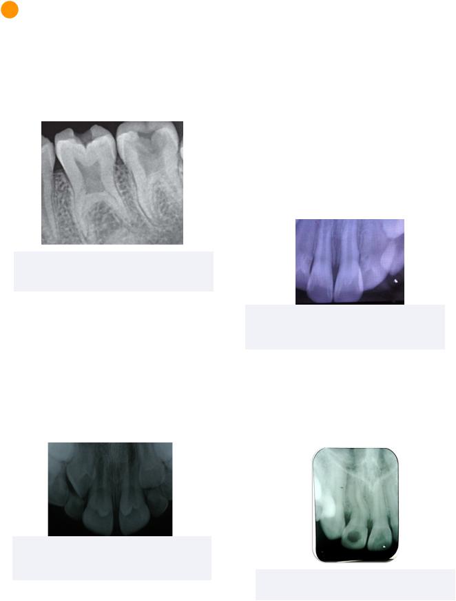
Taurodontism
Taurodontism refers to a tooth with short roots and an enlarged pulp chamber and elongated crowns.This trait is commonly linked to type IV amelogenesis imperfecta.
Figure 3.01 Taurodontism
Adapted from 2021 Chetty M., et al. Licensed CCBY 4.0
Dens Evaginatus
Dens evaginatus refers to the presence of an extra cusp containing enamel, dentin, and pulpal tissue. This is an important aspect to consider during operative dentistry in order to avoid pulpal encroachment.
Dens evaginatus may also be referred to as a talon cusp in anterior teeth.
Figure 3.02 Talon Cusp
2020 Rafael Alberto dos Santos Maia et al. Licensed CCBY 3.0
9
Dens Invaginatus (Dens in Dente)
Dens invaginatus is caused by an invagination of inner enamel epithelium (IEE) during tooth development.
After invagination, the IEE continues its normal differentiation into ameloblasts which secrete enamel in their invaginated state. This often results in the formation of a tunnel into the internal structure of the tooth which facilitates bacterial penetration and caries development. It must be diagnosed radiographically.
Commonly Affected Teeth: Permanent
Maxillary Lateral Incisors
Figure 3.03 Dens Invaginatus
2015 Nagaveni NB, et al. Licensed CCBY 4.0
Dilaceration
Dilaceration refers to an abnormal bend in the tooth root that commonly occurs due to trauma of the primary tooth affecting development of the permanent tooth.
Figure 3.02 Dilacerated Lateral Incisor
2018, Pauly G., et al. Licensed CCBY 4.0
INBDE Booster | Booster PrepTM

Pediatrics
4 Tooth Structural Anomalies
Enamel Hypoplasia
Enamel hypoplasia is a form of underdeveloped enamel tissue following a disturbance in the apposition stage of tooth development.
Turner’s Hypoplasia
A type of enamel hypoplasia following an infection or trauma induced inflammatory response to the primary tooth.
This results in a disturbance to the ameloblasts of the developing permanent tooth and thus only affects the permanent dentition.
This may be visualized as a brownish tinge on the enamel of permanent teeth.
Congenital Syphilis
Congenital syphilis in the fetus of mothers infected with syphilis can be commonly associated with enamel hypoplasia defects such as Hutchinson’s incisors and Mulberry molars.
This condition develops in utero and thus affects both primary and permanent teeth.
•Hutchinson’s Incisors: Hypoplastic triangular notch on incisal edges
•Mulberry Molars: globular rounded enamel cusps seen on occlusal surfaces of molars
Enamel Hypocalcification
Enamel hypocalcification occurs with a failure in maturation stage of tooth development. It is often clinically present as white spot lesions of abnormal mineralized enamel of teeth.
10
Amelogenesis Imperfecta (AI)
This condition consists of an alteration of enamel that has an autosomal dominant, recessive, or X-linked expression pattern of a disturbance in the bell stage.
This results in little to no enamel in both primary and permanent teeth, while the dentin and pulpal tissues remain unchanged.
Treatment options in these cases may include full-coverage crowns for aesthetic purposes.
Dentinogenesis Imperfecta (DI)
This condition consists of an alteration of dentin in a autosomal dominant expression pattern by affecting the bell stage of development.
Dentinogenesis imperfecta results in all teeth from both primary and permanent dentitions to have short roots, bell-shaped or bulbous crowns, constricted DEJ, and obliterated pulps. Extra-orally, it may also be accompanied by blue sclera and is commonly linked to osteogenesis imperfecta.
Treatment options for this condition consists of full-coverage crowns for aesthetic purposes.
INBDE Pro Tip: AI and DI are common concepts in the INBDE.
Amelogenesis |
Dentinogenesis |
Imperfecta |
Imperfecta |
|
|
• Affects enamel |
• Affects dentin |
• Autosomal |
• Autosomal |
dominant, |
Dominant |
recessive, or X- |
expression |
linked expression |
|
|
|
•Affects bell stage
•Affects all teeth in both dentitions
INBDE Booster | Booster PrepTM
Pediatrics
Regional Odontodysplasia
Regional odontodysplasia is a condition affecting all dental tissues in a quadrant of teeth in both primary and permanent dentition.
Affected teeth are often referred to as ghost teeth because they display short roots, open apices and enlarged pulp chambers.
Treatment includes supporting eruption to promote healthy alveolar bone and extracting affected teeth once erupted.
Figure 4.02 Regional Odontodysplasia
Adapted from 2014 by Song. CC BY 3.0
Concrescence
Concrescence occurs when there is a union of two adjacent teeth by cementum. It is commonly linked to hypercementosis and may result in difficulties with eruption of extraction of the affected teeth.
Most Commonly Affected: Maxillary Molars
Enamel Pearl
An enamel pearl is a piece of enamel found on the root surface which consequently blocks the attachment of Sharpey’s fibres to the tooth.
This results in periodontal pocketing in the patient and cannot be removed with scaling.
Most Commonly Affected: Molars
11
Dentin Dysplasia
Dentin dysplasia (DD) is an autosomal dominant condition leading to an alteration of dentin. It is often clinically presented in all teeth from both dentitions in two types:
Type I: Radicular Dentin Dysplasia
Type I DD affects the radicular dentin of teeth, resulting in short roots and chevron shaped pulps. This is often accompanied by premature mobility and exfoliation of the teeth.
Type II: Coronal Dentin Dysplasia
Type II DD affects the coronal enamel and is clinically determined by its thistle-shaped pulps, bulbous crowns, thin root. It can also be considered a form of dentinogenesis imperfecta.
Treatment options for this condition is limited as these teeth are not well suited for restoration.
Figure 4.01 Type I Radicular Dentin Dysplasia
Adapted from 2021 by Papagiannis A, et al. Licensee MDPI, Basel, Switzerland CC BY 4.0
INBDE Pro Tip: The types of dentin dysplasia are high yield topics.To remember the types of dentin dysplasia, remember that type I results in short roots since it is a lower (or shorter) number than 2.
INBDE Booster | Booster PrepTM
Pediatrics |
12 |
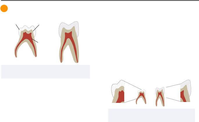
Primary Tooth Anatomy
1 General Characteristics
Pearly Enamel |
Thin Enamel |
and Dentin |
|
|
Large Pulp |
|
Spaces |
Figure 1.01 Cross Section of Primary Tooth (Left) and
Permanent Tooth (Right)
Mineralized Layers
The enamel and dentin layers are relatively thinner in primary teeth than in permanent teeth. The enamel thickness in primary and permanent teeth are on average 1mm and 2mm, respectively.
Additionally, the enamel thickness is relatively uniform in primary teeth. In permanent teeth the enamel thickness varies depending on the region of the tooth.
As a result of the thinner mineralized layers, primary teeth are more prone to tooth wear, caries, and faster caries progression.
Pulp Space
Primary teeth also have relatively larger pulp systems than permanent teeth. This makes them more prone to pulp involvement with caries progression, as well as pulp exposure with mechanical preparations.
Colour
Primary teeth are whiter than permanent teeth due to their thinner enamel.
Tooth Shape
Primary teeth have clinical crowns with a larger width to height ratio. This means that they are wider mesio-distally and shorter inciso-cervically relative to their successors.
Enamel Rod Orientation
In permanent teeth, the enamel rods are perpendicular to the DEJ and orient apically within the cervical third of the crown. In primary teeth, the enamel rods are uniformly facing occlusal in direction.
Figure 1.02 Enamel Cross Section of Primary Tooth
(Left) and Permanent Tooth (Right)
Cervical Bulge
Primary teeth have a cervical bulge or ridge which is most prominent at the primary first molar.
Root Shape
The roots of primary teeth are more divergent than those of permanent teeth, as they are required to accommodate the succedaneous teeth. Permanent teeth have roots of varying morphologies, including straight and convergent.
Additionally, the roots of primary teeth have small or absent root trunks. This means that the bifurcation of the roots occurs immediately or shortly after the CEJ of the tooth.
INBDE Booster | Booster PrepTM