

Pediatrics
Primary Canine Loss
The primary canines are important to maintaining the arch length. The arch length is the distance along the midline from the mesial contact point of the central incisors and the mesial contact point of the permanent first molars.
Premature loss of the canines causes lingual tipping or collapse of the incisors and eventual loss of arch length.
Figure 2.02 Lower Lingual Holding Arch
Intervention methods include lower lingual holding arch (LLHA) or Nance holding arm that are stabilized by the permanent first molars. Both of these appliances have an anteriorly extending strong wire to prevent lingual tipping of the incisors. However, the Nance is indicated for the maxillary arch as it features an acrylic pad on the wire for placement on the palate. Since the Nance is partially tissue borne, some argue it is not as effective as a lingual holding arch.
The permanent incisors must erupt before appliances are banded to prevent trapping the incisors which may erupt lingually.
Figure 2.03 Nance Holding Arm
25
Primary First Molar Loss
Premature loss of primary first molars are not critical to the space maintenance of primary dentition. However, appliances such as a band and loop, lower lingual holding arch, or
Nance can be utilized to maintain space. All of these appliances utilize a strong wire originating from the banded abutment tooth to maintain the exfoliated tooth space.
Figure 2.04 Band and Loop
Primary Second Molar Loss
The primary second molars (E)’s are said to be the key’s to the Leeway space. Premature loss can be critical to crowding and early intervention is recommended.
A common appliance utilized for this purpose is the distal shoe featuring a banded primary first molar that extends subgingivally to guide the unerupted permanent first molar. If the permanent first molar is already erupted, a lower lingual holding arch or Nance can be utilized.
Figure 2.05 Distal Shoe
INBDE Booster | Booster PrepTM

Pediatrics
3 Variables in Space Management
Eruption Pattern Variations
The eruption pattern and timing is generally conserved between individuals. However, variations can lead to spacing problems.
Permanent Lower Second Molar
If the permanent lower second molar erupts before the second premolar, a loss of leeway space for the second premolar will occur. This may lead to second premolar impaction that can be prevented by using a space maintainer.
Permanent Upper Canine
If erupted before or alongside the first premolar, the canine may be forced labially.
Asymmetrical Eruption
After eruption, the same tooth on the opposite side of the dental arch can be expected to erupt within 6 months. If this does not occur, extraction of the primary tooth may be indicated to maintain the midline.
Root Development
After the permanent tooth crown completes calcification, the root development and eruptive movement begins. At this point, the clast cells become activated and the bone remaining between the primary tooth and permanent tooth begins to get resorbed. On average, the tooth pierces bone and gingiva with ⅔ root formation and ¾ root formation, respectively.
Space maintenance is not necessary if there is no bone remaining between the primary tooth and underlying permanent tooth because it indicates the permanent tooth is close to eruption
26
Rule of Seven
Premolars are expected to erupt between the ages of 10-11. This indicates that the root resorption of the overlying primary molars must occur 2-3 years prior to eruption, between the ages of 7-9 years old.
Following this information, the rule of seven states that:
•A primary molar lost before the age of 7 will lead to a delay in premolar eruption
•A primary molar lost after the age of 7 will lead to an accelerated eruption of the premolar
INBDE Pro Tip: The rule of seven is a high yield topic for the INBDE.
Space Closure
Space closure mostly occurs within the first 6 months following tooth loss. The first 4-8 weeks demonstrate the most movement and tipping of neighbouring teeth due to the activated inflammatory mediators.
Additionally, active eruption of a neighbouring tooth increases the space loss.
INBDE Booster | Booster PrepTM
Pediatrics
4 Ectopic Eruption
Ectopic eruption refers to the eruption of permanent teeth along their unintended path. This may occur due to several reasons and may vary in severity.
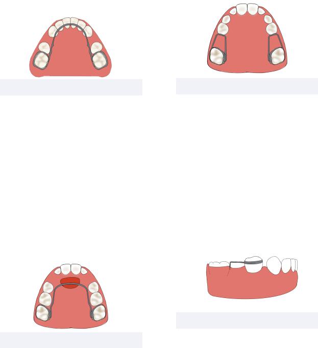
Ectopic Eruption of Incisors
Lingual eruption of the incisors may lead to a double row of teeth during development. This problem generally resolves with continued growth, unless the primary incisors become over-retained.
Lateral eruption of teeth may occurs due to early exfoliation of the primary lateral incisor. If this premature exfoliation was unilateral, the contralateral primary lateral must be extracted to avoid midline deviation.
Figure 4.01 Lingual eruption of Incisors
27
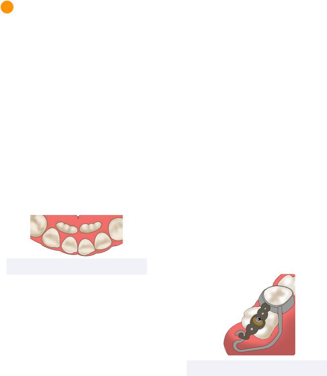
Ectopic Eruption of Molars
Mesial eruption of permanent molar eruptions is very common, with the maxillary first molar being most susceptible. Mesial eruption may lead to the permanent molar getting impacted underneath the distal of the primary second molar.
If the level of impaction is less than 1mm, an elastomeric spacer may be inserted between the primary second molar and permanent first molar to promote distal movement of the impacted tooth.
However, if the impaction is more severe a Halterman appliance may be indicated to shift the permanent first molar distally and re-create the lost space. It may also be indicated to extract the primary second molar and maintain the newly created space with a space maintaining appliance, such as a Nance.
Ectopic Eruption of Premolars
Distal eruption occurs most commonly with the mandibular second premolar that may only resorbs the distal root of the primary second molar. This leads to prolonged exfoliation of the primary second molar which deviates the eruption pathway of the second premolar.
Buccal or lingual eruption is also very common and must be managed by extraction of the primary molar, if it is not ready to exfoliate within a few weeks.
Figure 4.02 Halterman Appliance
INBDE Booster | Booster PrepTM
Pediatrics |
28 |
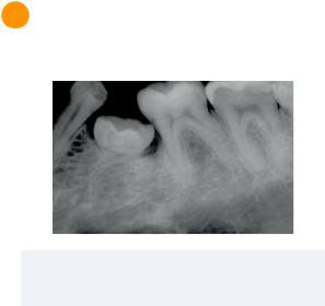
4 Ankylosis of Primary Molars
Figure 4.03 Ankylosed Primary Molar 2019 Lopes RDC, et al. CCBY 4.0
Ankylosis occurs when teeth become histologically fused to the underlying bone.
Prevalence
•Ethnicity: African-American (1%), Caucasian (4%)
•Dental Arch: more common in mandible
•Teeth: Mandibular first primary molars most common
•More common in primary first molars than primary second molars
Diagnosis
A critical clinical sign of ankylosed molars are when the crowns are infra-occluded and below the plane of occlusion.
Additional signs include:
•Lack of mobility
•Hollow sound upon tapping
•Radiographic loss of periodontal ligament space
Treatment
Treatment is often unnecessary. However, if neighbouring teeth begin to drift, extraction and space maintenance may be indicated.
INBDE Booster | Booster PrepTM