

Pediatrics |
17 |
Primary Tooth Treatment
Primary teeth have features which make them more susceptible to decay and rapid decay progression. Treatment options in pediatrics are unique to address these characteristics.
1 Early Childhood Caries (ECC)
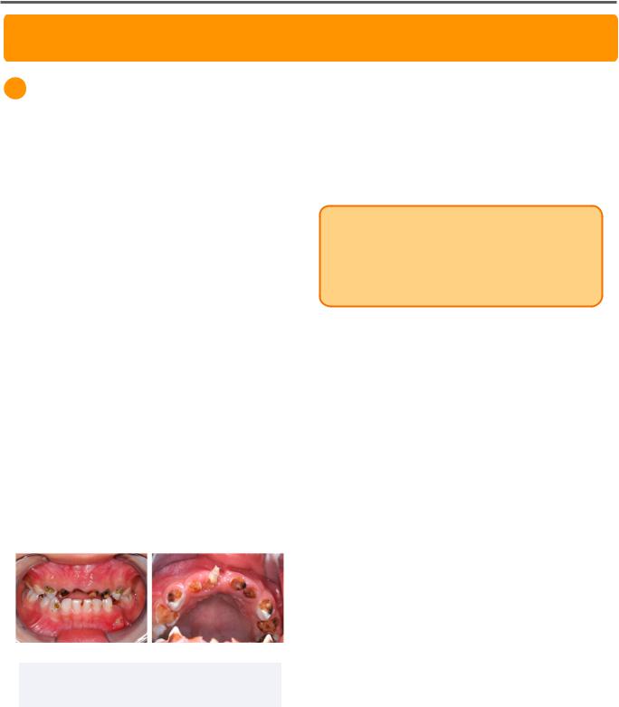
Early childhood caries are a form of widespread caries of primary teeth which occur in infants or young children. It is diagnosed when any dmft is seen on a patient <6 years of age.
Caries in ECC patients are often seen on the labial surfaces of the maxillary incisors, as well as the lingual and buccal surfaces of maxillary and primary molars.
Causes
ECC is also referred to as baby bottle syndrome because it commonly affects children who sleep with their bottle. For this reason, bottle or breastfeeding before bed should be stopped after the first primary tooth erupts.
Other causes may include excessive fruit juice consumption following constipation, or chronic high-sucrose antibiotic use with ear infections.
Figure 1.01 Severe Early Childhood Caries
Adapted from 2020 Chai H.H, et al., CCBY 4.0
Prevention
Caries risk can be mitigated by implementing cup drinking and a first dental visit by the age of 1. Additionally, toothpaste should be introduced at age 2 as a smear and increased to a pea size between the ages of 3 and 6.
INBDE Pro Tip: Pediatric patients should visit a dentist early, as primary teeth are more susceptible to caries. To remember this, utilize the phrase:
First tooth, First birthday = First (Dental) Visit.
Fluoride for Children
Fluoride supplementation is only used for children at risk for caries and living in nonfluoridated areas on a prescription only basis. The method and dosage depends on age and regular exposure to fluoride.
Fluoride Drops
Recommended for children ≤3 years old to mitigate chewing and swallowing of tablets.
Fluoride Tablets or Lozenges
Recommended for children between the ages of 3 and 6 years old.
Fluoride Mouth Rinse
Recommended for children over the age of 6 to minimize the chances of ingestion. Fluoride rinses are available in daily solutions at 0.05% NaF and weekly solutions of 0.2% NaF.
INBDE Booster | Booster PrepTM

Pediatrics
2 Restoration Options
Amalgam Restorations
The cavity preparation for amalgam restorations in primary teeth should be conservative in depth, but made at least 1.5mm deep.
In primary teeth, the cavity preparations should be extended into susceptible pits and fissures. They must have rounded line internal line angles, smooth and flat pulpal floor, and 90˚ cavosurface margins.
For proximal caries, the isthmus width must be ⅓ of the intercuspal distance of the facial and lingual cusps.
Extension to Pits ⅓ Intercuspal and Fissures Distance
1.5mm Depth
Figure 2.01 Amalgam Preparation in Primary
Tooth
Composite Resin Restorations
Composite resin restorations allow for more conservative preparations as they provide micro-mechanical retention through etch, prime, and bond systems. However, they require dry, isolated operating fields which may be difficult to achieve in pediatric patients.
The moisture sensitive technique makes composite resin restorations more susceptible to an inadequate marginal seal. The gingival margin is the most susceptible to failure.
18
Stainless Steel Crown
Stainless steel crowns are an effective restorative options for primary teeth with extensive caries or caries past the axial line angle. They are also the restoration of choice following a pulpotomy, pulpectomy, or fractured primary crown. However, they are ineffective as long-term restorations for permanent teeth.
After removal of defects, the crown preparation is conservative with 1mm occlusal reduction and rounded reduction adequate enough to break contacts with adjacent teeth. It is important to maintain the cervical bulge of the primary tooth as it provides retention for the stainless steel crown.
The pre-formed or modified stainless steel crown can be snapped onto the prepared tooth that allows it to function temporarily until exfoliation.
1mm Occlusal Reduction 
Figure 2.02 Stainless Steel Crown (Left) and
Preparation (Right)
Left: Trikkelle, CC BY 3.0
INBDE Booster | Booster PrepTM

Pediatrics
Strip Crown
Strip crowns are an aesthetic restorative option for primary anterior teeth with inter-proximal caries and potential incisal edge involvement. However, it requires adequate remaining tooth structure for bonding.
Preparations generally require a 1mm incisal reduction and breaking contacts with adjacent teeth. They are largely dictated by preparation design and caries involvement.
Strip crowns are fabricated using a celluloid crown form that is filled with composite resin after being trimmed and modified with a vent hole.
Figure 2.03 Anterior Strip Crown Placement
Adapted from Jeong M., et al. 2013, CCBY 3.0
19
3 Endodontic Treatment
Signs and Symptoms
The clinical signs and symptoms important to determining the need for endodontic therapy often overlap with normal physiologic changes in primary teeth.
Pain and Sensitivity
Pain and sensitivity is a symptom of endodontic involvement of a tooth. The pain or sensitivity may be triggered by hot or cold stimuli, or may even be spontaneous.
However, mild sensitivity may occur in primary teeth approximate to their exfoliation.
Furcation Radiolucency
Furcation radiolucency is a sign of pulp necrosis that is specific to primary teeth. Pulp necrosis in permanent teeth manifests as a periapical radiolucency around the apices of roots.
Figure 3.01 Primary Mandibular Second Molar with Furcation Involvement
Adapted from Arikan V., et al. 2016, CCBY 3.0
A similar radiolucency may also present when the successor permanent tooth approximates the primary tooth. To eliminate this as a differential diagnosis, it is important to consider additional signs and symptoms indicating endodontic treatment.
INBDE Pro Tip: Furcation radiolucency is a common INBDE topic, as it is an endodontic symptom specific to primary teeth.
INBDE Booster | Booster PrepTM

Pediatrics
Mobility
Mobility may present as a clinical symptom indicating endodontic therapy, but may also present as primary tooth roots resorb and prepare for exfoliation.
Fistula or Abscess
Fistula or abscesses are specific to true infections as they are clinical manifestations of pus accumulating in the periodontal tissues. This sign does not overlap with regular physiologic growth and development of dentition.
Root Resorption
Root resorption may present pathologically with extensive caries or physiologically as a part of the exfoliation process.
Physiologic root resorption is regulated by the dental follicle and stellate reticulum of the successive permanent tooth. The mineralization of the permanent roots is accompanied by the activation of clast cells that resorb bone and primary roots to create an eruption path. This process occurs two to three years prior to exfoliation and eruption.
INBDE Pro Tip: Unlike indirect and direct pulp cap methods, calcium hydroxide is not indicated in pulpotomy as it is an irritant that may induce internal root resorption in primary teeth.
20
Indirect Pulp Cap
Indirect pulp caps are a treatment option for deep caries approximating the pulp. During the preparation, a thin layer of dentin is maintained to prevent healthy pulp exposure.
Calcium
Hydroxide
Figure 3.02 Indirect Pulp Cap
This thin layer of dentin is reinforced with calcium hydroxide or resin-modified glass ionomer prior to restoration with an appropriate material. This process promotes the formation of tertiary dentin.
Direct Pulp Cap
Direct pulp caps are indicated for pinpoint pulp exposures caused by a trauma within 24 hours, or by a carious of mechanical exposure less than 2mm.
Calcium hydroxide is placed directly on the healthy exposed pulp and reinforced by resinmodified glass ionomer prior to restoration with an appropriate material.
RMGI |
Calcium |
|
Hydroxide |
Figure 3.02 Indirect Pulp Cap
Although this process is intended to promote tertiary dentin formation, it is uncommonly performed as it may cause internal root resorption in primary teeth.
INBDE Booster | Booster PrepTM