

Pediatrics
2 Maxillary Tooth Characteristics
**Only INBDE high-yield facts have been included in this section
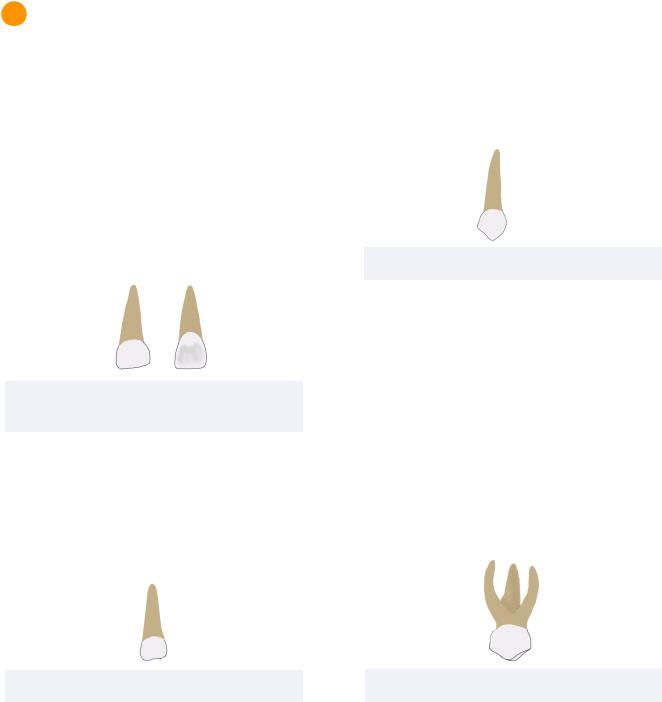
Primary Maxillary Central Incisor
Dimensions
The maxillary central incisors are the widest anterior teeth in the mesio-distal dimension for both primary and permanent dentitions.
The primary maxillary central incisor is the only anterior tooth with a width greater than its height.
Figure 2.01 Primary Maxillary Central Incisor from Labial View (Left) and Palatal View (Right)
Morphological Features
The primary maxillary central incisor also displays:
• Prominent labial and lingual cervical ridges
Primary Maxillary Lateral Incisors
Figure 2.02 Primary Maxillary Lateral Incisor
**no high yield facts are seen in the INBDE
Primary Maxillary Canine
Dimensions
The primary maxillary canine is the widest anterior tooth in the facial-lingual dimension.
13
The cusp of this tooth is also longer and sharper than the mandibular canine and permanent maxillary canine.
Morphological Features
The mesial cusp ridge is longer than the distal
Figure 2.03 Primary Maxillary Canine
cusp ridge, similar to as seen in the maxillary first premolar. Consequently, the cusp tip is offset distally from the midline.
Primary Maxillary First Molar
Morphological Features
The crown of the primary maxillary first molar resembles the crown of the permanent maxillary first premolar with a slight distal extension.
The crown also demonstrates a prominent mesio-facial cervical bulge that is accompanied by a prominent mesio-facial
Figure 2.04 Primary Maxillary First Molar
cervical ridge occlusally. This results in a CEJ that is more apically located on the mesial portion of the tooth.
Root Form
The root form of this tooth resembles that of the permanent maxillary molars. In the primary maxillary first molar, there is a palatal, mesiobuccal, and disco-buccal root.
INBDE Booster | Booster PrepTM
Pediatrics |
14 |
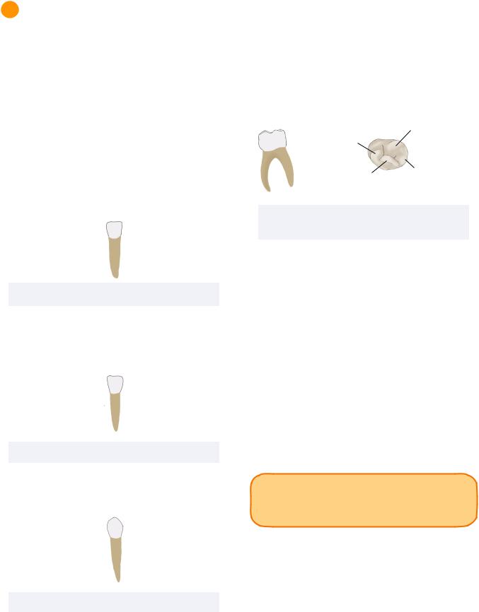
Cusp of
Carabelli
Figure 2.05 Primary Maxillary Second Molar from
Buccal view (Left) and Palatal View (Right)
Primary Maxillary Second Molar
The primary maxillary second molar is the last primary tooth to erupt.
Dimensions
The primary maxillary second molar is the widest primary tooth in the facial-lingual dimension.
Morphological Features
The primary maxillary second molar is the only primary tooth demonstrating a cusp of Carabelli, oblique ridge, and disto-lingual groove. If present, the cusp of Carabelli is located palatal to the mesio-lingual cusp tip. These features make the crown resembles that of the permanent maxillary first molar and not of its successor.
INBDE Booster | Booster PrepTM
Pediatrics
3 Mandibular Tooth Characteristics
**Only INBDE high-yield facts have been included in this section
Primary Mandibular Central Incisor
Dimensions
The primary mandibular central incisor is the smallest primary tooth in the facial-lingual dimension.
Morphological Features
As in the permanent dentition, the mandibular central incisor of primary dentition is the most symmetrical.
Figure 3.01 Primary Mandibular Central Incisor
Primary Mandibular Lateral Incisor and Canine
**no high yield facts are seen in the INBDE
Figure 3.02 Primary Mandibular Lateral Incisor
Primary Mandibular Canine
**no high yield facts are seen in the INBDE
Figure 3.03 Primary Mandibular Canine
15
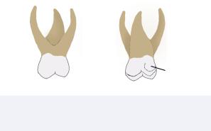
Primary Mandibular First Molar
The primary mandibular first molar is the most unique tooth in the entire human dentition. It is often said to resemble a combination of a mandibular first premolar mesially and a mandibular second premolar distally.
Disto-Buccal
Cusp
Mesio-Buccal
Cusp
Mesio-Lingual |
Disto-Lingual |
|
Cusp |
||
Cusp |
||
|
Figure 3.04 Primary Mandibular First Molar from Buccal View (Right) and Occlusal View (Left)
Morphological Features
The primary mandibular first molar has the most prominent mesio-facial cervical bulge that is accompanied by an S-shaped CEJ that is more cervically located on the mesial side.
The transverse ridge is also the most distinct on this primary tooth. This is neighboured by a distal triangular ridge and 4 cusps with 4 coinciding pulp horns.
The mesio-lingual cusp is cone-shaped and is the highest and sharpest cusp of the tooth. However, the mesio-buccal cusp is the largest in size.
INBDE Pro Tip: Although the mesiolingual cusp is the highest and sharpest, it is
the mesio-buccal cusp that is the largest.
Root Form
Similar to the permanent mandibular molars, the primary mandibular molars have a mesial and buccal root.
INBDE Booster | Booster PrepTM

Pediatrics
Primary Mandibular Second Molar
Dimension
The primary mandibular second molar is the widest primary tooth in the mesio-distal dimension.
Figure 3.05 Primary Mandibular Second Molar
Morphological Features
The crown resembles a permanent mandibular first molar. However, in the primary mandibular second molar, the mesio-buccal, disto-buccal, and distal cusps are relatively equal in size. The primary mandibular second molar has 5 cusps.
Root Form
Similar to the permanent molars, the primary mandibular second molar has a two root form. The mesial root is bigger than the distal root and also contains two root canals.
4 Natal and Neonatal Teeth |
16 |
|
Natal and neonatal teeth are commonly the primary mandibular incisors which are expected to erupt at 6 months of age. These teeth may cause nursing difficulties.
Natal teeth: refer to teeth present at birth Neonatal teeth: refer to teeth that erupt within the first 30 days
Riga-Fede Disease
This disease refers to natal or neonatal teeth induced ulcerations on the ventral surface of the tongue. Treatment may include smoothing of the incisors or extractions.
INBDE Pro Tip: Riga-Fede (‘feed’) can cause difficulties with feeding.
INBDE Booster | Booster PrepTM