

Pediatrics
Pediatric Soft Tissue
1 Healthy Gingiva Features
Healthy gingiva in children differ from adults as the physiologic changes of the oral cavity that occur during growth also occur in the soft tissue.
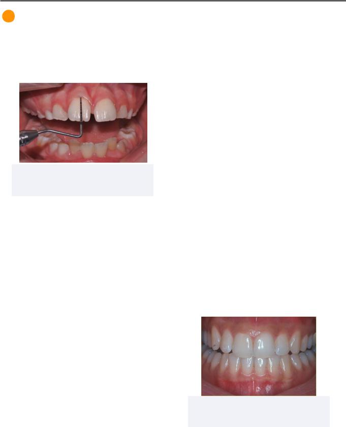
Figure 1.01 Healthy Pediatric Gingiva
2019 Valletta R, et al. Licensee MDPI, Basel, Switzerland. CCBY 4.0
Colour
Children
Gingiva in children are more red in colour. This is because the epithelium is thinner, less keratinized, and receives greater blood supply.
Adults
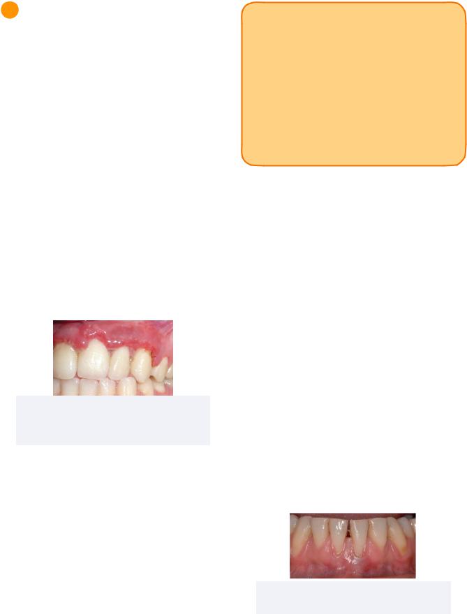
The gingiva is a coral pink colour.
Contour
Children
The gingiva in children have rounded, rolled margins. This is due to the prominent cervical ridges of the crowns, as well as the edema and activated inflammatory mediators which are present during eruption.
Adults
The gingival margins of adults are often knifeedge in health.
29
Consistency
Children
In children, the gingiva is flabby and less dense due to the lower density of fibres and lack of organized collagen in the connective tissue.
Adults
In adults, the gingiva is firm and resilient.
Texture
Children
The gingiva texture in children is smooth and lacks stippling because of the shorter and flatter dental papilla.
Adults
In adults, the gingiva texture is stippled and resembles an “orange-peel.”
Sulcus
Children
The sulcus in children are deeper because the less resilient soft tissue is easier to separate from the tooth.
Adult
In adults, the sulcus are more shallow with good periodontal health.
Figure 1.02 Healthy Adult Gingiva
2007 Crawford J. CCBY 3.0, via
INBDE Booster | Booster PrepTM
Pediatrics
2 Soft Tissue Pathology
Gingivitis
Gingivitis affects up to 70% of children over age 7. It is frequently seen in adolescents.
The presence of plaque is a required precursor to developing gingivitis. However, it may be exacerbated by mouth breathing, crowded teeth, erupting teeth, and braces.
In adolescents, hormones may contribute to the gingival inflammation and lead to adolescent gingivitis at the labial papilla. This form of gingivitis peaks at puberty.
It is recommended that parental help with oral hygiene continues until the age of 8 or adequate manual dexterity is developed to ensure proper plaque removal.
Acute Necrotizing Ulcerative Gingivitis
Figure 2.02 Acute Necrotizing Ulcerative
Gingivitis in Adult
2017 Malek R., et al. CCBY 3.0
Acute necrotizing gingivitis is most commonly seen in adults, but may also be seen in children.
The symptoms may include painful, inflamed, and bleeding gums which are accompanied by fever, necrotic tissue, pseudomembrane on the marginal gingiva, blunted papilla, and fetid breath.
Treatment may include debridement, oxidizing mouth rinses, and antibiotics.
30
INBDE Pro Tip: Each term in acute necrotizing ulcerative gingivitis refers to a group of symptoms of the condition.
Acute: refers to pain, fever
Necrotizing: refers to necrotic tissue, fetid breath
Ulcerative: refers to the pseudomembranous gingival margins,
Gingivitis: refers to the bleeding, inflamed gums and blunted papilla
Reduced Attached Gingiva
Attached gingiva is an important component of the periodontium that is firmly attached to the alveolar bone. It is more robust than the free gingiva and oral mucosa.
Reduced attached gingiva is diagnosed under the criteria that the band of attached gingiva is less than 2mm wide. It is most commonly caused by labial eruption of the tooth, but can also be caused by proclination of teeth, or gingival recession.
For reduced attached gingiva caused by labially positioned or proclined teeth, orthodontics can be utilized to lingually reposition the teeth and restore the attached gingiva. However, in some cases orthodontics can also exacerbate the condition.
Although treatment is not always necessary, intervention may include orthodontics, as well as a free gingival or connective tissue graft.
Figure 2.03 Reduced Attached Gingiva in Adult
2018 Nemcovsky C.E., et al. CCBY 4.0
INBDE Booster | Booster PrepTM
Pediatrics
Eruption Cyst
An eruption cyst is a bump on the crest of the alveolar ridge at the expected position of a tooth. It is most common in children around the incisors and mandibular first molars.
Treatment is usually not indicated. However, diagnosis can be confirmed with a radiograph and excised surgically if symptomatic.
Figure 2.04 Reduced Attached Gingiva in Adult
Edelweiss Publications CCBY 4.0
High Frenum
Figure 2.05 High Frenum
2018 Monnet-Corti V., et al. CCBY 4.0
A high frenum is defined by a frenum that is attached towards a more coronal aspect of the alveolar bone.
A high frenum may apply additional apical forces on the neighbouring gingiva and subsequently cause gingival recession. They may also be accompanied by a notch in the alveolar bone at the point of insertion, as well as a diastema.
Treatment indicates closure of the diastema prior to frenectomy. This is important because a frenectomy can heal with scar tissue that may provide elastic rebound to the teeth even after orthodontic correction of the diastema.
31
Periodontitis
Periodontitis is characterized by the loss of attachment and bone in the periodontium. It is less common in children.
Localized Aggressive Periodontitis
This form of periodontitis often involves the
first permanent molars and permanent incisors.
It is often accompanied by increased aggregatibacter actinomycetemcomitans (a.a) bacterial counts and is most commonly seen in African-American children.
Treatment includes surgical intervention and antibiotics.
Generalized Aggressive Periodontitis
This form of periodontitis involves the entire dentition and is often associated with increase plaque and calculus. It is uncommon in children.
Treatment options include surgical intervention and antibiotics.
Pre-Pubertal Periodontitis
This form of periodontitis is localized to the primary molars. It is most commonly seen in
African-American children.
Treatment options include debridement and antibiotics.
INBDE Booster | Booster PrepTM
Pediatrics |
32 |
Dental Trauma
Pediatric dentistry is a multidisciplinary branch of dentistry which encompasses many aspects of other dental specialties. It may provide a good overview of other sections in your preparation for the INDBE.
1 Dental Trauma
Dental trauma refers to any injury of the teeth, periodontium, or oral soft tissue.
Prevalence
•More common in boys than girls
•Most common in maxillary anteriors
•More common with increased overjet of >6mm
Ellis and Davey Classification of Fractures
The Ellis and Davey classification is commonly used in endodontics to specify the extent of dental trauma. The classification system has been generally summarized in the table below.
Class |
Description |
Trauma |
|
Type |
|||
|
|
||
|
|
|
|
|
Simple or extensive |
|
|
|
fracture of crown |
|
|
Class I/II |
involving considerable |
Minor |
|
|
to no dentin, but no |
|
|
|
pulp. |
|
|
|
|
|
|
|
Extensive fracture of |
|
|
|
crown involving dentin |
|
|
Class III/IV |
and pulp; potentially |
Moderate |
|
|
leading to loss of |
|
|
|
vitality. |
|
|
|
|
|
|
Class V to |
Fracture of root or full |
|
|
crown, loss of teeth, or |
Major |
||
VIII |
|||
displacement of tooth. |
|
||
|
|
||
|
|
|
2 Major Injuries
Major injuries often require extensive and highcost interventions with a multi-displinary and specialty approach. These teeth often have a poor prognosis and require a long term followup.
Luxations - Intrusion, Extrusion, Lateral Luxation
Intrusion
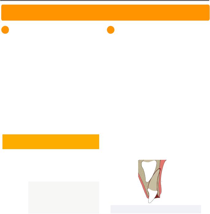
Intrusion is a type of luxation in which the tooth is forced apical into the periodontal tissues.
Due to the labial positioning of primary anterior teeth, intrusion injuries may be pushed against the successor teeth. The potential damage to underlying permanent teeth is dependent on the stage of tooth development during which the injury occurs.
Figure 2.01 Intrusion Luxation
Damages may include hypoplasia, hypocalcification, and dilaceration if injury occurs during the apposition, calcification, and root formation phases, respectively.
INBDE Booster | Booster PrepTM