- •Bud Stage
- •Initiation
- •Cap Stage
- •Bell Stage
- •Apposition
- •Maturation
- •Summary
- •Primary Teeth
- •Permanent Teeth
- •2-3 Rule
- •Supernumerary Teeth
- •Congenitally Missing Teeth
- •Microdontia
- •Macrodontia
- •Fusion
- •Gemination
- •Taurodontism
- •Dens Evaginatus
- •Dens Invaginatus (Dens in Dente)
- •Dilaceration
- •Enamel Hypoplasia
- •Amelogenesis Imperfecta (AI)
- •Dentinogenesis Imperfecta (DI)
- •Regional Odontodysplasia
- •Concrescence
- •Enamel Pearl
- •Dentin Dysplasia
- •Primary Maxillary Central Incisor
- •Primary Maxillary Lateral Incisors
- •Primary Maxillary Canine
- •Primary Maxillary First Molar
- •Primary Maxillary Second Molar
- •Primary Mandibular Central Incisor
- •Primary Mandibular Lateral Incisor and Canine
- •Primary Mandibular Canine
- •Primary Mandibular First Molar
- •Primary Mandibular Second Molar
- •Prevention
- •Fluoride for Children
- •Amalgam Restorations
- •Composite Resin Restorations
- •Stainless Steel Crown
- •Strip Crown
- •Signs and Symptoms
- •Indirect Pulp Cap
- •Direct Pulp Cap
- •Pulpotomy
- •Pulpectomy
- •Extraction
- •Summary
- •Primate Space
- •Leeway Space
- •Interdental Space
- •Primary Incisor Loss
- •Primary Canine Loss
- •Primary First Molar Loss
- •Primary Second Molar Loss
- •Eruption Pattern Variations
- •Root Development
- •Rule of Seven
- •Space Closure
- •Ectopic Eruption of Incisors
- •Ectopic Eruption of Premolars
- •Ectopic Eruption of Molars
- •Colour
- •Contour
- •Consistency
- •Texture
- •Sulcus
- •Gingivitis
- •Acute Necrotizing Ulcerative Gingivitis
- •Reduced Attached Gingiva
- •Eruption Cyst
- •High Frenum
- •Periodontitis
- •Luxations - Intrusion, Extrusion, Lateral Luxation
- •Intrusion
- •Extrusion
- •Avulsion
- •Alveolar and Crown-Root Fracture
- •Concussion
- •Craze Lines and Enamel Fractures
- •Enamel and Dentin Fractures
- •Subluxation
- •Extensive Tooth Structure Involvement
- •Medical History
- •Prevention
- •Pediatric Behaviour Types
- •Frankl Rating Scale
- •Autism Spectrum
- •Anticipatory Guidance
- •Familiarization
- •Functional Inquiry
- •Pre-Visit Imagery
- •Knee-to-Knee Exam
- •Systematic Desensitization
- •Distraction
- •Picture Exchange Communication System (PECS)
- •Behaviour Shaping
- •Treatment Deferral
- •Protective Stabilization
- •Aversive Conditioning
- •Minimal Sedation - Anxiolysis
- •Moderate Sedation - Conscious Sedation
- •Deep Sedation - IV Sedation
- •General Anesthesia
- •Nitrous Sedation
- •Local Anesthesia

Pediatrics
INBDE Pro Tip:
Primary teeth begin calcification in the order of:
A, D, B, C, E beginning at 14 weeks in utero at 1 week intervals.
Permanent teeth begin calcification at 6 month intervals from birth. The sequence of calcification occurs in the order of:
6, (1, L2, 3), U2, 4, 5, 7
3 Eruption Dates
The eruption times for teeth exist within a range. It is accepted that eruption will occur within a 6 month window of the provided dates. However, the order of eruption is more important for development than the dates themselves.
**All eruption times are from the ADA website
Primary Teeth
Tooth |
Eruption Time |
|
|
|
|
Mandibular Central |
6 months |
|
Incisor (A) |
||
|
||
|
|
|
Maxillary Central |
8 months |
|
Incisor (A) |
||
|
||
|
|
|
Maxillary Lateral |
9 months |
|
Incisor (B) |
||
|
||
|
|
|
Mandibular Lateral |
10 months |
|
Incisor (B) |
||
|
||
|
|
|
Maxillary Primary First |
13 months |
|
Molar (D) |
||
|
||
|
|
|
Mandibular Primary |
14 months |
|
First Molar (D) |
||
|
||
|
|
|
Maxillary Canine (C) |
16 months |
|
|
|
|
Mandibular Canine (C) |
17 months |
|
|
|
|
Maxillary Primary |
23 months |
|
Second Molar (E) |
||
|
||
|
|
|
Mandibular Primary |
26 months |
|
Second Molar (E) |
||
|
||
|
|
5
Figure 3.01 Eruption order of primary teeth
This figure is a useful visualization for memorizing the eruption sequence of primary teeth. The teeth are represented by their orthodontic designations and their order of eruption by the arrows in blue.
Permanent Teeth
For the permanent teeth, it is generally accepted that the mandibular teeth erupt before the corresponding maxillary teeth. Additionally, the corresponding teeth generally erupt earlier for females than in males.
The eruption of teeth often occurs symmetrically in that the eruption on one side will usually be followed by the eruption of the same tooth on the opposite side within a few months. If this does not occur, it may be suspected that the tooth is impacted or missing.
INBDE Booster | Booster PrepTM

Pediatrics
Tooth |
Eruption Time |
|
|
|
|
Maxillary/Mandibular |
|
|
1st Molar (6), |
|
|
|
6 years old |
|
Mandibular Central |
|
|
Incisor (1) |
|
|
|
|
|
Maxillary Central |
|
|
Incisor (1), |
|
|
|
7 years old |
|
Mandibular Lateral |
|
|
Incisor (2) |
|
|
|
|
|
Maxillary Lateral |
8 years old |
|
Incisors (2) |
||
|
||
|
|
|
Mandibular Canine |
9 years old |
|
(3) |
||
|
||
|
|
|
Maxillary/Mandibular |
10 years old |
|
1st Premolar (4) |
||
|
||
|
|
|
Maxillary 2nd |
10.5 years old |
|
Premolar (5) |
||
|
||
|
|
|
Maxillary Canine (3), |
|
|
Mandibular 2nd |
11 years old |
|
Premolar (5), |
||
Mandibular 2nd |
|
|
Molar (7) |
|
|
|
|
|
Maxillary 2nd Molar |
12 years old |
|
(7) |
||
|
||
|
|
|
Maxillary/Mandibular |
17 years old |
|
Molar (8) |
||
|
||
|
|
6
2-3 Rule
The 2-3 rule refers to the ⅔ of the root that is developed in erupting teeth. After initial eruption, the root takes 2-3 years to complete its development.
Figure 3.02 2-3 Rule
This figure is a useful visualization for memorizing the eruption sequence of permanent teeth. The teeth are represented by their orthodontic designations and are grouped by colour if they erupt at the same time.
INBDE Booster | Booster PrepTM

Pediatrics |
7 |
Developmental Disturbances
1 Tooth Number Anomalies
Disturbances resulting in missing or supernumerary teeth occurs in the initiation or bud stages.
Supernumerary Teeth
Supernumerary teeth refers to teeth that are present in addition to the typical set of teeth. They may require removal as they may block the normal eruption of permanent teeth.
Incidence: 3% of population
Most commonly affected: Mesiodens Mesiodens are extra teeth which develop close to the midline. They are often found palatally.
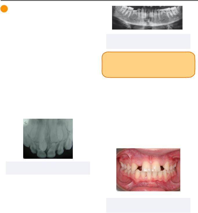
Congenitally Missing Teeth
Figure 1.02 Missing Second premolars
2015 Medio et al. Licensed CC BY 4.0.
INBDE Pro Tip: The order of most commonly congenitally missing teeth is a frequently appearing concept in the INBDE.
Missing teeth are addressed depending on the tooth missing and whether it is missing unior bilaterally.
A missing lateral incisor can be replaced with canine substitution or prosthetic replacement. If a second premolar is missing unilaterally, the second premolar may be extracted on the opposite side and closed with orthodontic treatment to provide symmetry.
Figure 1.01 Supernumerary Teeth (Mesiodens)
Albert, Public domain, via Wikimedia Commons.
Commonly Affected Perm Teeth (most to least): third molars, mandibular second premolars, maxillary laterals, maxillary second premolars
Commonly Affected Primary Teeth: Primary
Maxillary Lateral Incisor
Figure 1.03 Missing Lateral Incisors
2014 Sergio Paduano et al. Licensed CC BY 4.0.
INBDE Booster | Booster PrepTM

Pediatrics
2 Tooth Size Anomalies
Anomalies of size can occur due to disturbances in multiple stages of tooth development. Microdontia and macrodontia occur as a result of disturbances in the bell stage, whereas fusion and gemination occur due to disturbance in the cap stage.
Microdontia
Microdontia refers to the presence of small teeth and can either be generalized or localized in nature.
•Generalized: commonly seen with down syndrome, pituitary dwarfism, ectodermal dysplasia
•Localized: most common form
Figure 2.01 Localized Microdontia (Peg-Shaped
Lateral Incisor)
Adapted from 2020 by Zafar S.A, et al. Licensee MDPI, Basel, Switzerland CC BY 4.0
Peg-shaped lateral incisors are a common form of localized microdontia. It is an autosomal dominant trait which may vary in severity.
Macrodontia
Macrodontia refers to big teeth but do not include fusion or gemination traits. Like microdontia, it can also be generalized or localized in nature.
•Generalized: commonly seen with pituitary gigantism, pineal hyperplasia with hyperinsulinism
•Localized: commonly seen with hemifacial hyperplasia
8
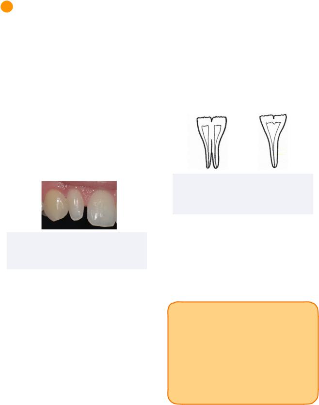
Fusion
Fusion occurs when two adjacent developing teeth merge together in the cap stage. When this occurs, the tooth count is one less than a typical set of teeth. Although the two teeth are fused, they each contain their own separate root canal system.
Commonly Affected Teeth: Primary teeth of anterior region
Figure 2.01 Diagrammatic Representation of Fusion (Left) and Gemination (Right)
Adapted from 2021, Pandya-Sharpe et al. Licensed CCBY 4.0
Gemination
Gemination occurs when one developing tooth develops two crowns with a shared root canal system. Although it may clinically resemble a fused tooth, the tooth count is normal despite the two seemingly-conjoined crowns.
INBDE Pro Tip: Take note of the differences in fusion and gemination:
•Fusion results in a decreased tooth count while gemination does not alter the tooth count.
•Fused teeth each contain their own separate root canal system, while gemination results in a shared root canal system.
INBDE Booster | Booster PrepTM
