

Pediatrics |
1 |
Tooth Development and Eruption
Pediatric dentistry is a multidisciplinary branch of dentistry which encompasses many aspects of other dental specialties. It may provide a good overview of other sections in your preparation for the INDBE.
1 Odontogenesis |
Dental Lamina |
|
Oral Epithelium |
Tooth development is dependent on genetic |
|
control and environmental factors, such as - |
Ectomesenchyme |
nutrition, trauma, infection, excess fluoride |
|
intake. It is composed of sequential events |
|
identified as initiation, bud, cap, bell, and |
|
erupted tooth stages. |
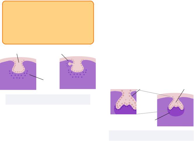
Figure 1.01 Initiation stage |
|
Initiation
The initiation stage begins 6 weeks in utero and is also referred to as the thickening stage. During this stage, the tooth development tissues are composed of the outer layer of oral epithelium, dental lamina, and ectomesenchyme.
All three of these tissue types are derived from the ectoderm. However, the ectomesenchyme is derived specifically from the neural crest cells of the ectoderm which has physiological characteristics which resemble mesenchymal origin.
•Oral Epithelium - superficial-most layer of developing mouth for both upper and lower arch forms
•Dental Lamina - row of thickened oral epithelium that will eventually become the developing teeth
•Ectomesenchyme - provides signal for oral epithelium to multiply into dental lamina.
Failure in this first step can lead to defects of congenitally missing or supernumerary teeth
Bud Stage
The bud stage begins 8 weeks in utero. It is characterized by a quickly developing dental lamina which begins to invade the underlying mesenchyme. This results in the formation of a dental placode which histologically resembles the shape of a proliferating bud.
All primary teeth and permanent molars arise from the dental lamina. However, permanent incisors, canines, and premolars, arise from their primary predecessor when a secondary bud develops from the initial bud.
The continuous proliferation of the dental placode into the mesenchyme results in a condensing mesenchyme which is also a characteristic of the bud stage.
Failure in the bud stage also leads to congenitally missing teeth or supernumerary teeth.
INBDE Booster | Booster PrepTM
Pediatrics
INBDE Pro Tip:
It is only the succedaneous permanent teeth which develop from a secondary bud of their primary predecessor.
The primary teeth and permanent molars are not replacing any teeth and thus develop from a primary bud.
Dental Lamina |
Secondary Bud |
Condensing
Mesenchyme
Figure 1.02 Bud stage
Non-succedaneous teeth develop from a single bud (Right). Succedaneous teeth develop from a secondary bud that arises from the primary bud of the developing primary tooth. (Left)
Cap Stage
The cap stage occurs 9 weeks in utero when the bud continues to proliferate and begins to differentiate into various layers.
Each dental placode grows to form an enamel organ which will ultimately form the enamel. The enamel organ consists of the outer enamel epithelium, inner enamel epithelium, stellate reticulum, and enamel knot.
•Outer Enamel Epithelium (OEE) - outermost cell layer
•Inner Enamel Epithelium (IEE) - cell layer underlying OEE
•Stellate Reticulum - star-shaped cells between the OEE and IEE
•Enamel Knot - thickened signalling centers of the developing tooth that will eventually form the cusp tips
2
The condensed mesenchyme will aggregate to form the dental papilla that will eventually form the dentin and pulp. Histologically, the enamel organ will resemble a cap over the dental papilla.
The dental follicle is defined as the surrounding sac of the dental papilla and enamel organ.
Early defects in this stage can lead to congenitally missing or supernumerary teeth. However, it is also likely to result in cysts, odontomas, gemination, fusion, or dens in dente.
OEE |
Enamel Organ |
Stellate
 Reticulum
 IEE
IEE
Dental Papilla
Figure 1.03 Cap stage
Bell Stage
The bell stage occurs 11 weeks in utero and is characterized by two simultaneously occurring phases of histodifferentiation and morphodifferentiation.
Histodifferentiation
In this phase, the tissues differentiate into distinct cell types which will be able to form the final tooth tissues.
•Inner Enamel Epithelium - differentiate into ameloblasts of tall columnar shape cells
•Dental Papilla - differentiate into odontoblasts of tall columnar shape cells
The interface between the ameloblasts and odontoblasts will form the dentinoenamel junction (DEJ), as it is where the enamel and dentin will be secreted.
INBDE Booster | Booster PrepTM
Pediatrics
Defects in this stage can lead to amelogenesis imperfecta or dentinogenesis imperfecta.
Morphodifferentiation
In this phase, the shape and size of the eventual crown is determined.
Defects in this phase can lead to size and shape abnormalities such as peg laterals or macrodontia.
|
OEE |
|
Ameloblasts |
Dental Papilla |
Odontoblasts |
|
Cervical Loop |
Figure 1.04 Early Bell Stage
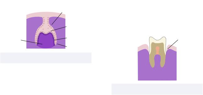
Apposition
Apposition occurs 14 weeks in utero and is defined by the deposition of initial tooth tissue matrices.
1.Odontoblasts begin to deposit dentin matrix of collagen that signals for ameloblast secretion.
The remaining dental papilla will eventually form the pulp.
2.Ameloblasts will deposit enamel matrix of amelogenin which signals root development at the cervical loop.
3.The cervical loop is the junction of the IEE and OEE which grows deep to form an extension called the Hertwig’s epithelial root sheath (HERS).
4.The HERS stimulates odontoblasts to secrete radicular dentin as part of the root.
With continued development, the HERS will eventually disintegrates to form clusters of cells called the epithelial rests of Malassez which remain in the periodontal ligament of the final tooth.
3
5.The stellate reticulum collapses to form the reduced enamel epithelium (REE) when the IEE and OEE combine. The REE lays along the superficial border of the forming enamel and protects the tooth during development.
Post-eruption, the REE will form the junctional epithelium.
Disruptions in this stage through trauma or localized infection can lead to localized defects in the enamel called Turner’s hypoplasia. Other defects may include enamel hypoplasia, enamel pearls, or concrescence.
Junctional
Epithelium
 Pulp
Pulp
 Radicular
Dentin
Figure 1.05 Apposition stage
Maturation
Maturation occurs after 14 weeks in utero at various time points for different teeth and is the longest stage. It is also referred to as calcification or mineralization. During this stage, the final deposition of enamel and dentin occurs.
The calcification process begins at the cusp tips and incisal edges at the previous enamel knots and proceeds in a cervical direction. Calcification takes 2 years to complete for a primary tooth crown and 4-5 years to complete for a permanent tooth crown, without including the time required for complete root formation.
INBDE Booster | Booster PrepTM

Pediatrics
Disturbances in this stage are more common as the critical time period is much longer for the maturation stage. Some of them include - enamel hypomineralization, fluorosis , tetracycline.
Fluorosis is a common defect occurring with systemic fluoride ingestion >1ppm that disturbs ameloblasts. It leads to enamel matrix defects that can lead to a mottled enamel appearance. It commonly affects children from their second trimester (14 weeks in utero) through 8 years old, while their teeth are forming.
Tetracycline is an antibiotic which can cause intrinsic staining. It binds calcium and is eventually incorporated into hydroxyapatite of the dentin. It also affects children from their second trimester (14 weeks in utero) through 8 years old, while their teeth are forming.
INBDE Pro Tip:
The later the disturbance occurs along the developmental process, the more minor the defect is.
Summary
Tooth Germ |
Cell Type |
Tissue |
|
|
|
|
|
Enamel Organ |
Ameloblast |
Enamel |
|
|
|
|
|
|
Odontoblast |
Dentin |
|
Dental Papilla |
|
|
|
Central Cell |
Pulp |
||
|
|||
|
|
|
|
|
Cementoblast |
Cementum |
|
|
|
|
|
Dental Follicle |
Osteoblast |
Alveolar Bone |
|
|
|
|
|
|
Fibroblast |
Periodontal |
|
|
Ligament |
||
|
|
||
|
|
|
4
2 Calcification Dates
Calcification of primary tooth roots are completed at 3-4 years of age.
|
|
Calcification |
|
Tooth |
Start Date of |
||
|
|
Crown |
|
|
|
|
|
|
Central |
14 weeks in |
|
|
Incisors (A) |
utero |
|
|
|
|
|
|
First Molars |
15 weeks in |
|
|
(D) |
utero |
|
|
|
|
|
Primary Teeth |
Lateral |
16 weeks in |
|
Incisors (B) |
utero |
||
|
|||
|
|
|
|
|
Canines (C) |
17 weeks in |
|
|
utero |
||
|
|
||
|
|
|
|
|
Second |
18 weeks in |
|
|
Molars (E) |
utero |
|
|
|
|
|
|
First Molars |
Birth |
|
|
(6) |
||
|
|
||
|
|
|
|
|
All anterior |
|
|
|
teeth except |
|
|
|
maxillary |
6 months |
|
|
laterals (1, L2, |
|
|
|
3) |
|
|
Permanent |
|
|
|
Maxillary |
12 months |
||
Teeth |
Laterals (U2) |
||
|
|||
|
|
|
|
|
First |
18 months |
|
|
Premolars (4) |
||
|
|
||
|
|
|
|
|
Second |
24 months |
|
|
Premolars (5) |
||
|
|
||
|
|
|
|
|
Second |
30 months |
|
|
Molars (7) |
||
|
|
||
|
|
|
|
INBDE Booster | Booster PrepTM