


406 CHAPTER 9 Upper tract obstruction
Hydronephrosis
Hydronephrosis is dilatation of the renal pelvis and calyces (Fig. 9.1). When combined with dilatation of the ureters it is known as hydroureteronephrosis.
Obstructive nephropathy is damage to the renal parenchyma resulting from obstruction to the flow of urine anywhere along the urinary tract.
Dilatation of the renal pelvis and calyces can occur without obstruction and thus hydronephrosis should not be taken to necessarily imply the presence of obstructive uropathy.
Ultrasound
•False negative (i.e., obstruction present, no hydronephrosis)—acute onset of obstruction; in the presence of an intrarenal collecting system; with dehydration; misdiagnosis of dilatation of the calyces as renal cortical cysts (in acute ureteric colic, ultrasonography fails to detect hydronephrosis in up to 35% of patients with proven acute obstruction on IVP)
•False positive (i.e., hydronephrosis, no obstruction)—capacious extrarenal pelvis; parapelvic cysts; vesicoureteric reflux; high urine flow
Diagnostic approach to the patient with hydronephrosis
Hydronephrosis may present either as an incidental finding on an ultrasound or CT done because of nonspecific symptoms or it may be identified in a patient with a raised creatinine or presenting with flank pain.
Symptoms, if present, will depend on the rapidity of onset of obstruction of the kidney (if that is the cause of the hydronephrosis); whether obstruction is complete or partial, unilateral or bilateral; and whether the obstruction to the ureter is extrinsic to the ureter or within its lumen.
Causes of hydronephrosis are listed in Box 9.1.
History
•Severe flank pain suggests a more acute onset of obstruction and, if very sudden in onset, a ureteric stone may well be the cause. Pain induced by a diuresis (e.g., following consumption of alcohol) suggests a possible UPJ obstruction (See Chapter 7, p. 346).
•Anuria (the symptom of bilateral ureteric obstruction or complete obstruction of a solitary kidney)
•If renal function is impaired, symptoms of renal failure may be present (e.g., nausea, lethargy, anorexia).
•Extrinsic causes of obstruction (e.g., compression of the ureters by retroperitoneal malignancy) usually have a more insidious onset, whereas intrinsic obstruction (ureteric stone) is often present with severe pain of very sudden onset.
•An increase in urine output may be reported by the patient that is due to poor renal concentrating ability.
•Obstruction in the presence of bacterial urinary tract infection—signs and symptoms of pyelonephritis (flank pain and tenderness, fever) or sepsis
HYDRONEPHROSIS 407
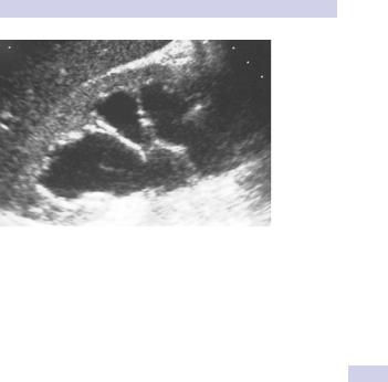
Figure 9.1 Hydronephrosis as seen on renal ultrasonography.
Examination
•Measure blood pressure—elevated in high-pressure chronic retention (HPCR) due to benign prostatic obstruction (caused by fluid overload).
•Bilateral edema (due to fluid overload)
•Abdominal examination—percuss and palpate for an enlarged bladder.
•In men, DRE (?prostate or rectal cancer), and in women, vaginal examination (?cervical cancer)
•Check serum creatinine to determine the functional effect of the hydronephrosis.
•Renal ultrasonography (if not already done)
IVP findings in renal obstruction
•An obstructive (dense) nephrogram
•A delay in filling of the collecting system with contrast material
•Dilatation of the collecting system
•An increase in renal size
•Rupture of fornices (junction between renal papilla and its calyx) with urinary extravasation
•Ureteric dilatation and tortuosity
•A standing column of contrast material in the ureter
Unilateral hydronephrosis
Obtain a KUB X-ray (a ureteric stone may be seen), or CTU (or IVP) if a stone is suspected.
•If no stone is seen, but hydronephrosis is confirmed and ureter is nondilated, the obstruction must be at the UPJ. In the absence of a ureteric stone visible on CTU, the diagnosis must be UPJ obstruction.
•If no stone is seen and the ureter is dilated as well as kidney, ureteric TCC is likely. Arrange for retrograde pyelography to identify site of obstruction, and ureteroscopy/ureteric biopsy.

408 CHAPTER 9 Upper tract obstruction
Bilateral hydronephrosis
•If the patient is in retention or has a substantial post-void residual urine volume, pass a catheter. If the elevated creatinine falls (and the hydronephrosis improves), the diagnosis is bladder outlet obstruction (BOO), due, for example, to BPH, prostate cancer, urethral stricture, or detrusor-sphincter dyssynergia. If the creatinine remains elevated, the obstruction affecting both ureters is higher upstream.
•Obtain TRUS and prostatic biopsy if prostate cancer is suspected on DRE, and a CT scan to look for malignant bilateral ureteric obstruction or abdominal aortic aneurism (AAA).

HYDRONEPHROSIS 409
Box 9.1 Causes of hydronephrosis
Unilateral
•Obstructing ureteric stone
•UPJ obstruction
•Obstructing clot in ureter
•Obstructing ureteric TCC
•Any of the causes listed below when the pathological process has not yet extended to involve both ureters
Bilateral
•Bladder outlet obstruction (BOO)
•BPH
•Prostate cancer
•Urethral stricture
•Detrusor-sphincter dyssynergia (DSD)
•Posterior urethral valve
•Bilateral ureteric obstruction at their level of entry into the bladder
•Locally advanced cervical cancer
•Locally advanced prostate cancer
•Locally advanced rectal cancer
•Poor bladder compliance (often combined with DSD): neuropathic bladder (spinal cord injury, spina bifida); post-pelvic radiotherapy
•Periureteric inflammation
•From adjacent bowel involved with inflammatory bowel disease (e.g., Crohn’s, ulcerative colitis) or diverticular disease
•Retroperitoneal fibrosis
•Idiopathic (diagnosed following exclusion of other causes)
•Periarteritis—aortic aneurysm, iliac artery aneurysm
•Post-irradiation
•Drugs—methysergide, hydralazine, haloperidol, LSD, methyldopa, B-blockers, phenacetin, amphetamines
•Malignant—retroperitoneal malignancy (lymphoma, metastatic disease from e.g., breast cancer), post-chemotherapy
•Chemicals—talcum powder
•Infection—TB, syphilis, gonorrhea, chronic UTI
•Sarcoidosis
•Bilateral UPJ obstruction (uncommon)
•Hydronephrosis of pregnancy (partly due to smooth-muscle relaxant effect of progesterone, partly obstruction of ureters by fetus)
•Hydronephrosis in association with an ileal conduit (a substantial proportion of patients with ileal conduit urinary diversion have bilateral hydronephrosis, in the absence of obstruction)
•Bilateral ureteric stones (rare)