

Биоинженерия / ТИ_печень(органы_ЖКТ) / Bioengineering_Liver_Transplantation
.pdfBioengineering 2019, 6, 35
higher in the control group, but did not reach statistical significance. Postoperative complications ≥ Grade III according to Dindo-Clavien were similar in both groups. Surrogates of complicated clinical courses like length of ICU stay and length of hospital stay did not show di erences among the groups.
Table 2. Secondary outcome parameters: values given as median and range, where appropriate, in brackets.
Parameter |
OPAL (n = 57) |
Control (n = 59) |
p-Value |
Retransplantation (n) |
2 |
- |
- |
|
|
|
|
Early allograft dysfunction (EAD) (n) |
14 |
12 |
0.58 |
|
|
|
|
Recipient ICU stay (days) |
3 (1–45) |
3 (1–41) |
0.97 |
|
|
|
|
Post Tx dialysis (n) |
5 |
9 |
0.28 |
|
|
|
|
Recipient hospital stay (days) |
20 (2–114) |
18 (1–85) |
0.07 |
|
|
|
|
30-day mortality (n) |
3 |
2 |
0.62 |
|
|
|
|
In-hospital mortality (n) |
5 |
5 |
0.95 |
|
|
|
|
Postop. Comlications (n) |
22 |
17 |
|
Dindo-Clavien IIIa |
7 |
5 |
|
Dindo-Clavien IIIb |
8 |
3 |
0.26 |
Dindo-Clavien IVa |
5 |
6 |
|
Dindo-Clavien IVb |
2 |
3 |
|
|
|
|
|
Rejection within 3 months (n) |
6 |
8 |
0.61 |
|
|
|
|
Serum levels of TNF alpha were found to be significantly reduced after oxygen persu ation:
11.1± 1.6 versus 5.9 ± 0.4 pg/ml; mean ± SEM, control versus OPAL p < 0.05.
3.6.Patient and Graft Outcome
Death censored graft survival in the treatment group was 89% after one, three, and five years. The control group demonstrated a graft survival of 87%, 84% and 82% after one, three, and five years, respectively (p > 0.05).
Patient survival in the complete study cohort was 80% and 70% after one and five years, respectively. Patient survival in the treatment group was 77%, 74%, and 74% after one, three and five years.
Accordingly, the control group demonstrated patient survival rates of 83%, 76%, and 66%. Comparison between groups demonstrated non-significant di erences (p = 0.56).
Overall cause of death in the recipients after transplantation was in descending order: Tumor recurrence (10.4%), sepsis (8.6%), and HCV reinfection (6.9%).
3.7. Association of Clinical Parameters with Development of EAD
Logistic regression was performed to identify parameters associated with the development of EAD as a clinical marker for inferior allograft function. Results are displayed in Table 3.
For the treatment group, no clinical parameter was delineated as independent predictor for development of EAD after liver transplantation. In contrast, history of cardiopulmonary resuscitation and last donor ALT were significantly and independently associated with the development of EAD in the control group. Summarizing, oxygen persu ation showed a positive e ect concerning the development of EAD in the case of donors with history of cardiopulmonary resuscitation and of donors with elevated ALT levels.
30

Bioengineering 2019, 6, 35
Table 3. Early allograft dysfunction; censored for treatment arm: Nominal logistic analysis, multivariate analysis and likelihood ratio test (p-values).
Parameter |
OPAL |
Control |
|||
|
Univariate |
Multivariate |
Univariate |
Multivariate |
|
|
|
|
|
|
|
Donor Age (years) |
0.81 |
|
0.41 |
|
|
|
|
|
|
|
|
Donor Age > 70years |
0.7 |
|
0.65 |
|
|
|
|
|
|
|
|
Donor BMI |
0.09 |
3.26 |
0.67 |
0.14 |
|
0.07 |
0.71 |
||||
|
|
|
|||
|
|
|
|
|
|
Donor cause of death |
0.31 |
|
0.23 |
|
|
|
|
|
|
|
|
Donor ICU stay (days) |
0.49 |
|
0.79 |
|
|
|
|
|
|
|
|
Donor cardiopulmonary resuscitation |
0.47 |
0.12 |
0.07 |
6.02 |
|
0.73 |
0.01 |
||||
|
|
|
|||
|
|
|
|
|
|
Donor AST (U/L) |
0.87 |
|
0.1 |
|
|
|
|
|
|
|
|
Donor ALT U/L) |
0.38 |
2.54 |
0.03 |
5.25 |
|
0.11 |
0.02 |
||||
|
|
|
|||
|
|
|
|
|
|
Donor γGT (U/L) |
0.73 |
0.97 |
0.03 |
1.25 |
|
0.32 |
0.26 |
||||
|
|
|
|||
|
|
|
|
|
|
Donor Bilirubin (μmol/L) |
0.26 |
|
0.29 |
|
|
|
|
|
|
|
|
Donor Risk Index |
0.77 |
|
0.4 |
|
|
|
|
|
|
|
|
Allograft histology: macrosteatosis |
0.18 |
|
0.67 |
|
|
|
|
|
|
|
|
Allograft histology: fibrosis |
0.48 |
|
0.76 |
|
|
|
|
|
|
|
|
Preservation solution |
0.13 |
1.75 |
0.07 |
1.67 |
|
0.19 |
0.2 |
||||
|
|
|
|||
|
|
|
|
|
|
Cold Ischemia Time (h) |
0.13 |
|
0.11 |
|
|
|
|
|
|
|
|
Warm Ischemia Time (min) |
0.61 |
|
0.66 |
|
|
|
|
|
|
|
|
Duration of Surgical Procedure (min) |
0.89 |
|
0.86 |
|
|
|
|
|
|
|
|
Recipient Age (years) |
0.36 |
|
0.6 |
|
|
|
|
|
|
|
|
Recipient BMI |
0.37 |
1.39 |
0.08 |
3.24 |
|
0.24 |
0.07 |
||||
|
|
|
|||
|
|
|
|
|
|
Lab-MELD score |
0.52 |
|
0.43 |
|
|
|
|
|
|
|
|
3.8. Association of Clinical Parameters with Patient Survival
Cox proportional hazard analysis was performed to identify parameters associated with long term patient survival (Table 4).
None of the analyzed factor demonstrated an independent association with the patient survival in the treatment group. On the contrary, two factors were significantly and independently associated with the patient survival in the control group: allograft macrosteatosis as a marker for graft quality and the MELD score of the recipients as a marker for severity of the underlying liver disease.
Quite of interest in this analysis was the strong tendency of donor age with the patient survival in the control group. Further analysis demonstrated that an advanced donor age of more than 70 years was significantly associated with the patient survival in univariable analysis. This was not observed in the treatment group. Due to the retroactive character of this analysis, donor age > 70 years was not introduced in the multivariable cox proportional model.
These findings could be interpreted as a positive e ect of the revitalization treatment in the case of older livers (from donors > 70 years of age) or macrosteatotic livers, proposing selection criteria for the use of this method in the corresponding instances.
31

Bioengineering 2019, 6, 35
Table 4. Cox proportional hazard analysis (p-values), censored for long term patient survival and treatment arm.
Parameter |
OPAL |
Control |
|||
|
Univariate |
Multivariate |
Univariate |
Multivariate |
|
|
|
|
|
|
|
Donor age(years) |
0.63 |
0.138 |
0.051 |
0.48 |
|
0.71 |
0.49 |
||||
|
|
|
|||
|
|
|
|
|
|
Donor age > 70 years |
0.22 |
|
0.047 |
|
|
|
|
|
|
|
|
Donor BMI |
0.97 |
|
0.6 |
|
|
|
|
|
|
|
|
Donor cause of death |
0.17 |
|
0.99 |
|
|
|
|
|
|
|
|
Donor ICU stay (days) |
0.29 |
|
0.3 |
|
|
|
|
|
|
|
|
Donor cardiopulmonary resuscitation |
0.46 |
|
0.19 |
|
|
|
|
|
|
|
|
Donor AST U/L) |
0.87 |
|
0.53 |
|
|
|
|
|
|
|
|
Donor ALT (U/L) |
0.2 |
|
0.12 |
|
|
|
|
|
|
|
|
Donor γGT (U/L) |
0.67 |
|
0.69 |
|
|
|
|
|
|
|
|
Donor Bilirubin μmol/L) |
0.2 |
|
0.18 |
|
|
|
|
|
|
|
|
Donor Risk Index |
0.35 |
|
0.65 |
|
|
|
|
|
|
|
|
Allograft histology: macrosteatosis |
0.46 |
0.38 |
0.07 |
6.2 |
|
0.54 |
0.01 |
||||
|
|
|
|||
|
|
|
|
|
|
Allograft histology: fibrosis |
0.09 |
1.17 |
0.44 |
3.69 |
|
0.28 |
0.06 |
||||
|
|
|
|||
|
|
|
|
|
|
Preservation solution |
0.37 |
|
0.7 |
|
|
|
|
|
|
|
|
Cold Ischemia Time (h) |
0.55 |
0.05 |
0.09 |
3.55 |
|
0.83 |
0.06 |
||||
|
|
|
|||
|
|
|
|
|
|
Warm Ischemia Time (min) |
0.83 |
|
0.27 |
|
|
|
|
|
|
|
|
Duration of Surgical Procedure (min) |
0.28 |
|
0.9 |
|
|
|
|
|
|
|
|
Recipient Age (years) |
0.71 |
|
0.17 |
|
|
|
|
|
|
|
|
Recipient BMI |
0.48 |
|
0.34 |
|
|
|
|
|
|
|
|
MELD |
0.49 |
0.48 |
0.054 |
5.06 |
|
0.49 |
0.03 |
||||
|
|
|
|||
|
|
|
|
|
|
3.9. Subgroup Analysis
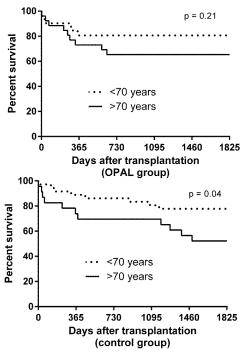
The impact of advanced donor age was further investigated in a subgroup analysis. Patient survival was compared in recipients transplanted with organs from donors with age > 70 years to recipients transplanted with organs from donors with age < 70 years in the treatment group (n = 26) and control group (n = 23), respectively. Results are depicted in Figure 3.
In the treatment group patient survival was 80% after one and five years when transplantation was carried out with younger donors. In donors aged 70 or more the patient survival was 70% and 65% after one and five years, without statistical significant di erences between groups. However, the same analysis in the control group demonstrated significantly worse survival after transplantation of allografts from donors aged 70 years or more with one and five year patient survival of 70% and 48%. Donors aged younger than 70 years led to a patient survival of 85% and 75% after one and five years, respectively.
We also evaluated maximal serum values of AST in the population receiving macrosteatotic livers (>20%) and found an accentuated trend towards a benefit in the OPAL group: 987 (271–3016) U/L versus 2498 (890–4332) U/L. However, because of the small number of patients (n = 12 in the OPAL group, n = 6 in the control group) di erences were not evaluated for significance.
32
Bioengineering 2019, 6, 35
Figure 3. Five year patient survival in the treatment group (oxygen persu ation (OPAL)) and in the standard care group (control) according to recipient age (<70 years versus ≥70 years).
4. Discussion
This randomized controlled single center study investigated for the first time the impact of oxygen persu ation as adjunct in liver preservation on early allograft injury and dysfunction upon repefusion. The primary endpoint was aminotransferase peak of AST within the first three days after liver transplantation. While AST values were lower in the treatment group, statistical significance was not reached.
Assessment of secondary endpoints, such as early allograft dysfunction, primary non-function, and patient survival, showed a positive e ect of the treatment on the development of EAD in the case of donors with history of cardiopulmonary resuscitation and of elevated ALT levels. Benefits with regard to patient survival were also present for marginal liver grafts with macrosteatosis or originating from donors aged > 70 years.
Furthermore a moderate, but significant reduction of TNF-alpha release could be documented for the entire collective.
Thus, a distinct clinical benefit for application of retrograde oxygen persu ation as reconditioning tool immediately before liver transplantation could be delineated in concrete subcategories of particularly endangered donor grafts. This comes to confirm the corresponding literature from experimental studies, as the concept of retrograde oxygen persu ation in liver allograft reconditioning is based on scientifically high grade research which has been published in the past decades: Initial reports by Isselhard [22] and Ross [23] demonstrated convincing results in animal kidneys already in the 1970s. Subsequently, the first clinical pilot project was successfully initiated in renal transplantation [24]. Experimental applications in liver allografts followed thereafter [25,26]. This research demonstrated significant reduction of non-parenchymal cell injury and vascular endothelial dysfunction after cold preservation of the liver by gaseous oxygen [27] as well as reduction of proteolysis leading to improved
33
Bioengineering 2019, 6, 35
functional outcome after transplantation [28]. Gaseous oxygenation resulted in normalization of vascular resistance and reduced release of hepatocellular enzymes. Further investigations showed prevention of functional and ultrastructural impairments by venous oxygen persu ation [29] in steatotic rat livers.
In a porcine model, gaseous oxygen persu ation prevented primary non-function of livers after extended cold storage times and improved one week survival of the recipient from 0% up to 83% [11].
Based on these premises one might have expected a less equivocal result upon clinical usage oxygen persu ation. However, in contrast to the controlled experimental situation, a broader heterogeneity of included donor livers as well as recipient health status may influence the outcome after transplantation in the clinical setting.
Some of the donor organs might have had lesser needs for additional treatment than others, and, although distributed evenly across the groups, a notable fraction of not so marginal grafts might have obscured the benefit of oxygen persu ation in less resilient livers.
Such e ect would not have been predictable, as extensive human studies are needed to delineate the specific influence of preservation techniques on each group of allografts. The subgroup analysis presented in this studies might be taken as hint that oxygen persu ation might be most e ective in older donor allografts. While the study was not powered to prove this, the influence of donor age on patient survival in the control group, which was not traceable in the treatment group, suggests reduced damage to the allograft after oxygen persu ation.
In addition, results of multivariable studies demonstrated that history of donor cardiopulmonary resuscitation as well as elevated ALT levels in the donor contributed to development of EAD in the control group but not in the treatment group. This might indicate that after reconditioning of pre-damaged allografts, such risk factors lost their impact. The same was observed for patient survival: allograft macrosteatosis and MELD score were delineated as risk factors only in the control group. Against the background of similar patients and allografts transplanted in both groups, this resembles a surrogate of improved allografts after treatment by oxygen persu ation.
It should be of interest that the data at hand demonstrates excellent safety of the method applied. Serious adverse events related to treatment were not observed and all endpoints were qualitative similar to the common standard of cold storage.
Organ preservation is a science virtually lacking relevant clinical progress over more than 20 years. Nowadays, several approaches of machine perfusion have been taken from experimental projects to first clinical applications [30–32]. These studies demonstrated feasibility and safety of such new
methods and suggested beneficial e ects on clinical outcomes like peak of aminotransferase after transplantation, development of EAD and maybe even biliary complications. Most importantly, these pilot projects led to the initiation of randomized controlled trials comparing these new methods with static cold storage. So far, no clear benefits in term of graft and patient survival could be demonstrated.
This again underscores the importance to identify more precisely those subgroups of allografts that need or may benefit from reconditioning measures and those who will not.
Compared with the more sophisticated methods of machine perfusion, the simple insu ation of gaseous oxygen excels by its ease of use and the unmatched cost-e ectiveness may furthermore allow for a less critical application in attempt to rescue questionable liver grafts.
5. Conclusions
In conclusion, this randomized controlled trial demonstrated safety of venous oxygen persu ation of liver allografts immediately before transplantation as reconditioning tool. A clinical benefit could be demonstrated in concrete subcategories of less than optimal donor organs. Pending success of alternative new preservation methods might justify further clinical evaluation of oxygen persu ation as safe, cheap, and easy applicable reconditioning method in liver allografts subgroups.
Author Contributions: A.G. participated in performance of research, coordinated the collection of data and samples; A.G. and T.M. were responsible for the experimental therapy; D.P.H. and G.S. collected, analyzed and
34
Bioengineering 2019, 6, 35
interpreted data; J.T., T.B., F.S., and A.P. were responsible for the clinical conduct of the study; J.B. participated in data collection; A.P. and T.M. designed this study with help from other authors; All authors participated in the revision of the manuscript
Funding: The study was supported by the German Research Foundation (DFG MI 470/14/2).
Acknowledgments: We thank Mrs. Ose and her team for their continuous support, data monitoring and conducting some of the statistical analysis.
Conflicts of Interest: The authors declare no conflict of interest.
References
1.Nickkholgh, A.; Weitz, J.; Encke, J.; Sauer, P.; Mehrabi, A.; Büchler, M.W.; Schmidt, J.; Schemmer, P. Utilization of extended donor criteria in liver transplantation: a comprehensive review of the literature. Nephrol. Dial. Transplant. 2007, 22, viii29–viii36. [CrossRef] [PubMed]
2.Briceño, J.; Marchal, T.; Padillo, J.; Solórzano, G.; Pera, C. Influence of marginal donors on liver preservation injury. Transplantation 2002, 74, 522–526. [CrossRef]
3.Tector, A.J.; Mangus, R.S.; Chestovich, P.; Vianna, R.; Fridell, J.A.; Milgrom, M.L.; Sanders, C.; Kwo, P.Y. Use of Extended Criteria Livers Decreases Wait Time for Liver Transplantation Without Adversely Impacting Posttransplant Survival. Ann. Surg. 2006, 244, 439–450. [CrossRef] [PubMed]
4.Markmann, J.F.; Markmann, J.W.; Markmann, D.A.; Bacquerizo, A.; Singer, J.; Holt, C.D.; Gornbein, J.; Yersiz, H.; Morrissey, M.; Lerner, S.M.; et al. Preoperative factors associated with outcome and their impact on resource use in 1148 consecutive primary liver transplants. Transplantation 2001, 72, 1113–1122. [CrossRef] [PubMed]
5.Moore, D.E.; Feurer, I.D.; Spero , T.; Gorden, D.L.; Wright, J.K.; Chari, R.S.; Pinson, C.W. Impact of Donor, Technical, and Recipient Risk Factors on Survival and Quality of Life After Liver Transplantation. Arch. Surg. 2005, 140, 273. [CrossRef]
6.Hoyer, D.P.; Paul, A.; Gallinat, A.; Molmenti, E.P.; Reinhardt, R.; Minor, T.; Saner, F.H.; Canbay, A.; Treckmann, J.W.; Sotiropoulos, G.C.; et al. Donor information based prediction of early allograft dysfunction and outcome in liver transplantation. Liver Int. 2015, 35, 156–163. [CrossRef]
7.Minor, T.; Saad, S.; Nagelschmidt, M.; Koetting, M.; Fu, Z.; Paul, A.; Isselhard, W. Successful transplantation of porcine livers after warm ischemic insult in situ and cold preservation including postconditioning with gaseous oxygen. Transplantation 1998, 65, 1262–1264. [CrossRef]
8.Minor, T.; Paul, A. Hypothermic reconditioning in organ transplantation. Curr. Opin. Organ Transplant. 2013, 18, 161–167. [CrossRef]
9.Fuller, B.J.; Busza, A.L.; Proctor, E. Possible resuscitation of liver function by hypothermic reperfusion in vitro after prolonged (24-hour) cold preservation–a 31p nmr study. Transplantation 1990, 50, 511–512. [CrossRef] [PubMed]
10.Koetting, M.; Lüer, B.; E erz, P.; Paul, A.; Minor, T. Optimal time for hypothermic reconditioning of liver grafts by venous systemic oxygen persu ation (vsop) in a large animal model. Transplantation 2011, 91, 42–47. [CrossRef] [PubMed]
11.Minor, T.; Koetting, M.; Kaiser, G.; E erz, P.; Lüer, B.; Paul, A. Hypothermic Reconditioning by Gaseous Oxygen Improves Survival After Liver Transplantation in the Pig. Am. J. Transplant. 2011, 11, 2627–2634. [CrossRef] [PubMed]
12.Minor, T.; Stegemann, J.; Hirner, A.; Koetting, M. Impaired autophagic clearance after cold preservation of fatty livers correlates with tissue necrosis upon reperfusion and is reversed by hypothermic reconditioning. Liver Transplant. 2009, 15, 798–805. [CrossRef]
13.Kim, J.S.; NItta, T.; Mohuczy, D.; O’Malley, K.A.; Moldawer, L.L.; Dunn, W.A., Jr.; Behrns, K.E. Impaired autophagy: A mechanism of mitochondrial dysfunction in anoxic rat hepatocytes. Hepatology 2008, 47, 1725–1736. [CrossRef] [PubMed]
14.Gustafsson, Å.B.; Gottlieb, R.A. Recycle or Die: The Role of Autophagy in Cardioprotection. J. Mol. Cell. Cardiol. 2008, 44, 654–661. [CrossRef]
15.Treckmann, J.; Minor, T.; Saad, S.; Özcelik, A.; Malagó, M.; Broelsch, C.E.; Paul, A. Retrograde oxygen persu ation preservation of human livers: A pilot study. Liver Transplant. 2008, 14, 358–364. [CrossRef]
35

Bioengineering 2019, 6, 35
16.Khorsandi, S.E.; Jitraruch, S.; Fairbanks, L.; Cotoi, C.; Jassem, W.; Vilca-Melendez, H.; Prachalias, A.; Dhawan, A.; Heaton, N.; Srinivasan, P. The e ect of anterograde persu ation on energy charge and hepatocyte function in donation after cardiac death livers unsuitable for transplant. Liver Transplant. 2014, 20, 698–704. [CrossRef]
17.Minor, T. Vascular oxygen persu ation for preservation and reconditioning of marginal liver grafts. In Organ Preservation and Reengineering, 1st ed.; Uygun, K., Lee, C.E., Eds.; Artech House: Norwood, MA, USA, 2011; pp. 125–134.
18.Minor, T.; Pütter, C.; Gallinat, A.; Ose, C.; Kaiser, G.; Scherag, A.; Treckmann, J.; Paul, A. Oxygen persu ation as adjunct in liver preservation (OPAL): Study protocol for a randomized controlled trial. Trials 2011, 12, 234. [CrossRef] [PubMed]
19.Oltho , K.M.; Kulik, L.; Samstein, B.; Kaminski, M.; Abecassis, M.; Emond, J.; Shaked, A.; Christie, J.D. Validation of a current definition of early allograft dysfunction in liver transplant recipients and analysis of risk factors. Liver Transplant. 2010, 16, 943–949. [CrossRef] [PubMed]
20.Braat, A.E.; Blok, J.J.; Putter, H.; Adam, R.; Burroughs, A.K.; Rahmel, A.O.; Porte, R.J.; Rogiers, X.; Ringers, J.; European Liver and Intestine Transplant Association (ELITA) and Eurotransplant Liver Intestine Advisory Committee (ELIAC). The Eurotransplant Donor Risk Index in Liver Transplantation: ET-DRI. Am. J. Transplant. 2012, 12, 2789–2796. [CrossRef]
21.Charlson, M.E.; Pompei, P.; Ales, K.L.; MacKenzie, C. A new method of classifying prognostic comorbidity in longitudinal studies: Development and validation. J. Chronic Dis. 1987, 40, 373–383. [CrossRef]
22.Isselhard, W.; Witte, J.; Denecke, H.; Berger, M.; Fischer, J.H.; Molzberger, H.; Freiberg, C.; Ammermann, D.; Brunke, M. Function and metabolism of canine kidneys after aerobic ischemia by retrograde persu ation with gaseous oxygen. Exp. Med. 1974, 164, 35–44. [CrossRef]
23.Ross, H.; Escott, M.L. Gaseous oxygen perfusion of the renal vessels as an adjunct in kidney preservation. Transplantation 1979, 28, 362–364. [CrossRef]
24.Rolles, K.; Foreman, J.; Pegg, D.E. A pilot clinical study of retrograde oxygen persu ation in renal preservation.
Transplantation 1989, 48, 339–342.
25.Minor, T.; Isselhard, W. Venous oxygen insu ation to prevent reoxygenation injury after ischemia of a solid organ. Transplantation 1994, 58, 121–123. [PubMed]
26.Minor, T.; Klauke, H.; Vollmar, B.; Isselhard, W.; Menger, M.D. Biophysical aspects of liver aeration by vascular persu ation with gaseous oxygen. Transplantation 1997, 63, 1843–1846. [CrossRef] [PubMed]
27.Minor, T.; Isselhard, W.; Klauke, H. Reduction in nonparenchymal cell injury and vascular endothelial dysfunction after cold preservation of the liver by gaseous oxygen. Transpl. Int. 1996, 9, S425–S428. [CrossRef]
28.Minor, T.; Klauke, H.; Nagelschmidt, M.; Isselhard, W. Reduction of proteolysis by venous-systemic oxygen persu ation during rat liver preservation and improved functional outcome after transplantation. Transplantation 1997, 63, 365–368. [CrossRef]
29.Minor, T.; Akbar, S.; Tolba, R.; Dombrowski, F. Cold preservation of fatty liver grafts: prevention of functional and ultrastructural impairments by venous oxygen persu ation. J. Hepatol. 2000, 32, 105–111. [CrossRef]
30.Dutkowski, P.; Polak, W.G.; Muiesan, P.; Schlegel, A.; Verhoeven, C.J.; Scalera, I.; DeOliveira, M.L.; Kron, P.; Clavien, P.-A. First comparison of hypothermic oxygenated perfusion versus static cold storage of human donation after cardiac death liver transplants: An international-matched case analysis. Ann. Surg. 2015, 262, 764–771. [CrossRef]
31.Hoyer, D.P.; Mathe, Z.; Gallinat, A.; Canbay, A.C.; Treckmann, J.W.; Rauen, U.; Paul, A.; Minor, T. Controlled oxygenated rewarming of cold stored livers prior to transplantation: First clinical application of a new concept. Transplantation 2016, 100, 147–152. [CrossRef] [PubMed]
32.Watson, C.J.E.; Kosmoliaptsis, V.; Randle, L.V.; Russell, N.K.; Gri ths, W.J.H.; Davies, S.; Mergental, H.; Butler, A.J. Preimplant Normothermic Liver Perfusion of a Suboptimal Liver Donated After Circulatory Death. Am. J. Transplant. 2015, 16, 353–357. [CrossRef] [PubMed]
©2019 by the authors. Licensee MDPI, Basel, Switzerland. This article is an open access article distributed under the terms and conditions of the Creative Commons Attribution (CC BY) license (http://creativecommons.org/licenses/by/4.0/).
36

bioengineering
Review
Towards Bioengineered Liver Stem Cell
Transplantation Studies in a Preclinical Dog Model
for Inherited Copper Toxicosis
Hedwig S. Kruitwagen, Hille Fieten and Louis C. Penning *
Department of Clinical Sciences of Companion Animals, Faculty of Veterinary Medicine, Utrecht University, 3584CM Utrecht, The Netherlands; H.S.Kruitwagen@uu.nl (H.S.K.); H.Fieten@uu.nl (H.F.)
* Correspondence: L.C.Penning@uu.nl
Received: 26 June 2019; Accepted: 17 September 2019; Published: 25 September 2019
Abstract: Wilson Disease is a rare autosomal recessive liver disorder in humans. Although its clinical presentation and age of onset are highly variable, hallmarks include signs of liver disease, neurological features and so-called Kayser-Fleischer rings in the eyes of the patient. Hepatic copper accumulation leads to liver disease and eventually to liver cirrhosis. Treatment options include life-long copper chelation therapy and/or decrease in copper intake. Eventually liver transplantations are indicated. Although clinical outcome of liver transplantations is favorable, the lack of suitable donor livers hampers large numbers of transplantations. As an alternative, cell therapies with hepatocytes or liver stem cells are currently under investigation. Stem cell biology in relation to pets is in its infancy. Due to the specific population structure of dogs, canine copper toxicosis is frequently encountered in various dog breeds. Since the histology and clinical presentation resemble Wilson Disease, we combined genetics, gene-editing, and matrices-based stem cell cultures to develop a translational preclinical transplantation model for inherited copper toxicosis in dogs. Here we describe the roadmap followed, starting from the discovery of a causative copper toxicosis mutation in a specific dog breed and culminating in transplantation of genetically-engineered autologous liver stem cells.
Keywords: copper toxicosis; stem cell transplantation; Wilson Disease; preclinical large animal model
1. Introduction
The trace element copper is indispensable for various biochemical processes [1]. At the same time, the transition element copper (reduced as Cu+ and oxidized as Cu2+) is involved in chemical reactions leading to the production of reactive oxygen species. Therefore, its intracellular free concentrations need to be regulated within very narrow boundaries [2]. Regulation occurs at the level of cellular uptake, intracellular binding and distribution, and lastly cellular excretion. Copper is imported into cells mainly via Copper Transporter 1 (Ctr1) [3]. Once copper is inside the cell, copper binding proteins ensure that the free copper levels remain very low. These chaperone proteins include Cytochrome c Oxidase Copper Chaperone (Cox17), Copper Chaperone for Superoxide Dismutase (CCS), and Antioxidant protein1 (ATOX1) [1,2]. Intracellularly copper can be sequestered by glutathione, and metallothionein. Excretion is mediated via P-type ATPases, ATP7A and ATP7B [4]. Transport through the blood stream is mediated via ceruloplasmin. Copper related diseases in humans include Menke’s Disease (copper deficiency disorder), Wilson Disease (copper accumulation), Indian childhood cirrhosis [5], endemic Tyrolean infantile cirrhosis [6], and idiopathic copper toxicosis [7]. The causative mutations for Wilson Disease are in the ATP7B gene [8,9]. The ATP7B protein is responsible for excretion of bound intrahepatic copper into the bile. Biliary copper excretion accounts for as much as 95% of the total body copper excretion. There is a wide variation in clinical presentation of Wilson Disease (WD) and a large number of mutations has been reported (well over 500; http://www.wilsondisease.med.ualberta.ca/database.asp) [10–12].
Bioengineering 2019, 6, 88; doi:10.3390/bioengineering6040088 37 |
www.mdpi.com/journal/bioengineering |
Bioengineering 2019, 6, 88
Phenotypical variation also occurs within the same genotype, hinting to the e ect of modifier genes. In contrast to hepatic copper overload, Menkes Disease (MD) is presented with impaired copper absorption in various organs. Wilson Disease is a rare X-linked copper deficiency disorder caused by mutations in the ATP7A gene [13–17]. The limited genotype-phenotype correlation and the rarity of both WD and MD urge for innovative clinical approaches to improve the quality of life of people su ering from Wilson’s or Wilson Disease. What makes dogs (Canis lupus familiaris), especially for WD, so well-suited? In order to fully appreciate the potential of these animals, some insights into the canine population structure are necessary. Ever since dogs were domesticated, they have been under severe artificial breeding selection, for instance for behavioral traits and/or specific morphological features [18]. This resulted in isolated genetic populations of dog breeds [19]. The limited genetic variation within breeds, and at the same time a large genetic variation over all breeds, provides a gold-mine for geneticists. Whereas the genetic variation over the various breeds remained intact, the reduced genetic variability within breeds worked as a genetic amplifier and o ered researchers a genetic dissection microscope [19]. Together with the selection for a unique trait, such as excessive muscle formation, short limbs or a specific coat color, an increased risk for the development of specific disorders with a simple and/or complex inheritance pattern arose within breeds. Exploiting the downside of inbreeding may therefore be instrumental for the discovery of causative and modifier genes involved in complex diseases and/or rare diseases such as inherited copper toxicosis.
Next to the above mentioned genetic-argument, other research advantages reside within dogs. Dogs are of comparable size of humans and they share similar environmental exposures. Especially the size allows to design and test procedures at a humanized size with highly comparable anatomical arrangements. This is an obvious advantage for preclinical studies, for instance related to liver transplantation. In this respect, the readers might be aware that the first liver transplantations were performed in dogs [20].
In summary both genetic and technical arguments are in favor to utilize dogs as important preclinical models for inherited copper toxicosis. However, there is more to come. Veterinarians have been confronted with sheep and dogs presenting with copper related disorders already for decades [21–24]. Deleterious levels of hepatic copper are described in several dog breeds including Bedlington terriers, Skye terriers, West-Highland White terriers, Dobermanns, Dalmatians and Labrador retrievers [25–30]. Pedigree analysis of most breeds revealed a complex mode of inheritance of copper-mediated hepatitis. Therefore, the phenotypic expression is not dependent on one single genetic factor, but on mutations in more genes and also environmental factors are deemed influential in the phenotypic presentation. As an exception to the rule of thumb that copper toxicosis is a complex genetic disorder, a simple autosomal recessive mode of inheritance is observed in the Bedlington terrier [31].
2. A Roadmap towards a Relevant Preclinical Model Animal for Liver Stem Cell Transplantations
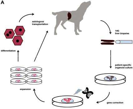
Although rodent models for WD have been instrumental to dissect molecularly how mutations in the ATP7B gene lead to hepatic copper accumulation, their size does not allow for longitudinal studies (individual animals followed for long time and consecutive measurements). This prompted us to investigate the genetic background of inherited copper toxicosis in several dog breeds. One important feature of a clinical model is the knowledge of the genetic cause and preferentially a simple breeding strategy to acquire su cient number of experimental animals. In line with this, a similar progression of the disease in time strengthens the validity of the model. Another aspect is the feasibility to obtain su cient and genetically gene-corrected liver stem cells, preferentially autologous to minimize the risk of rejection of the transplanted cells. Lastly, in view of the clinical application in human medicine, mode of cell transplantation must be similar to the one preferred in human medicine. All these steps will be outlined in more detail below. Figure 1 depicts the strategy followed for functional liver recovery after autologous genetically-engineered liver stem cell transplantation in a COMMD1 deficient dog.
38
Bioengineering 2019, 6, 88
Figure 1. The strategy followed for functional liver recovery after autologous genetically-engineered liver stem cell transplantation in a COMMD1 deficient dog.
2.1. Requirement 1. Copper Accumulation in Bedlington Terriers Is caused by a Deletion of exon-2 in the COMMD1-gene
By means of mapping studies and positional cloning, a 13kB deletion covering exon-2 of the commd1 gene was discovered as the causative mutation of Bedlington terrier copper toxicosis [31,32].
In the following years, the involvement of this mutation in other copper storage diseases was investigated. It turned out that this mutation was not causative for either Indian Childhood Cirrhosis (ICC), Endemic Tyrolean Infantile Cirrhosis (ETIC), nor Idiopathic copper toxicosis (ICT) [33].
Furthermore, whether or not the murr1 mutations are somehow involved in WD is a matter of debate [34–36]. The intracellular interaction between the ATP7B protein and the COMMD1 protein (previously known as murr1) explains the similarities in WD and Bedlington terrier copper toxicosis [37]. The COMMD1 protein, COpper Metabolism Murr1 domain-containing protein had an unknown function at the time it was discovered. To unravel its function, among others yeast-two hybrid screens were used with COMMD1 as bait. Of interest, a direct COMMD1-ATP7B interaction occurs which was confirmed in cell lines [37]. In WD the interaction between ATP7B and COMMD1 is enhanced and leads to lower ATP7B stability. This interaction partially explains the similar phenotypes of WD in men and copper toxicosis in Bedlington terriers. At present a plethora of functions are related to the COMMD1 protein, including sodium transport via epithelial sodium channel (ENaC), tra cking of cystic fibrosis transmembrane conductance regulator (CFTR), inhibition of Cu/Zn -SOD, NFk-B signalling, Hypoxia Inducing Factor (HIF1) regulation and HIV-replication [37–41]. COMMD1 depletion leads to increased serum Low Density Lipoprotein (LDL) levels, due to mis localization of the LDL-receptor and consequently a reduced uptake of LDL particles [42]. One of the common themes of COMMD1 action seems to be related to protein degradation via ubiquitination, at least
39