

Биоинженерия / ТИ_печень(органы_ЖКТ) / Bioengineering_Liver_Transplantation
.pdfBioengineering 2019, 6, 95
networks remains a challenge for most tissue engineers. Hence, there are considerable types of 3D printing methods that are expected to overcome current limitations. 3D bioprinting o ers the ability to develop highly complex 3D patterns with living cells that mimic organ level functions, and it has therefore been applied in organs-on-chips and organs engineering. The main bioprinting techniques are extrusion [27], inkjet [28] and laser-induced forward transfer (LIFT) [29,30], each one possessing several advantages and disadvantages.
2.1. Laser Bioprinting
Laser-induced forward transfer (LIFT) is a technique presented more than 30 years ago by Bohandy et al. [31]. Briefly, a pulsed laser beam is applied on a donor slide (or ribbon) covered with a laser-energy-absorbing layer (e.g., gold or titanium) containing the desired material (e.g., cells, hydrogels and growth factors), followed by the evaporation of the material; this results in a high-pressure bubble jetting toward the receiving substrate that is placed underneath the donor slide, as shown in Figure 1.
Figure 1. Schematic representation of laser-induced forward transfer (LIFT) setup.
For the direct printing of cells, the use of LIFT is proposed because it enables the printing of bio-inks within a wide range of viscosities (1–300 mPa s) [32] and at high speeds while cell viability is preserved (>90%).
LIFT is a nozzle-free technique and therefore does not have the problems of nozzle clogging with cells or biological materials, which are some major drawbacks of other bioprinting technologies. Moreover, this technique o ers printing cell concentrations up to 1 × 10⁸ cells/mL with a very high resolution [33].
The use of LIFT for the printing of functional biomaterials can be traced back to 2003 [34], while the development of 2D cell structures was first proposed in 2008 [35]. Regarding the use of lasers for the printing of 3D structures, the first report was published in 2011 by M. Gruene et al. [36], while in 2012, Koch et al. [37] published the printing of multiple cell lines in order to create epidermal tissue. These multiple cell lines were previously proven to be resistant to damage during the laser-assisted printing process [38]. The proliferation of cells over a period of 10 days was studied, and the ability of 3D printed cells to form real tissue was demonstrated. It is critical to know how the laser process a ects cell viability as well as phenotypes. Catros et al. [39] studied the e ects of laser pulse energy, extracellular matrix (ECM) thickness and viscosity of the bioink on cell viability. Cell viability 24 h post-printing was measured to compare di erent printing settings. It was concluded that while higher laser energy
80
Bioengineering 2019, 6, 95
leads to more cell fatality, increasing film thickness as well as bioink viscosity results in increased cell viability. Moreover, another laser group investigated the e ects of bioink viscosity, laser energy and printing speed on printing resolution [32]. It was shown that a microscale resolution and 5 kHz printing speed were within reach. This work is another proof for the applicability of printing cells and biomaterials via LIFT printing to engineer miniaturized tissue layouts with de novo high cell density and microscale organization. An interesting study was demonstrated by Keriquel et al. [40], whereby in vivo laser bioprinting was used to deposit nano-hydroxyapatite in a mouse calvaria 3D defect model as a proof of concept. In the future, study materials that can directly integrate into a patient’s tissue could be used. Finally, incorporating the patients’ own cells may facilitate the applicability of these types of constructs to contribute to both the structural and functional components of the tissue.
2.2. Inkjet Bioprinting
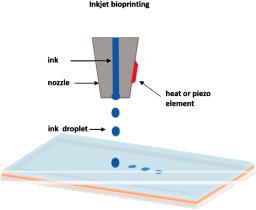
Inkjet-based bioprinting is a noncontact technique in which droplets of cells or biomaterials are patterned into desired substrates.
The drop-on demand inkjet bioprinters are the most common ones, and they consist of thermal, piezoelectric, and electrostatic inkjet nozzles [41]. A schematic diagram of inkjet printing is shown in Figure 2.
Figure 2. Schematic representation of inkjet printing.
With respect to the construction of cellular structures, inkjet bioprinters are normally used for the printing of matrices for the cell growth, such as small sca olds. Di erent inkjet printheads with multiple nozzles have been developed to increase printing speed and fabricate larger cellular constructs [42].
However, inkjet bioprinters also have limitations on material viscosity (ideally below 10 centipoise) due to the excessive force required to eject drops using solutions at higher viscosities [43]. Another major disadvantage of this technique is the di culty in achieving biologically relevant cell densities. Often, low cell concentrations are used to facilitate droplet formation (less than 10 million cells/mL) [44]. To provide a higher concentration of cells, the inhibition of some hydrogels can be generated by adding crosslinking agents. However, the requirement for crosslinking agents often slows the bioprinting process and involves the chemical modification of naturally occurring ECM materials, which changes both their chemical and material properties [45]. Despite these disadvantages, inkjet bioprinting has notable benefits, including low cost, high speed and biocompatibility with a broad range of biological materials [46]. Significant studies of inkjet printing have included the regeneration of functional tissues, such as skin and cartilage, in situ [47,48]. With the advantages of high throughput digital control and high resolution, this technique enables the direct placement of cells, biological factors
81
Bioengineering 2019, 6, 95
and biomaterial sca olds directly into skin or cartilage lesions. Inkjet-based bioprinting facilitates the successful deposition of either primary cells or stem cell types with uniform density, and it maintains high cell viability and function after printing. These studies have shown the ability of inkjet bioprinting to regenerate functional constructs.
2.3. Extrusion Bioprinting
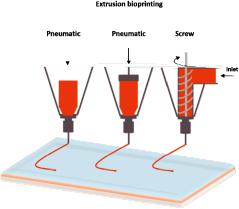
The extrusion-based bioprinting technique is characterized by a temperature-controlled biomaterial dispensing system driven by a pneumatic pressure or a mechanical piston, as demonstrated in Figure 3. Schematic representation of extrusion bioprinting. The printing system generates continuous biomaterial filaments, instead of droplets, that are deposited in two dimensions; filaments are placed along the x- and z-axes and then move higher in the y-axis. The final product is a 3D structure. This technique provides the ability to deposit very high cell densities as well biological material such as hydrogels and biocompatible copolymers. Several groups have used sole cells or multicellular cell spheroids and allowed for their self-assembly into the desired 3D structures using extrusion bioprinters [49–51]. Pioneer work using this approach is currently being performed at the Wyss Institute under Prof J. Lewis [52]. Each print head is equipped with an on-board temperature controller to adjust the temperature depending on the material that is being printed, enabling sequential layer-by-layer printing and avoiding contamination between di erent materials.
Figure 3. Schematic representation of extrusion bioprinting.
However, a major disadvantage of extrusion bioprinting is that cell viability is lower than that with inkjet-based bioprinting (40–86%). The decreased cell survival rate possibly results from the shear stresses inflicted on cells in viscous fluids [53].
Extrusion-based bioprinting approaches have been also used for the generation of multiple tissue types, including aortic valves [54] and in vitro pharmokinetic models [55].
A review of the outstanding research works using the above printing techniques for liver and liver tissue engineering is presented.
A brief review of the above mentioned bioprinting techniques is presented in Table 1. A brief review of common bioprinting techniques.
82

Bioengineering 2019, 6, 95
Table 1. A brief review of common bioprinting techniques.
|
Laser Assisted |
Inkjet |
Extrusion |
|
|
Bioprinting |
|||
|
|
|
||
|
|
|
|
|
|
High resolution, |
Ability to print low |
Simple, capable of |
|
|
printing various |
|||
|
deposition of |
viscosity biomaterials, |
||
|
biomaterials, ability to |
|||
|
biomaterials in solid or |
fast fabrication speed, |
||
Advantages |
print high cell densities, |
|||
liquid phase, and nozzle |
low cost, high resolution, |
|||
|
multi-material printing, |
|||
|
free and non-contact |
multi-material printing, |
||
|
and ability to control |
|||
|
printing. |
Simple operation. |
||
|
ejection speed. |
|||
|
|
|
||
|
|
|
|
|
|
High cost, thermal |
Inherent inability to |
|
|
|
damage due to |
provide a continuous |
|
|
|
nanosecond/femtosecond |
flow, poor functionality |
Only applicable for |
|
|
laser irritation, metallic |
for vertical structures, |
viscous liquids, gelation |
|
Drawbacks |
residuals possible |
low cell densities, |
and solidification, and |
|
damage of tissue from |
clogging of nozzle, |
limited material selection |
||
|
||||
|
use of laser lights, slow |
imposing thermal or |
(shear thinning |
|
|
printing speed, and |
acoustic stress to cells, |
ability required). |
|
|
di culty in handling |
and limited variety |
|
|
|
heterogenous cells. |
of bioink. |
|
|
|
|
|
|
|
Speed |
Medium |
Fast |
Slow |
|
|
|
|
|
|
Cell viability |
<85% |
~80% |
>90% |
|
|
|
|
|
|
Resolution |
10 μm |
50 μm |
100 μm |
|
|
|
|
|
|
Cell density |
Medium |
Low |
High |
|
|
|
|
|
|
Viscosity |
1–300 mPa s |
<10 mPa s |
30–6 × 10⁷ mPa s |
|
Scalability |
Low |
Low |
Low–Medium |
|
|
|
|
|
|
Structural integrity |
Low |
Low |
High |
|
|
|
|
|
|
Cost |
High |
Low |
Low–Medium |
|
|
|
|
|
3. Tissue and Liver Bioprinting
As previously mentioned, the liver is considered one of the most significant organs in the human body due to its special characteristics. It plays a major role in metabolism with numerous functions, including the regulation of glycogen storage, the decomposition of red blood cells, plasma protein synthesis, hormone production, and the detoxification of chemicals [56,57]. In anatomy, the liver is divided into four lobes. The right lobe, which is much bigger than the left lobe, involves two minor lobes—the quadrate and caudate lobes. Blood is supplied to the liver through two di erent vessels. The hepatic artery supplies arterial blood from the heart to the liver, and the hepatic portal vein carries blood consisting of nutrients and toxins from the intestines to the liver [57].
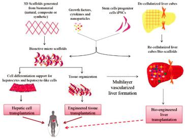
The liver has an extensive regeneration capacity due to the high proliferation ability of hepatocytes, even if it is subjected to vast damages. The tissue engineering of the liver is not new, and there are several groups that have worked on the engineering of liver tissues and bioartificial livers as early as 1996 [58,59]. Therefore, various tissue bioprinting techniques have been used to fabricate biomimetic liver tissues—even a whole liver. A schematic representation of the key approaches used for liver tissue engineering is demonstrated in Figure 4 [59].
83
Bioengineering 2019, 6, 95
Figure 4. Schematic diagram of liver tissue engineering. Solid lines show already ongoing approaches, whereas dotted lines indicate proposed mechanisms [59].
3.1. Micropatterned 2D and 3D Liver Models
Over the past few decades, liver tissue engineering has made significant progress towards the establishment of in vitro liver models for both fundamental pathophysiological studies and drug screening. The sources of cells used for these in vitro liver models include primary hepatocytes, hepatic cell lines isolated from tumors or liver slices, and stem cell-derived hepatic cells [60,61]. Gri th et al. [62] fabricated a vascularized liver on a small scale using the inkjet printing technique. They were pioneers in investigating the role of sca old architecture from biodegradable polyesters using a manufacturing technique amenable to scaling-up, commercial production, and culture conditions for achieving hepatic function in long-term perfusion cultures.
Monolayer culture, organoid culture and co-culture platforms have been established using culture plates [63], commercially available wells [64], dielectrophoresis micropatterning [65] and physical mask-based additive photopatterning methods [60]. However, the liver specific functions of hepatocytes cultured in such platforms are functional only for weeks of in vitro culture [63,66]. Therefore, liver constructs that better mimic the native environment and help maintain in vitro liver functions is in great demand.
3D bioprinting technology, with its potential to pattern cells and biomaterials in a precise manner, provides a great tool to achieve novel and biomimetic in vitro liver models with increasing structural complexity.
3.2. 3D Bioprinting for Liver Models
3D printing is a scientific field with innovative techniques that o er remarkable benefits in terms of the vascular network formation of liver tissues and organs due to their feasibility, variety of available printing methods, and precise controllability. With the appearance of bioprinting, the constructions of functional tissue livers or mini liver organs have become an impending reality. Currently, many researchers are contributing to the improvement of 3D printed vascular networks on a best e ort basis for their introduction into the medical field.
Many researchers that have worked on tissue engineering have successfully achieved to fabricate biomimetic 3D printed vascularized liver constructs with their own unique properties such as rapid
84
Bioengineering 2019, 6, 95
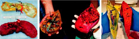
restoration ability even after considerable damage [67]. In an earlier work by Cheng et al. [68], 30 layers of a hepatocyte/gelatin mixture were laminated into a high spatial structure using a 3D rapid prototyping technology. The 3D hepatocyte/gelatin pattern remained viable and performed biological functions in the construct for more than two months. In an e ort to develop personalized tissues and organs for precision medicine, Organovo, harnessing the advantages of 3D bioprinting, used a syringe-based extrusion printer to develop 3D printed human liver tissues that can remain fully functional and stable for up to 28 days. The researcher demonstrated a multicellular liver structure involving hepatocytes, hepatic stellates, and endothelial cells (ECs). 3D liver tissues possessed critical liver functions, including albumin production, cholesterol biosynthesis, fibrinogen and transferrin production, and inducible cytochrome (CYP) 1A2 and CYP 3A4 activities. These in vitro models of 3D vascularized livers could potentially be implanted into patients to replace their damaged livers [69]. In 2013, the first human liver was synthetically reproduced and validated against the actual native liver at the time of surgery by Zein et al. [70]. Specifically, successful 3D synthetic livers were printed along with their complex network of vascular and biliary structures which replicated the native livers for six patients, three living donors, and three respective recipients. Prior to the transplantation, the dimensions of the donor and recipient livers were recorded in detail, including the diameters of veins to fabricate a vascularized liver using the inkjet printing technique and based on each patient’s individual computed tomography (CT) scan and magnetic resonance imaging (MRI). To implement external vascularization, the authors utilized a permanent adhesive to attach to the liver lobe (Figure 5). These results demonstrate the potential e cacy of a 3D printed synthetic liver with a vascular network in the human body as a valuable tool for drug delivery, a substitute for treating partially or irreversibly damaged liver tissue, and a tool for potentially minimizing intraoperative complications. That was the first human liver to have been synthetically reproduced and validated against the actual native liver at the time of surgery.
Figure 5. (a) Side view of a 3D printed liver and extracted liver of a patient, where long, short, and double arrows indicate hepatic artery, hepatic vein, and portal vein, respectively. (b) Right lobes of 3D printed and extracted livers with indications of the hepatic artery (single arrows) and portal vein (double arrows). (c) Cross-sectional views of 3D printed and extracted livers with indications of hepatic vein (single arrows) and portal vein (dotted arrows) [70].
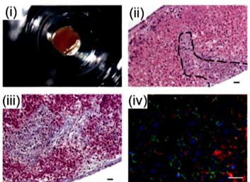
Nguyen et al. [71], established a novel bioprinted human mini liver tissue from the co-culture of primary human hepatocytes, hepatic stellate cells (HSC) and human umbilical vein endothelial cells (HUVEC) cells to test clinical drug-induced toxicity in vitro using an inkjet 3D bioprinter. A histological analysis showed the presence of distinct intercellular hepatocyte junctions, cluster of di erentiation 31 (CD31+) endothelial networks, and desmin-positive, smooth muscle actin-negative quiescent stellates, mimicking the in vivo human drug response at the tissue level (Figure 6). A major challenge in liver tissue engineering is the proliferation, long-term culture and maintenance of hepatocyte function ex vivo of primary hepatocytes [38].
85
Bioengineering 2019, 6, 95
Figure 6. Organovo’s mini liver tissue: (i) A macroscopic image of liver tissue housed in a 24-well transwell, (ii) Hematoxylin and eosin (HE) staining of a tissue cross-section, (iii) extracellular matrix (ECM) deposition assessed by Masson’s trichrome staining, and (iv) Iimmunohistochemistry (IHC) staining of the parenchymal compartment for E-cadherin (green) and albumin (red) [71].
A recent study by our team [72] utilized the LIFT technique to laser print hepatocyte cancer cell line Huh7 on porous collagen-Glycosaminoglycan (GAG) sca olds, which are biomaterials with established applications in re-generative medicine implants. The results showed the benefits of the laser bioprinting technique for the precise placement and immobilization of hepatocyte cells into porous collagen sca olds for novel custom-made implants for regenerative medicine applications.
Arai et al. [73] used an inkjet 3D bioprinter to fabricate a 3D culture system using an artificial sca old for studying the liver-specific functions of hepatocytes. The printed construct expressed liver-specific proteins and receptors such as albumin, MPR2, and asialoglycoprotein receptor (ASGPR), thus proving the functionality of the printed liver tissue. The work by Matsusaki et al. [74] demonstrated that high cell activities and high cell–cell interactions of the fabricated 3D human liver chip from HepG2/HUVECs laden fibronectin and gelatin using inkjet printing technology were analogous to the native liver structure due to the hierarchical sandwich structures.
In another study by Y Kim et al. [75], mouse primary hepatocytes (isolated from the livers of six-to-eight weeks old mice) were printed into a 3D liver tissue construct using an extrusion-based bioprinting system. Cells were viable for 14 days, with liver-specific gene expressions, namely albumin, hepatocyte nuclear factor 4 alpha (HNF-4α), forkhead box protein A3 (Foxa3), and asialoglycoprotein receptor 1 (ASGR1), increasing gradually up to day 14. In another study, Lee et al. [76] developed 3D structures from polycaprolactone (PCL) with improved mechanical properties for liver tissue regeneration by using a multi-head tissue building printing system. A co-cultured 3D microenvironment of primary rat hepatocytes (HCs), human umbilical vein endothelial cells (HUVECs), and human lung fibroblasts (HLFs) were successfully established and maintained to study liver cells proliferation. The results of this work suggested that the employed co-cultured microenvironment promoted heterotypic cellular interaction within a 3D construct. Similarly, Skardal et al. [77] utilized a 3D bioprinting platform to fabricate liver tissue, which has high potential for influencing how future drug and toxicology screening and personalized medicine approaches are performed. Measurable levels of both albumin and urea as well as common soluble biomarkers for liver were tested, and these remained relatively consistent throughout the culture period. Moreover, this group developed a 3D liver
86

Bioengineering 2019, 6, 95
tissue model containing primary human hepatocytes and liver stellate cells supported by bioinks, and they tested the functional indicators. Specifically, these constructs were maintained in culture for six days, and liver functionality was examined by exposing the constructs to a hepatic toxicant, acetaminophen (APAP, 100 μM), and measuring the levels of albumin, urea, α-GST (alpha Glutathione S-Transferase), and lactic acid dehydrogenase (LDH) in the media over time. An analysis of both urea and albumin levels showed a significant decrease until day 15 for the acetaminophen-treated conditions. In addition, the levels of α-GST, a detoxification protein, increased at day nine, and the levels of lactic acid dehydrogenase (LDH), a marker of liver damage, also peaked due to printing-related stress but decrease to nominal levels by day six. Finally, histological staining presented a greater cellularity in untreated constructs, while drug-treated conditions showed a loss of cellularity. In the future, these models could be used for drug screening, disease modeling, and precision medicine applications [78]. An interesting decellularized extracellular matrix (dECM) bioink derived from a native liver was demonstrated by Lee et al. [79]. The proposed bioink, in combination with the 3D bioprinting technology, could be a suitable biomechanical and biochemical microenvironment for liver tissue function. Specifically, the cell-printed mixtures consisted of dECM bioink seeded with human bone marrow-derived mesenchymal stem cells (BMSCs) and liver cancer cells (human hepatocellular carcinoma), as well as PCL polymer for 3D structural support, with control constructs prepared with a collagen bioink. The resulting cell-laden printed bioink was evaluated and compared with those in commercial collagen bioink. An analysis of liver-specific functions of these constructs by assessing albumin and urea levels presented that the dECM bioink enhanced liver cell functions. Moreover, the level expression of key transcription factor HNF4A (Hepatocyte nuclear factor 4 alpha) was particularly upregulated in the liver dECM group to more than twice the level seen for the collagen, and the level expression of transcriptional markers HNF1A and HNF3B (Hepatocyte nuclear factor 3-beta) was significantly higher in the liver dECM group.
A recent study by Kurreck et al. [80] utilized the extrusion bioprinting technique to print a 3D tissue model composed of bioinks and human bipotent hepatic progenitor cells (HepaRG) with established applications in virus biology. A short summary of recent outstanding bioprinting studies is presented in Table 2.
Table 2. A short summary of outstanding recent liver bioprinting studies.
Printing Method |
Cell Type/Bioink |
Achievements |
Reference |
|
|
|
The laminated hepatocytes |
|
|
Extrusion bioprinting |
Hepatocytes Gelatin |
remained viable and performed |
[68] |
|
biological functions for more |
||||
|
|
|
||
|
|
than 2 months |
|
|
|
|
|
|
|
|
|
Viable up to 28 days |
|
|
|
|
(% not mentioned) |
|
|
|
|
Inkjet bioprinting |
|
|
|
Primary human hepatocytes, |
Galactosylated alginate |
|
|
Extrusion-based |
hepatic stellates, HUVEC cells, |
(12 mg/mL) |
|
|
and non-parenchymal |
Primary mouse hepatocytes |
[71] |
||
bioprinting |
||||
cells/NovoGelR 2.0 hydrogel |
(isolated from the liver tissue of |
|
||
|
|
|||
|
(concentration not mentioned) |
male 6–8-weeks-old ICR 12 mice) |
|
|
|
|
Data not available >85% after 2 |
|
|
|
|
Days test of hepatotoxicity of |
|
|
|
|
trovafloxacin and Levofloxacin |
|
|
|
|
|
|
|
|
Primary mouse hepatocytes |
|
|
|
|
(isolated from the liver tissue |
|
|
|
Inkjet bioprinting |
of male 6-to-8-week-old ICR |
>85% after 2 days |
[73] |
|
|
12 mice)/Galactosylated |
|
|
|
|
alginate (12 mg/mL) |
|
|
|
|
|
|
|
87

Bioengineering 2019, 6, 95
Table 2. Cont.
Printing Method |
Cell Type/Bioink |
Achievements |
Reference |
|
|
|
Multilayered organ tissue model |
|
|
Inkjet bioprinting |
HUVEC |
test of hepatotoxicity of |
[74] |
|
|
|
troglitazone (Rezulin) |
|
|
|
|
|
|
|
|
Primary mouse hepatocytes |
|
|
|
Extrusion-based |
(isolated from the livers of 6–8 |
Viable up to 14 days |
[75] |
|
bioprinting |
weeks old mice)/Alginate |
(% not mentioned) |
||
|
||||
|
(3% w/v) |
|
|
|
|
|
|
|
|
|
HepG2, |
|
|
|
Extrusion bioprinting |
BMMSCs/decellularized |
Liver tissue model |
[79] |
|
|
extracellular matrix (dECM) |
|
|
|
|
|
|
|
|
|
hiPSCs |
|
|
|
|
(human-induced pluripotent |
|
|
|
|
stem cell lines, RCi-22 and |
|
|
|
Microvalve bioprinting |
RCi-50); |
>55% after 1 day |
[81] |
|
hESCs |
||||
|
|
|
||
|
human embryonic stem cell |
|
|
|
|
lines, RC-6 and |
|
|
|
|
RC-10)/Alginate (1.5% w/v) |
|
|
|
|
|
|
|
|
Extrusion-based |
Primary hepatocytes |
Viable up to 60 days (% not |
[82] |
|
bioprinting |
mentioned) |
|||
|
|
|||
|
|
|
|
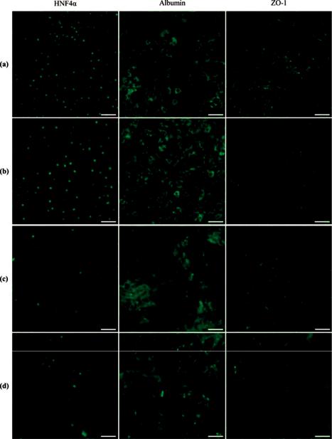
An alternative approach to liver tissue fabrication is the use of stem cells. Concerning the hepatic di erentiation of induced pluripotent stem cells (iPSCs) to liver-specific cell lines. The first successful work on bioprinting a mini-liver from both human-induced pluripotent stem cells (hiPSCs) and human embryonic stem cells (hESCs), which have matured to be hepatocyte-like cells, was reported by Faulkner-Jones et al. using a valve-based bioprinting system which did not adversely a ect cell viability (~84%) [83]. The group built a 3D alginate matrix, and the analysis was carried out after 21 days of di erentiation protocol, revealing peak albumin secretion that meant the construct was hepatic in nature [81], as shown in Figure 7 [84]. Recently, Choi et al. [85] used a nozzle 3D bioprinter to fabricate a liver-mimicking architecture using primary hepatocytes, and they demonstrated the benefits of co-cultured primary hepatocytes and mesenchymal stem cells (MSCs). This research indicated that the expression of hepatic genes and proteins was higher for up to seven days in the 3D hepatic architecture, and that the primary hepatocyte cell morphology was stable.
Most 3D-bioprinted tissues demonstrate liver-specific functions in addition to injury response. Several companies and research groups have created living constructs that mimic native liver structures and functions [86–89].
There is an acute demand for livers, and the fabrication of liver tissue or liver will definitely alleviate this problem. Liver tissue and organoids can also be used in other assays such as drug testing and liver disease studies. As with mature hepatocytes, hepatocyte-like cells obtained from stem cells tend to quickly functionally deteriorate under in vitro conditions. The liver structure is complex with a modular microenvironment; thus, it is di cult to model native liver tissue [87]. Recently, Kizawa et al. [82] printed a liver tissue by the spheroid assembly of primary hepatocytes (1 × 10⁴ cells/mL) that maintained functionality up to 60 days by using a sca old-free 3D bioprinting technology from Cyfuse Biomedical (NA1002, Cyfuse Biomedical), as demonstrated in Figure 8. The human 3D bioprinted liver construct also maintained the expression of many drug transporter proteins and metabolic enzymes for many weeks.
88
Bioengineering 2019, 6, 95
Figure 7. Fluorescence images of printed human-induced pluripotent stem cells (hiPSC)-derived hepatocytes showing hepatocyte marker expression in green: (a,b) human embryonic stem cells (hESC)-derived hepatocyte-like cells (HLCs) (RC-10): (a) Non-printed control; (b) printed results; (c,d) hiPSC-derived HLCs (RCi-22); (c) non-printed control; (d) printed results (scale bars 50 μm) [81].
89