

Биоинженерия / ТИ_печень(органы_ЖКТ) / Bioengineering_Liver_Transplantation
.pdfBioengineering 2019, 6, 95
Figure 8. Self-organization in bio-printed human liver tissues. (A) Hematoxylin and eosin stain (HE) staining shows structure of bio-printed liver tissue on day 50. (B) Immunostaining with the MRP2 antibody detected bile acid transporters (day 50). (C) Immunostaining with, cluster of di erentiation 31 (CD31) antibody detected blood vessel-like and sinusoid-like structures (day 14). (D) Terminal deoxynucleotidyl transferase dUTP nick end labeling (TUNEL) staining detected little apoptosis (day 60). (E) Immunostaining with the OAT2/8 antibody detected drug uptake transporters (day 44). (F) Immunostaining with MRP2 antibody showed tissue distribution (day 44). (G) Masson’s trichrome staining shows collagen accumulation (day 50). Black bars represent 50 μm [82].
Tissue engineers have continued to improve the quality of their human liver creations. The creation of living mini-organs is a relatively new area of science with the potential to replace animal models that are not always accurate. Organoid systems are the recently developed 3D bioengineered platforms for studying assays such as drug toxicity testing and metabolic diseases. Organoids are cell-derived in vitro 3D organ models that allow for the study of biological processes and also have important e ects for clinical use in an environment that mimics endogenous cell organization and organ structures. These models overcome the major constraints of 2D tissue models and provide prolonged cell viability and functionality [90]. These in vitro culture systems contain a self-renewing stem cell population which di erentiates into multiple, organ-specific cell types that exhibit a spatial organization similar to the corresponding organ and are capable of recapitulating some functions of that organ, thus providing a highly physiologically relevant system.
Organoids have been formed via several di erent methods, e.g., spinner flask cultures [91], utilizing rotating cultures [92], stationary cultures in hanging drops with well-known 96or 384-well
90
Bioengineering 2019, 6, 95
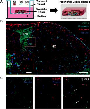
plates [93], and cell growth on non-adherent surfaces [94]. The utilization of engineering tools such as biomaterial sca olds, microfluidics and bioprinting has enabled greater control over the cellular environment, which has increased the accurate prediction of clinically relevant outcomes and the longevity of liver functions in vitro. For example, Norona et al. [95] fabricated a 3D bioprinted liver tissue housed in a 24-well Transwell (Corning Inc, Corning, NY, USA) that can recapitulate drug-, chemical-, and Transforming growth factor β1 (TGF-β1)-induced fibrogenesis at the cellular, molecular, and histological levels, as demonstrated in Figure 9. Taking into consideration the above characteristics, these bioprinted in vitro tissue models of human liver demonstrate the utility of novel 3D bioprinted tissues to further evaluate compound-induced liver fibrosis in a more defined and systematic way.
Figure 9. 3D bioprinted tissue exhibits a compartmentalized architecture and maintains hepatic stellate cells in a quiescent-like phenotype. (A) Illustration of a transverse cross-section of bioprinted tissue on a transwell insert comprising hepatocytes (HCs) and compartmentalized endothelial cells (ECs) and hepatic stellate cells (HSCs). (B) The organization of non-parenchymal cells (NPCs) is depicted with CD31 and vimentin staining to mark ECs and HSCs, respectively. Albumin is used to denote the hepatocellular compartment (HC). Scale bar =100 μm, inset scale bar =25 μm. (C) HSC activation status was examined using desmin (generic marker) and Alpha-smooth muscle actin (α-SMA) (activation marker). Quiescent HSCs are denoted with white arrows. Scale bar = 50 μm [95].
3.3. Liver-on-Chip Platforms
In contrast to static models, perfusion systems or cell microfluidic platforms can allow for the automated control over several conditions such as culture medium, pH, temperature, fluid pressures, cell shear stress, nutrient supply, and waste removal. Microfluidic systems have been implemented in engineering liver tissues [96]. Significant applications of microfluidics in tissue engineering technology
91
Bioengineering 2019, 6, 95
include cell culture and making gradient biomaterials [97]. For these reasons, microfluidic cell platforms are preferable for mimicking the native and dynamic cellular environment compared to static cell culture systems [98]. Moreover, these systems remain precise long term and could provide information on tissue responses to various conditions over time scales that are clinically relevant [99].
The microarchitecture of the liver is crucial to liver function [100]. Hepatocytes interact with mesenchymal cells, stellate cells, Küp er cells, macrophages, and lymphocytes [101]. A main feature of the liver is the perfusion of fluid. When compared to a conventional cell culture, liver function can be enhanced in a microfluidic chip [102].
Furthermore, some diseases or injury states have also been supported inside a microfluidic chamber for pharmaceutical testing [103,104]. Recently, polydimethylsiloxane (PDMS)-based microfluidic devices have been made obtainable by using multiple chambers to mimic the sinusoidal architecture of the liver. For example, Kang et al. [105] used their system to analyze the viral replication for hepatotropic hepatitis B virus. Moreover, they demonstrated that primary rat hepatocytes maintained normal morphology and produced urea for 30 days when they were cultivated on one side of a transwell membrane, while immortalized bovine aortic endothelial cells were cultivated on the other side of the membrane that was subjected to dual-channel microfluidic perfusion. Another group [106] developed a system to model alcohol injury. Their liver injury-on-chip system was made by two chambers for seeding of hepatocytes and stellate cells, as well as three more chambers for miniature aptamer-modified electrodes to monitor liver cell signaling. This system makes it possible to monitor the paracrine crosstalk between co-cultured cell types communicating via the same signaling.
Additionally, the advantages of perfusion on the functions of liver co-cultures is that perfusion can drive the cells to gradients of oxygen, nutrients, and hormones, which have been shown to lead to liver parenchyma or di erential functions in hepatocytes across the length of the sinusoid [107]. Allen et al. [108] fabricated a perfusion bioreactor platform with oxygen gradients that was used to induce an in vivo-like zonal pattern of CYP450s and acetaminophen toxicity in rat hepatocyte cultures. This bioreactor system could provide useful information about the maintenance of liver zonation in order to get deeper insight into the mechanism of metabolism and toxicity.
In contrast to an oxygen gradient, McCarty et al. [109] demonstrated a gradient of exogenous hormone (insulin and glucagon) onto a rat hepatocyte monolayer using a microfluidic device. Utilizing this advanced control system, they demonstrated the in vitro creation of hepatocyte carbohydrate, nitrogen, alcohol degradation, and drug conjugation metabolic zonation. This useful type of system could be essential for the development of in vitro liver disease models.
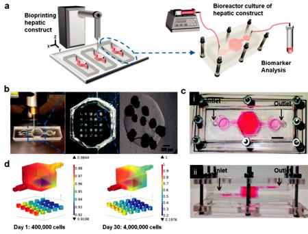
Only a few reports have been published which combine direct printing techniques with on-chip technologies for the fabrication of organs on chips. Direct printing into a microfluidic chamber to build a liver-on-a-chip platform was also demonstrated by Bhise et al. [110]. Droplets of HepG2 spheroid- Gelatin-methacryloyl (GelMA) mixture were printed on a glass slide within the cell culture chamber of a bioreactor, followed by immediate UV cross linking. The engineered hepatic construct remained functional during the 30-day culture period and showed a drug response similar to published data (Figure 10).
Another printing technique utilizing micro valves integrated with microfluidic chips was studied by Chang et al. [111] in order to fabricate reproducible three-dimensional cell-encapsulated alginate-based, tissue-engineered constructs in chambers for drug screening platforms in planetary environments.
92
Bioengineering 2019, 6, 95
Figure 10. (a) Schematic of the hepatic bioreactor culture platform integrated with a bioprinter and biomarker analysis module. (b) Bioprinting photocrosslinkable Gelatin-methacryloyl (GelMA) hydrogel-based hepatic construct within the bioreactor as a dot array. (c) Top-view (i) and side-view (ii) of the assembled bioreactor with the inlet and outlet fluidic ports as indicated. Scale bar = 1 mm. (d) Oxygen concentration gradient in the bioreactor, considering the oxygen uptake of, case A: 400,000 hepatocytes on day one (16,000 cells per dot), and case B: 4000,000 hepatocytes on day 30 (160,000 cells per dot) [110].
Liver platforms are being integrated with di erent cell lines for liver tissue fabrication. It has been researched that perfused hepatocyte-endothelial co cultures show a greater rate of production of drug metabolites relative to static controls [112]. An interesting in vitro hepatic model was demonstrated by Khetani et al. [60] for drug screening and modeling liver diseases using engineered micropatterned co-cultures of induced pluripotent stem cell-derived human hepatocyte-like cells (iHeps) and 3T3-J2 murine embryonic fibroblasts with a Matrigel. This in vitro model of human liver was maintained for several weeks in culture. Moreover, Cho et al. [113] developed a controlling co-cultured microenvironment to study the heterotypic cell interactions of hepatocytes on a patterned fibroblast layer using microfabricated PDMS stencils. The liver-specific functions of the hepatocytes including intracellular albumin staining and E-cadherin expression were increased as a result of enhanced heterotypic contact in the co culture system. In other similar research, primary human hepatocytes along with human endothelial (EA.hy926), immune (U937) and stellate (LX-2) cells were co-cultured in a microfluidic device. This study described a relevant liver model which was maintained for weeks in order to investigate liver studies and the microfluidic integration technology with other organs [114]. Other approaches to create artificial, three-dimensional hepatic tissue constructs and the regeneration of injured livers reported the co culture systems of hepatic stellate cells (HSCs) [115] and both HSCs and ECs [116]. Several liver platforms have already been fabricated with the aim of the reliable replication of liver physiology and metabolism to benefit the pharmaceutical industry in drug discovery and development. The performance of current liver platforms needs to be improved to
93
Bioengineering 2019, 6, 95
further mimic the physiology and function of liver in the body. Future advances in this area could emerge from the combinatory use of existing technologies to move toward a liver model with a more complete functionality.
4. Sca olds Fabrication Methods
Sca olds are 3D artificial biostructures which are used in tissue engineering as well-defined matrices for cell adhesion and proliferation. A high porous architecture and a controllable porous size are key parameters for accommodating di erent types of cells, whereas porosity has a crucial role in attachment and migration of transplanted cells. Depending on the fabrication method and the raw material, the porous size varies between 100 and 500 μm in order to be suitable for applications such as bone regeneration [117], cardiac tissues [118] and cells proliferation [119].
Its biocompatibility, mechanical properties, and chemical properties make the material suitable for medical applications and cell culture. Towards the fabrication of 3D sca olds, several approaches have been used, such as two-photon polymerization, selective laser sintering, and 3D printing techniques (inkjet and extrusion printing).
4.1. Laser-Based Methods
The main purpose for the fabrication of 3D structures that are aimed to be used as a matrix for the selective placement and growth of cells is the printing of biocompatible polymers for the creation of a 3D shape. Two photon polymerization, a widely used method for developing 3D materials suitable for cell growth and proliferation, is based on the irradiation of a monomer with a laser beam to trigger a cross-linking process by two photon absorption in selected depths [120]. As the desired structure forms by the selective polymerization o ered by the laser beam, the non-polymerized monomer is subsequently removed by extensive washing procedures.
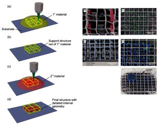
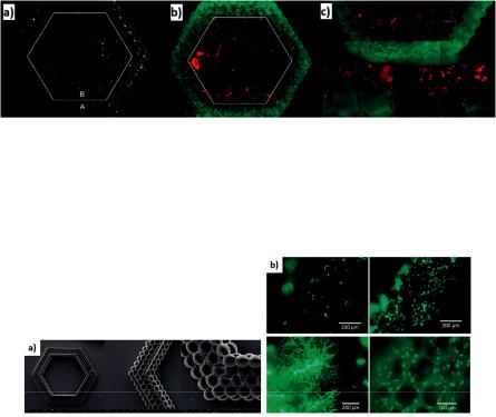
The use of lasers for the creation of biopolymer sca olds enables the easy tuning of the porosity of the final 3D structure by the alteration of the irradiation conditions, as explained by Rekštyte et al. [121]. In the reported study, 3D polymeric porous sca olds with size porosity of micrometers were obtained with the use of four di erent combinations of materials and a large variety of fabrication parameters (Figure 11).
Figure 11. Direct writing laser procedure (left). Final structures of fabricated sca olds consist of two di erent polymeric materials (right) [121].
94
Bioengineering 2019, 6, 95
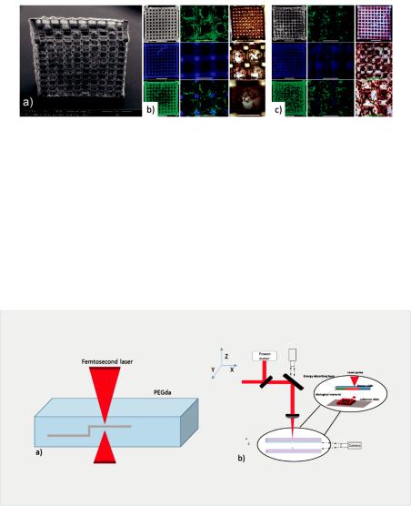
In addition, Ovsianikov et al. at 2011 [122] created gelatin-based sca olds with methacrylamide groups for the development of adipose tissue and transplants for plastic surgeries. The results verified the stability of the material and their ability to support ASC adhesion and proliferation from seven to twenty-two days as shown in Figure 12.
Figure 12. (a) SEM image of fabricated gelatin sca old. (b,c) Fluorescence microscopy pictures for the 2 photon polymerization sca old after seven days and 22 days [122].
3D hydrogel sca olds created by two-photon polymerization (2PP) for the support of Henrietta Lacks (HELA) cells’ culture for tissue engineering applications were also reported by Y.C Zheng et al. [123]. The starting material consisted of an aqueous solution of 3,6-bis[2-(1-methyl- pyridinium)vinyl]-9-pentyl-carbazole diiodide (BMVPC), cucurbit [7] uril (CB7), and polyethylene glycol diacrylate (PEGDA) was used as a monomer for 2PP.
Another advantage that laser-based techniques o er is the use of lasers for the creation of the 3D matrix and the selective deposition of cells with high precision. Ovsianikov et al. [120] presented this approach by utilizing lasers to polymerize an acrylated poly(ethyleneglycol) (PEG) monomer for the fabrication of a cell sca old and the direct laser printing of two di erent types of cells on the fabricated sca old (Figure 13).
Figure 13. (a) Schematic representation of two photon polymerization process of an acrylated poly(ethyleneglycol) (PEG). (b) Schematic representation of LIFT technique for the printing of cells on the fabricated sca old [120].
The final structure of this study had a hexagonal shape with six layers of cylinders along the diameter of the shape (Figure 14). Vascular smooth muscle cells (VSMCs) were laser printed at the outer perimeter of the sca old, while EC cells were deposited at the inner perimeter (Figure 14).
95
Bioengineering 2019, 6, 95
Figure 14. (a) SEM images of 2PP fabricated sca olds. (b,c) Fluorescence microscopy images after the deposition of two cell lines in the same sca old [120].
The combination of the laser-based 2PP technique with the micromolding technique resulted in the accelerated duration of the fabricated sca olds, according to A. Koroleva et al. [124] (Figure 15). In this structure, human pulmonary microvascular endothelial cells (HPMEC) were cultured for seven days and migrated into the fabricated fibrin gel sca old (Figure 15).
Figure 15. (a) Fabricated fibrin gel sca old with hexagonal shape. (b) Cells cultured for seven days [124].
Another laser-based technique used for the fabrication of cell sca olds is called selective laser sintering [125]. This is a layer-by-layer approach in which a laser beam is used to selectively sinter particles of a polymeric material in order to create layers with specific geometric characteristics [126].
4.2. Inkjet Printing
Inkjet printing is one of the most popular 3D printing techniques for the fabrication of structures of a great variety of materials. This technique enables the printing of picoliter droplets according to a software design in order to create 2D or 3D structures, and it can be either a continuous or a drop-on-demand (DOD) printing approach. The DOD inkjet printing technique has been widely used to create arrays of small liquid biodroplets [127,128]. The mechanism based on a thermal approach or a piezoelectric approach are the two main printing mechanisms with a DOD inkjet printer. The thermal inkjet printer contains of a thermal actuator which heats up the printing head, consequently generating a bubble of gas which, upon expansion, ejects a droplet of liquid to a receiver substrate [129]. On the other hand, the piezoelectric printers consist of a piezoelectric actuator which surrounds the ink chamber. An increase of the voltage across of the piezoelectric actuator initiates the formation of droplets during the flow of the ink [130].
Ink jet printing techniques have been frequently used for the control of cells growth in a matrix by the printing of protein solutions [131], the printing of cells [131], or the printing of the 3D sca old. In the field of inkjet printing of sca olds, impressive results were presented by Xu et al. [132] with the printing of a functional 3D sca old for cardiac tissue application (Figure 16a). Also, Duan et al. who printed valve network (Figure 16b) [54].
96
Bioengineering 2019, 6, 95
Figure 16. (a): Cardiac tissue by Xu et al. [132]. (b): Printed valve network by Duan et al. [54].
Moreover, there have been studies that utilize the inkjet printing technique for the creation of a 3D polymer matrix and the precise deposition of cells in a twostep procedure [133].
Even though the inkjet printing technique is an established technique for the printing of materials that can be used as 3D sca olds for cell cultures, such as biodegradable polymers [134–136] and natural polymers [133,137], most of the reported 3D liver cell cultures by the use of sca olds are created by more verified techniques in the industry such as microextrusion printing. Microextrusion printing is an additive manufacturing method for creating 3D micro-structures in a layer-by-layer manner. This is enabled by the continuous microprinting of polymeric materials for the creation of individual layers. These types of printers consist of a piston, upon which extraction deposits the biomaterial through a micro-needle. To our knowledge, the only study in which a piezoelectric inkjet printer was used for creating sca olds for liver cultures was presented by Arai et al. [73]. The novelty of that work was the architecture of the final structure—a sandwich shape of two galactosylated alginate (GA)-gel sheets on the top and bottom and hepatocytes cells in-between the two layers. This design provided the opportunity to regulate the polarity of the hepatocytes.
5. Sca olds for Liver Tissue Engineering
As mentioned previously, the liver is one of the most important and largest organs in human body, and it plays a significant role in metabolic functions. Many groups have studied the generation of 3D liver structures for potential liver regeneration applications. The use of 3D polymeric structures for generating cell cultures and liver models has facilitated the overcoming of the limitations of 2D cell culture models such as the uncontrollable cells’ polarity and non-directed cells’ attachment. A wide range of biocompatible polymers has been used as starting materials for building stable matrices for the growth and proliferation of cancer and primary hepatic cell lines. Lewis et al. [138] investigated the creation of a 3D porous gelatin sca old using a pneumatic extrusion piston-driven EnvisionTEC (GmbH) 3D-Bioplotter.
Six di erent single layers with di erent porous sizes were precisely placed in such a way to create two variable geometries (di ering in the strength of the connection between the layers), towards the final 3D sca old. The sca olds were used as matrix for the cell culture of the di erentiated hepatocyte-derived carcinoma cell line (Huh7). After, the fabrication of the optimum sca old geometry, the viability and the functionality of the seeded Huh7 cells were studied for seven days. Towards this goal, a comparison was made between the two geometries of the 3D sca olds and the 2D models. The results revealed almost the same viability of the 2D and 3D cell cultures; however, the 3D structures enabled an increase in the hepatic functions of the cells, mainly due to the strong connection between the pores of the structure. These lateral architectural 3D models have been proven suitable to use
97
Bioengineering 2019, 6, 95
for studying the specific functions of hepatocytes such as albumin secretion, CYP activity, and bile transport, because they provide an appropriate environment for well-defined cells [138]. Furthermore, a micro-extrusion bioprinter (INKREDIBLE+) was used by Hiller et al. to create a 3D structure by printing a mixed ink consisting of: hydrogels (alginate, gelatin), human extracellular matrix (hECM) and human HepaRG liver cells. The selected cell line was used in thus metabolic study due to its morphology and metabolic characteristics. The aim of this study was to test the metabolic activity and the viability of the cells for structures with variable concentration of hECM. The hECM substance changes the mechanical characteristics of the 3D microstructures. It consists of collagen type I, which improves the properties of the sca old but, in high concentrations, has a negative e ect on cell functionality [80]. Another group (Kim et al.) used alginate and isolated mouse primary hepatocytes to create a 3D bio-printed structure. The process combined the use of a micro-syringe with a three dimensional motion stage to create the final 3D structure in a layer-by-layer manner. The final shape of the structure was adjusted by scanning parameters such as velocity and pressure. The main goal of this study was to create hepatocyte cell culture networks which demonstrated a high viability of the cells after 14 days with good hepatic functionality [75]. The same year, Kang et al. [139] used mouse-induced hepatocyte-like cells (miHeps) by pluripotent stem cells (PSC) for the development of a 3D structure made by miHeps and aginate using extrusion printing. Five layers of cells and alginate printing solution built the final 3D cell culture which was placed in a mouse in vivo. The implant was examined 14 and 28 days after the surgery, presenting results that indicated that the in vivo transplanted sca old was more functional than the in vitro model. Lee et al. [76] created 3D sca olds with improved mechanical properties for a 3D hepatocytes cell culture environment. In this study, the sca old was made by polycaprolactone (PCL) as a starting material by using a homemade printer with multiple deposition heads-multihead tissue/organ building (MtoBS system). The layer-by-layer printed structure consisted of PCL and hydrogel layers with a mixture of collagen and three di erent cell lines—HCs, HUVECs, and HLFs. The final structures had the ability to support multiple functional cell lines which could maintain their hepatic functions for 10 days. Jeon et al. fabricated 3D alginate sca olds with cancer hepatic cells (HepG2) using a micro extrusion printer and tested the proliferation and the viability of hepatocytes on the 3D structure for three weeks. The histology and immunohistochemistry of the final cultures were investigated along with their ability to support operational cells [140]. Gong et al. designed and fabricated well defined 3D chitosan–gelatin (C/G) sca olds which consisted of polymeric channels and pores using both an indirect method called the solid freeform fabrication (SFF) and freeze drying methods. Two thermoplastic materials were used for forming the mold of the sca old. Chitosan and gelatin were used as matrix materials, and the final structure was initiated by the freeze-drying process, which also led to the creation of micro pores. The tuning of the freeze-drying parameters resulted in the optimum shape and morphology of the sca old. The functionality of the developed sca old structure was tested for the culturing of HepG2 cell line [141].
6. Conclusions
In summary, 3D bioprinting technology enables the fabrication of biomimetic tissues and implants with the use of biomaterials, growth factors, and living cells, which can either be printed in a specific pattern for the development of the final tissue structure or, in many cases, can be printed on an already existing 3D matrix (sca old). Furthermore, in the field of tissue engineering, an bioartificial liver is considered one of the most promising tools as a therapeutic method for severe liver diseases and, in the field of regenerative medicine, for drug testing. The most commonly used direct writing techniques for the printing of cells are laser-based techniques, inkjet printing, and microextrusion printing. Those techniques are mainly chosen because they can easily adapt to the cultivation environment, can create high-resolution cell structures, and, in many cases, can also be used to create 3D sca olds for the cell growth. In the field of liver tissue engineering, a lot of work has been done with the use of the above-mentioned techniques towards; however, greater e ort is required to solve problems
98
Bioengineering 2019, 6, 95
encountered in the inability to replicate the actual 3D living liver tissue environment. Finally, the combination of a 3D bioprinting technique with a microfluidic control can be a promising method for controlled drug delivery systems and for future regenerative medicine.
Author Contributions: Individual contributions of authors are as follows: Writing—original draft preparation, C.K., V.L.; Writing—review and editing, M.C., I.Z.; Supervision, I.Z.
Funding: This research was funded by State Scholarships Foundation: ‘Reinforcement of Postdoctoral Researchers’.
Acknowledgments: The work was supported by the IKY scholarships program, which is co-financed by the European Union (European Social Fund-ESF) and Greek national funds through the action entitled ‘Reinforcement of Postdoctoral Researchers’, in the framework of the Operational Programme ‘Human Resources Development Program, Education and Lifelong Learning’ of the National Strategic Reference Framework (NSRF) 2014–2020 and by ESPA 2014–2020 program through the action entitled Synergy of ELI-LASERLAB Europe, HIPER&IPERION-CH.gr in the framework of the NTUA’s Participation in the project HELLAS-CH (MIS 5002735).
Conflicts of Interest: The authors declare no conflict of interest.
References
1.Kruth, J. Material Incress Manufacturing by Rapid Prototyping Techniques. CIRP Ann. 1991, 40, 603–614. [CrossRef]
2.Heller, T.B.; Hill, R.M.; Saggal, A.F. Method of and Apparatus for Forming a Solid Three-Dimensional Article from a Liquid Medium. U.S. Patent 5,071,337, 10 December 1991.
3.Hull, C.W. Apparatus for Production of Three-Dimensional Objects by Stereolithography. U.S. Patent 4,575,330, 11 March 1986.
4.Nakamura, M.; Iwanaga, S.; Henmi, C.; Arai, K.; Nishiyama, Y. Biomatrices and biomaterials for future developments of bioprinting and biofabrication. Biofabrication 2010, 2, 014110. [CrossRef] [PubMed]
5.Murphy, S.V.; Atala, A. 3D bioprinting of tissues and organs. Nat. Biotechnol. 2014, 32, 773–785. [CrossRef] [PubMed]
6.Munaz, A.; Vadivelu, R.K.; John, J.S.; Barton, M.; Kamble, H.; Nguyen, N.T. Three-dimensional printing of biological matters. J. Sci. Adv. Mater. Devices 2016, 1, 1–17. [CrossRef]
7.Derakhshanfar, S.; Mbeleck, R.K.; Xu, K.; Zhang, X.; Zhong, W.; Xing, M. 3D bioprinting for biomedical devices and tissue engineering: A review of recent trends and advances. Bioact. Mater. 2018, 3, 144–156. [CrossRef] [PubMed]
8.Bertassoni, L.E.; Cecconi, M.; Manoharan, V.; Nikkhah, M.; Hjortnaes, J.; Cristino, A.L.; Barabaschi, G.; Demarchi, D.; Dokmeci, M.R.; Yang, Y.; et al. Hydrogel bioprinted microchannel networks for vascularization of tissue engineering constructs. Lab Chip 2014, 14, 2202–2211. [CrossRef] [PubMed]
9.Moya, M.L.; Hsu, Y.H.; Lee, A.P.; Hughes, C.C.; George, S.C. In Vitro Perfused Human Capillary Networks. Tissue Eng. Part C Methods 2013, 19, 730–737. [CrossRef] [PubMed]
10.Li, X.; Chen, Y.; Li, P.C.H. A simple and fast microfluidic approach of same-single-cell analysis (SASCA) for the study of multidrug resistance modulation in cancer cells. Lab Chip 2011, 11, 1378–1384. [CrossRef]
11.Li, X.J.; Nie, Z.H.; Cheng, C.M.; Goodale, A.B.; Whitesides, G.M. Paper-based electrochemical ELISA. Proc. Micro Total Anal. Syst. 2010, 14, 1487–1489.
12.Salieb-Beugelaar, G.B.; Simone, G.; Arora, A.; Philippi, A.; Manz, A. Latest Developments in Microfluidic Cell Biology and Analysis Systems. Anal. Chem. 2010, 82, 4848–4864. [CrossRef]
13.Jang, K.; Sato, K.; Igawa, K.; Chung, U.I.; Kitamori, T. Development of an osteoblast-based 3D continuous-perfusion microfluidic system for drug screening. Anal. Bioanal. Chem. 2008, 390, 825–832. [CrossRef] [PubMed]
14.Chang, C.C.; Boland, E.D.; Williams, S.K.; Hoying, J.B. Direct-write bioprinting three-dimensional biohybrid systems for future regenerative therapies. J. Biomed. Mater. Res. Part B Appl. Biomater. 2011, 98, 160–170. [CrossRef] [PubMed]
15.Kurobe, H.; Maxfield, M.W.; Breuer, C.K.; Shinoka, T. Concise Review: Tissue-Engineered Vascular Grafts for Cardiac Surgery: Past, Present, and Future. Stem Cells Transl. Med. 2012, 1, 566–571. [CrossRef] [PubMed]
16.Freeman, R.B.; Ste ck, D.E., Jr.; Guidinger, M.K.; Farmer, D.G.; Berg, C.L.; Merion, R.M. Liver and Intestine Transplantation in the United States 1998–2006. Am. J. Transplant. 2008, 8, 958–976. [CrossRef] [PubMed]
99