

Биоинженерия / ТИ_печень(органы_ЖКТ) / Bioengineering_Liver_Transplantation
.pdfBioengineering 2019, 6, 81
paracrine signaling interactions involving various cytokines and growth factors, that stimulate regeneration and neoangiogenesis [86,87].
Endothelial progenitor cells (EPCs) can be found in both peripheral blood vessels and bone marrow, and their main function is to participate in the neovascularization of damaged tissue [88,89]. In the context of cell therapy for liver diseases, one animal study demonstrated that the transplantation of EPCs led to a lessening of liver fibrosis [79]. ESCs are also able to promote hepatocyte proliferation and increase matrix metalloproteinase activity [90]. All these e ects are related to an increased secretion of specific growth factors [91,92].
Another promising cell treatment for liver diseases is based on mesenchymal stem cells (MSCs), a population of multipotent progenitors capable of di erentiating towards adipogenic, osteogenic and hepatogenic lineages, with a low immunogenicity [93]. Bone marrow is considered the main source of MSCs [94], but alternative sources are being examined, such as adipose tissue [95], placenta, amniotic fluid, umbilical cord blood, and umbilical cord [96,97]. Our research has focused on umbilical cord MSCs. We demonstrated in an animal model that, when systematically administered, these cells can repair acute liver injury [97]. The ability of the same cells to repair tissue damage was also demonstrated in a chemically-induced intestinal injury in immunodeficient mice [98]. We and other authors have demonstrated that MSCs have the capacity to provide both metabolic and trophic support due to their potential for hepatocytic di erentiation, and their secretion of anti-inflammatory, anti-apoptotic, immunomodulatory, and pro-proliferative factors [97–99]. This leads to liver function being restored via the repair of damaged tissue, the suppression of inflammation, and the stimulation of endogenous regeneration through paracrine e ects [100].
Cell-based therapy using HPCs could potentially regenerate the liver during chronic diseases. Multiple protocols have been established for isolating HPCs in fetal and rodent models, and cell di erentiation protocols are available for progenitor cells derived from the human liver or biliary tree [101–103]. Due to the low number of these cells in the liver, the use of autologous HPCs is probably unfeasible. The use of expanded fetal or syngeneic HPCs is more likely, though this approach raises questions regarding the engraftment rate of transplanted cells, and the need for immunosuppressant therapy. Despite the theoretical feasibility of such approaches, we still have only a limited understanding of HPCs, their precise role in liver pathophysiology, and how the entire process of regeneration/di erentiation is regulated. Given the possible disadvantages of HPC activation, which might exacerbate disease progression or prompt the onset of cancer [104], all these issues warrant further study and careful examination before any therapeutic approaches could be applicable.
3.1.5. Hepatic Organoids
Considered as a bridge between liver cell therapy and liver bioengineering, hepatic organoids are functional three-dimensional (3D) in vitro models of the liver consisting of a spherical monolayer of epithelium that preserves the key physiological features of the liver [105]. Liver organoids are typically obtained by isolating and expanding stem cells or hepatic progenitor cells.
Liver organoids show a limited spontaneous di erentiation during maintenance and expansion. For this reason, protocols for establishing organoids were divided into two steps. The first relied on proliferation culture conditions for the establishment and expansion of hepatic organoids. Then, in a second step, proliferative signals were removed, and di erentiation towards hepatocyte-like cells was induced. These culture conditions enabled organoids to be obtained with 30–50% fulfilling hepatic characteristics [106], but without the complete functional repertoire of adult hepatocytes—a drawback shared by HPC-to-hepatocyte di erentiation.
Di erentiated hepatic organoids transplanted into mouse models of liver failure have demonstrated a capacity for engraftment and repopulation of the damaged liver, with partial rescue of liver function [105]. Equivalent human liver organoids transplanted into mice with acute liver damage were able to produce human albumin and alpha-1-antitrypin, with secretion levels comparable with those after the transplantation of adult hepatocytes [101].
10
Bioengineering 2019, 6, 81
Three-dimensional liver tissue has also been engineered by using human iPSCs to derive hepatocytes in co-culture with mesenchymal and endothelial cells [107]. When transplanted into mice, these liver buds were vascularized and matured to synthesize serum proteins and carry out detoxifying functions.
Current research is aiming for the clinical application of liver buds suitable for hepatic administration via the portal vein in patients in need of a liver transplant [108]. Among the di erent cell sources, adult stem cells directly derived from hepatic tissue are preferred. Indeed, drawbacks of human iPSCs or trans-di erentiated cells used in the design of clinical solutions concern their exposure to genetic modifications through reprogramming factors, and their genomic instability, particularly in long-term cultures [109].
Moreover, liver organoids provide a novel platform for research on: 1) liver development and regeneration; 2) detoxification and metabolism; 3) liver disease modelling; and 4) adult stem cell biology.
3.2. Liver Tissue Bioengineering
Tissue engineering could o er various solutions for reducing the waiting list by creating biocompatible sca olds and extracorporeal liver devices suitable for either in vitro or in vivo applications [110].
In the last two decades, a growing number of studies demonstrated that 3D cultures have a number of advantages over traditional two-dimensional (2D) cell cultures [110,111]. A physiologically 3D microenvironment is crucial to the development of in vitro tissue models, particularly for such complex tissues as the liver, in which the interaction between hepatocytes, hepatic stellate cells, and extracellular matrix (ECM) creates the microenvironment of the hepatic lobules [112].
The search for e cient biocompatible sca olds aims to create organic or polymeric constructs that mimic the liver ECM and replicate functional characteristics such as cell adhesion, viability, growth, and proliferation. The principal strategies are based on biomaterials such as polymer-based 3D constructs, decellularized ECM, or bioprinting 3D constructs.
Another recent approach involves the development of bioreactors to improve various functions of hepatocytes that are seeded in constructs. In bioreactors, a real 3D microenvironment niche is created to improve cell attachment, growth, and proliferation, with a marked improvement in liver metabolism and function [110]. A more sophisticated technology is the liver-on-chip: A combination of bio-reactor techniques and microfluidic devices to sustain the phenotype of hepatocytes and liver-specific functions in long-term culture [113].
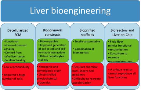
Below we provide an overview of such bioengineering approaches, and Figure 3 shows the main pros and cons of each of them.
3.2.1. Decellularized Extracellular Matrix
A new approach to liver regenerative medicine involves generating 3D organs with a decellularized, native liver biosca old that can be repopulated with parenchymal and non-parenchymal cells [114]. The liver’s native ECM has a complex composition and topography, serving as a structure for cell-ECM adhesion, interaction, and polarity, with implications for the regulation of cell morphology, proliferation, di erentiation, and viability interactions [115]. Donor organs unsuitable for transplantation are used to create whole-liver sca olds which are subsequently reseeded with healthy cells to create transplantable liver grafts. The sca olds maintain the native liver architecture and ECM composition, which allows for proper cell homing and function. Decellularization techniques were introduced in the 1980s [116], but the concept of whole-organ decellularization was developed later by Ott and colleagues in mice hearts [117]. This technique was later adapted for liver engineering purposes [118], with the preservation of the chemical composition and structure of the ECM with structurally intact vessels, and bile ducts. This biosca old was then recellularized with hepatocytes and endothelial cells. The recellularized graft transplanted in vivo and perfused ex vivo demonstrated mature liver functions. Further improvements in the technique were obtained over the years, such us multistep cell seeding,
11
Bioengineering 2019, 6, 81
the use of stem cells (MSCs, fetal hepatocytes, iPSCs) [119–121], optimization of the decellularization cocktail, and perfusion without any thrombus formation [122]. The feasibility of this technique was also demonstrated in larger animal models [123], and even in humans [124], bringing the approach to clinical scale.
Figure 3. Main pros (green boxes) and cons (red boxes) of the principal liver bioengineering approaches. ECM: extracellular matrix.
All these studies demonstrated that decellularized livers hold great potential as a therapeutic approach, but numerous pitfalls remain. First, the technique allows for the successful seeding and culture of hepatocytes, but colonization of the bile duct with functional cells and the achievement of an intact vascular network remain to be perfected. Another important issue before whole liver biosca olds can be used in clinical practice is the lack of a suitable source of cells, which should be readily available and renewable because successful liver recellularization demands hundreds of millions of cells. The limited availability and inability to expand primary hepatocytes has led researchers in the field to search for a new cell source. Although many groups have attempted to overcome the problem by using fetal liver cells, stem cells or iPSCs, the production of such huge numbers of hepatocytes is still far beyond current technical capability.
Another hurdle that should be promptly addressed is “sample to sample” variation due to the unique condition of each donor deriving from the use of discarded livers [125]. The next goals of bioengineering research will be to solve these problems.
3.2.2. Biopolymer Constructs
In modern tissue engineering, e orts are being made to make natural biomaterials mimic the natural hepatic ECM. The main components of these sca olds are collagen and hyaluronic acid. The latter strongly supports cell attachment, proliferation, di erentiation, growth, and migration. Immature and mature hepatocytes express CD44, the surface receptors for hyaluronic acid, so biopolymers with hyaluronic acid and its derivatives have more adhesive power for hepatocytes. They can retain hepatocyte viability for 4 weeks [126].
12
Bioengineering 2019, 6, 81
Other natural biomaterials used in the construction of bioactive sca olds are alginate, chitin, chitosan, silk, Matrigel®, and sponge. Matrigel® is a sca old consisting of a mixture of ECM proteins derived from the basal membranes of murine chondrosarcoma, which contains laminin, heparan sulfate proteoglycan, and collagen type IV [127]. It has been used in numerous studies to culture hepatocytes and induce the hepatic di erentiation of stem cells [97,128].
Although hydrogels formed by natural biomaterials such as alginate and Matrigel® are biocompatible and improve the generation of cell-to-cell and cell-to-matrix interactions, they have some important limits that prevent their clinical application. The main shortcomings of such biomaterials are their uncontrollable physicochemical properties, degradability, lack of regenerative ability, and inconsistent mechanical properties. Moreover, due to the xenogenic and tumorigenic origin of Matrigels, they are not an optimal support for clinical applications in liver bioengineering [129].
By comparison with natural biomaterials, synthetic materials o er a wide range of properties and a better control over them. Sca olds containing biodegradable polymers, such as polylactic acid, polyglycolic acid, polyanhydrides, polyfumarates, polyorthoesters, polycaprolactones, poly-L-lactic acid, and polycarbonates facilitate cell regeneration, transplantation, and degradation on time [130]. The biocompatibility of bioengineered matrices and sca old adhesion properties could also be improved by chemically modifying these polymers (e.g., by incorporating proteins and special bioactive domains), stimulating cell attachment and migration, and thereby facilitating liver tissue repair [131].
While natural and synthetic materials support the successful culture of hepatocytes, these constructs fail to perfectly reproduce the microenvironment of the liver essential to a functional liver cell activity. For this reason, their therapeutic potential is limited.
3.2.3. Bioprinted Sca olds
Although the use of biomaterials in 3D culture has improved the settings for liver tissue engineering, it has some limitations. These include the di culty of creating complex biological structures and designs due to size, material, compositional, and technological constraints [132]. An innovative solution to these problems involves using bioprinted sca olds, tissue-mimicking constructs created by means of a bioprinting process with biocompatible materials (i.e., bio-inks) [133]. Advances in bioprinting technology have enabled the creation of more complex 3D structures using combinations of di erent biomaterials and cell types [134]. The chance to totally customize the prints also guarantees the complete personalization of such sca olds and their applications. The available bioprinting modalities include extrusion, inkjet, and laser-assisted bioprinting [135]. Extrusion bioprinting, the most often-used bioprinting modality in biomedical research, allows for a strong degree of customization with few restrictions on the cells used [134]. The choice of biomaterials is more restrictive, however, as they are either easy to print or ideal for cell culture, but typically not both [136]. The ideal characteristics of bio-inks for extrusion bioprinting are viscosity to enable printing, associated with an adequate elasticity to maintain their structure, while also maintaining cell viability and supporting cell function [132].
The most common biomaterials used for bioprinting are collagen, alginate, polyethylene glycol (PEG), hyaluronic acid, fibrin, gelatin, or polycaprolactone, each with unique properties [133]. With the exception of collagen, these biomaterials need the addition of a cross-linker that could adversely a ect the cells. For this reason, they should be appropriately balanced to guarantee the best biocompatibility of the bio-ink being used [133]. Although collagen is an ideal material for in-vivo-like tissue replication, it is a poor bio-ink because it has a timeand temperature-sensitive cross-linking [137]. A multi-component hybrid bio-ink is therefore a potential solution for achieving ideal physiological relevance and bio-printability. Unfortunately, durable 3D construct fabrication requires the incorporation of chemical stabilizers, such as polycaprolactone, showing the limitations of bio-inking technologies in mimicking both the biochemical composition and the complex 3D structure of the liver.
Another important challenge in 3D bioprinting is how to fabricate and mimic cellular microenvironments from molecular to macroscopic scales for tissue engineering and regenerative medicine. Using this approach, the researcher aims to create a whole functional liver suitable for
13
Bioengineering 2019, 6, 81
transplantation, but some important issues, such as vascularization, should be addressed before this methodology can really be implemented.
3.3. Bioreactor Systems
Despite the great progress made in biomaterial development for tissue engineering, some challenges need to be overcome. The most important limiting parameter in tissue engineering and bioprinting concerns vascularization [138]. Without suitable vascularization, cells are subject to hypoxia, toxemia, apoptosis, and immediate cell death. The bioreactor approach aims to overcome this limitation. In fact, the bioreactor involves a designed or programmed fluid flow as an integral part of the culture format. The flow in perfusion bioreactors enables a continuous exchange of nutrients, a better oxygen delivery, and a physiological shear stress, influencing cell function in ways that are impossible to achieve in static culture formats [139].
The evolution of bioreactor technologies has paralleled advances in the development of functional biomaterial sca olds [140]. The sca old not only provides an adhesion surface for cells, but also profoundly influences cell shape and gene expression relevant to cell growth and liver-specific functions. Moreover, when placed as a separation between cells and the medium, the sca olds act as a modulator for water and nutrient transport from the medium to the cells, and discharge waste metabolites from the cells to the medium [141].
Four principal types of bioreactors have been used for liver cell culture: 1) flat plate and monolayer; 2) hollow fiber; 3) perfused beds and sca olds; and 4) encapsulation and suspension. With the exception of type 1, the other bioreactors enable the 3D monoculture or co-culture of hepatocytes under tissuespecific mechanical forces (pressure, shear stress, flow) [142,143]. Some of these bioreactors have been used as bioartificial livers, charged with various types of liver cells, as a bridge for patients with acute liver failure awaiting transplantation [144]. Now the challenge is to use cell-based bioreactors as in vitro screening systems for drug toxicity, metabolism evaluation and potential clinical treatments.
Some parameters are crucial to hepatocyte vitality and functionality, including various biophysical factors such as oxygenation, hemodynamics, and shear stress. Perfusion in bioreactor devices enables the establishment of oxygen gradients and hepatic zonation, resulting in graded CYP expression and metabolism [145,146]. A controlled oxygen gradient from 25 to 70 mmHg inside a hepatic bioreactor creates a functional hepatocyte zonation similar to what is observed in vivo. Cell oxygenation could be partially controlled by varying the medium flow rate, but may consequently exert a shear stress on the hepatocytes. Flow rate should be carefully controlled since cell damage can occur. Hydrodynamic stress induces ECM remodeling, sca old degradation and changes in tissue composition, influencing the device’s structural and mechanical properties. On the other hand, low flow rates limit the oxygen supply, lead to nutrient deficiency, and reduce cell viability and survival probability [146].
The co-culture of hepatocytes and non-parenchymal cells is important for the reorganization of hepatocytes in culture by secreting cytokines, nitric oxide, and matrix components [145,147]. Co-culture is also useful for inducing liver-specific functions, preserving maximal levels of functional adhesion molecule expression, and reducing the number of cells needed for a bioartificial liver [144].
The main limitation of the bioreactors is that not all critical liver functions can be replicated on the desired level as yet. For this reason, based on the present state of the art, a unique bioreactor that can faithfully reproduce all liver functions is still lacking.
3.4. Micro-Bioreactors and Liver-on-Chip
The combination of nanotechnology, microchips, and microfluidics in a single device has great potential for applications in liver tissue engineering. Various strategies have been developed to obtain micro-bioreactors. Microsystems technology has been used to fabricate 2D or 3D culture devices by using di erent types of materials, like silicon, silicone elastomer, and biocompatible and biodegradable polymers. Such systems typically exhibit laminar flow, similar to the environment in vivo, and allow
14
Bioengineering 2019, 6, 81
the creation of microfluidic channels with larger surface-to-volume ratios suitable for oxygen and nutrition supply [113].
Other interesting systems that exploit microfluidic technology are the so-called “liver-on-chip” devices [148]. These systems consist of microchambers containing engineered tissue and living cell cultures interconnected by a microfluidic network. Such organs on chips enable the study of human phys-iology in an organ-specific context, and the development of novel in vitro disease models. They have the potential to serve as replacements for animals used in drug development, toxin testing, and screening for biothreats and chemical warfare agents [149].
4. Conclusions
In conclusion, regenerative medicine and bioengineering are cutting-edge technologies that look promising as a final solution to the treatment of end-stage liver diseases. A better understanding of liver regeneration and the development of in vitro systems that successfully mimic hepatocyte expansion and di erentiation will make autologous cell therapy a feasible alternative to liver transplantation. The current scenario is also moving towards the successful development of whole bioengineered livers and their e ective use in clinical practice in lieu of liver transplantation.
Author Contributions: Writing—original draft preparation: D.B.; writing—review and editing: F.R.P. and P.B.
Funding: This research received no external funding.
Acknowledgments: Authors are grateful to Marina Minnaja Foundation for co-funding Debora Bizzaro postdoctoral fellowship.
Conflicts of Interest: The authors declare no conflict of interest.
References
1.Manns, M.P.; Burra, P.; Sargent, J.; Horton, R.; Karlsen, T.H. The Lancet-EASL Commission on liver diseases in Europe: overcoming unmet needs, stigma, and inequities. Lancet 2018, 392, 621–622. [CrossRef]
2.Germani, G.; Theocharidou, E.; Adam, R.; Karam, V.; Wendon, J.; O’Grady, J.; Burra, P.; Senzolo, M.; Mirza, D.; Castaing, D.; et al. Liver transplantation for acute liver failure in Europe: Outcomes over 20 years from the ELTR database. J. Hepatol. 2012, 57, 288–296. [CrossRef] [PubMed]
3.Burra, P.; Berenguer, M.; Pomfret, E. The ILTS Consensus Conference on NAFLD/NASH and liver transplantation. Transplantation 2018. [CrossRef]
4.Pang, Y.; Horimoto, Y.; Sutoko, S.; Montagne, K.; Shinohara, M.; Mathiue, D.; Komori, K.; Anzai, M.; Niino, T.; Sakai, Y. Novel integrative methodology for engineering large liver tissue equivalents based on three-dimensional sca old fabrication and cellular aggregate assembly. Biofabrication 2016, 8, 16. [CrossRef] [PubMed]
5.Su, Z.C.; Li, P.S.; Wu, B.G.; Ma, H.; Wang, Y.C.; Liu, G.X.; Zeng, H.L.; Li, Z.Z.; Wei, X. PHBVHHx sca olds loaded with umbilical cord-derived mesenchymal stem cells or hepatocyte-like cells di erentiated from these cells for liver tissue engineering. Mater. Sci. Eng. C Mater. Biol. Appl. 2014, 45, 374–382. [CrossRef]
6.Tremblay, K.D.; Zaret, K.S. Distinct populations of endoderm cells converge to generate the embryonic liver bud and ventral foregut tissues. Dev. Biol. 2005, 280, 87–99. [CrossRef]
7.Houssaint, E. Di erentiation of the mouse hepatic primordium. I. An analysis of tissue interactions in hepatocyte di erentiation. Cell Di er. 1980, 9, 269–279. [CrossRef]
8.Medlock, E.S.; Haar, J.L. The liver hemopoietic environment: I. Developing hepatocytes and their role in fetal hemopoiesis. Anat. Rec. 1983, 207, 31–41. [CrossRef] [PubMed]
9.Lemaigre, F.P. Development of the biliary tract. Mech. Dev. 2003, 120, 81–87. [CrossRef]
10.Kung, J.W.C.; Currie, I.S.; Forbes, S.J.; Ross, J.A. Liver Development, Regeneration, and Carcinogenesis.
J. Biomed. Biotechnol. 2010. [CrossRef]
11.Tanimizu, N.; Miyajima, A. Notch signaling controls hepatoblast di erentiation by altering the expression of liver-enriched transcription factors. J. Cell Sci. 2004, 117, 3165–3174. [CrossRef] [PubMed]
12.McCright, B.; Lozier, J.; Gridley, T. A mouse model of Alagille syndrome: Notch2 as a genetic modifier of Jag1 haploinsu ciency. Development 2002, 129, 1075–1082. [PubMed]
15
Bioengineering 2019, 6, 81
13.Suzuki, A.; Iwama, A.; Miyashita, H.; Nakauchi, H.; Taniguchi, H. Role for growth factors and extracellular matrix in controlling differentiation of prospectively isolated hepatic stem cells. Development 2003, 130, 2513–2524. [CrossRef] [PubMed]
14.Jochheim, A.; Cieslak, A.; Hillemann, T.; Cantz, T.; Scharf, J.; Manns, M.P.; Ott, M. Multi-stage analysis of di erential gene expression in BALB/C mouse liver development by high-density microarrays. Di erentiation 2003, 71, 62–72. [CrossRef] [PubMed]
15.Zhao, R.; Duncan, S.A. Embryonic development of the liver. Hepatology 2005, 41, 956–967. [CrossRef] [PubMed]
16.Forbes, S.J.; Rosenthal, N. Preparing the ground for tissue regeneration: from mechanism to therapy. Nat. Med. 2014, 20, 857–869. [CrossRef]
17.Riehle, K.J.; Dan, Y.Y.; Campbell, J.S.; Fausto, N. New concepts in liver regeneration. J. Gastroenterol. Hepatol. 2011, 26, 203–212. [CrossRef]
18.MacDonald, R.A. Lifespan of liver cells: autoradiographic study using tritiated thymidine in normal, cirrhotic, and partially hepatectomized rats. Arch. Int. Med. 1961, 107, 335–343. [CrossRef]
19.Gilgenkrantz, H.; Collin de l’Hortet, A. New insights into liver regeneration. Clin. Res. Hepatol. Gastroenterol. 2011, 35, 623–629. [CrossRef]
20.Michalopoulos, G.K. Hepatostat: Liver Regeneration and Normal Liver Tissue Maintenance. Hepatology 2017, 65, 1384–1392. [CrossRef]
21.Taub, R. Liver regeneration: From myth to mechanism. Nat. Rev. Mol. Cell Biol. 2004, 5, 836–847. [CrossRef] [PubMed]
22.Aravinthan, A.; Scarpini, C.; Tachtatzis, P.; Verma, S.; Penrhyn-Lowe, S.; Harvey, R.; Davies, S.E.; Allison, M.; Coleman, N.; Alexander, G. Hepatocyte senescence predicts progression in non-alcohol-related fatty liver disease. J. Hepatol. 2013, 58, 549–556. [CrossRef] [PubMed]
23.Fausto, N.; Campbell, J.S.; Riehle, K.J. Liver regeneration. J. Hepatol. 2012, 57, 692–694. [CrossRef]
24.Farber, E. Similarities in the sequence of early histological changes induced in the liver of the rat by ethionine, 2-acetylamino-fluorene, and 3’-methyl-4-dimethylaminoazobenzene. Cancer Res. 1956, 16, 142–148. [PubMed]
25.Lazaro, C.A.; Rhim, J.A.; Yamada, Y.; Fausto, N. Generation of hepatocytes from oval cell precursors in culture. Cancer Res. 1998, 58, 5514–5522. [PubMed]
26.Dunsford, H.A.; Karnasuta, C.; Hunt, J.M.; Sell, S. Di erent lineages of chemically-induced hepatocellular-carcinoma in rats defined by monoclonal-antibodies. Cancer Res. 1989, 49, 4894–4900. [PubMed]
27.Theise, N.D.; Saxena, R.; Portmann, B.C.; Thung, S.N.; Yee, H.; Chiriboga, L.; Kumar, A.; Crawford, J.M. The canals of Hering and hepatic stem cells in humans. Hepatology 1999, 30, 1425–1433. [CrossRef]
28.Stueck, A.E.; Wanless, I.R. Hepatocyte Buds Derived From Progenitor Cells Repopulate Regions of Parenchymal Extinction in Human Cirrhosis. Hepatology 2015, 61, 1696–1707. [CrossRef]
29.Roskams, T.A.; Theise, N.D.; Balabaud, C.; Bhagat, G.; Bhathal, P.S.; Bioulac-Sage, P.; Brunt, E.M.; Crawford, J.M.; Crosby, H.A.; Desmet, V.; et al. Nomenclature of the finer branches of the biliary tree: Canals, ductules, and ductular reactions in human livers. Hepatology 2004, 39, 1739–1745. [CrossRef]
30.Zajicek, G.; Oren, R.; Weinreb, M. THE STREAMING LIVER. Liver 1985, 5, 293–300. [CrossRef]
31.Roskams, T.; Yang, S.Q.; Koteish, A.; Durnez, A.; DeVos, R.; Huang, X.W.; Achten, R.; Verslype, C.; Diehl, A.M. Oxidative stress and oval cell accumulation in mice and humans with alcoholic and nonalcoholic fatty liver disease. Am. J. Pathol. 2003, 163, 1301–1311. [CrossRef]
32.Kuwahara, R.; Kofman, A.V.; Landis, C.S.; Swenson, E.S.; Barendswaard, E.; Theise, N.D. The hepatic stem cell niche: Identification by label-retaining cell assay. Hepatology 2008, 47, 1994–2002. [CrossRef] [PubMed]
33.Kubes, P.; Mehal, W.Z. Sterile Inflammation in the Liver. Gastroenterology 2012, 143, 1158–1172. [CrossRef] [PubMed]
34.Schattenberg, J.M.; Galle, P.R.; Schuchmann, M. Apoptosis in liver disease. Liver Int. 2006, 26, 904–911. [CrossRef] [PubMed]
35.Haga, S.; Terui, K.; Zhang, H.Q.; Enosawa, S.; Ogawa, W.; Inoue, H.; Okuyama, T.; Takeda, K.; Akira, S.; Ogino, T.; et al. Stat3 protects against Fas-induced liver injury by redox-dependent and -independent mechanisms. J. Clin. Investig. 2003, 112, 989–998. [CrossRef]
16
Bioengineering 2019, 6, 81
36.Li, W.; Liang, X.P.; Kellendonk, C.; Poli, V.; Taub, R. STAT3 contributes to the mitogenic response of hepatocytes during liver regeneration. J. Biol. Chem. 2002, 277, 28411–28417. [CrossRef] [PubMed]
37.Jeong, W.I.; Park, O.; Radaeva, S.; Gao, B. STAT1 inhibits liver fibrosis in mice by inhibiting stellate cell proliferation and stimulating NK cell cytotoxicity. Hepatology 2006, 44, 1441–1451. [CrossRef]
38.Sun, Z.L.; Klein, A.S.; Radaeva, S.; Hong, F.; El-Assal, O.; Pan, H.N.; Jaruga, B.; Batkai, S.; Hoshino, S.; Tian, Z.G.; et al. In vitro interleukin-6 treatment prevents mortality associated with fatty liver transplants in rats. Gastroenterology 2003, 125, 202–215. [CrossRef]
39.Bizzaro, D.; Crescenzi, M.; Di Liddo, R.; Arcidiacono, D.; Cappon, A.; Bertalot, T.; Amodio, V.; Tasso, A.; Stefani, A.; Bertazzo, V.; et al. Sex-dependent di erences in inflammatory responses during liver regeneration in a murine model of acute liver injury. Clin. Sci. (Lond.) 2018, 132, 255–272. [CrossRef]
40.Yates, F.E.; Herbst, A.L.; Urquhart, J. Sex di erence in rate of ring A reduction of delta 4–3-keto-steroids in vitro by rat liver. Endocrinology 1958, 63, 887–902. [CrossRef]
41.Marcos, R.; Lopes, C.; Malhao, F.; Correia-Gomes, C.; Fonseca, S.; Lima, M.; Gebhardt, R.; Rocha, E. Stereological assessment of sexual dimorphism in the rat liver reveals di erences in hepatocytes and Kup er cells but not hepatic stellate cells. J. Anat. 2016, 228, 996–1005. [CrossRef] [PubMed]
42.Tsukamoto, I.; Kojo, S. The sex di erence in the regulation of liver regeneration after partial hepatectomy in the rat. Biochim. Biophys. Acta 1990, 1033, 287–290. [CrossRef]
43.Imamura, H.; Shimada, R.; Kubota, M.; Matsuyama, Y.; Nakayama, A.; Miyagawa, S.; Makuuchi, M.; Kawasaki, S. Preoperative portal vein embolization: an audit of 84 patients. Hepatology 1999, 29, 1099–1105. [CrossRef] [PubMed]
44.Francavilla, A.; Eagon, P.K.; DiLeo, A.; Polimeno, L.; Panella, C.; Aquilino, A.M.; Ingrosso, M.; Van Thiel, D.H.; Starzl, T.E. Sex hormone-related functions in regenerating male rat liver. Gastroenterology 1986, 91, 1263–1270. [CrossRef]
45.Yamaguchi, M.; Yu, L.; Nazmy El-Assal, O.; Satoh, T.; Kumar Dhar, D.; Yamanoi, A.; Nagasue, N. Androgen metabolism in regenerating liver of male rats: evidence for active uptake and utilization of testosterone. Hepatol. Res. 2001, 20, 114–127. [CrossRef]
46.Starzl, T.E.; Marchioro, T.L.; Porter, K.A.; Brettschneider, L. Homotransplantation of the liver. Transplantation 1967, 5, 790–803. [CrossRef] [PubMed]
47.Adam, R.; Karam, V.; Delvart, V.; O’Grady, J.; Mirza, D.; Klempnauer, J.; Castaing, D.; Neuhaus, P.; Jamieson, N.; Salizzoni, M.; et al. Evolution of indications and results of liver transplantation in Europe. A report from the European Liver Transplant Registry (ELTR). J. Hepatol. 2012, 57, 675–688. [CrossRef] [PubMed]
48.Dutkowski, P.; Linecker, M.; DeOliveira, M.L.; Mullhaupt, B.; Clavien, P.A. Challenges to Liver Transplantation and Strategies to Improve Outcomes. Gastroenterology 2015, 148, 307–323. [CrossRef]
49.Kim, W.R.; Therneau, T.M.; Benson, J.T.; Kremers, W.K.; Rosen, C.B.; Gores, G.J.; Dickson, E.R. Deaths on the liver transplant waiting list: An analysis of competing risks. Hepatology 2006, 43, 345–351. [CrossRef]
50.Toniutto, P.; Zanetto, A.; Ferrarese, A.; Burra, P. Current challenges and future directions for liver transplantation. Liver Int. 2017, 37, 317–327. [CrossRef] [PubMed]
51.Bartlett, D.C.; Newsome, P.N. Hepatocyte cell therapy in liver disease. Expert Rev. Gastroenterol. Hepatol. 2015, 9, 1261–1272. [CrossRef]
52.Fisher, R.A.; Strom, S.C. Human hepatocyte transplantation: Worldwide results. Transplantation 2006, 82, 441–449. [CrossRef] [PubMed]
53.Huebert, R.C.; Rakela, J. Cellular Therapy for Liver Disease. Mayo Clin. Proc. 2014, 89, 414–424. [CrossRef] [PubMed]
54.Ferrer, J.R.; Chokechanachaisakul, A.; Wertheim, J.A. New Tools in Experimental Cellular Therapy for the Treatment of Liver Diseases. Curr. Transplant. Rep. 2015, 2, 202–210. [CrossRef] [PubMed]
55.Du eld, J.S.; Forbes, S.J.; Constandinou, C.M. Selective depletion of macrophages reveals distinct, opposing roles during liver injury and repair. J. Clin. Investig. 2005, 115, 56–65. [CrossRef] [PubMed]
56.Fallowfield, J.A.; Mizuno, M.; Kendall, T.J. Scar-associated macrophages are a major source of hepatic matrix metalloproteinase-13 and facilitate the resolution of murine hepatic fibrosis. J. Immunol. 2007, 178, 5288–5295. [CrossRef] [PubMed]
17
Bioengineering 2019, 6, 81
57.Ramachandran, P.; Pellicoro, A.; Vernon, M.A.; Boulter, L.; Aucott, R.L.; Ali, A.; Hartland, S.N.; Snowdon, V.K.; Cappon, A.; Gordon-Walker, T.T.; et al. Di erential Ly-6C expression identifies the recruited macrophage phenotype, which orchestrates the regression of murine liver fibrosis. Proc. Natl. Acad. Sci. USA 2012, 109, E3186–E3195. [CrossRef] [PubMed]
58.Thomas, J.A.; Pope, C.; Wojtacha, D.; Robson, A.J.; Gordon-Walker, T.T.; Hartland, S.; Ramachandran, P.; Van Deemter, M.; Hume, D.A.; Iredale, J.P.; et al. Macrophage Therapy for Murine Liver Fibrosis Recruits Host E ector Cells Improving Fibrosis, Regeneration, and Function. Hepatology 2011, 53, 2003–2015. [CrossRef]
59.Thomson, J.A.; Itskovitz-Eldor, J.; Shapiro, S.S.; Waknitz, M.A.; Swiergiel, J.J.; Marshall, V.S.; Jones, J.M. Embryonic stem cell lines derived from human blastocysts. Science 1998, 282, 1145–1147. [CrossRef]
60.Reubino , B.E.; Pera, M.F.; Fong, C.Y.; Trounson, A.; Bongso, A. Embryonic stem cell lines from human blastocysts: somatic di erentiation in vitro. Nat. Biotechnol. 2000, 18, 399–404. [CrossRef]
61.Brolen, G.; Sivertsson, L.; Bjorquist, P.; Eriksson, G.; Ek, M.; Semb, H.; Johansson, I.; Andersson, T.B.; Ingelman-Sundberg, M.; Heins, N. Hepatocyte-like cells derived from human embryonic stem cells specifically via definitive endoderm and a progenitor stage. J. Biotechnol. 2010, 145, 284–294. [CrossRef]
62.Hay, D.C.; Fletcher, J.; Payne, C.; Terrace, J.D.; Gallagher, R.C.J.; Snoeys, J.; Black, J.R.; Wojtacha, D.; Samuel, K.; Hannoun, Z.; et al. Highly e cient di erentiation of hESCs to functional hepatic endoderm requires ActivinA and Wnt3a signaling. Proc. Natl. Acad. Sci. USA 2008, 105, 12301–12306. [CrossRef] [PubMed]
63.Woo, D.H.; Kim, S.K.; Lim, H.J.; Heo, J.; Park, H.S.; Kang, G.Y.; Kim, S.E.; You, H.J.; Hoeppner, D.J.; Kim, Y.; et al. Direct and Indirect Contribution of Human Embryonic Stem Cell-Derived Hepatocyte-Like Cells to Liver Repair in Mice. Gastroenterology 2012, 142, 602–611. [CrossRef] [PubMed]
64.Duan, Y.Y.; Catana, A.; Meng, Y.; Yamamoto, N.; He, S.Q.; Gupta, S.; Gambhir, S.S.; Zerna, M.A. Di erentiation and enrichment of hepatocyte-like cells from human embryonic stem cells in vitro and in vivo. Stem Cells 2007, 25, 3058–3068. [CrossRef] [PubMed]
65.Lavon, N.; Yanuka, O.; Benvenisty, N. Di erentiation and isolation of hepatic-like cells from human embryonic stem cells. Di erentiation 2004, 72, 230–238. [CrossRef] [PubMed]
66.Basma, H.; Soto-Gutierrez, A.; Yannam, G.R.; Liu, L.P.; Ito, R.; Yamamoto, T.; Ellis, E.; Carson, S.D.; Sato, S.; Chen, Y.; et al. Di erentiation and Transplantation of Human Embryonic Stem Cell-Derived Hepatocytes. Gastroenterology 2009, 136, 990–999. [CrossRef]
67.Yamamoto, H.; Quinn, G.; Asari, A.; Yamanokuchi, H.; Teratani, T.; Terada, M.; Ochiya, T. Di erentiation of embryonic stem cells into hepatocytes: Biological functions and therapeutic application. Hepatology 2003, 37, 983–993. [CrossRef] [PubMed]
68.Tolosa, L.; Caron, J.; Hannoun, Z.; Antoni, M.; Lopez, S.; Burks, D.; Castell, J.V.; Weber, A.; Gomez-Lechon, M.J.; Dubart-Kupperschmitt, A. Transplantation of hESC-derived hepatocytes protects mice from liver injury.
Stem Cell Res. Ther. 2015, 6. [CrossRef]
69.Zhu, S.Y.; Rezvani, M.; Harbell, J.; Mattis, A.N.; Wolfe, A.R.; Benet, L.Z.; Willenbring, H.; Ding, S. Mouse liver repopulation with hepatocytes generated from human fibroblasts. Nature 2014, 508, 93–97. [CrossRef]
70.Takahashi, K.; Yamanaka, S. Induction of pluripotent stem cells from mouse embryonic and adult fibroblast cultures by defined factors. Cell 2006, 126, 663–676. [CrossRef]
71.Kia, R.; Sison, R.L.; Heslop, J.; Kitteringham, N.R.; Hanley, N.; Mills, J.S.; Park, B.K.; Goldring, C.E. Stem cell-derived hepatocytes as a predictive model for drug-induced liver injury: are we there yet? Br. J. Clin. Pharmacol. 2013, 75, 885–896. [CrossRef] [PubMed]
72.Gomez-Lechon, M.J.; Tolosa, L. Human hepatocytes derived from pluripotent stem cells: a promising cell model for drug hepatotoxicity screening. Arch. Toxicol. 2016, 90, 2049–2061. [CrossRef] [PubMed]
73.Zhao, T.B.; Zhang, Z.N.; Rong, Z.L.; Xu, Y. Immunogenicity of induced pluripotent stem cells. Nature 2011, 474, 212–215. [CrossRef] [PubMed]
74.Tolosa, L.; Pareja, E.; Gomez-Lechon, M. Clinical Application of Pluripotent Stem Cells: An Alternative Cell-Based Therapy for Treating Liver Diseases? Transplantation 2016, 100, 2548–2557. [CrossRef] [PubMed]
75.Araki, R.; Uda, M.; Hoki, Y.; Sunayama, M.; Nakamura, M.; Ando, S.; Sugiura, M.; Ideno, H.; Shimada, A.; Nifuji, A.; et al. Negligible immunogenicity of terminally di erentiated cells derived from induced pluripotent or embryonic stem cells. Nature 2013, 494, 100. [CrossRef] [PubMed]
76.Forbes, S.J.; Newsome, P.N. New horizons for stem cell therapy in liver disease. J. Hepatol. 2012, 56, 496–499. [CrossRef] [PubMed]
18
Bioengineering 2019, 6, 81
77.Zhan, Y.; Wang, Y.; Wei, L.; Chen, H.; Cong, X.; Fei, R.; Gao, Y.; Liu, F. Di erentiation of hematopoietic stem cells into hepatocytes in liver fibrosis in rats. Transplant. Proc. 2006, 38, 3082–3085. [CrossRef] [PubMed]
78.Terai, S.; Takami, T.; Yamamoto, N.; Fujisawa, K.; Ishikawa, T.; Urata, Y.; Tanimoto, H.; Iwamoto, T.; Mizunaga, Y.; Matsuda, T.; et al. Status and Prospects of Liver Cirrhosis Treatment by Using Bone Marrow-Derived Cells and Mesenchymal Cells. Tissue Eng. Part B Rev. 2014, 20, 206–210. [CrossRef]
79.Nakamura, T.; Torimura, T.; Sakamoto, M.; Hashimoto, O.; Taniguchi, E.; Inoue, K.; Sakata, R.; Kumashiro, R.; Murohara, T.; Ueno, T.; et al. Significance and therapeutic potential of endothelial progenitor cell transplantation in a cirrhotic liver rat model. Gastroenterology 2007, 133, 91–107. [CrossRef] [PubMed]
80.Dollé, L.; Best, J.; Mei, J.; Al Battah, F.; Reynaert, H.; van Grunsven, L.A.; Geerts, A. The quest for liver progenitor cells: A practical point of view. J. Hepatol. 2010, 52, 117–129. [CrossRef]
81.Rossi, L.; Challen, G.A.; Sirin, O.; Lin, K.K.-Y.; Goodell, M.A. Hematopoietic stem cell characterization and isolation. Methods Mol. Biol. 2011, 750, 47–59. [CrossRef]
82.Lagasse, E.; Connors, H.; Al-Dhalimy, M.; Reitsma, M.; Dohse, M.; Osborne, L.; Wang, X.; Finegold, M.; Weissman, I.L.; Grompe, M. Purified hematopoietic stem cells can di erentiate into hepatocytes in vivo. Nat. Med. 2000, 6, 1229–1234. [CrossRef] [PubMed]
83.Yannaki, E.; Athanasiou, E.; Xagorari, A.; Constantinou, V.; Batsis, L.; Kaloyannidis, P.; Proya, E.; Anagnostopoulos, A.; Fassas, A. G-CSF-primed hematopoietic stem cells or G-CSF per se accelerate recovery and improve survival after liver injury, predominantly by promoting endogenous repair programs. Exp. Hematol. 2005, 33, 108–119. [CrossRef] [PubMed]
84.Vainshtein, J.M.; Kabarriti, R.; Mehta, K.J.; Roy-Chowdhury, J.; Guha, C. Bone Marrow-Derived Stromal Cell Therapy in Cirrhosis: Clinical Evidence, Cellular Mechanisms, and Implications for the Treatment of Hepatocellular Carcinoma. Int. J. Radiat. Oncol. Biol. Phys. 2014, 89, 786–803. [CrossRef] [PubMed]
85.Austin, T.W.; Lagasse, E. Hepatic regeneration from hematopoietic stem cells. Mech. Dev. 2003, 120, 131–135. [CrossRef]
86.Thorgeirsson, S.S.; Grisham, J.W. Hematopoietic cells as hepatocyte stem cells: A critical review of the evidence. Hepatology 2006, 43, 2–8. [CrossRef] [PubMed]
87.Larrivée, B.; Karsan, A. Involvement of Marrow-Derived Endothelial Cells in Vascularization. In Bone Marrow-Derived Progenitors; Handbook of Experimental Pharmacology; Kauser, K., Zeiher, A.M., Eds.; Springer: Berlin/Heidelberg, Germany, 2007.
88.Asahara, T.; Masuda, H.; Takahashi, T.; Kalka, C.; Pastore, C.; Silver, M.; Kearne, M.; Magner, M.; Isner, J.M. Bone marrow origin of endothelial progenitor cells responsible for postnatal vasculogenesis in physiological and pathological neovascularization. Circ. Res. 1999, 85, 221–228. [CrossRef] [PubMed]
89.Asahara, T.; Murohara, T.; Sullivan, A.; Silver, M.; vanderZee, R.; Li, T.; Witzenbichler, B.; Schatteman, G.; Isner, J.M. Isolation of putative progenitor endothelial cells for angiogenesis. Science 1997, 275, 964–967. [CrossRef]
90.Wang, L.; Wang, X.D.; Xie, G.H.; Hill, C.K.; DeLeve, L.D. Liver sinusoidal endothelial cell progenitor cells promote liver regeneration in rats. J. Clin. Investig. 2012, 122, 1567–1573. [CrossRef]
91.Taniguchi, E.; Kin, M.; Torimura, T.; Nakamura, T.; Kumemura, H.; Hanada, S.; Hisamoto, T.; Yoshida, T.; Kawaguchi, T.; Baba, S.; et al. Endothelial progenitor cell transplantation improves the survival following liver injury in mice. Gastroenterology 2006, 130, 521–531. [CrossRef]
92.Ueno, T.; Nakamura, T.; Torimura, T.; Sata, M. Angiogenic cell therapy for hepatic fibrosis. Med. Mol. Morphol. 2006, 39, 16–21. [CrossRef]
93.Dominici, M.; Le Blanc, K.; Mueller, I.; Slaper-Cortenbach, I.; Marini, F.C.; Krause, D.S.; Deans, R.J.; Keating, A.; Prockop, D.J.; Horwitz, E.M. Minimal criteria for defining multipotent mesenchymal stromal cells. The International Society for Cellular Therapy position statement. Cytotherapy 2006, 8, 315–317. [CrossRef] [PubMed]
94.Bianco, P.; Riminucci, M.; Gronthos, S.; Robey, P.G. Bone marrow stromal stem cells: Nature, biology, and potential applications. Stem Cells 2001, 19, 180–192. [CrossRef] [PubMed]
95.Rodriguez, A.M.; Elabd, C.; Amri, E.Z.; Ailhaud, G.; Dani, C. The human adipose tissue is a source of multipotent stem cells. Biochimie 2005, 87, 125–128. [CrossRef] [PubMed]
96.Araujo, A.B.; Furlan, J.M.; Salton, G.D.; Schmalfuss, T.; Rohsig, L.M.; Silla, L.M.R.; Passos, E.P.; Paz, A.H. Isolation of human mesenchymal stem cells from amnion, chorion, placental decidua and umbilical cord: comparison of four enzymatic protocols. Biotechnol. Lett. 2018, 40, 989–998. [CrossRef] [PubMed]
19