

Биоинженерия / ТИ_печень(органы_ЖКТ) / Bioengineering_Liver_Transplantation
.pdfBioengineering 2019, 6, 81
97.Burra, P.; Arcidiacono, D.; Bizzaro, D.; Chioato, T.; Di Liddo, R.; Banerjee, A.; Cappon, A.; Bo, P.; Conconi, M.T.; Parnigotto, P.P.; et al. Systemic administration of a novel human umbilical cord mesenchymal stem cells population accelerates the resolution of acute liver injury. BMC Gastroenterol. 2012, 12. [CrossRef] [PubMed]
98.Banerjee, A.; Bizzaro, D.; Burra, P.; Di Liddo, R.; Pathak, S.; Arcidiacono, D.; Cappon, A.; Bo, P.; Conconi, M.T.; Crescenzi, M.; et al. Umbilical cord mesenchymal stem cells modulate dextran sulfate sodium induced acute colitis in immunodeficient mice. Stem Cell Res. Ther. 2015, 6. [CrossRef]
99.Christ, B.; Bruckner, S.; Winkler, S. The Therapeutic Promise of Mesenchymal Stem Cells for Liver Restoration. Trends Mol. Med. 2015, 21, 673–686. [CrossRef] [PubMed]
100.Andrzejewska, A.; Lukomska, B.; Janowski, M. Concise Review: Mesenchymal Stem Cells: From Roots to Boost. Stem Cells 2019, 37. [CrossRef]
101.Huch, M.; Gehart, H.; van Boxtel, R.; Hamer, K.; Blokzijl, F.; Verstegen, M.M.A.; Ellis, E.; van Wenum, M.; Fuchs, S.A.; de Ligt, J.; et al. Long-Term Culture of Genome-Stable Bipotent Stem Cells from Adult Human Liver. Cell 2015, 160, 299–312. [CrossRef]
102.Lanzoni, G.; Cardinale, V.; Carpino, G. The Hepatic, Biliary, and Pancreatic Network of Stem/Progenitor Cell Niches in Humans: A New Reference Frame for Disease and Regeneration. Hepatology 2016, 64, 277–286. [CrossRef] [PubMed]
103.Schmelzer, E.; Zhang, L.; Bruce, A.; Wauthier, E.; Ludlow, J.; Yao, H.L.; Moss, N.; Melhem, A.; McClelland, R.; Turner, W.; et al. Human hepatic stem cells from fetal and postnatal donors. J. Exp. Med. 2007, 204, 1973–1987. [CrossRef] [PubMed]
104.Libbrecht, L. Hepatic progenitor cells in human liver tumor development. World J. Gastroenterol. 2006, 12, 6261–6265. [CrossRef] [PubMed]
105.Huch, M.; Dorrell, C.; Boj, S.F.; van Es, J.H.; Li, V.S.; van de Wetering, M.; Sato, T.; Hamer, K.; Sasaki, N.; Finegold, M.J.; et al. In vitro expansion of single Lgr5+ liver stem cells induced by Wnt-driven regeneration. Nature 2013, 494, 247–250. [CrossRef] [PubMed]
106.Huch, M.; Boj, S.F.; Clevers, H. Lgr5(+) liver stem cells, hepatic organoids and regenerative medicine. Regen Med. 2013, 8, 385–387. [CrossRef] [PubMed]
107.Takebe, T.; Sekine, K.; Enomura, M.; Koike, H.; Kimura, M.; Ogaeri, T.; Zhang, R.R.; Ueno, Y.; Zheng, Y.W.; Koike, N.; et al. Vascularized and functional human liver from an iPSC-derived organ bud transplant. Nature 2013, 499, 481–484. [CrossRef] [PubMed]
108.Willyard, C. The boom in mini stomachs, brains, breasts, kidneys and more. Nat. News 2015, 523, 520–522. [CrossRef] [PubMed]
109.Huch, M.; Koo, B.K. Modeling mouse and human development using organoid cultures. Development 2015, 142, 3113–3125. [CrossRef]
110.Shafiee, A.; Atala, A. Tissue Engineering: Toward a New Era of Medicine. Annu. Rev. Med. 2017, 68, 29–40. [CrossRef]
111.Ehrbar, M.; Sala, A.; Lienemann, P.; Ranga, A.; Mosiewicz, K.; Bittermann, A.; Rizzi, S.C.; Weber, F.E.; Lutolf, M.P. Elucidating the role of matrix sti ness in 3D cell migration and remodeling. Biophys. J. 2011, 100, 284–293. [CrossRef]
112.Esch, M.B.; Prot, J.M.; Wang, Y.I.; Miller, P.; Llamas-Vidales, J.R.; Naughton, B.A.; Applegate, D.R.; Shuler, M.L. Multi-cellular 3D human primary liver cell culture elevates metabolic activity under fluidic flow. Lab Chip 2015, 15, 2269–2277. [CrossRef] [PubMed]
113.Skardal, A.; Shupe, T.; Atala, A. Organoid-on-a-chip and body-on-a-chip systems for drug screening and disease modeling. Drug Discov. Today 2016, 21, 1399–1411. [CrossRef] [PubMed]
114.Chen, F.M.; Liu, X.H. Advancing biomaterials of human origin for tissue engineering. Prog. Polym. Sci. 2016, 53, 86–168. [CrossRef] [PubMed]
115.Maghsoudlou, P.; Georgiades, F.; Smith, H.; Milan, A.; Shangaris, P.; Urbani, L.; Loukogeorgakis, S.P.; Lombardi, B.; Mazza, G.; Hagen, C.; et al. Optimization of Liver Decellularization Maintains Extracellular Matrix Micro-Architecture and Composition Predisposing to E ective Cell Seeding. PLoS ONE 2016, 11. [CrossRef]
116.Andrews, E. Damage of Porcine Aortic-Valve Tissue Caused by the Surfactant Sodiumdodecylsulphate.
Thorac. Cardiovasc. Surg. 1986, 34, 340–341. [CrossRef]
20
Bioengineering 2019, 6, 81
117.Ott, H.C.; Matthiesen, T.S.; Goh, S.K.; Black, L.D.; Kren, S.M.; Neto , T.I.; Taylor, D.A. Perfusion-decellularized matrix: using nature’s platform to engineer a bioartificial heart. Nat. Med. 2008, 14, 213–221. [CrossRef] [PubMed]
118.Uygun, B.E.; Soto-Gutierrez, A.; Yagi, H.; Izamis, M.L.; Guzzardi, M.A.; Shulman, C.; Milwid, J.; Kobayashi, N.; Tilles, A.; Berthiaume, F.; et al. Organ reengineering through development of a transplantable recellularized liver graft using decellularized liver matrix. Nat. Med. 2010, 16, 814–820. [CrossRef] [PubMed]
119.Kadota, Y.; Yagi, H.; Inomata, K.; Matsubara, K.; Hibi, T.; Abe, Y.; Kitago, M.; Shinoda, M.; Obara, H.; Itano, O.; et al. Mesenchymal stem cells support hepatocyte function in engineered liver grafts. Organogenesis 2014, 10, 268–277. [CrossRef]
120.Park, K.M.; Hussein, K.H.; Hong, S.H.; Ahn, C.; Yang, S.R.; Park, S.M.; Kweon, O.K.; Kim, B.M.; Woo, H.M. Decellularized Liver Extracellular Matrix as Promising Tools for Transplantable Bioengineered Liver Promotes Hepatic Lineage Commitments of Induced Pluripotent Stem Cells. Tissue Eng. Part A 2016, 22, 449–460. [CrossRef]
121.Ogiso, S.; Yasuchika, K.; Fukumitsu, K.; Ishii, T.; Kojima, H.; Miyauchi, Y.; Yamaoka, R.; Komori, J.; Katayama, H.; Kawai, T.; et al. E cient recellularisation of decellularised whole-liver grafts using biliary tree and foetal hepatocytes. Sci. Rep. 2016, 6, 35887. [CrossRef]
122.Devalliere, J.; Chen, Y.B.; Dooley, K.; Yarmush, M.L.; Uygun, B.E. Improving functional re-endothelialization of acellular liver sca old using REDV cell-binding domain. Acta Biomater. 2018, 78, 151–164. [CrossRef]
123.Wu, Q.; Bao, J.; Zhou, Y.J.; Wang, Y.J.; Du, Z.G.; Shi, Y.J.; Li, L.; Bu, H. Optimizing Perfusion-Decellularization Methods of Porcine Livers for Clinical-Scale Whole-Organ Bioengineering. BioMed Res. Int. 2015. [CrossRef]
124.Verstegen, M.M.A.; Willemse, J.; van den Hoek, S.; Kremers, G.J.; Luider, T.M.; van Huizen, N.A.; Willemssen, F.; Metselaar, H.J.; Ijzermans, J.N.M.; van der Laan, L.J.W.; et al. Decellularization of Whole Human Liver Grafts Using Controlled Perfusion for Transplantable Organ Biosca olds. Stem Cells Dev. 2017, 26, 1304–1315. [CrossRef]
125.Mattei, G.; Magliaro, C.; Pirone, A.; Ahluwalia, A. Decellularized Human Liver Is Too Heterogeneous for Designing a Generic Extracellular Matrix Mimic Hepatic Sca old. Artif. Organs 2017, 41, E347–E355. [CrossRef]
126.Turner, W.S.; Schmelzer, E.; McClelland, R.; Wauthier, E.; Chen, W.; Reid, L.M. Human hepatoblast phenotype maintained by hyaluronan hydrogels. J. Biomed. Mater. Res. Part B Appl. Biomater. 2007, 82B, 156–168. [CrossRef]
127.Richert, L.; Binda, D.; Hamilton, G.; Viollon-Abadie, C.; Alexandre, E.; Bigot-Lasserre, D.; Bars, R.; Coassolo, P.; LeCluyse, E. Evaluation of the e ect of culture configuration on morphology, survival time, antioxidant status and metabolic capacities of cultured rat hepatocytes. Toxicol. In Vitro 2002, 16, 89–99. [CrossRef]
128.Fu, R.H.; Wang, Y.C.; Liu, S.P.; Huang, C.M.; Kang, Y.H.; Tsai, C.H.; Shyu, W.C.; Lin, S.Z. Di erentiation of Stem Cells: Strategies for Modifying Surface Biomaterials. Cell Transplant. 2011, 20, 37–47. [CrossRef]
129.Kleinman, H.K.; Martin, G.R. Matrigel: basement membrane matrix with biological activity. Semin Cancer Biol. 2005, 15, 378–386. [CrossRef]
130.Jain, E.; Damania, A.; Kumar, A. Biomaterials for liver tissue engineering. Hepatol. Int. 2014, 8, 185–197. [CrossRef]
131.Wang, H.P.; Shi, Q.; Guo, Y.N.; Li, Y.N.; Sun, T.; Huang, Q.; Fukuda, T. Contact assembly of cell-laden hollow microtubes through automated micromanipulator tip locating. J. Micromech. Microeng. 2017, 27. [CrossRef]
132.Jammalamadaka, U.; Tappa, K. Recent Advances in Biomaterials for 3D Printing and Tissue Engineering.
J. Funct. Biomater. 2018, 9. [CrossRef]
133.Hospodiuk, M.; Dey, M.; Sosnoski, D.; Ozbolat, I.T. The bioink: A comprehensive review on bioprintable materials. Biotechnol. Adv. 2017, 35, 217–239. [CrossRef]
134.Liu, W.J.; Zhang, Y.S.; Heinrich, M.A.; De Ferrari, F.; Jang, H.L.; Bakht, S.M.; Alvarez, M.M.; Yang, J.Z.; Li, Y.C.; Trujillo-de Santiago, G.; et al. Rapid Continuous Multimaterial Extrusion Bioprinting. Adv. Mater. 2017, 29. [CrossRef]
135.Murphy, S.V.; Atala, A. 3D bioprinting of tissues and organs. Nat. Biotechnol. 2014, 32, 773–785. [CrossRef]
136.Murphy, S.V.; Skardal, A.; Atala, A. Evaluation of hydrogels for bio-printing applications. J. Biomed. Mater. Res. Part A 2013, 101, 272–284. [CrossRef]
137.Skardal, A.; Atala, A. Biomaterials for Integration with 3-D Bioprinting. Ann. Biomed. Eng. 2015, 43, 730–746. [CrossRef]
21

Bioengineering 2019, 6, 81
138.Arslan-Yildiz, A.; El Assal, R.; Chen, P.; Guven, S.; Inci, F.; Demirci, U. Towards artificial tissue models: past, present, and future of 3D bioprinting. Biofabrication 2016, 8. [CrossRef]
139.Li, X.; He, J.K.; Liu, Y.X.; Zhao, Q.; Wu, W.Q.; Li, D.C.; Jin, Z.M. Biomaterial Sca olds with Biomimetic Fluidic Channels for Hepatocyte Culture. J. Bionic Eng. 2013, 10, 57–64. [CrossRef]
140.Gri th, L.G.; Swartz, M.A. Capturing complex 3D tissue physiology in vitro. Nat. Rev. Mol. Cell Biol. 2006, 7, 211–224. [CrossRef] [PubMed]
141.Zavan, B.; Brun, P.; Vindigni, V.; Amadori, A.; Habeler, W.; Pontisso, P.; Montemurro, D.; Abatangelo, G.; Cortivo, R. Extracellular matrix-enriched polymeric sca olds as a substrate for hepatocyte cultures: in vitro and in vivo studies. Biomaterials 2005, 26, 7038–7045. [CrossRef] [PubMed]
142.Gerlach, J.C.; Encke, J.; Hole, O.; Muller, C.; Ryan, C.J.; Neuhaus, P. BIOREACTOR FOR A LARGER SCALE HEPATOCYTE IN-VITRO PERFUSION. Transplantation 1994, 58, 984–988. [CrossRef]
143.Lee, M.Y.; Kumar, R.A.; Sukumaran, S.M.; Hogg, M.G.; Clark, D.S.; Dordick, J.S. Three-dimensional cellular microarray for high-throughput toxicology assays. Proc. Natl. Acad. Sci. USA 2008, 105, 59–63. [CrossRef]
144.Andria, B.; Bracco, A.; Cirino, G.; Chamuleau, R.A.F.M. Liver Cell Culture Devices. Cell Med. 2010, 1, 55–70. [CrossRef]
145.Allen, J.W.; Khetani, S.R.; Bhatia, S.N. In vitro zonation and toxicity in a hepatocyte bioreactor. Toxicol. Sci. 2005, 84, 110–119. [CrossRef]
146.Lee-Montiel, F.T.; George, S.M.; Gough, A.H.; Sharma, A.D.; Wu, J.F.; DeBiasio, R.; Vernetti, L.A.; Taylor, D.L. Control of oxygen tension recapitulates zone-specific functions in human liver microphysiology systems. Exp. Biol. Med. 2017, 242, 1617–1632. [CrossRef]
147.Goulet, F.; Normand, C.; Morin, O. Cellular interactions promote tissue-specific function, biomatrix deposition and junctional communication of primary cultured hepatocytes. Hepatology 1988, 8, 1010–1018. [CrossRef]
148.Baudoin, R.; Corlu, A.; Griscom, L.; Legallais, C.; Leclerc, E. Trends in the development of microfluidic cell biochips for in vitro hepatotoxicity. Toxicol. In Vitro 2007, 21, 535–544. [CrossRef]
149.Banaeiyan, A.A.; Theobald, J.; Paukstyte, J.; Wolfl, S.; Adiels, C.B.; Goksor, M. Design and fabrication of a scalable liver-lobule-on-a-chip microphysiological platform. Biofabrication 2017, 9, 015014. [CrossRef]
©2019 by the authors. Licensee MDPI, Basel, Switzerland. This article is an open access article distributed under the terms and conditions of the Creative Commons Attribution (CC BY) license (http://creativecommons.org/licenses/by/4.0/).
22

bioengineering
Article
Oxygen Persu ation in Liver Transplantation Results
of a Randomized Controlled Trial
Anja Gallinat 1, Dieter Paul Hoyer 1, Georgios Sotiropoulos 1, Jürgen Treckmann 1, Tamas Benkoe 1, Jennifer Belker 1, Fuat Saner 1, Andreas Paul 1 and Thomas Minor 2,*
1General, Visceral and Transplantation Surgery, University Hospital Essen, University Essen-Duisburg, 45147 Essen, Germany; anja.gallinat@uk-essen.de (A.G.); dieter.hoyer@uk-essen.de (D.P.H.); georgios.sotiropoulos@uk-essen.de (G.S.); Juergen-Walter.treckmann@uk-essen.de (J.T.); tamas.benkoe@uk-essen.de (T.B.); jennifer.belker@uk-essen.de (J.B.); fuat.saner@uk-essen.de (F.S.); andreas.paul@uk-essen.de (A.P.)
2Surgical Research Department, General, Visceral and Transplantation Surgery, University Hospital Essen, University Essen-Duisburg, 45147 Essen, Germany
* Correspondence: thomas.minor@uk-essen.de; Tel.: +49-(0)201-723-2713
Received: 14 March 2019; Accepted: 25 April 2019; Published: 27 April 2019
Abstract: Oxygen persu ation has shown experimentally to favorably influence hepatic energy dependent pathways and to improve survival after transplantation. The present trial evaluated oxygen persu ation as adjunct in clinical liver preservation. A total of n = 116 adult patients (age: 54 (23–68) years, M/F: 70/46), were enrolled in this prospective randomized study. Grafts were randomized to either oxygen persu ation for ≥2 h (O2) or mere cold storage (control). Only liver grafts from donors ≥55 years and/or marginal grafts after multiple rejections by other centers were included. Primary endpoint was peak-aspartate aminotransferase (AST) level until post-operative day 3. Standard parameters including graftand patient survival were analyzed by uniand multivariate analysis. Both study groups were comparable except for a longer ICU stay (4 versus 3 days) of the donors and a higher recipient age (57 versus 52 years) in the O2-group. Serum levels of TNF alpha were significantly reduced after oxygen persu ation (p < 0.05). Median peak-AST values did not di er between the groups (O2: 580 U/L, control: 699 U/L). Five year graftand patient survival was similar. Subgroup analysis demonstrated a positive e ect of oxygen persu ation concerning the development of early allograft dysfunction (EAD), in donors with a history of cardiopulmonary resuscitation and elevated ALT values, and concerning older or macrosteatotic livers. This study favors pre-implantation O2-persu ation in concrete subcategories of less than optimal liver grafts, for which oxygen persu ation can be considered a safe, cheap and easy applicable reconditioning method.
Keywords: liver transplantation; oxygen persu ation; reconditioning; randomized controlled trail
1. Introduction
The worldwide growing organ shortage led to an adjustment of thresholds to accept organs for transplantation. In the field of liver transplantation the numbers of donors aging > 65 years has increased more than ten-fold from 1991 to 2001 in the United Network for Organ sharing as well as the European Liver Transplant registry [1]. Likewise acceptance of steatotic organs and other risk a icted organs has been increasing. Former studies demonstrated inferior outcomes for such organs [2–4]. Organs derived from suboptimal donors carry higher susceptibility of preservation/ischemia and reperfusion injuries, which ultimately results in higher rates of early allograft dysfunction after liver transplantation, a complication associated with reduced graft and patient survival [5,6].
Therefore, organ preservation techniques need to be adapted, contributing to the demands of suboptimal grafts. Optimized preservation techniques present a valuable opportunity to decrease
Bioengineering 2019, 6, 35; doi:10.3390/bioengineering6020035 23 |
www.mdpi.com/journal/bioengineering |
Bioengineering 2019, 6, 35
ischemic organ injury and increase the number of viable donor organs and advance the total pool of organ donors.
The attenuation of ischemic organ damage can be elegantly achieved by the provision of gaseous oxygen during static cold storage. Development of preservation damage most likely depends on adequate redox and intracellular signal homeostasis. Venous oxygen persu ation [7] is thought to replenish depleted cellular energy stores in a simple and applicable way. As the majority of preservation/reperfusion injury arises at the time of the warm reperfusion of the organ [8,9], an end-ischemic adaption of the preservation method carries the possibility to prime the organs for this critical period of the transplantation process. Indeed, the optimal treatment time for a hypothermic reconditioning of cold stored liver grafts by gaseous oxygen persu ation was previously evaluated in a large animal model, demonstrating the best results after 2 h of end-ischemic reconditioning [10,11]. The mechanism includes the stabilization of cell and organ integrity by reduction of ischemia induced failure of cellular autophagy [12], which leads to an increased regenerative potential of the cells during the reperfusion period to clear impaired cell organelles and reprocess denaturated proteins [13,14].
First clinical applications of this end-ischemic organ persu ation demonstrated feasibility and safety in the clinical setting [15]. Subsequently, Khorsandi and coworkers [16] were able to confirm gaseous oxygen insu ation to improve hepatic energy homeostasis at the end of ischemic preservation in human discard livers.
Based on these encouraging observations, a randomized controlled trial was created to systematically address, whether 2 h of gaseous oxygen persu ation of the isolated liver graft immediately prior to transplantation will improve early graft function upon reperfusion and mitigate adverse e ects associated with preservation/reperfusion injury as compared to standard cold storage.
2. Patients and Methods
2.1. Study Design
The present study was carried out at a single center. It was designed as randomized, controlled, single blinded clinical study and comprised two arms (treatment versus control) (ISRCTN00167887). The study was approved by the local ethics committee (Ethics committee University Hospital Essen, AZ 09-4281) and followed the Declaration of Helsinki. The study was supported by the German Research Foundation (DFG MI 470/14/2).
2.2. Study Population
Only patients who met all inclusion and no exclusion criteria were included in the trial. Inclusion criteria for the allografts were met when the organ was allocated by the “rescue o er”
mechanism by EUROTRANSPLANT (see below) or when donor age was 55 years or older. (In contrast to the originally foreseen donor age criterion of >65 years, the required donor age has been reduced from 65 years to 55 years in order to cope with unexpectedly low numbers of organ o ers at the time of enrollment and to safeguard the timely completion of the study. A respective o cial amendment been approved by the local authorities.)
For the inclusion criteria of the liver transplant recipients the following requirements had to be met:
•Adult patients (>18 years of age)
•Recipients undergoing the first liver transplantation
•Willingness and ability to attend regular follow up examinations
•Written informed consent
Exclusion criteria for the recipients included high urgency listing, participation in other clinical trials and positivity for HIV.
24
Bioengineering 2019, 6, 35
2.3. Study Procedures
After acceptance of organ o ers all livers were inspected at the local transplant center by an experienced transplant surgeon. Liver zero-biopsies were done by the acting implant surgeon, whenever the macroscopic appearance of the liver deemed questionable. Thus, in some cases, livers were transplanted without prior histology.
When found to be suitable for transplantation randomization was initiated after verification of all inclusion and exclusion criteria. Randomization was technically realized by a web-interface organized by the center for clinical trials Essen with 1:1 randomization ratio as per computer-generated randomization schedule. Variable block sizes were used with patient level stratification for Model for End-Stage Liver Disease (MELD) score (3 groups: <20, 20–30, and >30).
After randomization to one of the study arms all procedures strictly followed the study protocol: For the treatment group, donor livers were subjected to 2 h of venous systemic oxygen persu ation (OPAL) as detailed previously [15,17]. Shortly, allografts were stored in ice-cold preservation solution during the procedure and backtable preparation was carried out as usual. Additionally, a catheter was inserted into the suprahepatic vena cava. An atraumatic clamp temporarily closed the infrahepatic vena cava. Filtered (membrane pore size of 5 μm) and humidified oxygen gas was then introduced via the catheter in the suprahepatic caval vein at a pressure limited to 18 mmHg to avoid barotraumata of the vasculature. An endosu ator (WISAP GmbH Sauerlach, Germany), which was technically modified for the use of oxygen instead of carbon dioxide, was utilized. When persu ation is started postsinusoidal venules become dilated due to the pressure applied to the hepatic venous system and gas bubbles rise up via the portal vain. Additionally, small pinpricks are set with a 27 gauge needle into dilated venules at the periphery of the liver lobes that also allow the oxygen to leave the microvasculature [17,18]. In contrast to the machine perfusion technologies, no liquid perfusion is involved in the oxygenation of the liver tissue, that takes place exclusively by gaseous di usion. The persu ation method hence does not require the use of additional oxygenators or disposable perfusion kits.
For the control group livers were kept simply cold stored until implantation after usual backtable procedures.
After transplantation all patients were observed for seven days on a daily basis. Additional follow up visits were carried out on the day of discharge, 3, 6, and 12 months after transplantation. Further follow up included common visits at our outpatient clinic.
2.4. Objectives and Endpoints
Primary endpoint was the peak value of serum aspartate aminotransferase (AST) during the first three days after liver transplantation. Secondary endpoints were graft and patient survival, rate of re-transplantation, early allograft function (EAD, see below), ICU stay, time of postoperative ventilation and dialysis as well as morbidity according to Dindo-Clavien Classification Grade ≥ 3. Moreover serum levels of TNF-alpha were determined to investigate the impact of hypothermic reconditioning on pro-inflammatory upregulation after reperfusion. Serum samples taken one hour after reperfusion were analyzed using commercial ELISA kits on a fluorescence micro plate reader (Tecan, Grailsheim, Germany) according to the instructions of the manufacturer (R&D Systems, Wiesbaden, Germany).
2.5. Surgical Procedure and Immunosuppression
All organ procurements were carried out by specialized local teams according to the standards of the local procurement organizations within the di erent EUROTRANSPLANT regions. Orthotopic liver transplantation was performed with vena cava replacement and end-to-end-anastomosis of portal vein, hepatic artery and bile duct. All patients were treated at the ICU after transplantation. The perioperative care was similar in both groups as well as the concept of immunosuppression. Intravenous corticosteroids (1000 mg methylprednisolone) were applied intraoperatively. Postoperatively,
25
Bioengineering 2019, 6, 35
tacrolimus (adjusted in accordance to the trough level of the drug) in combination with corticosteroids and mycophenolate mofetil were utilized.
2.6. Definition of Rescue Allocation
Livers refused by more than three di erent centers for allocated candidates with the highest MELD scores on the national waiting list were characterized as “organ rescue o ers”. These grafts were then either o ered to the nearest center with a suitable recipient or allocated to the first center to accept them (multiple-refusal/competitive rescue o er procedure). “Organ rescue o ers” were also occasionally encountered in instances of donor instability, prolonged cold ischemic times, or unfavorable logistic reasons.
2.7. Definition of Early Allograft Dysfunction (EAD)
Early Allograft Dysfunction (EAD) was defined as: Bilirubin ≥ 10 mg/dL on postoperative day 7 and/or INR ≥ 1.6 on postoperative day 7 and/or AST or ALT > 2000 IU/L within the first 7 days [19]. Each case was classified as “EAD” or “no-EAD.”
2.8. Clinical Factors for Outcome Analysis
The following donor factors: age, gender, BMI, cause of death (cerebrovascular accident, hypoxia, trauma, others), cold ischemic time, ICU length of stay, biopsy proven steatosis (macrovesicular and microvesicular), organ protection solution used during the procurement (HTK, UW), last laboratory values (AST, ALT, gGT, Bilirubin, Creatinine, Serum Sodium, INR) and the Donor Risk Index (DRI) [20]—for the calculation of the DRI “race” was always set to “Caucasian”. The following recipient factors were analyzed: age, gender, BMI, etiology of liver disease, laboratory Model for End stage Liver Disease (MELD) score before transplantation, time for surgical procedure, warm ischemic time, hospitality stay, and rejection within 3 month. Charlson Co-morbidity index was calculated according to Charlson et al. [21].
2.9. Monitoring, Data Safety Monitoring Board
All trial related procedures were monitored and controlled by the center for clinical trials Essen (ZKSE), according to ICH-GCP guidelines. Additionally, an independent data safety monitoring board (DSMB), consisting of a clinician, scientist and statistician closely followed the proper conduct of the trial and all severe adverse events (SAE). SAE were defined as life threatening or deadly events or events that entail permanent injuries or require prolongation of hospital stay. None of the occurring complications (the frequency of which did not di er between the groups) gave reason for intervention by the safety board.
2.10. Sample Size Calculation and Statistical Analysis
The sample size calculation was performed to detect a relative e ect of p =0.66 (which is comparable to a mean di erence of ~0.6 in units of standard deviations of a standard normal distribution) for the primary endpoint (maximum AST value during the first three days after transplantation) with a power of 0.8 when significance is set to a = 0.05 (two-sided). A drop-out rate of 10% was assumed, resulting in a sample size of 58 patients per groups (total 116 patients).
Data were expressed as mean and standard error of the mean or median and range values, as appropriate. Categorical variables were analyzed by chi-squared test. Continuous variables were analyzed by the Student t test or the Mann–Whitney U test. Treatment groups and clinical parameters were linked to the development of EAD after transplantation by univariable and multivariable logistic regression analysis censored for treatment groups. Factors with a p-value < 0.1 in either group were introduced into the respective multivariable model. Patient survival was calculated using the Kaplan–Meier method and compared with the log-rank test. Univariable and multivariable Cox
26
Bioengineering 2019, 6, 35
Proportional Hazard Analyses were carried out to delineate independent predictors of patient survival. Long-term patient survival was censored for patient death and treatment arm in order to investigate the impact of study procedures on the early outcome after transplantation and intervention. p < 0.05 was considered to be statistically significant. Statistical analyses were performed using JMP (version 10.0.0 SAS, SAS Institute Inc., Cary, NC, USA) and SPSS (IBM SPSS Statistics 24, IBM®, Armonk, NY, USA).
3. Results
3.1. Recruitment and Follow Up
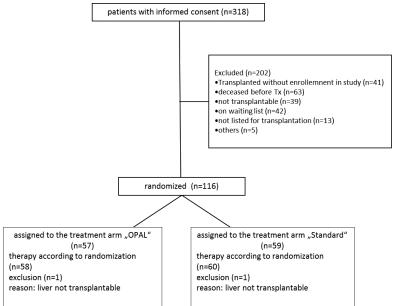
Patients were enrolled and transplanted from 09/2011 to 12/2013. According to the study protocol 116 patients were recruited. Of these, 57 patients were randomized into the treatment group and 59 patients randomized into the control group. One year follow up was 100%. Graft and patient survival were analyzed for a total of 5 years after transplantation.
Only one patient was lost to follow up with a functioning allograft after more than 2 years after transplantation.
Median follow up time was 1466 days (1–2028 days). Enrollment of patients is shown in Figure 1.
Figure 1. CONSORT diagram illustrating the study enrollment.
Following randomization 2 patients (1 in each treatment arm) were excluded as the donor organ has been considered inacceptable for transplantation after inspection by the responsible surgeon.
3.2. Donor, Recipient, and Perioperative Characteristics
Mean age of donor organs was 63 (±1.26) years. Half (50%) of the donors were male. Median donor ICU treatment before organ procurement was 3.0 (1–19) days. The median DRI was 1.8 ± 0.3. Cold preservation was applied for 452 ± 13.4 min. The warm ischemic time during the surgical procedure was 30 ± 0.6 min.
Recipients had a mean age of 53.2 ± 0.8 years and were predominantly male (60.3%). Indications (or a combination of indications) for OLT included cirrhosis related to alcoholic cirrhosis (31.9%),
27

Bioengineering 2019, 6, 35
viral hepatitis (26.7%), hepatocellular carcinoma (26.7%), NASH (6.9%) and others (25.9%). The mean labMELD before liver transplantation was 14.6 ± 0.6. Median duration of the surgical procedure was 257 (155–661) min.
Further details regarding donor, recipient and perioperative characteristics in both study arms are given in Table 1.
Table 1. Donor and recipient data; values given as median and range, where appropriate, in brackets.
Parameter |
OPAL (n = 57) |
Control (n = 59) |
p-Value |
|
Donor Age (years) |
64 (30–95) |
63 |
(28–84) |
0.57 |
|
|
|
|
|
Donor Gender (m/f) (%) |
56/44 |
|
44/56 |
0.27 |
|
|
|
|
|
Donor BMI (kg/m2) |
25(19–42) |
26 |
(19–51) |
0.38 |
Donor ICU stay (days) |
3 (1–16) |
4 |
(1–19) |
0.02 |
|
|
|
|
|
Donor Cause of death (n) |
|
|
|
|
Cerebrovascular |
37 |
|
39 |
0.8 |
Hypoxia |
10 |
|
13 |
|
Trauma |
5 |
|
4 |
|
Others |
5 |
|
3 |
|
|
|
|
|
|
Donor aspartate aminotransferase (AST) (U/L) |
48 (9–501) |
41 |
(9–607) |
0.8 |
|
|
|
|
|
Donor ALT (U/L) |
32 (6–956) |
33 |
(6–282) |
0.87 |
|
|
|
|
|
Donor γGT (U/L) |
46 (7–381) |
57 |
(6–416) |
0.29 |
|
|
|
|
|
Donor Sodium (μmol/L) |
149 (132–163) |
149 (132–169) |
0.71 |
|
|
|
|
|
|
Donor Creatinin (μmol/L) |
80 (32–689) |
81 (33–265) |
0.87 |
|
|
|
|
|
|
Donor Bilirubin (μmol/L) |
9 (3.4–30) |
8.2 (2.7–564) |
0.16 |
|
|
|
|
|
|
Donor INR |
1.13 (0.88–3.50) |
1.12 (0.87–5.60) |
0.81 |
|
|
|
|
|
|
Donor Risk Index |
1.83 (1.1–2.5) |
1.80 (1.1–2.5) |
0.55 |
|
|
|
|
|
|
Allograft Histology (n) |
49 |
|
47 |
|
Macrosteatosis (≥20%) |
13 |
|
6 |
0.09 |
Microsteatosis (%) |
50 (5–95) |
40 (0–90) |
0.36 |
|
Perfusion solution HTK/ UW (n) |
53/4 |
|
52/7 |
0.37 |
|
|
|
|
|
Cold Ischemia Time (min) |
443 (289–1090) |
390 (259–740) |
0.12 |
|
|
|
|
|
|
Recipient Age (years) |
57 (31/69) |
52 |
(24–67) |
0.046 |
|
|
|
|
|
Recipient Gender (m/f) (%) |
38/19 |
|
32/27 |
0.17 |
|
|
|
|
|
Recipient BMI (kg/m2) |
27 (18–44) |
25 |
(17–41) |
0.14 |
Underlying disease (%) |
|
|
|
|
Viral Hepatitis |
8 |
|
10 |
|
HCC |
14 |
|
16 |
0.83 |
linebreak Cholestative disease |
7 |
|
4 |
|
Alcohol |
14 |
|
18 |
|
NASH |
3 |
|
3 |
|
Others |
11 |
|
8 |
|
|
|
|
|
|
Charlson Co-morbidity Index |
4 (1–8) |
4 (2–8) |
0.25 |
|
|
|
|
|
|
Laboratory Model for End-Stage Liver Disease (MELD) |
13 (6–31) |
15 (6–40) |
0.16 |
|
|
|
|
|
|
In brief, clinically relevant characteristics were similar in both groups. Donor ICU stay was significantly shorter in the treatment group. Recipient age was significantly older in the treatment group. In both groups allocation was center based in 80%. Cold ischemia time was numerically longer in the treatment group (p > 0.05).
28
Bioengineering 2019, 6, 35
3.3. Surgical Study Procedures
Oxygen persu ation was applied for 137 (103–205) min in the treatment group. Treatment with persu ation did not result in any serious adverse event or allograft loss. Minor bleeding was observed from the pinpricks. These were not clinically relevant and stopped spontaneously or after minimal electrocoagulation. Median duration of the surgical procedure was 260 (176–460) min in the treatment arm and 250 (155–661) min in the control group. Warm ischemia time was similar, being 30 (16–41) min in the persu ation group and 29 (20–65) min in the control group. Statistical di erences regarding surgical study procedures were not observed between groups.
Transfusion of packed red blood cells (PRBs) was low and the same in both groups: 16 patients (28%) in the treatment group were transfused with a median of 2 PRBs compared to 25 patients (42%) transfused with a median of 2 PRBs in the control group.
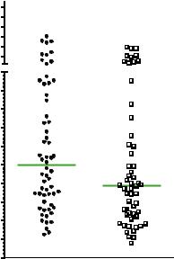
3.4. Primary Endpoint
Peak AST values within the first three days after liver transplantation were higher in the control group compared to the treatment group. However, this did not reach statistical significance (1246 (310–8064) versus 972 (194–17577), control versus OPAL; cf Figure 2).
$ 6 7 P D[ ' 8 /
|
P HGLDQ Q |
|
|
||
|
||
|
|
|
|
|
|
|
|
|
|
|
|
|
|
|
|
|
|
|
|
|
|
|
|
|
|
|
|
|
|
|
|
|
|
|
|
|
|
|
|
|
F R Q WUR O |
2 3 |
$ / |
Figure 2. Peak values of AST during the first 3 days after transplantation.
3.5. Secondary Endpoints
For secondary endpoints several assessments of patient death and early graft function were compared between groups. Details are depicted in Table 2.
Rates of 30-day mortality and In-hospital mortality were not statistically di erent between groups. Few retransplantations were necessary in the present study: One patient developed primary non-function and died after retransplantation. Another patient in the treatment group developed arterial thrombosis one month after transplantation and was successfully retransplanted. This patient is now well and alive. Statistical comparison of PNF and retransplantation rates was not performed due to the low number of events. Early Allograft Dysfunction occurred in every fifth to fourth patient in both groups. Rate of postoperative acute kidney failure and the necessity for hemodialysis was
29