

Биоинженерия / Биомеханика_микрофлюидные_устройства / статьи / 37_herricks2011
.pdfPublished on 11 July 2011. Downloaded by Universita Degli Studi di Napoli Federico II on 18/07/2013 15:37:23.
|
View Article Online / Journal Homepage / Table of Contents for this issue |
Lab on a Chip |
Dynamic Article LinksC |
Cite this: Lab Chip, 2011, 11, 2994 |
|
www.rsc.org/loc |
TECHNICAL NOTE |
A microfluidic system to study cytoadhesion of Plasmodium falciparum infected erythrocytes to primary brain microvascularendothelial cells
Thurston Herricks,*a Karl B. Seydel,bcd George Turner,e Malcolm Molyneux,d Robert Heyderman,d Terrie Taylorbc and Pradipsinh K. Rathod.a
Received 14th February 2011, Accepted 21st June 2011
DOI: 10.1039/c1lc20131j
The cellular events leading to severe and complicated malaria in some Plasmodium falciparum infections are poorly understood. Additional tools are required to better understand the pathogenesis of this disease. In this technical report, we describe a microfluidic culture system and image processing algorithms that were developed to observe cytoadhesion interactions of P. falciparum parasitized erythrocytes rolling on primary brain microvascularendothelial cells. We isolated and cultured human primary microvascular brain endothelial cells in a closed loop microfluidic culture system where
a peristaltic pump and media reservoirs were integrated onto a microscope stage insert. We developed image processing methods to enhance contrast of rolling parasitized erythrocytes on endothelial cells and to estimate the local wall shear stress. The velocity of parasitized erythrocytes rolling on primary brain microvascularendothelial cells was then measured under physiologically relevant wall shear stresses. Finally, we deployed this method successfully at a field site in Blantyre, Malawi. The method is a promising new tool for the investigation of the pathogenesis of severe malaria.
Background |
|
|
|
|
|
|
Directly observing cell-cell interactions within affected organs |
|||||||||||||
Cellular events associated |
with malaria |
mortality |
are highly |
in vivo is generally not feasible owing to technical and ethical |
||||||||||||||||
constraints, though imaging capillary flow in the rectal mucosa |
||||||||||||||||||||
varied and poorly understood.1 As a malaria parasite invades |
||||||||||||||||||||
has been performed in patients with malaria, but as impairment |
||||||||||||||||||||
and grows within an erythrocyte, it extensively alters the cell’s |
||||||||||||||||||||
of blood flow in this region is not a recognized feature of severe |
||||||||||||||||||||
plasma membrane by exporting |
proteins to the erythrocyte |
|||||||||||||||||||
malaria, this method can only approximate pathogenic processes |
||||||||||||||||||||
surface. These proteins facilitate cytoadhesion to ligands |
||||||||||||||||||||
in other organs.5 |
In |
vitro experimental models |
with varying |
|||||||||||||||||
expressed by vascular endothelial cells.2–4 Autopsy results from |
degrees of complexity have been developed to help understand |
|||||||||||||||||||
patients with fatal malaria infections have revealed that para- |
||||||||||||||||||||
parasitized |
erythrocytes-endothelial cell interactions. Initially, |
|||||||||||||||||||
sitized erythrocytes often accumulate in the deep capillary beds |
||||||||||||||||||||
static adhesion assays were used to observe and characterize |
||||||||||||||||||||
of the brain, kidney, lungs, or intestine by cytoadhering to |
||||||||||||||||||||
parasite-protein |
and |
parasite-endothelial |
cell |
interactions.6 |
||||||||||||||||
endothelial |
cells that express tissue-specific |
ligands.1 As the |
Introduction of |
parallel-plate |
flow chambers |
|
permitted |
the |
||||||||||||
parasitized |
erythrocytes accumulate |
in |
an |
organ’s capillary |
|
|||||||||||||||
ability to |
study |
cytoadhesion |
of parasitized |
erythrocytes |
to |
|||||||||||||||
network, they are thought to cause |
both |
an inflammatory |
||||||||||||||||||
specific surface-adsorbed proteins, transgenic mammalian cell |
||||||||||||||||||||
response and mechanical impairment of blood flow, which can |
||||||||||||||||||||
lines, and vascular endothelial cell lines.7–13 |
Parallel-plate flow |
|||||||||||||||||||
ultimately lead to organ failure.1 |
Detailed characterization of |
chambers offered the additional advantage of well defined fluid |
||||||||||||||||||
parasite-endothelial cytoadhesive |
events |
that lead |
to organ |
|||||||||||||||||
dynamics. |
Experiments |
using |
parallel-plat |
flow |
chambers |
|||||||||||||||
failure is crucial to understanding the pathogenesis of compli- |
||||||||||||||||||||
revealed the phenomenon of parasitized erythrocytes rolling or |
||||||||||||||||||||
cated P. falciparum infections. |
|
|
|
|
|
|||||||||||||||
|
|
|
|
|
‘tank-treading’ over surfaces and cells displaying specific ligands. |
|||||||||||||||
|
|
|
|
|
|
|
|
|||||||||||||
|
|
|
|
|
|
|
|
These tools have identified ligands such as ICAM-1, CD-36, and |
||||||||||||
aUniversity of Washington, Department of Chemistry, Seattle, WA, USA |
chondroitin-4-sulfate that wild type P. falciparum isolates have |
|||||||||||||||||||
strong binding interactions and these interactions have been |
||||||||||||||||||||
bBlantyre Malaria Project, University of Malawi College of Medicine, |
||||||||||||||||||||
Blantyre, Malawi |
|
|
|
|
|
|
associated with the pathological symptoms of complicated |
|||||||||||||
cMichiganStateUniversity, College of Osteopathic Medicine, East Lansing, |
malaria infections.8,14 |
|
|
|
|
|
|
|
|
|
||||||||||
MI, USA |
|
|
|
|
|
|
|
In spite of these general advances, mechanistic links of how |
||||||||||||
dMalawi-Liverpool-Wellcome |
Trust |
Clinical |
Research |
Programme, |
||||||||||||||||
interactions of host-parasite receptors relates to malaria path- |
||||||||||||||||||||
College of Medicine, Malawi, |
and The Liverpool |
School |
of Tropical |
|||||||||||||||||
ogenesis remain weak at |
best. Over time, |
cultured |
parasites |
|||||||||||||||||
Medicine, University of Liverpool, Liverpool, UK |
|
|
||||||||||||||||||
|
|
|
|
|
|
|
|
|
|
|
|
|
|
|||||||
eGames4You LLC, Scottsdale, AZ, USA |
|
|
|
|
undergo antigenic variation in expression of |
their surface |
||||||||||||||
|
|
|
|
|
|
|
||||||||||||||
2994 | Lab Chip, 2011, 11, 2994–3000 |
|
|
|
|
|
This journal is ª The Royal Society of Chemistry 2011 |
|
|||||||||||||
Published on 11 July 2011. Downloaded by Universita Degli Studi di Napoli Federico II on 18/07/2013 15:37:23.
|
|
|
|
|
|
|
|
|
|
|
View Article Online |
||
adhesion proteins which complicates investigation of specific |
averaging technique was developed to enhance contrast of slow |
|
|||||||||||
parasite-ligand interactions.15,16 |
Consequently, replicating |
rolling parasitized erythrocytes. The frame averaging technique |
|||||||||||
disease pathogenesis in the laboratory models is difficult when |
was then used to augment image tracking methods available in |
||||||||||||
there is a time lag between collecting parasites and endothelial |
the software Meta Morph. Image analysis techniques were |
||||||||||||
cells from patients in an endemic area and then performing |
developed to estimate the wall shear stress by measuring the |
||||||||||||
experiments in a distant laboratory. An important technical |
velocity of erythrocytes moving through the field of view. |
||||||||||||
challenge in understanding malaria pathogenesis is to study |
Together these tools were used, at a field site in Africa, to |
||||||||||||
interactions between primary endothelial cells and fresh para- |
demonstrate the feasibility of observing recently isolated para- |
||||||||||||
site isolates. We reported development of a microfluidic device |
sitized erythrocytes rolling on primary brain micro- |
||||||||||||
with which we observed rolling of parasitized erythrocytes on |
vascularendothelial cells under a range of biologically relevant |
||||||||||||
transgenic |
CHO |
cells |
expressing |
ICAM-1 |
and |
CD36 |
in |
wall shear stresses. |
|
|
|
||
a laboratory setting.17 A suite of technical and conceptual |
|
|
|
|
|||||||||
changes |
were required |
to make this approach suitable |
for |
Materials and methods |
|||||||||
a field setting. |
|
|
|
|
|
|
|||||||
|
|
|
|
|
|
|
|
|
|
||||
Microfluidic devices offer a flexible platform for performing |
Endothelial cell isolation and culture |
||||||||||||
a wide variety of experiments aimed at studying the behavior of |
Primary micro-vascular brain endothelial cells were collected |
||||||||||||
particles |
and cells under flow conditions |
similar |
to those |
||||||||||
from a fatal cerebral malaria infection using procedures per- |
|||||||||||||
observed |
in the microcirculation. In particular, microfluidic |
||||||||||||
formed in accordance with the Malawi College of Medicine |
|||||||||||||
devices have been utilized to study reduced deformability of P. |
|||||||||||||
Research Ethics Committee approval and approval from the |
|||||||||||||
falciparum |
parasitized |
erythrocytes.18,19 Previous endothelial |
|||||||||||
Michigan State University IRB. Informed consent was obtained |
|||||||||||||
culture methods have used a technique by which adhesive cells |
|||||||||||||
from the appropriate family members before autopsy. A 2 cm3 |
|||||||||||||
were initially cultured on glass cover-slips and then assembled |
portion of frontal lobe was collected at time of autopsy and |
||||||||||||
into a flow chamber.20 |
Several groups have described micro- |
||||||||||||
placed in 40 mL of PBS with ABAM (GIBCO BRL, Grand |
|||||||||||||
fluidic devices that have been developed specifically to study |
|||||||||||||
Island, NY). The tissue was transferred to a sterile petri dish and |
|||||||||||||
cytoadhesion events. Some early examples of these microfluidic |
|||||||||||||
minced using sterile scissors and forceps. The suspension was |
|||||||||||||
studies have investigated interactions of platelets and erythro- |
|||||||||||||
transferred to a 15 mL conical and centrifuged at 800 G for |
|||||||||||||
cytes with |
endothelial |
cells in microfluidic environments.21–24 |
|||||||||||
minutes. The pellet was resuspended in 5 mL of digestion buffer |
|||||||||||||
Examples of other novel devices which incorporate variable flow |
|||||||||||||
(25 mM HEPES, 5 mM glucose, 120 mM NaCl, 50 mM KCl, |
|||||||||||||
regimes have been utilized for studies involving platelets and |
|||||||||||||
1 mM calcium chloride, 1.5% bovine serum albumin, and 5 mg |
|||||||||||||
particles |
adherent |
to either endothelial cells or to |
adsorbed |
||||||||||
mL 1 collagenase/dispase). This suspension was incubated at |
|||||||||||||
protein.25,26 In some cases these microfluidic devices have been |
|||||||||||||
37 C for 1 h with occasional shaking. The solution was then |
|||||||||||||
utilized to |
culture |
endothelial cells for angiogenesis experi- |
|||||||||||
centrifuged and the pellet re-suspended in 5mL of endothelial cell |
|||||||||||||
ments.27 |
Recently described microfluidic culture systems have |
||||||||||||
media and passed through a 210 mm filter. The filtrate was |
|||||||||||||
used gravity, syringe |
pumps, or |
intermittent pumping |
to |
||||||||||
washed twice in PBS/ABAM and then re-suspended in 5 mL |
|||||||||||||
exchange media for cell culture. Flow through the microfluidic |
|||||||||||||
0.05% Trypsin-EDTA (GIBCO-BRL) and incubated for 15 min |
|||||||||||||
devices is governed by Poiseuille’s Law which relates the pressure |
|||||||||||||
at 37 C. The reaction was stopped by adding 5 mL of endothelial |
|||||||||||||
difference across the microfluidic channels to the fluid flow rate. |
|||||||||||||
cell media, and centrifuged. The pellet was re-suspended in |
|||||||||||||
These microfluidic systems are pumped by creating a pressure |
|||||||||||||
endothelial cell media and filtered through a 110 mm filter. The |
|||||||||||||
differential across a channel which then drives fluid flow through |
|||||||||||||
filtrate was washed once and re-suspended in 7 mL of endothelial |
|||||||||||||
the microfluidic device from the high pressure region to the lower |
|||||||||||||
cell media and cultured in a T-25 vented flask at 37 C, 5% CO2 |
|||||||||||||
pressure. |
|
|
|
|
|
|
|
|
|||||
|
|
|
|
|
|
|
|
with daily media changes. |
|
|
|||
In this paper, we describe a microfluidic culture system (mF- |
|
|
|||||||||||
|
|
|
|
||||||||||
CS) and image analysis methods developed to study cytoadhe- |
Malaria parasite culture |
|
|
|
|||||||||
sion of malaria parasite isolates to primary human brain |
|
|
|
||||||||||
|
|
|
|
||||||||||
microvascularendothelial cells at a field site in Blantyre Malawi. |
The malaria parasite culture was isolated from a cerebral malaria |
||||||||||||
|
|
|
|
|
|
|
|
|
|||||
The mF-CS consisted of the microfluidic device (mF-D), tubing, |
patient via venipuncture and then maintained in RPMI 1640 |
||||||||||||
a miniature peristaltic pump, a stepper motor, pressure sensor, |
supplemented with 2.5 wt % Albumax II and gentamicin. |
||||||||||||
|
|
|
|
|
|
|
|
|
|||||
stepper motor controller, and stage mount. The mF-D developed |
Malaria parasites were cultured as described previously at 2% |
||||||||||||
for this study was simple to construct with a minimum number of |
hematocrit in O+ blood under a blood gas mixture of 2.5% O2, |
||||||||||||
fluidic connections, so the devices could be manufactured in large |
|||||||||||||
5% CO2, and 92.5% N2. |
28 |
For this study only a single parasite |
|||||||||||
numbers. The mF-CS was a closed loop so that connections were |
|
||||||||||||
isolate was used. |
|
|
|
||||||||||
maintained as the device was moved from cell culture incubator |
|
|
|
|
|||||||||
to the microscope stage. The mF-D is designed to allow reliable |
Microfluidic device fabrication |
||||||||||||
loading of microfluidic channels with a minimum number of |
|||||||||||||
|
|
|
|
||||||||||
endothelial cells, thus permitting multiple experiments with |
PDMS mF-D were fabricated using single layer replica |
||||||||||||
minimum culturing of primary endothelial cells. The system was |
modeling. Channel patterns were designed using AutoCAD |
||||||||||||
designed to provide continuous perfusion of the microfluidic |
(Autodesk) based on a simplified version of c-cup shaped |
||||||||||||
channels over several days so that endothelial cells would |
cellular traps published previously.29 The channels between the |
||||||||||||
multiply and grow within the channels under flow. A frame |
cellular traps were 100mm wide and 1 mm long. These long |
||||||||||||
|
|
|
|
|
|
|
|||||||
This journal is ª The Royal Society of Chemistry 2011 |
|
|
|
|
Lab Chip, 2011, 11, 2994–3000 | 2995 |
||||||||
Published on 11 July 2011. Downloaded by Universita Degli Studi di Napoli Federico II on 18/07/2013 15:37:23.
View Article Online
channels are the capillary regions designed to observe parasiteendothelial cell interactions. Positive photo masks were generated (Photo Sciences Inc.) and wafers were patterned using AZ4620 photo resist using standard protocols. The wafers were then etched in a Bosche deep reactive ion etch process (Oxford Instruments ICP 380) to produce channel depths of 30 mm. The silicon masters were vapor primed with the release agent (tridecafluoro-1,1,2,2,-tetrahydrooctly)-trichlorosilane (Gelest) in a vacuum desiccator overnight. PDMS was then mixed 1 : 10 monomer to hardener and poured over the wafers and cured at 140 C for 50 min. The channels were then released and punched using a sharpened 14 gauge blunt needle (Small Parts). Glass cover slips (Gold Seal #3334) and PDMS channels were rinsed with ethanol and then exposed to oxygen plasma of
10 watts for 40 s (Harrik Plasma Cleaner PDC-001). After |
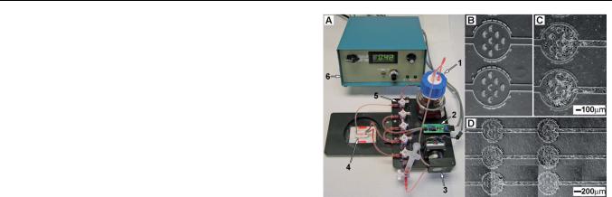
Fig. 1 An image of the microfluidic culture system (mF-CS) (A). The |
|||
exposure to the plasma, the cover slip and PDMS channel were |
media reservoir (1), pressure sensor/amplifier (2), stepper motor/peri- |
|||
brought into conformal contact to create the mF-D. Connec- |
staltic pump head (3), microfluidic device (mF-D) (4), and manifold (5) |
|||
tions were made to the channels using 15 gauge 45 blunt |
were all integrated onto a microscope stage insert. Only electrical |
|||
syringe needles (Small Parts) and 1.14 mm silicon auto-analysis |
connections to the stepper motor controller (6) unit need to be removed |
|||
to transfer from incubator to the microscope stage. Loading cells into |
||||
tubing (Cole-Parmer). Stop cocks and manifolds (Cole-Parmer) |
||||
channels (B). Cells proliferated in the device after 48 h (C). Endothelial |
||||
were used to control flow through the device. |
||||
cells after 72 h formed a lawn over the entirety of the microfluidic channel |
||||
|
||||
|
while under continuous flow (D). The markings around the loading |
|||
Pump description |
chambers are indications of chamber position on the chip. Up to four mF- |
|||
|
CS could be maintained in an incubator for independent experiments at |
|||
The pump system operates by maintaining a constant pressure |
any given time. |
|
|
|
across the mF-D. A manifold (Cole-Parmer) is configured such |
|
|
|
|
that the pressure sensor can measure the pressure drop across the |
|
|
|
|
mF-D. A microcontroller compares a voltage from the pressure |
T-25 flask. Approximately 1–4 105 cells were then placed in |
|||
sensor to a set point voltage. The microcontroller was pro- |
||||
grammed so that if there is a difference between the set point |
250 mL of media and introduced into the channels by removing |
|||
a connection, pulling the endothelial cell suspension into the |
||||
voltage and the sensor voltage, the controller moves a stepper |
||||
connector and then replacing the connector. The channels were |
||||
motor (Lin Engineering) connected to a peristaltic pump head |
||||
perfused initially at a pressure of 3.5 kPa for 20 min to move the |
||||
(Watson-Marlow 400A/F) to either pump fluid toward or away |
||||
majority of cells through the channels and trap endothelial cells |
||||
from the mFD. A 3 mL syringe with at least a 1 mL air-bubble is |
||||
in the c-cups (Fig. 1B). After the |
cells were loaded into the |
|||
placed between the peristaltic pump and the mFD. The air-bubble |
||||
channels the driving pressure was reduced to 0.7 kPa to allow |
||||
in the syringe acts both as a bubble trap and as a pressure |
||||
continuous perfusion. The later resulted in a wall shear stress of |
||||
dampener to reduce pressure fluctuations associated with stepper |
||||
0.5 Pa within the narrow channels between the loading areas of |
||||
motor movement. The pump controller was designed using the |
||||
the channels. The endothelial cells were cultured for 2–4 days |
||||
Luminary Micro Stellaris Stepper Motor Reference Design Kit |
||||
until they reached confluence (Fig. 1C–1D). |
||||
(Texas Instruments) modified to accept input from an analog |
||||
|
|
|
||
pressure sensor. The stepper motor microcontroller was pro- |
|
|
|
|
grammed by Games4you LLC using the IAR Embedded Work |
Microfluidic cytoadhesion experiments |
|||
bench to modify the code provided in the Stepper Motor |
||||
|
|
|
||
Reference Design Kit. The pressure sensor amplifier was |
P. falciparum parasites were cultured as previously described.28 |
|||
designed using PCB artist (Advanced Circuits) using a surface |
When the parasite cultures were primarily in the trophozoite |
|||
mount pressure sensor (Honneywell26PC01SMT) and an |
stage, cells were adjusted to 5% parasitemia and 2% hematocrit. |
|||
instrumentation amplifier (INA114 Texas Instruments). All |
Parasites were introduced to the device by removing one of the |
|||
components were mounted on anodized aluminum stage inserts |
two upstream connectors and drawing about 200 mL of parasite |
|||
designed for a Prior motorized stage so that they could be viewed |
culture into the connector. After replacing the connector, flow |
|||
on an inverted microscope. All components could be easily |
through the device was restarted. Cell flows in channels were |
|||
transferred to a cell culture incubator without disconnecting any |
viewed using a Nikon TE2000-S (Nikon USA) with a 20X |
|||
fluid connections. |
ELWD DIC lens. Sequential image sequences or image stacks |
|||
|
were recorded using a Photometrics Cool Snap EZ camera. |
|||
Microfluidic endothelial cell culture |
Videos were recorded using 1 ms exposure times using Meta |
|||
Morph version 7.4 (Molecular Devices). Parasitized erythrocytes |
||||
|
||||
All channel tubing, manifolds, and media reservoirs were either |
were identified by the presence hemozoin and obvious contrast |
|||
sterilized with ethanol or autoclaved, and assembled as shown |
differences between parasitized and normal erythrocytes. Data |
|||
(Fig. 1A). Endothelial cells were passaged using 0.25% trypsin- |
shown are accumulated over 5 |
independent experiments |
||
EDTA when cell cultures reached about 80% confluence in a |
(Fig. 4B). |
|
|
|
|
|
|||
2996 | Lab Chip, 2011, 11, 2994–3000 |
This journal is ª The Royal Society of Chemistry 2011 |
|
||
Published on 11 July 2011. Downloaded by Universita Degli Studi di Napoli Federico II on 18/07/2013 15:37:23.
|
View Article Online |
||
Frame-averaging contrast enhancement |
augmented standard methods of object tracking available |
|
|
The raw images stacks were processed to facilitate data analysis |
through the MetaMorph object tracking plug-in (Fig. 2C). |
||
|
|
||
on adhered cells. When necessary, frame averaging was used to |
Erythrocyte velocity averaging |
||
enhance contrast by removing background cells and channel |
|||
|
|
||
features so that slower moving cells were visualized in the image |
The mean velocity of erythrocytes was averaged using a routine |
||
sequences. The slowly moving cells were usually rolling parasit- |
scripted in MATLAB version 7.5.0 R2007b (Math Works) using |
||
ized erythrocytes but occasionally uninfected cells caught on |
functions available in the Image Processing Tool Box. Meta- |
||
a physical obstruction (Fig. 2A). Background subtraction was |
morph image sequence stacks had their backgrounds removed |
||
performed using either journal functions available in Meta- |
using the method described in the previous section. The images |
||
morph. Sequential images collected in the image stack were |
were converted to binary images by setting a threshold value. The |
||
averaged to generate frame-averaged background image. This |
resulting binary images were filtered such that any pixels that |
||
background image was subtracted from each frame in the image |
were present on two consecutive frames were removed. This had |
||
stack such that the absolute value of the difference between the |
the effect of removing background information which was not in |
||
background and each frame was recovered. The result produced |
motion from frame to frame. Then two interrogation regions |
||
animage where moving objects appear white against a black |
were chosen. The interrogation regions were defined as a span- |
||
background. The contrast for rolling and slowly moving cells was |
ning all rows of the image and enough pixel columns so the |
||
further enhanced by averaging three sequential images of this |
interrogation region was the same size or slightly larger than the |
||
background subtracted stack. The procedure of averaging three |
erythrocytes imaged. The position of the initial interrogation |
||
sequential frames was repeated for each frame in the image |
region was chosen with a priori knowledge of the flow direction |
||
sequence. The resulting images enhanced the contrast from any |
(Fig. 3A). The second interrogation region was displaced by |
||
moving cell whose position overlapped the same pixels for three |
certain distance in direction of flow in the next sequential image |
||
consecutive frames (Fig. 2B). Rapidly moving cells which did not |
(Fig. 3B). The total number of white pixels in each interrogation |
||
occupy the same pixels for at least two consecutive frames had |
region was identified. Next the difference in the number of pixels |
||
their contrast reduced. The image processing technique |
between the two interrogation regions was determined for each |
||
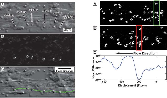
Fig. 2 A typical DIC image of cells cultured in the 100 mm wide and 30 mm high microfluidic channels (A). Tracking parasitized erythrocytes using Meta Morph was difficult in situations where the cells were obfuscated by endothelial cells. Contrast enhancement used averaging of three consecutive frames to improve contrast of rolling cells (B) and facilitated reliable tracking of those cells (C). The advantage is this technique can improve contrast in many situations where visually tracking cells is difficult and addition of fluorescent probes is not practical.
Fig. 3 Measuring velocity of flowing erythrocytes in two consecutive frames. The background has been removed and thresholding applied to observe moving erythrocytes. The green box in (A) is held fixed while the red interrogation region in the next sequential frame (B) is displaced a certain number of pixels from the initial interrogation region. The difference in the number of white pixels between the two interrogation regions is determined. This difference is averaged for each displacement over all sequential images in the image stack (C). The minimum of this mean difference corresponds to the average distance that a cells traverse between sequential images. The average cell velocity is determined by the mean time between images and the image magnification.
This journal is ª The Royal Society of Chemistry 2011 |
Lab Chip, 2011, 11, 2994–3000 | 2997 |
Published on 11 July 2011. Downloaded by Universita Degli Studi di Napoli Federico II on 18/07/2013 15:37:23.
|
|
|
|
View Article Online |
||
consecutive frame. These differences were then averaged over all |
beyond, until the endothelial cells had overgrown the channels, |
|
||||
consecutive frames in the movie. This process was repeated for |
which generally occurred after 5–6 days of continuous culture. |
|||||
different displacement values of the downstream interrogation |
|
|
||||
window. When the difference between the two interrogation |
Bubbles |
|||||
frames reached a minimum was where roughly the same numbers |
Although the mF-CS was a closed loop system, care was taken to |
|||||
of erythrocytes were in the consecutive image frames (Fig. 3C). |
||||||
avoid bubbles that would obstruct flow or compromise the |
||||||
The average time between consecutive images allowed calcula- |
||||||
endothelial cell culture. The most problematic bubbles are those |
||||||
tion of the cells mean velocity. The wall shear stress was then |
||||||
that form in the tubing between the channel and the syringe, |
||||||
calculated using the depth of focus for the 20X lens which is |
||||||
because they interfere with channel perfusion. Bubbles that form |
||||||
approximately 3.6 mm. This method is insensitive to the parabolic |
||||||
elsewhere in the fluid circuit either become trapped in the syringe |
||||||
velocity profile across width of |
the microfluidic channel. |
|||||
or pumped into the reservoir. We found that a minimum driving |
||||||
However, a good approximation of wall |
shear stresses |
was |
||||
pressure drop of 1.5 kPa prevented bubbles in the inlet tubing at |
||||||
obtained. |
|
|
|
|||
|
|
|
the University of Washington in Seattle (elevation 20 m, ambient |
|||
|
|
|
|
|||
|
|
|
|
lab temperature 20–25 C). However, in Blantyre, Malawi |
||
Results and discussion |
|
|
|
(elevation 1000m, ambient laboratory temperature 25–30 C), |
||
|
|
|
|
only 0.7 kPa was required. Bubble formation is related to dis- |
||
Requirements and capabilities |
|
|
|
solved gases in the media. The lower atmospheric pressure and |
||
|
|
|
|
|||
The mF-CS described here presents a novel solution to micro- |
higher temperature in Malawi may combine to reduce bubbles in |
|||||
the tubing. |
||||||
fluidic studies in malaria research |
field |
settings. First, |
the |
|||
Lower driving pressures, and thus lower flow rates, could be |
||||||
approach provided a closed loop |
system |
that integrates |
all |
|||
achieved by removing the reservoir. In this manner the system |
||||||
pumping onto the microscope stage. It was not necessary to |
||||||
was completely closed and pressurized by the air bubble in the |
||||||
disrupt fluid or pneumatic connections to transfer the culture |
||||||
syringe, which expanded as the temperature equilibrated in the |
||||||
system from the incubator to the microscope although it was |
||||||
incubator to 37 C. The higher overall system pressure may have |
||||||
possible to change electrical connections easily (Fig. 1). Sterility |
||||||
helped to reduce bubble nucleation and growth. Gas diffusion |
||||||
of the channels was easily maintained during routine culture in |
||||||
through both the PDMS and the silicon tubing appeared to be |
||||||
the mF-CS. The closed loop configuration prevented contami- |
||||||
sufficient to buffer the media as the endothelial cells grew well |
||||||
nation in the relatively dusty environment of the African field |
||||||
with the mF-CS configured in this manner. |
||||||
site. Incubator space in any laboratory is often limited so the |
||||||
|
|
|||||
ability to use multiple syringe pumps for several channels is not |
|
|
||||
feasible. Second, the pumping system maintained a constant |
Tracking rolling parasitized erythrocytes |
|||||
|
|
|||||
pressure across the channel so that perfusion was maintained for |
The design of the mF-CS was optimized to provide a stable |
|||||
experiments that lasted several days. The pumping system was |
||||||
environment to quantify cytoadhesion of parasitized erythro- |
||||||
also integrated with the Windows-PC used to record image data |
||||||
cytes to endothelial cells. Uninfected erythrocytes were not |
||||||
so that the flow rates could be manipulated from a computer |
||||||
observed to roll on the endothelial cells but rather occasionally |
||||||
interface. This microfluidic culture system allowed easy control |
||||||
bind and then release without rolling. The tracking methods |
||||||
of flow conditions as endothelial cells grew and changed the flow |
||||||
offered in Meta Morph failed to reliably track rolling parasites, |
||||||
environment of the channels. |
|
|
|
|||
|
|
|
particularly when the parasite was obfuscated either by the |
|||
|
|
|
|
|||
|
|
|
|
endothelial cells or in some cases by a high concentration of |
||
Using the microfluidic system in the field |
|
erythrocytes. To overcome this problem, we averaged three |
||||
|
consecutive frames together so that the contrast of slowly moving |
|||||
|
|
|
|
|||
Growing cells to confluence |
|
|
|
cells (such as rolling parasitized erythrocytes) was enhanced over |
||
The mF-D used in this paper used c-shaped cups designed to trap |
that of faster moving unbound uninfected cells and the immobile |
|||||
background (Fig. 2). This permitted the identification of rolling |
||||||
cells at specific points in the channels where cells were immobi- |
||||||
cells without perturbing the experiment by adding and washing |
||||||
lized long enough to adhere to the channel walls and prolif- |
||||||
away fluorescent dyes or antibody probes. Frame averaging |
||||||
erate.29 These channel designs were optimized using laboratory |
||||||
worked well to increase contrast, but it occasionally created an |
||||||
cell lines in the US before being used to culture primary endo- |
||||||
effect where the rolling parasites would appear to disappear and |
||||||
thelial cells obtained from fatal cerebral malaria infections at the |
||||||
reappear or ‘‘strobe’’ when they moved farther than the cell width |
||||||
field site in Malawi. The c-shaped structures worked well for |
||||||
in three consecutive frames. When this was observed, visual |
||||||
reliably loading primary endothelial cells (Fig. 1B). Once the mF- |
||||||
inspection of both the original image sequence and the frame |
||||||
D was loaded, the endothelial cells would migrate from the traps |
||||||
averaged image sequence was required to properly track the cells |
||||||
against the fluid flow, continuing to migrate and multiply until |
||||||
position. Overall the frame averaging technique worked very well |
||||||
they reached confluence after about 2–4 days (Fig. 1C and 1D). |
||||||
to augment the tracking methods available in Meta Morph. |
||||||
The dimensions of the channels between the c-cup regions were |
||||||
|
|
|||||
chosen to allow the endothelial cells to migrate yet be still large |
Estimating local wall shear stress |
|||||
enough that flow was not restricted excessively by the presence of |
||||||
|
|
|||||
the proliferating endothelial cells. The pumping system allowed |
In all mF-D with cells, local flow can vary greatly as the cells |
|||||
for continuous perfusion until the cells reached confluence and |
colonize and grow within the device channels. As cells proliferate |
|||||
|
|
|
|
|
||
2998 | Lab Chip, 2011, 11, 2994–3000 |
|
|
|
This journal is ª The Royal Society of Chemistry 2011 |
|
|

Published on 11 July 2011. Downloaded by Universita Degli Studi di Napoli Federico II on 18/07/2013 15:37:23.
|
|
|
|
|
|
|
|
|
View Article Online |
|||
in the micro-channels, the cross-sectional area of the channel |
the overall pressure drop across the channel or the overall flow |
|
||||||||||
decreases, resulting in unequal flow through different sections of |
rate through the channel based on an infusion pump. |
|
|
|||||||||
the device. A method to estimate the local wall shear stress was |
|
|
|
|
|
|
|
|
|
|
|
|
developed using erythrocytes as particles for tracking fluid |
Rolling velocity versus wall shear stress |
|
|
|
|
|||||||
velocities. Particle image velocimetry (PIV) has been performed |
A calibration plot showed that the average velocity of erythro- |
|||||||||||
using the erythrocytes to estimate the local flow environment.30,31 |
||||||||||||
PIV is measured using specialized high-speed cameras, laser light |
cytes increased with |
the measured pressure |
drop across |
the |
||||||||
channels (Fig. 4A). The range of velocities in the calibration plot |
||||||||||||
sources, and image correlation methods to describe the velocity |
||||||||||||
covers |
the velocities |
observed |
in experiments at a constant |
|||||||||
field across the field of view. In this study, an average velocity |
||||||||||||
applied pressure. Reduced flow rates with higher pressure are due |
||||||||||||
estimation was used. The average velocity method uses conven- |
||||||||||||
to endothelial cells growing in the channels and reducing the |
||||||||||||
tional optics and cameras and is more appropriate for field use. |
||||||||||||
effective cross-sectional area. Erythrocyte velocities of 5–20 mm s 1 |
||||||||||||
The average velocity method does not resolve the parabolic |
||||||||||||
have been observed in capillaries, which translates to wall shear |
||||||||||||
velocity field across the width of the micro-channel. Therefore, |
||||||||||||
stresses range of 0.1 to 0.7 kPa.32,33 These wall shear stresses are |
||||||||||||
this method is restricted in areas where flow is relatively laminar |
||||||||||||
in the same range observed in the mF-D described here.34–37 The |
||||||||||||
and unperturbed by obstructions. The method provides a useful |
||||||||||||
rolling velocity |
of |
individual |
parasitized |
erythrocytes |
was |
|||||||
estimate of local velocities when particles are present in at least |
||||||||||||
observed |
over |
a |
variety of wall shear |
stresses (Fig. 4B). As |
||||||||
two consecutive images across the field of view. Over the velocity |
||||||||||||
expected, |
the |
rolling |
velocity increased |
with |
the applied |
wall |
||||||
range measured, the average velocity appears to be linearly |
||||||||||||
shear stress and at higher shear stresses fewer parasitized eryth- |
||||||||||||
related to the pressure applied across the channel (Fig. 4A). This |
||||||||||||
rocytes tended to adhere. The relatively few number of parasit- |
||||||||||||
technique provides a reasonable estimate of the local flow rate |
||||||||||||
ized |
erythrocytes observed |
rolling |
on |
endothelial |
cells |
|||||||
through the channels, and is superior to relying on a measure of |
||||||||||||
underscores the difficulty and unique characteristics of working |
||||||||||||
|
||||||||||||
|
with fresh parasite field isolates. Normally for similar experi- |
|||||||||||
|
ments in non-endemic countries, parasite cultures are selected to |
|||||||||||
|
enrich for expression of adhesive characteristics before binding |
|||||||||||
|
experiments are performed. In an effort to keep the microfluidic |
|||||||||||
|
system as close to physiologic conditions as possible, a field |
|||||||||||
|
parasite isolate was chosen to directly demonstrate that the |
|||||||||||
|
techniques described capture parasite-endothelial cell interac- |
|||||||||||
|
tions over a variety of flow conditions. While a detailed investi- |
|||||||||||
|
gation across multiple parasite isolates and primary brain |
|||||||||||
|
endothelial cells is not presented here, the present work |
|||||||||||
|
demonstrates that these microfluidic technologies are ready for |
|||||||||||
|
field applications. |
|
|
|
|
|
|
|
||||
|
Conclusion |
|
As the parasitized erythrocytes accumulate in the microcircula- |
|
tion it is important to understand the conditions under which |
|
they cytoadhere and how they migrate under various flow |
|
conditions. The mF-CS described here was developed for field |
|
experimentation to observe parasitized erythrocyte cytoadhesion |
|
to primary endothelial cells. Quantifying the rolling behavior of |
|
parasitized erythrocytes over a variety of shear stresses can help |
|
describe the behavior of parasitized cells in micro-circulation. |
|
These types of measurements could help future investigations |
|
into interactions between endothelial cells and parasitized |
|
erythrocytes. This report demonstrates that microfluidic systems |
|
can be utilized to perform experiments in a malaria-endemic |
|
area. Such a system can mimic the micro-circulatory conditions |
|
in the deep capillary beds of organs and may improve our |
|
understanding of malaria pathogenesis. The mF-CS and image |
|
analysis tools described here provide a promising new resource |
|
for investigating how cytoadhesion contributes to severe malarial |
Fig. 4 The mean erythrocyte velocity increased linearly with the applied |
infections. |
pressure drop across the device (A). The wall shear stress was estimated |
|
using the erythrocyte velocity and the depth of field of the objective lens. |
Acknowledgements |
The rolling velocity of parasitized erythrocytes’s increased as the esti- |
|
mated wall shear stress increased (B). Each dot indicates an individual |
This work was supported by the NIH under the following |
parasitized erythrocyte. |
grants R21 AI081234 (P.K.R.), K23AI079402 (K.B.S), and |
|
|
This journal is ª The Royal Society of Chemistry 2011 |
Lab Chip, 2011, 11, 2994–3000 | 2999 |
Published on 11 July 2011. Downloaded by Universita Degli Studi di Napoli Federico II on 18/07/2013 15:37:23.
|
|
|
View Article Online |
||
U19AI089688 (P.K.R.). We specifically thank Jason Stage, Dave |
15 |
M. Frank, R. Dzikowski, B. Amulic and K. Deitsch, Mol. Microbiol., |
|
||
Tucker and the late George Turner of Games4you LLC for |
|
2007, 64, 1486–1498. |
|||
16 |
D. J. Roberts, A. G. Craig, A. R. Berendt, R. Pinches, G. Nash, |
||||
developing software for the microcontroller pump. |
|||||
|
K. Marsh and C. I. Newbold, Nature, 1992, 357, 689–692. |
||||
|
|
|
|||
|
|
17 |
M. Antia, T. Herricks and P. K. Rathod, PLoS Pathog., 2007, 3, e99. |
||
References |
18 |
J. P. Shelby, J. White, K. Ganesan, P. K. Rathod and D. T. Chiu, |
|||
|
|
|
Proc. Natl. Acad. Sci. U. S. A., 2003, 100, 14618–14622. |
||
1 |
L. H. Miller, D. I. Baruch, K. Marsh and O. K. Doumbo, Nature, |
19 |
T. Herricks, M. Antia and P. K. Rathod, Cell Microbiol., 2009. |
||
|
2002, 415, 673–679. |
20 |
B. M. Cooke, S. Usami, I. Perry and G. B. Nash, Microvasc. Res., |
||
2 |
L. H. Miller, S. Usami and S. Chien, J. Clin. Invest., 1971, 50, 1451– |
|
1993, 45, 33–45. |
||
|
1455. |
21 |
T. D’Amico Oblak, P. Root and D. M. Spence, Anal. Chem., 2006, 78, |
||
3 |
A. M. Dondorp, E. Pongponratn and N. J. White, Acta Trop., 2004, |
|
3193–3197. |
||
|
89, 309–317. |
22 |
W. Karunarathne, C. J. Ku and D. M. Spence, Integr. Biol., 2009, 1, |
||
4 |
H. A. Cranston, C. W. Boylan, G. L. Carroll, S. P. Sutera, |
|
655–663. |
||
|
J. R. Williamson, I. Y. Gluzman and D. J. Krogstad, Science, 1984, |
23 |
D. H. Kotsis and D. M. Spence, Anal. Chem., 2003, 75, 145–151. |
||
|
223, 400–403. |
24 |
D. M. Spence, N. J. Torrence, M. L. Kovarik and R. S. Martin, |
||
5 |
A. M. Dondorp, C. Ince, P. Charunwatthana, J. Hanson, A. van |
|
Analyst, 2004, 129, 995–1000. |
||
|
Kuijen, M. A. Faiz, M. R. Rahman, M. Hasan, E. Bin Yunus, |
25 |
S. Usami, H. H. Chen, Y. Zhao, S. Chien and R. Skalak, Ann. |
||
|
A. Ghose, R. Ruangveerayut, D. Limmathurotsakul, K. Mathura, |
|
Biomed. Eng., 1993, 21, 77–83. |
||
|
N. J. White and N. P. Day, J. Infect. Dis., 2008, 197, 79–84. |
26 |
J. M. Rosano, N. Tousi, R. C. Scott, B. Krynska, V. Rizzo, |
||
6 |
I. Ljunstrom, H. Perlmann, M. Schlichthere, A. Scherf and M. |
|
B. Prabhakarpandian, K. Pant, S. Sundaram and M. F. Kiani, |
||
|
Wahlgren, ed., Methods in Malaria Research, Manassas, Virginia, |
|
Biomed. Microdevices, 2009. |
||
|
2004. |
27 |
S. Chung, R. Sudo, P. J. Mack, C. R. Wan, V. Vickerman and |
||
7 |
B. M. Cooke, A. R. Berendt, A. G. Craig, J. MacGregor, |
|
R. D. Kamm, Lab Chip, 2009, 9, 269–275. |
||
|
C. I. Newbold and G. B. Nash, Br. J. Haematol., 1994, 87, 162–170. |
28 |
W. Trager and J. B. Jensen, Science, 1976, 193, 673–675. |
||
8 |
G. B. Nash, B. M. Cooke, K. Marsh, A. Berendt, C. Newbold and |
29 |
Z. Wang, M. C. Kim, M. Marquez and T. Thorsen, Lab Chip, 2007, 7, |
||
|
J. Stuart, Blood, 1992, 79, 798–807. |
|
740–745. |
||
9 |
C. J. McCormick, A. Craig, D. Roberts, C. I. Newbold and |
30 |
Y. Sugii, S. Nishio and K. Okamoto, Ann. N. Y. Acad. Sci., 2002, 972, |
||
|
A. R. Berendt, J. Clin. Invest., 1997, 100, 2521–2529. |
|
331–336. |
||
10 |
C. Newbold, P. Warn, G. Black, A. Berendt, A. Craig, B. Snow, |
31 |
Y. Sugii, S. Nishio and K. Okamoto, Physiol. Meas., 2002, 23, 403– |
||
|
M. Msobo, N. Peshu and K. Marsh, Am. J. Trop. Med. Hyg., 1997, |
|
416. |
|
|
|
57, 389–398. |
32 |
C. M. Rovainen, T. A. Woolsey, N. C. Blocher, D. B. Wang and |
||
11 |
K. R. Hughes, G. A. Biagini and A. G. Craig, Molecular and |
|
O. F. Robinson, J. Cereb. Blood Flow Metab., 1993, 13, 359–371. |
||
|
biochemical parasitology, 169, pp. 71–78. |
33 |
A. C. Ngai and H. R. Winn, Am. J. Physiol., 1996, 270, H1712–1717. |
||
12 |
S. J. Chakravorty, K. R. Hughes and A. G. Craig, Biochem. Soc. |
34 |
M. Oshima, T. Kobayashi and K. Takagi, Ann. N. Y. Acad. Sci., 2002, |
||
|
Trans., 2008, 36, 221–228. |
|
972, 337–344. |
||
13 |
D. J. Bridges, J. Bunn, J. A. van Mourik, G. Grau, R. J. Preston, |
35 |
H. H. Lipowsky, S. Kovalcheck and B. W. Zweifach, Circ. Res., 1978, |
||
|
M. Molyneux, V. Combes, J. S. O’Donnell, B. de Laat and |
|
43, 738–749. |
||
|
A. Craig, Blood, 115, pp. 1472–1474. |
36 |
P. Ganesan, S. He and H. Xu, Microvasc. Res., 80, pp. 99–109. |
||
14 |
J. D. Smith and A. G. Craig, Curr. Issues Mol. Biol., 2005, 7, 81–93. |
37 |
P. Ganesan, S. He and H. Xu, Ann. Biomed. Eng., 38, pp. 1566–1585. |
||
3000 | Lab Chip, 2011, 11, 2994–3000 |
This journal is ª The Royal Society of Chemistry 2011 |