

Биоинженерия / Биомеханика_микрофлюидные_устройства / статьи / 49_sundararaghavan2009
.pdfARTICLE
Neurite Growth in 3D Collagen Gels With Gradients of Mechanical Properties
Harini G. Sundararaghavan,1 Gary A. Monteiro,1 Bonnie L. Firestein,2 David I. Shreiber1y
1Department of Biomedical Engineering, Rutgers, The State University of New Jersey; telephone: 732-445-4500 ext 6312; fax: 732-445-3753; e-mail: shreiber@rci.rutgers.edu
2Department of Cell Biology and Neuroscience, Rutgers, The State University of New Jersey
Received 24 March 2008; revision received 7 July 2008; accepted 22 July 2008
Published online 8 August 2008 in Wiley InterScience (www.interscience.wiley.com). DOI 10.1002/bit.22074
ABSTRACT: We have designed and developed a microfluidic system to study the response of cells to controlled gradients of mechanical stiffness in 3D collagen gels. An ‘H’-shaped, source–sink network was filled with a type I collagen solution, which self-assembled into a fibrillar gel. A 1D gradient of genipin—a natural crosslinker that also causes collagen to fluoresce upon crosslinking—was generated in the cross-channel through the 3D collagen gel to create a gradient of crosslinks and stiffness. The gradient of stiffness was observed via fluorescence. A separate, underlying channel in the microfluidic construct allowed the introduction of cells into the gradient. Neurites from chick dorsal root ganglia explants grew significantly longer down the gradient of stiffness than up the gradient and than in control gels not treated with genipin. No changes in cell adhesion, collagen fiber size, or density were observed following crosslinking with genipin, indicating that the primary effect of genipin was on the mechanical properties of the gel. These results demonstrate that (1) the microfluidic system can be used to study durotactic behavior of cells and (2) neurite growth can be directed and enhanced by a gradient of mechanical properties, with the goal of incorporating mechanical gradients into nerve and spinal cord regenerative therapies.
Biotechnol. Bioeng. 2009;102: 632–643.2008 Wiley Periodicals, Inc.
KEYWORDS: nerve regeneration; gradients; durotaxis; microfluidics; tissue engineering
yAssistant Professor. Correspondence to: D.I. Shreiber
Contract grant sponsor: New Jersey Commission on Spinal Cord Research Contract grant number: 03-3028-SCR-E-0; 05-2907-SCR-E-0
Contract grant sponsor: Paralyzed Veterans of America Research Foundation Contract grant number: 2401
Contract grant sponsor: National Science Foundation (NSF-IGERT on Integratively Designed Biointerfaces)
Contract grant number: DGE 033196
Contract grant sponsor: Charles and Johanna Busch Biomedical Research Foundation Contract grant sponsor: National Science Foundation
Contract grant number: IBN-0548543
Additional Supporting Information may be found in the online version of this article.
Introduction
The mechanical stiffness of tissue substrates and/or the surrounding network of extracellular matrix has proven to be a crucial regulator of cellular functions (Arora et al. 1999; Balgude et al. 2001; Discher et al. 2005). Growth and movement of several cell types can be dictated by the substrate/matrix stiffness. Lo et al. (2000) first reported the preferential movement of fibroblasts with respect to mechanical stiffness and coined the term ‘‘durotaxis’’ to describe this phenomenon. Since then, quantitative differences in cell motility and process growth have been identified for neurons (Flanagan et al., 2002; Leach et al., 2007), smooth muscle cells (Peyton and Putnam, 2005), and epithelial cells (Saez et al., 2007). In addition, many other phenotypic and functional phenomena have been observed for these and other cells that affect proliferation, differentiation, matrix synthesis and degradation, and tractionmediated events (Georges and Janmey, 2005).
Directed cell migration is fundamental in many physiologic and pathologic processes such as tissue morphogenesis, wound healing, and tumorigenesis, and is also desired frequently in several tissue engineering applications. Of particular interest is the directed growth of neurites for regeneration of peripheral and central nervous system tissue. During development, axons are guided by attractive and repulsive soluble chemotactic cues and adhesion-based haptotactic cues, as well as contact guidance fields established by glia, aligned ECM proteins, and other axons, all of which are naturally presented in a three dimensional environment. Approaches to regenerating peripheral nerves and spinal cord tissue have attempted to include these directional cues to orient neurite growth (Schmidt and Leach, 2003). Another parameter, mechanical stiffness, has been shown to significantly affect neurite outgrowth (Flanagan et al., 2002; Georges and Janmey, 2005), and has been used to enhance growth isotropically by tuning matrix stiffness to entice neurite growth (Balgude et al., 2001), with improved growth occurring generally on or in more compliant systems. However, gradients of stiffness in a 3D,
632 Biotechnology and Bioengineering, Vol. 102, No. 2, February 1, 2009 |
2008 Wiley Periodicals, Inc. |
tissue-like system have not been employed to orient and potentially enhance growth via durotaxis.
Several approaches have been devised to probe the influence of substrate/network stiffness on cellular behavior. Simple techniques involve functionalizing poly(acrylamide) gels of different concentrations and/or crosslinking density with proteins that foster cell attachment (Lo et al., 2000), or coating these gels with a thin 3D layer of collagen in or on which the cells are cultured (Discher et al., 2005) to examine the response to uniform presentation of stiffness. Microfabrication techniques have been used to develop more elaborate systems comprising, for example, calibrated, elastomeric microposts of different dimensions, which maintain different bending properties to present a 2D substrate of varying stiffness to the cells (Tan et al., 2003), or gradients of stiffness generated through photo-initiated crosslinking of a 2D substrate (Burdick et al., 2004).
We have developed a system to generate stable 1D gradients of mechanical properties through a 3D collagen gel. Microfluidic networks are pre-filled with a type I collagen solution, which is allowed to self-assemble. Gradients of genipin, a cell-compatible, fast-acting crosslinking agent (Sung et al., 2003), are generated through the collagen gel using a source–sink network for a defined period of time to establish a 1D gradient through the 3D gel. Collagen crosslinked with genipin fluoresces at 630 nm when excited by light at 590 nm (Almog et al., 2004). In a previous study, we found that the intensity of the fluorescence correlates well with the degree of crosslinking and storage modulus of the collagen gel (Sundararaghavan et al., 2008). As such, genipin-generated patterns of crosslinks are directly visualized via fluorescence, and interpreted as a gradient of stiffness. We demonstrate the functionality of these gradients by biasing and enhancing neurite outgrowth from chick dorsal root ganglia (DRGs). Potential effects of genipin-mediated crosslinking on cell adhesion and collagen fiber size and density were examined in separate assays and judged to minimally contribute to the observed neurite growth.
Methods
Microfluidic Networks
A simple, ‘H’-shaped, ‘‘source–sink’’ arrangement was used to generate gradients in a cross-channel connecting source- to-sink (Fig. 1). Channel dimensions for the source–sink network were selected by simulating flow in networks with a computational fluid dynamics package (ESI-CFD Huntsville, AL) to achieve uniform gradients across the width of the cross-channel, which showed that the source and sink channels should be at least 2 wider than the cross-channel. Source and sink channels were 500 mm wide 100 mm deep and were connected by a 5 mm long, 150 mm wide, and 100 mm deep channel (Fig. 1A). Microfluidic networks were fabricated using standard photolithography techniques
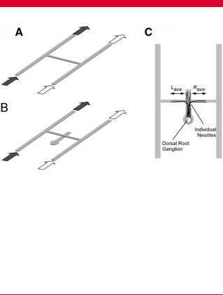
Figure 1. Schematic of ‘H’-shaped microfluidics network used to create gradients. A: After filling the network with collagen solution and allowing the collagen to self-assemble into a gel, a gradient of genipin is created in the cross-channel by supplying culture medium with genipin in the source inlet (dark arrows) and medium alone in the sink channel (white arrows). B: To introduce cells into the gradient, a second network (facing channel-side up) comprising a straight channel and circular well is filled with collagen and a DRG placed in the well. After self-assembly, the ‘H’-network is placed on top of the straight channel, creating a collagen-collagen interface at the intersection. The gradient is then formed in the cross-channel as in (A). C: Top view of the network shown in (B) with neurite growth drawn in. Neurite growth is quantified by counting the number of processes and the average length of processes that extend from the DRG and grow up and into the cross-channel in the left (L) and right (R) directions.
(Whitesides et al., 2001) at Bell Labs/Lucent Technologies (Murray Hill, NJ) through a grant from the New Jersey Nanotechnology Consortium. The photomask was printed from an AutoCAD drawing of the network design. A silicon wafer was spin-coated with SU-8 negative photoresist (Microchem, Newton, MA) and baked for 5 min at 658C followed by 10 min at 1008C. The photoresist was exposed to UV light through the photomask using a Quintel 2001 CT Mask Alignment/Exposure system. The coated wafer was baked again and immersed in SU-8 developer for 12 min to clear un-reacted photoresist and form the final ‘‘master.’’ A poly(dimethyl siloxane) solution (PDMS; Dow Corning, Midland, MI) was poured over the master and baked overnight at 508C to produce a negative relief. The PDMS was removed, the design was cut out of the mold, and holes were punched for the inlet and outlet using a blunt 19-gauge syringe. The PDMS and a clean glass slide were plasma treated and bonded together to form the final device. The inlets were connected to a syringe pump (Harvard Apparatus, Cambridge, MA) using polyethylene tubing (Small Parts, Miami Lakes, FL).
Collagen Preparation
Type I collagen solutions were prepared as previously described (Shreiber et al., 2001) by mixing 20 mL 1 M Hepes buffer, 140 mL 0.1 N NaOH, 100 mL 10 PBS, 52 mL of PBS
Sundararaghavan et al.: Neurite Growth in Mechanical Gradients |
633 |
Biotechnology and Bioengineering
(Invitrogen, Carlsbad, CA), and 677 mL of a 3.0 mg/mL type I collagen solution (Elastin Products Co., Owensville, MO) to make a 2.0 mg/mL collagen solution. The collagen solution self-assembled into a fibrillar gel upon incubation at 378C.
Generation of Gradients of Mechanical Properties
The microfluidic networks were first filled uniformly with a type I collagen solution using a syringe pump operating at 0.1 mL/min while viewing the network with an upright tissue culture microscope to ensure that the network was filled properly with no bubbles. After inspection, the filled microfluidic network was transferred to a humidified, 378C, 5% CO2 incubator, and the collagen was allowed to selfassemble for at least 1 h. The source solution was then changed to culture medium (DMEM þ 10% FBS (Atlanta Biologicals, Lawrenceville, GA), 1% glutamine, 1% penicillin/streptomyosin (Sigma, St. Louis, MO)) plus a defined concentration of genipin (Challenge Bioproducts Co., Taichung, Taiwan) of either 1 or 10 mM, while the sink solution was changed to the same medium without genipin. These solutions were flowed gently through the fibrillar gel-filled microfluidic network at 0.3 mL/min at 378C for 12 h. To remove the remaining genipin from the network following the desired incubation period, the inlets were switched to medium without genipin. The cross-channel was flushed by actuating only one randomly selected inlet syringe at 0.3 mL/min for 3 h, after which medium was again delivered through both inlets.
Gradient Evaluation
Gradients of genipin-mediated crosslinking were verified by examining the fluorescence intensity emitted by the crosslinked collagen (590 nm Exc, 630 nm Em), which indirectly verified the pattern of mechanical properties. Our previous study confirmed that fluorescence intensity strongly correlates with the storage modulus measured in shear using parallel plate rheometry for a range of fluorescence intensities generated by crosslinking gels for 2–12 h with 0–10 mM genipin in 24-well plates (Sundararaghavan et al., 2008). This calibration was repeated for 0, 0.1, 0.5, and 1 mM genipin crosslinked for 12 h in single channel microfluidic networks with the same channel depth as used above. Collectively from these studies, the storage modulus in shear at 0.1 Hz (the lowest frequency tested) ranges from 57 3 Pa for untreated collagen to 377 25 Pa for collagen crosslinked with 1 mM genipin and 797 50 Pa (average SE) for 10 mM genipin for 12 h of crosslinking. Following rinsing, networks were transferred to a computercontrolled stage and imaged using an Olympus IX81 inverted microscope (Olympus, Melville, NY). The fluorescence intensity in the cross-channel was quantified using Olympus Microsuite Image Analysis Software (Olympus).
Neurite Outgrowth Assay
To evaluate if the changes in stiffness can induce phenotypic changes in cellular behavior, neurite outgrowth from chick DRGs was evaluated in the presence of durotactic gradients. The microfluidic system was modified to include a small well for DRG culture. A second network was generated that comprised a 1 mm diameter well connected to a straight 500 mm wide and 100 mm deep channel (Fig. 1B). This network was place upside down (channels facing up) on a glass slide and filled with collagen solution. DRGs were isolated from E8 chick embryos (Charles River Laboratories, Willmington, MA), and a single DRG was placed in the collagen-filled circular well. The network was transferred to a 378C incubator to facilitate self-assembly, entrapping the DRG in the collagen gel. The ‘‘source–sink’’ network was then plasma treated and bonded to the gel-filled underlying network such that the middle of the cross-channel of the ‘‘source–sink’’ intersected with the straight channel of the underlying network approximately 1,000 mm from the circular well containing the DRG. The top network was filled with collagen solution, which was allowed to gel. Gels were then treated with genipin and rinsed as described above. Gradients of crosslinking were again confirmed by visualizing the gradient of fluorescence. Networks were transferred to a humidified, 378C, 5% CO2 incubator and perfused with fresh medium (DMEM supplemented with 10% FBS and 100 ng/mL NGF (R&D Systems, Minneapolis, MN)) via gravity flow. DRGs were cultured in the networks for 5 days to allow neurites to grow through the collagen gel and extend up and into the cross-channel a significant distance, potentially in either direction.
To visualize neurite growth after 5 days in culture, the neurites were stained immunohistochemically in the networks for neurofilament proteins. Inlet solutions were changed to 4% paraformaldehyde for 3 h to fix the collagen and cells, then changed to a rinse buffer comprising 1% BSA þ 0.5% Triton in PBS for 3 h. Inlet solutions were changed to a 10% goat serum blocking solution in rinse buffer for 4 h, and then to an anti-neurofilament antibody cocktail of 1:200 a-NF 200 and 1:1,000 a-NF 68 (Sigma) overnight. Networks were rinsed for 4 h, and inlets were switched to a 1:400 dilution of goat anti-mouse Alexa 488 secondary antibody (Molecular Probes/Invitrogen, Eugene, OR) and incubated overnight. Devices were rinsed a final time for 4 h and then transferred to an inverted epifluorescence microscope for imaging.
Neurite growth was quantified as the number and length of neurites projecting up the stiffness gradient versus down the stiffness gradient, or in opposite directions for control experiments and uniform crosslinking where no gradient was present. For each device, digital images were taken with a 40 objective at the intersection of the explant and crosschannels and at the end of the individual growth cones. Using Olympus Microsuite Image Analysis Software, the (X, Y) coordinates were recorded for each growth cone and for the channel intersection to determine the distance of
634 |
Biotechnology and Bioengineering, Vol. 102, No. 2, February 1, 2009 |
growth in the gradient channel (Fig. 1C). For a given experiment, one experimental condition (0–1 mM for 12 h, 0–1 mM for 24 h, 0.5–0.5 mM for 12 h, or 1–1 mM for 12 h) and one control condition (0–0 mM) were performed, and in all cases, explants for the paired experiments were from the same chick embryo. Further elements of the data analysis for neurite outgrowth are presented in Results Section.
Adhesion Assay
Cell adhesion on collagen gels crosslinked to different degrees with genipin was tested using primary rat dermal fibroblasts as a uniform cell type, and with a mixed population of cells harvested from dissociated E8 chick embryo DRGs. DRG explants were suspended in a solution of 0.3% BSA (Sigma), 0.05% trypsin (Sigma) in HBSS (Lonza, Allendale, NJ) for 10 min at 378C, vortexing every 3 min. The sample was centrifuged for 2 min at 2,000 rpm. Cells were resuspended in 1 mL media (DMEM supplemented with 10% FBS, 100 ng/mL NGF) and triturated 10 times with a 1-mL pipette with a 200-mL pipette tip placed on top of a 1,000-mL pipette tip. Cells were counted using a hemocytometer. Collagen solution (100 mL, prepared as above) was pipetted into each well of a 24-well plate. The plate was then incubated at 378C for 60 min to allow the collagen to self-assemble. The collagen gel in each well was incubated in 600 mL genipin solution at 0, 0.5, 1, 5, or 10 mM in PBS for 12 h to allow crosslinking, during which time the plates were on a rocker to ensure rapid equilibration of genipin throughout the gel. The 0.5 mM condition was omitted for experiments with dissociated DRG cells. Each concentration was performed in triplicate in each plate. After 12 h, the gels were washed three times with PBS. Fibroblasts or dissociated DRG cells (100,000 cells/ well) were seeded on top of the gels and allowed to attach for 4 h. The gels were then washed twice with PBS, and the remaining cells were labeled using Calcein-AM (Invitrogen). Plates were transferred to the computer controlled stage of an Olympus IX81 inverted microscope operating in epifluorescence mode (480 nm Exc, 535 nm Em). Four images were taken at random per sample and the cells were counted manually. The adhesion experiment was repeated 3 times, and the average number of attached cells per field compared with ANOVA, with significance levels set at P <0.05.
Fibril Size and Density Assay
To evaluate the effects of the genipin crosslinking regimen on collagen fiber size and density (as an indirect assessment of gel porosity), straight microfluidic channels (500 mm wide, 100 mm deep, and 1cm long) were bonded to glass coverslips and filled with collagen solution that was spiked with FITC-labeled collagen (10%, v/v; Elastin Products), to allow for visualization of fibers. Following self-assembly, inlets were changed to genipin solutions at concentrations of
0, 1, or 10 mM for 12 h. Inlet solutions were then switched to PBS and devices were rinsed for 3 h. Devices were transferred to a Leica TCS SP2/MP confocal microscope (Leica Microsystems, Exton, PA). Images were taken at 63 with a 2 digital zoom at 488 nm excitation with a 500–535 nm emission bandpass filter. All image frames underwent two line and frame averaging. Three images were taken at random in each device. Each image was divided into nine equal squares. The average number and diameter of fibers was determined in three of the nine squares with the image analysis software. The analysis was repeated for three gels in each condition, and results were compared with ANOVA (significance set at P <0.05).
Results
Gradient Characterization
Exposure of fibrillar collagen within microfluidic networks to a gradient of genipin generated a gradient of crosslinks in the collagen, which was observed as a gradient of fluorescence intensity. The intensity profile in cross-channels was evaluated with image analysis tools (Fig. 2). A source–sink combination of 10–0 mM genipin generated a steeper gradient in intensity than a gradient from 1 to 0 mM. Uniform presentation of genipin produced uniform increases in intensity compared to 0 mM solutions (data not shown). When the underlying explant channel was included for neurite outgrowth experiments, the intensity plot increased at the intersection of the gradient and explant channels but returned to the original linear plot thereafter, indicating that the integrated signal of fluorescence intensity from the thicker collagen gel at the intersection was responsible for the increase, rather than an increase in crosslinking (Fig. 3). Based on the calibration of fluorescence intensity to storage modulus measured at 0.1 Hz and 1% shear strain amplitude (representative calibration shown in Fig. 2B), gels treated with 1 mM genipin for 12 h had an average gradient ( SEM) of 0.064 0.005 Pa/mm across the 5-mm long channel. When the exposure time was increased to 24 h, the gradient increased by 12% to 0.075 0.005 Pa/mm.
Neurite Outgrowth Assay
DRG explants were cultured in an underlying channel that intersects at the center of the cross-channel and therefore the approximate center of the gradient (e.g., 160 Pa for gradients generated by 12 h exposure to 0–1 mM genipin). In all conditions, several neurites (typically 15–30) grew from the underlying channel into the cross-channel, although most remained in the original channel (confocal micrograph shown in Fig. 4A). Those that entered the cross-channel could grow in either direction—either up or down the stiffness gradient, or for control cases, in uniformly
Sundararaghavan et al.: Neurite Growth in Mechanical Gradients |
635 |
Biotechnology and Bioengineering |
|
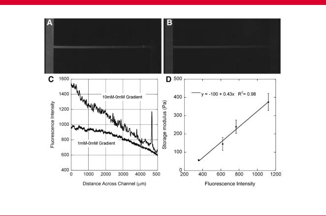
Figure 2. Representative gradients of fluorescence generated by exposure to a gradient of (A) 10 mM genipin–0 mM genipin or (B) 1 mM genipin–0 mM genipin for 12 h. C: Grayscale intensity values along the cross-channel for the images shown in (A) and (B). D: The intensity of fluorescence correlates to the stiffness of the gel, as shown in this representative calibration. The gradient of fluorescence is steeper when gels are exposed to a steeper gradient of genipin.
untreated or uniformly crosslinked channels. For each experiment, the average length within the cross-channel of the neurites that grew in each of the two directions was calculated by identifying the end of the growth cone (representative epifluorescent image taken with inverted microscope shown in Fig. 4B.) These were then plotted collectively for a given condition on the same set of axes, where the longer length (down the gradient) was plotted on the abscissa and the shorter length (up the gradient) on the ordinate. The data from each of the five experimental conditions was fit to a line that passed through the origin to evaluate the uniformity of growth. For completely uniform growth, the average lengths should be equal in the two directions, and the best fit line should have a slope of one. Biased growth would result in a shallower slope.
Data from the different experimental conditions are shown in Figure 5 as the average growth up and down the gradient ( SE) for each experiment and associated control experiment. In no case was growth exactly uniform ( F-test, P <0.05), and all points fell below the line that indicated perfectly symmetric growth. Control experiments were clustered near the ideal line and had slopes ( 95% confidence intervals) of 0.89 0.025 for assays not exposed to genipin, 0.90 0.059 for assays exposed to 0.5 mM genipin uniformly, and 0.83 0.068 for assays exposed to 1 mM genipin uniformly. In gradient experiments, the longer length was always in the direction of greater
compliance (down the gradient of stiffness). The slopes of the lines fitted through the 0–1 mM gradient experimental points and the origin were 0.45 0.209 for 12 h exposure and 0.57 0.230 for 24 h exposure, which were both substantially lower than the slopes of the control experiments and clearly below a slope of one.
As a measure of the strength of bias, the ratio of relative growth down the gradient (or in the ‘‘longer’’ direction for uniform 0.5 and 1.0 mM assays) versus the relative bias in the control for that experiment was taken and averaged across all experiments. On average, neurite bias was 2.51 0.77 times greater for 0–1 mM, 12 h exposure and 1.77 0.24 times greater for 0–1 mM, 24 h exposure. For uniform presentation, the average ratios were 1.00 0.03 for exposure to 0.5 mM genipin, and 1.04 0.05 for exposure to 1.0 mM genipin. The experimental conditions and relative growth biases are summarized in Table I.
No statistically significant differences in the number of neurites that extended in the two directions were observed for the untreated control or controls treated with uniform presentation of 0.5 or 1 mM genipin (Fig. 6). Significantly fewer neurons extended in the 1 mM uniform cases than in the untreated controls, but not the 0.5 mM uniform controls ( P ¼ 0.83). The number of neurites extending down the stiffness gradient was significantly greater than up the gradient for the 0–1 mM gradient, 12 h exposure experiments ( P ¼ 0.01); however, this trend was not observed for
636 |
Biotechnology and Bioengineering, Vol. 102, No. 2, February 1, 2009 |
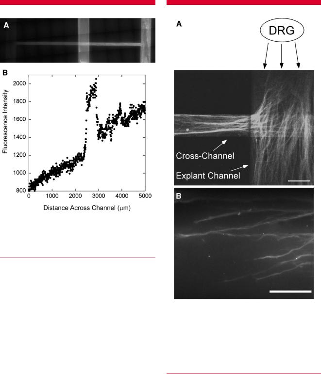
Figure 3. A: Representative gradient of fluorescence in modified networks with underlying explant generated by a gradient from 0 mM (left) to 10 mM (right) of genipin for 12 h. B: A roughly linear gradient of intensity is observed in the cross-channel, but includes a spike in intensity at the intersection of the two channels, after which intensity returns to the same linear contour. The increase in intensity is due to the integration of fluorescence through the thicker gel at the intersection.
the experiments treated with a gradient of genipin for 24 h ( P ¼ 1.0).
Significant variability in the magnitude of growth and in the number of neurites was observed in all conditions among different experiments. For example, while always more uniform, growth in some control experiments was double the growth in other experiments, and the day-to-day trends in growth in the experimental conditions typically paralleled that in the untreated controls. As such, the variability seemed to be linked to an experimental condition—most likely differences in the viability of explants from particular chick embryos. These day-to-day variations were accounted for in the statistical analysis of the length of neurites in gradient and uniform conditions by comparing the length of neurite growth among the control condition, up the gradient of stiffness, and down the gradient of stiffness for the 0–1 mM, 12 h exposure experiments with a two-way ANOVA using Type III sum of squares, where the gradient condition was a fixed effect and the day of the experiment was a random effect (Shreiber et al., 2001), followed by Scheffe’s post hoc test for pairwise
Figure 4. Images of neurite growth in the collagen gel-filled network. A DRG was placed within a collagen gel in the underlying explant channel and cultured for 5 days, at which time the networks were perfused with paraformaldehyde and then stained immunohistochemically for neurofilament proteins. A: As shown in this confocal micrograph, neurites grew from the DRG and either continued in the explant channel or grew up and into the cross-channel of the overlying ‘H’-shaped network. B: To measure the length of neurites in either directions, the growing end of neurites was identified from epifluorescent images taken with an inverted microscope with a 40 objective, and the length back to the intersection of the explant and crosschannels was determine based on the stage position and location within the image. Scale bars: (A) 150 mm and (B) 75 mm.
comparisons. Average length in gradient experiments is shown in Figure 7. The length of neurite growth down the stiffness gradient was significantly greater than growth in the control condition ( P ¼ 0.002), and growth in the control was significantly greater than growth up the stiffness gradient ( P ¼ 0.001). Similar results were observed in
Sundararaghavan et al.: Neurite Growth in Mechanical Gradients |
637 |
Biotechnology and Bioengineering |
|
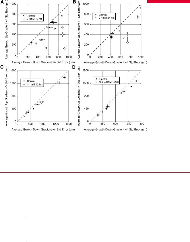
Figure 5. Growth in gradient and uniform presentation of stiffness along with associated controls. A: Stiffness gradient produced by exposure to 0–1 mM genipin for 12 h; (B) gradient from exposure to 0–1 mM genipin for 24 h; (C) uniform stiffness produced by exposure to 1 mM genipin for 12 h; (D) uniform stiffness from exposure to 0.5mM genipin for 12 h. Each point represents the average growth of neurites in a single network. The longer growth was always expressed as the x-coordinate. Thus, perfectly uniform growth would have a slope of one (dashed line), and biased growth would have a slope less than one. In no case was growth perfectly uniform, though in control experiments, the points fell very close to the dashed line. In gradient experiments, the direction of longer growth was always in the direction of lower stiffness, and these points deviated significantly from the line indicating uniform growth. In uniformly presented 1 and 0.5 mM generated stiffness, the points fell very close to the dashed line that indicates perfectly uniform growth and were similar to controls.
experiments where the gradient was generated with 24 h exposure. Growth down the stiffness gradient was significantly greater than the control growth ( P ¼ 0.001), and control growth was greater than growth up the stiffness
gradient ( P ¼ 0.016). Finally, growth in untreated controls was significantly greater than in gels that were crosslinked with a uniform presentation of 0.5 mM for 12 h ( P ¼ 0.038) and 1 mM genipin for 12 h ( P ¼ 1.4E 5).
Table I. Summary of growth assays.
Condition |
Storage modulus across microfluidic networka |
n |
Relative growth biasb |
|
|
|
|
1–0 mM, 12 h exposure |
377 25–57 2.8 Pa |
7 |
2.51 0.77 |
1–0 mM, 24 h exposure |
432 25–57 2.8 Pa |
5 |
1.77 0.24 |
1–1 mM, 12 h exposure |
377 25–377 25 Pa |
5 |
1.04 0.05 |
0.5–0.5 mM, 12 h exposure |
160 28–160 28 Pa |
4 |
1.00 0.03 |
aStorage modulus range estimated from calibration of storage modulus measured at 0.1 Hz and 1% shear strain to fluorescence intensity. Results are average SE.
bRelative growth bias calculated by taking the ratio of growth down:growth up the gradient of stiffness, normalizing by the same ratio from the matched, untreated control experiment, and averaging across all experiments. Results are average SE.
638 |
Biotechnology and Bioengineering, Vol. 102, No. 2, February 1, 2009 |
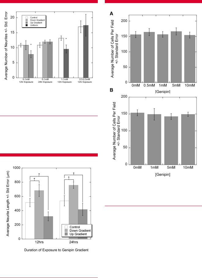
Figure 6. Neurite numbers in experimental cases were compared to matched controls performed on the same day with DRG explants from the same chick. Neurite number was significantly decreased up the gradient of stiffness compared to down the gradient and compared to control cases for 12 h exposure, but not 24 h exposure. Neurite number for uniformly treated 1 mM gels was less than matched controls, but no differences were detected for uniformly treated 0.5 mM gels. ANOVA followed by Scheffe’s post hoc test, P < 0.05.
Adhesion Assay
The influence of genipin-mediated crosslinking on the adhesion properties of collagen gels was evaluated with a simple detachment assay using dermal fibroblasts as a
Figure 7. Neurite lengths in gradient cases were compared to matched controls performed on the same day with DRG explants from the same chick. For both 12 and 24 h treated gels, the average growth down the gradient of stiffness was significantly greater than growth in control, untreated gels, which was greater than growth up the gradient of stiffness. Two-way ANOVA followed by Scheffe’s post hoc test, P <0.05.
Figure 8. Effects of genipin treatment on cell adhesion. Collagen gels were formed in 24 well plates and crosslinked with defined concentrations of genipin for 12 h and then rinsed extensively. A: Rat dermal fibroblasts or (B) cells from dissociated DRG explants were seeded on the gels and allowed to attach for 4 h. Detached cells were removed with rinsing, and the remaining cells were counted. In both cases, cell adhesion to collagen gels treated with 0–10 mM genipin for 12 h did not vary.
representative cell that could be uniformly presented (Fig. 8). No significant differences were observed in the adhesion of the fibroblasts to collagen gels crosslinked with up to 10 mM genipin for 12 h compared to the untreated controls (Fig. 8A; P ¼ 0.679). The assay was repeated with cells from dissociated DRGs (Fig. 8B), which represent a mixed population of primarily neurons, Schwann cells, and fibroblasts, and again, no significant differences in adherent cells were detected ( P ¼ 0.918). These results indicate that exposure to a gradient of genipin did not introduce significant changes to the adhesion profile presented to cells to influence cell behavior.
Sundararaghavan et al.: Neurite Growth in Mechanical Gradients |
639 |
Biotechnology and Bioengineering
Fibril Size and Density
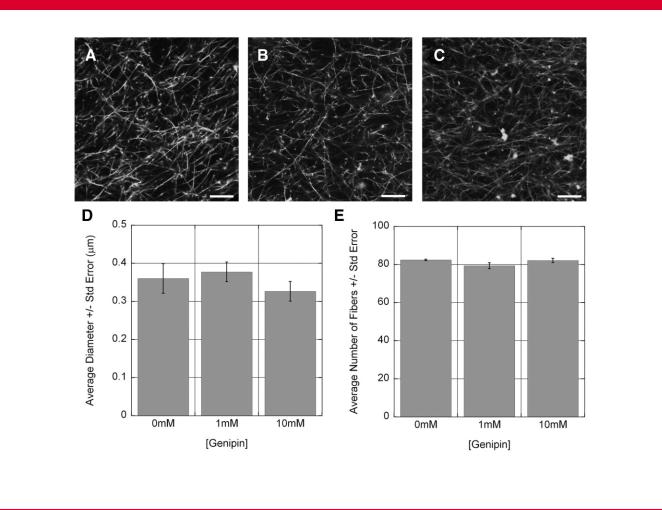
The influence of genipin-mediated crosslinking on collagen gel fiber morphology was estimated by evaluating the average size and density of collagen fibers in hydrated gels from high magnification epifluorescent images taken with confocal microscopy. Exposure to 1–10 mM genipin for 12 h did not produce any overt changes in fiber morphology or density. No significant differences were observed in the average fibril size (ANOVA, P ¼ 0.524) or fibril density ( P ¼ 0.194; Fig. 9). These results suggest that the gradient of genipin did not introduce a significant gradient of porosity to influence diffusion or present spatially varying steric hindrance.
Discussion
By developing an assay to study the response of cells to spatially varying mechanical properties in a 3D system, we hoped to improve the physiologic relevance of previous studies that demonstrate the importance of substrate mechanical properties in dictating cellular functions, and
move towards implementing patterned mechanical properties for tissue engineering applications. Previous studies have demonstrated neuritic preference for compliant substrates (Balgude et al., 2001; Fawcett et al., 1995; Leach et al., 2007) but have not shown how neurites respond to spatial changes in tissue or substrate mechanical properties. Using our system, we have shown that neurites preferentially grow down a gradient of stiffness within a 3D collagen gel.
The storage modulus in shear of our gels in the experiments where gradients were formed from 12 h of exposure to genipin ranges from 377 Pa ( 25) to 57 Pa ( 3) over the 5 mm channel, which is a gradient 0.064 Pa/mm. A typical growth cone is less than 5 mm, and it seems remarkable that the growth cone would maintain transducing elements capable of detecting a 0.35 Pa difference in the shear modulus. However, the thin filipodia that protrude from the growth cone and probe the environment can be as long as 50 mm and average 30 mm in chick DRG neurites (Buettner, 1994). Furthermore, the growth cone itself is merely the leading edge of a much longer neurite that, in this study, grows >1 mm within the cross-channel in 5 days. It is possible that the neuron integrates the stiffness information from filipodia and/or contact points along the
Effects of genipin treatment on collagen fiber morphology. Collagen solutions were spiked with fluorescent collagen, and gels treated with (A) 0 mM, (B) 1 mM, or (C) 10 mM for 12 h and imaged using confocal microscopy. Images were divided into a 3 3 grid, and the number of fibers and diameter of fibers counted in three boxes (the main diagonal). No significant differences were identified in either the fibril size (D) or the number of fibers (E). Scale bar: 10 mm.
640 |
Biotechnology and Bioengineering, Vol. 102, No. 2, February 1, 2009 |
trajectory of the whole or parts of the neurite to control the rate of growth. Although the length of neurites growing down a steeper gradient of stiffness appeared to be increased with a steeper gradient by 11% (Fig. 9; 24 h vs. 12 h genipin gradient exposure), the differences in the baseline control response prevented comparisons across these two conditions, and we cannot comment yet on the relationship to gradient steepness. We also note that the neurites likely sense the tensile properties of the system or individual fibers. The elastic modulus is approximately three times the shear modulus for incompressible materials, which results in approximately an order of magnitude difference in stiffness across the channel for the gradient conditions.
Rather than driving neurite growth with the gradient, it is also possible that the neurites are merely growing in a 3D environment with an optimal range of mechanical properties. Recent studies have shown that PC12 cells, an immortalized adrenocortical cell that exhibits a neuronlike phenotype when incubated in nerve growth factor, have a biphasic response to stiffness with shorter neurites on gels with a compliance 10 Pa, and increased growth and branching in gels of a higher compliance from 102 to 104 Pa (Leach et al., 2007). Other studies show that neurites from primary neurons sense differences between 200 Pa (‘‘soft’’) and 2 kPa (‘‘hard’’) substrates (Georges and Janmey, 2005). Combined with our results, these studies point to the possibility that the optimal stiffness for neurite growth lies between the stiffness at the middle of the cross-channel (e.g., 150–200 Pa for 12 h exposure experiments) and the stiffness at the sink ( 60 Pa), and that the enhanced growth down the stiffness gradient is because the stiffness of the gel in the control cases is sub-optimal. However, our results following uniform crosslinking with 0.5 mM demonstrate that the midpoint stiffness in gradient cases is sub-optimal for growth compared to control conditions, which further indicates that the gradient drives growth.
Genipin as a Crosslinking Agent
We generated changes in the mechanical properties of collagen gels using a naturally occurring compound, genipin, which we have previously shown to increase stiffness of collagen gels with increasing concentration and genipin exposure times (Sundararaghavan et al., 2008). Genipin has been shown to crosslink cellular and acellular tissues (Liang et al., 2004; Sung et al., 2001, 2003; Yerramalli et al., 2006), as well as biomaterials including gelatin microspheres (Liang et al., 2003), alginate–chitosan composites (Chen et al., 2004), and poly(ethylene)-glycol hydrogels (Moffat and Marra, 2004), and is increasingly observed in the literature for tissue engineering applications, including nerve regeneration, without adverse affects on cell behavior post-crosslinking. However, we have found the compound to be cytotoxic following direct exposure of cells to >1 mM genipin (Sundararaghavan et al., 2008), which limited our ability to create gradients with steeper slopes.
Genipin also has the unique feature of causing collagen fibers to fluoresce as it crosslinks, which was particularly beneficial for this study in that it allowed the gradients to be visualized during and after crosslinking as well as to be calibrated to mechanical properties. In principal, however, we have demonstrated the ability to impose a gradient of a soluble factor through a collagen-filled microfluidic network. Thus, any soluble crosslinking agent could be employed, such as aldehydes or enzymes (e.g., lysyl oxidase and transglutaminase). Moreover, the generation of a gradient of soluble factors also demonstrates that controlled chemotactic gradients can be generated through collagen gels using microfluidics.
Introduction of Alternate Guidance Fields Via
Genipin-Mediated Crosslinking
In addition to altering the stiffness, genipin may produce other changes in the collagen gel that could also drive or contribute to the biased neurite growth. Specifically, neurite behavior may be affected by direct exposure to genipin. Genipin-mediated crosslinking may produce changes in the adhesion properties of collagen to generate a haptotactic gradient, and/or genipin-mediated crosslinking may alter the porosity of collagen to potentially generate diffusion gradients of nutrients and/or sterically hinder growth.
The cytotoxic effects of higher (>1 mM) doses of genipin (Sundararaghavan et al., 2008) suggest that genipin may decrease cellular trophism, in general, and neurotrophism, specifically, since the DRG was exposed to genipin during gradient formation. Alternatively, the soluble genipin may act as a chemorepellant. However, the gradient of soluble genipin was maintained for as short as 12 h, and then the gels were rinsed extensively, well before any neurites reached the cross-channel. If genipin exposure decreased neurite outgrowth from DRGs, we may still expect biased growth away from the genipin source and down the stiffness gradient, but we would also expect that growth to be stunted compared to growth in control conditions, where DRGs were not exposed to genipin or genipin-crosslinked collagen for any period of time. We found that growth was enhanced down the gradient of stiffness compared to untreated controls, which demonstrates that the exposure to genipin or genipin crosslinked gels did not negatively affect neurite growth.
Separate sets of experiments were performed to evaluate the effects of genipin crosslinking on cell adhesion and on fiber density and thickness. We did not observe significant differences in the adhesion of fibroblasts or a mixture of cells from dissociated DRGs seeded on genipin-treated collagen gels versus control gels. Although we examined neurite growth through a 3D gel, probing attachment strength of cells entrapped within a 3D network of fibers is not trivial, and as such we evaluated cell attachment to a 2D fibrillar surface. While the influence of cell adhesion on cell behavior or outcome will vary between 2D and 3D systems—for example, cells entrapped in a 3D gel will not be ‘detached’
Sundararaghavan et al.: Neurite Growth in Mechanical Gradients |
641 |
Biotechnology and Bioengineering |
|