

Scratch technique for auscultating the liver. With stethoscope over the liver, lightly scratch the abdominal surface, moving toward the liver. The sound wili be intensified over the liver. (From Seidel HM, Ball JW, Dains JE, Benedict GW: Mosby's Guide to Physical Examination, 3rd ed. St. Louis, Mosby. 1995, with permission.)

A, The spleen is palpated bimanually with the patient in a supine position and the examiner at the patient's right side. The examiner's left hand is placed on the lower left rib cage, and the right hand explores for the spleen. (From Yang JC, Rickman LS, Bosser SK: The clinical diagnosis of splenomegaly. West J Med 155:47-52, 1991, with permission.)
B, The drawing shows the positioning of the examiner's hands during ballottement of the spleen.
(From Yang JC, Rickman LS, Bosser SK: The clinical diagnosis of splenomegaly. West J Med 155:47-52, 1991, with permission.) C, The spleen is palpated from above. (From Yang JC, Rickman LS, Bosser SK:
The clinical diagnosis of splenomegaly. West J Med 155:47-52, 1991, with permission.)

A, The drawing depicts Nixon's method of percussing the spleen. (From Yang JC, Rickman LS, Bosser SK: The clinical diagnosis of splenomegaly. West J Med 55:47-52, 1991, with permission.)

B, In Castell's method of percussing the spleen, the examiner percusses at the intersection of the left anterior axillary line and 9th intercostal space (marked). The lower diagonal line points to normal spleen. (From Yang JC, Rickman LS, Bosser SK: The clinical diagnosis of splenomegaly. West J Med 55:47-52, 1991, with permission.)

C. Traube's space is shown, as defined by Barkun et al. (From Yang JC, Rickman LS, Bosser SK: The clinical diagnosis of splenomegaly.
West J Med 155:47-52, 1991, with permission.)
The gallbladder can usually been palpated in the following clinical situations
•In carcinoma of the head of the pancreas
•In mucocele of the gallbladder
•In carcinoma of the gallbladder
GROSS ASCITES
•Dull in flanks
•Umbilicus transverse and/or hernia present
•Shifting dullness positive
•Fluid thrill positive

Technique for testing for a fluid wave. (From Swartz MH: Pocket Companion to Textbook of Physical Diagnosis. Philadelphia, W.B.
Saunders, 1995, with permission.)

Technique for testing for shifting dullness. The colored areas represent the areas of tympanv (From Swartz MH:
Diagnosis. |
|
Philadefphia |
permission.) |
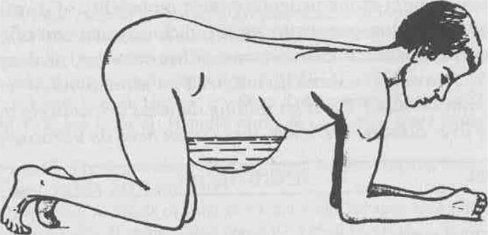
Patient positioning for eliciting the puddle sign. (From Dioguardi N, SannaGP: Modern! Aspetti di Semeiotica Medica. Milan, Societa Editrice Universe, 1975, with permission.)