

- •Foreword
- •Preface
- •Contents
- •1. Introduction to Pathology
- •2. Techniques for the Study of Pathology
- •6. Inflammation and Healing
- •8. Neoplasia
- •16. The Heart
- •17. The Respiratory System
- •18. The Eye, ENT and Neck
- •20. The Gastrointestinal Tract
- •24. The Female Genital Tract
- •25. The Breast
- •26. The Skin
- •27. The Endocrine System
- •28. The Musculoskeletal System
- •29. Soft Tissue Tumours
- •30. The Nervous System
- •Appendix
- •Further Readings
- •Index
461
Chapter 17 |
The Respiratory System |
LUNGS
NORMAL STRUCTURE
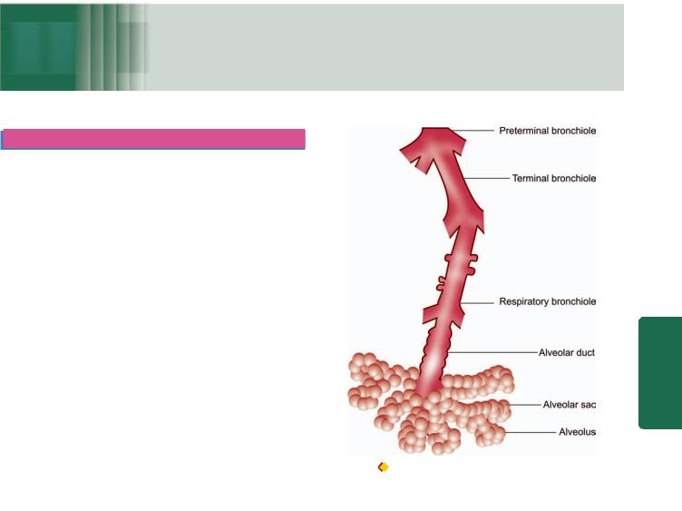
ANATOMY. The normal adult right lung weighs 375 to 550 gm (average 450 gm) and is divided by two fissures into three lobes—the upper, middle and lower lobes. The weight of the normal adult left lung is 325 to 450 gm (average 400 gm) and has one fissure dividing it into two lobes—the upper and lower lobes, while the middle lobe is represented by the lingula. The airways of the lungs arise from the trachea by its division into right and left main bronchi which continue to divide and subdivide further, eventually terminating into the alveolar sacs (Fig. 17.1).
The right main bronchus is more vertical so that aspirated foreign material tends to pass down to the right lung rather than to the left. The trachea, major bronchi and their branchings possess cartilage, smooth muscle and mucous glands in their walls, while the bronchioles have smooth muscle but lack cartilage as well as the mucous glands. Between the tracheal bifurcation and the smallest bronchi, about 8 divisions take place. The bronchioles so formed further undergo 3 to 4 divisions leading to the terminal bronchioles which are less than 2 mm in diameter. The part of the lung tissue distal to a terminal bronchiole is called an acinus. A cluster of about 5 acini supplied by terminal bronchioles and enclosed by visible fibrous septa is termed as the pulmonary lobule. An acinus consists of 3 parts:
1.Several (usually 3 to 5 generations) respiratory bronchioles originate from a terminal bronchiole.
2.Each respiratory bronchiole divides into several alveolar ducts.
3.Each alveolar duct opens into many alveolar sacs (alveoli) which are blind ends of the respiratory passages.
The lungs have double blood supply—oxygenated blood from the bronchial arteries and venous blood from the pulmonary arteries, and there is mixing of the blood to some extent. In case of blockage of one side of circulation, the supply from the other can maintain the vitality of pulmonary parenchyma. The bronchial veins drain the blood supplied by the bronchial arteries. The lungs have abundant intercommunicating lymphatics on the surface which drain into the subpleural plexus. Hilar and tracheobronchial lymph nodes receive the lymph and drain into the thoracic duct.
HISTOLOGY. The bronchi and their subdivisions up to bronchioles are lined by pseudostratified columnar ciliated epithelial cells, also called respiratory epithelium. These cells are admixed with mucus-secreting goblet cells which decrease in number as the bronchioles are approached. The
Figure 17.1 |
The structure of an acinus which is part of the lung |
distal to terminal bronchiole. It shows respiratory bronchiole, alveolar ducts and alveolar sacs.
mucosa of bronchi contains numerous submucosal mucous glands and neuroendocrine cells which are bronchial counterparts of the argentaffin cells of the alimentary tract (Chapter 20). The structure of bronchioles differs from that of bronchi and its subdivisions as well as from alveoli. They are lined by a single layer of pseudostratified columnar ciliated epithelium but no mucus cells and hence, unlike the bronchi, contain no mucus secretion on the surface. They contain some nonciliated Clara cells which secrete protein rich in lysozyme and immunoglobulins but unlike the alveoli contain no surfactant.
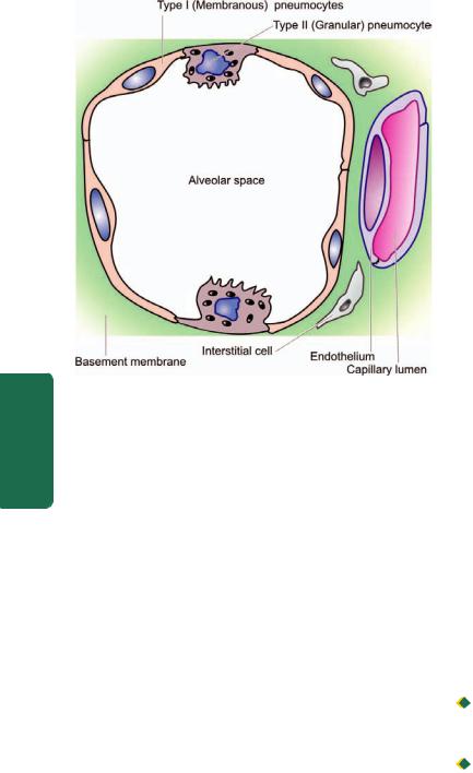
The alveolar walls or alveolar septa are the sites of exchange between the blood and air and have the following microscopic features (Fig. 17.2):
1.The capillary endothelium lines the anastomotic capillaries in the alveolar walls.
2.The capillary endothelium and the, alveolar lining epithelial cells are separated by the capillary basement membrane and some interstitial tissue. The interstitial tissue consists of scanty amount of collagen, fibroblasts, fine elastic fibres, smooth muscle cells, a few mast cells and mononuclear cells.
3.The alveolar epithelium consists of 2 types of cells: type I or membranous pneumocytes are the most numerous covering
System Respiratory The 17 CHAPTER
462
Pathology Systemic III SECTION
Figure 17.2 
 Histologic structure of alveolar wall (alveolar septa). It shows capillary endothelium, capillary basement membrane and scanty interstitial tissue and the alveolar lining cells (type I or membranous pneumocytes and type II or granular pneumocytes).
about 95% of alveolar surface, while type II or granular pneumocytes project into the alveoli and are covered by microvilli. Type II pneumocytes are essentially reserve cells which undergo compensatory hyperplasia when type I pneumocytes are injured and are also the source of pulmonary surfactant rich in lecithin. The main functions of coating of surfactant are to lower the surface tension of the alveolar lining cells and in maintaining the stability of the alveoli.
4.The alveolar macrophages belonging to mononuclearphagocyte system are present either free in the alveolar spaces or are attached to the alveolar cells.
5.The pores of Kohn are the sites of alveolar connections between the adjacent alveoli and allow the passage of bacteria and exudate.
FUNCTIONS. The primary functions of lungs is oxygenation of the blood and removal of carbon dioxide. The respiratory tract is particularly exposed to infection as well as to the hazards of inhalation of pollutants from the inhaled air and cigarette smoke. There exists a natural mechanism of filtering and clearing of such pollutants through respiratory epithelium, tracheobronchial lymphatics and alveolar macrophages. Besides, the lungs are the only other organ after heart through which all the blood of the body passes during circulation. Therefore, cardiovascular diseases have serious effects on the lungs, and conversely, diseases of the lungs which interfere with pulmonary blood flow have significant effects on the heart and systemic circulation.
Following groups of diseases of the respiratory tract are studied in this chapter:
1. Paediatric lung disease (congenital and acquired)
2.Pulmonary vascular disease
3.Pulmonary infections
4.Chronic obstructive pulomonary disease
5.Chronic restrictive pulmonary disease
6.Tumours of lungs.
PAEDIATRIC LUNG DISEASE
A number of congenital anomalies (e.g. agenesis, hypoplasia, heterotopic tissue, vascular anomalies, tracheal and bronchial anomalies, congenital pulmonary overinflation or lobar emphysema, congenital cysts and bronchopulmonary sequestration) and certain neonatal acquired lung diseases (respiratory distress syndrome or hyaline membrane disease, bronchopulmonary dysplasia, meconium aspiration syndrome, persistent foetal circulation, atelactasis, collapse and bronchiolitis) have been described. Some of the important conditions from point of view of pathology are discussed below.
CONGENITAL CYSTS
Developmental defects involving deficiency of bronchial or bronchiolar cartilage, elastic tissue and muscle result in congenital cystic disease of lungs. A single large cyst of this type occupying almost a lobe is called pneumatocele. Multiple small cysts are more common and give sponge-like appearance to the lung. The cysts are thin-walled and dilated and generally lined by flattened ciliated epithelium overlying a thin layer of supportive connective tissue. These cysts may contain air or may get infected and become abscesses. Cysts may rupture into bronchi producing haemoptysis, or into the pleural cavity producing pneumothorax.
BRONCHOPULMONARY SEQUESTRATION
Sequestration is the presence of lobes or segments of lung tissue which are not connected to the airway system. The blood supply of the sequestered area is not from the pulmonary arteries but from the aorta or its branches. Sequestration may be intralobar or extralobar.

 Intralobar sequestration is the sequestered bronchopulmonary mass within the pleural covering of the affected lung.

 Extralobar sequestration is the sequestered mass of lung tissue lying outside the pleural investing layer such as in the base of left lung or below the diaphragm. The extralobar sequestration is predominantly seen in infants and children and is often associated with other congenital malformations.
ACUTE RESPIRATORY DISTRESS SYNDROME (HYALINE MEMBRANE DISEASE)
Acute respiratory distress syndrome (ARDS) is a severe, at times life-threatening, form of progressive respiratory insufficiency which involves pulmonary tissues diffusely i.e. involvement of the alveolar epithelium, alveolar lumina and interstitial tissue. ARDS exists in 2 forms: neonatal and adult type. Both have the common morphological feature of formation of hyaline membrane in the alveoli and hence is

also termed as hyaline membrane disease (HMD). The two forms of ARDS have different clinical settings, response to treatment and consequences, etiology, pathogenesis but have similar morphology, and hence are discussed together below.
CLINICAL FEATURES AND CONSEQUENCES. These are as under:
 Neonatal ARDS occurring in newborn infants begins with dyspnoea within a few hours after birth with tachypnoea, hypoxia and cyanosis; in severe cases death may occur within a few hours.

 Adult ARDS is known by various synonyms such as shock-lung syndrome, diffuse alveolar damage (DAD), acute alveolar injury, traumatic wet lungs and post-traumatic respiratory insufficiency. The condition was first recognised in adults during World War II in survivors of non-thoracic injuries with shock. Adult ARDS also presents clinically by sudden and severe respiratory distress, tachypnoea, tachycardia, cyanosis and severe hypoxaemia.
ETIOLOGY. The two forms of ARDS have distinct etiology:

 Neonatal ARDS is primarily initiated by hypoxia, either shortly before birth or immediately afterward. It occurs in following clinical settings:
1.Preterm infants
2.Infants born to diabetic mothers
3.Delivery by caesarean section
4.Infants born to mothers with previous premature infants
5.Excessive sedation of the mother causing depression in respiration of the infant
6.Birth asphyxia from various causes such as coils of umbilical cord around the neck
7.Male preponderance (1.5 to 2 times) over female babies due to early maturation of female lungs
8.Finally, many cases of neonatal ARDS remain idiopathic.

 Adult ARDS may occur from the following causes:
1.Shock due to sepsis, trauma, burns
2.Diffuse pulmonary infections, chiefly viral pneumonia
3.Pancreatitis
4.Oxygen toxicity
5.Inhalation of toxins and irritants e.g. smoke, war gases, nitrogen dioxide, metal fumes etc.
6.Narcotic overdose
7.Drugs e.g. salicylates, colchicine
8.Aspiration pneumonitis
9.Fat embolism
10.Radiation.
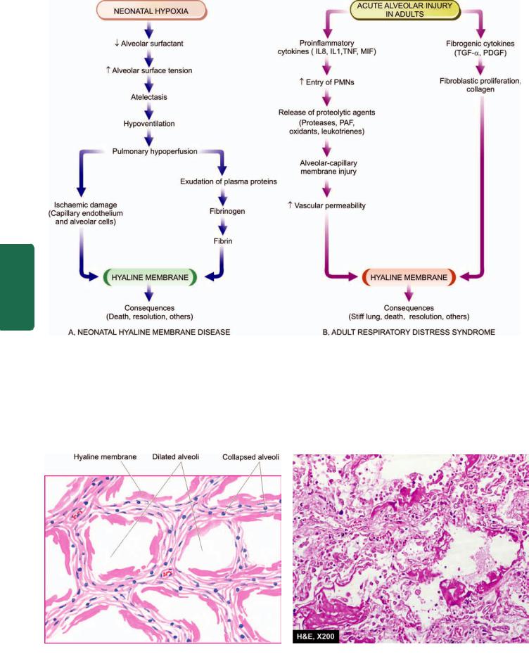
PATHOGENESIS. In both neonatal and adult type ARDS, there is damage to alveolocapillary wall triggered by etiologic factors listed above, and the final pathologic consequence of formation of hyaline membrane is also similar. However, how it occurs is different in the neonates than in adults. The sequence of events in the pathogenesis of both neonatal and adult ARDS is schematically illustrated in Fig. 17.3 and is outlined below:

 Neonatal ARDS. Entry of air into alveoli is essential for formation of hyaline membrane i.e. dead born infants do not develop HMD.
i) The basic defect in neonatal ARDS is a deficiency of pulmonary surfactant, normally synthesised by type II alveolar cells. The production of surfactant is normally increased shortly before birth but in prematurity and in neonatal hypoxia from any of the foregoing causes, its synthesis is decreased. The main function of alveolar surfactant being lowering of alveolar surface tension, its deficiency leads to increased alveolar surface tension which in turn causes
atelectasis.
ii)Atelectasis of the lungs results in hypoventilation, pulmonary hypoperfusion and ischaemic damage to capillary endothelium.
iii)This results in ischaemic necrosis of the alveolocapillary wall, exudation of plasma proteins including fibrinogen into the alveoli and eventually formation of hyaline membrane on the alveolar surface containing largely fibrin.
 Adult ARDS. The mechanism of acute injury by etiologic agents listed above depends upon the imbalance between pro-
inflammatory and anti-inflammatory cytokines:
i)Activated pulmonay macrophages release proinflammatory cytokines such as IL8, IL1, and tumour necrosis factor (TNF), while macrophage inhibitory factor (MIF) helps to sustain inflammation in the alveoli. Number of neutrophils in the alveoli is increased in acute injury. Neutrophils on activation release products which cause active tissue injury e.g. proteases, platelet activating factor, oxidants and leukotrienes.
ii)Besides the role of cytokines in acute injury, a few fibrogenic cytokines such as transforming growth factor-α (TGF-α) and platelet-derived growth factor (PDGF) play a role in repair process by stimulation of proliferation of fibroblast and collagen.
In either case, injury to the capillary endothelium leads to increased vascular permeability while injured pneumocytes, especially type 1, undergo necrosis. The net effect of injury to both capillary endothelium and alveolar epithelium is interstitial and intra-alveolar oedema, congestion, fibrin deposition and formation of hyaline membranes. As a result of coating of the alveoli with hyaline membranes, there is loss of surfactant causing collapse called ‘stiff lung’. There is an attempt at regeneration of alveolar cells by proliferation of type II alveolar cells so as to increase the secretion of surfactant.
MORPHOLOGIC FEATURES. Grossly, the lungs are normal in size. They are characteristically stiff, congested, heavy and airless so that they sink in water.
Microscopically, the important features are as follows
(Fig. 17.4):
1.There is presence of collapsed alveoli (atelectasis) alternating with dilated alveoli.
2.Necrosis of alveolar epithelial cells and formation of characteristic eosinophilic hyaline membranes lining the respiratory bronchioles, alveolar ducts and the proximal alveoli. The membrane is largely composed of fibrin admixed with cell debris derived from necrotic alveolar cells.
3.Interstitial and intra-alveolar oedema, congestion and intra-alveolar haemorrhages.
463
System Respiratory The 17 CHAPTER
464
III SECTION
Pathology Systemic
Figure 17.3 
 Schematic representation of sequence of events leading to the formation of hyaline membrane in neonatal and adult acute respiratory distress syndrome (ARDS).
4. |
Changes of bronchopneumonia may supervene. |
|
6. In organising stage, there is interstitial fibrosis |
|
5. |
With time, compensatory proliferation of pneumocytes |
|
obliterating alveolar spaces. |
|
into alveolar lumen may be seen as tufts of alveolar |
CONSEQUENCES. ARDS in both neonates and adults can |
|||
epithelium. |
||||
have following consequences: |
||||
|
|
|||
Figure 17.4 
 Histological appearance of hyaline membrane disease. There are alternate areas of collapsed and dilated alveolar spaces, many of which are lined by eosinophilic hyaline membranes.
1.Death. The mortality rate in neonatal ARDS is high (20 to 30%) and is still higher in babies under 1 kg of body weight. The stiff lung in adult ARDS fails to respond to oxygen therapy and is acutely serious and severe respiratory problem which may be fatal.
2.Resolution. Milder cases of neonatal ARDS recover with adequate oxygen therapy by ventilator-assist methods in a few days, while in adult ARDS control of the trigger which initiated it may result in resolution. The hyaline membrane is liquefied by the neutrophils and macrophages and thus absorbed. The cell debris in alveolar lumina are cleared by the macrophages and restore the normal aeration of the alveoli.
3.Other sequelae. Besides the two extremes—death and recovery, other long-term sequelae of ARDS are as under: i) Some cases of neonatal ARDS who recover may develop
bronchopulmonary dysplasia later on.
ii) In both neonatal and adult ARDS, there may be develop-
ment of desquamative interstitial pneumonia (DIP) due to pneumocytes proliferation supervened with inflammation. iii) Patients of adult ARDS who survive acute episodes may develop widespread interstitial fibrosis later and progress
to diffuse fibrosing alveolitis (Hamman Rich syndrome).
BRONCHOPULMONARY DYSPLASIA
Bronchopulmonary dysplasia occurs as a complication in infants treated for neonatal ARDS with oxygen and assisted ventilation. The toxicity of oxygen and barotrauma from high pressure of oxygen give rise to subacute or chronic fibrosing condition of the lungs termed bronchopulmonary dysplasia. The condition is clinically characterised by persistence of respiratory distress for upto 3 to 6 months.
Microscopically, there is organisation of hyaline membranes resulting in fibrous thickening of the alveolar walls, bronchiolitis, peribronchial fibrosis, and development of emphysema due to alveolar dilatation. Many bronchioles show squamous metaplasia.
ATELECTASIS AND COLLAPSE
Atelectasis in the newborn or primary atelectasis is defined as incomplete expansion of a lung or part of a lung, while pulmonary collapse or secondary atelectasis is the term used for reduction in lung size of a previously expanded and wellaerated lung. Obviously, the former occurs in newborn whereas the latter may occur at any age.
ATELECTASIS. Stillborn infants have total atelectasis, while the newborn infants with weak respiratory action develop incomplete expansion of the lungs and clinical atelectasis. The common causes are prematurity, cerebral birth injury, CNS malformations and intrauterine hypoxia.
Grossly, the lungs are small, dark blue, fleshy and noncrepitant
Microscopically, the alveolar spaces in the affected area are small with thick interalveolar septa. The alveolar spaces contain proteinaceous fluid with a few epithelial
squames and meconium. Scattered aerated areas of the lung are hyperinflated causing interstitial emphysema and pneumothorax.
COLLAPSE. Pulmonary collapse or secondary atelectasis in children and adults may occur from various causes such as compression, obstruction, contraction and lack of pulmonary surfactant. Accordingly, collapse may be of the following types:
1.Compressive collapse. Pressure from outside causes compressive collapse e.g. by massive pleural effusion, haemothorax, pneumothorax, intrathoracic tumour, high diaphragm and spinal deformities. Compressive collapse involves subpleural regions and affects lower lobes more than the central areas.
2.Obstructive/absorptive collapse. Obstruction of a bronchus or many bronchioles causes absorption of oxygen in the affected alveoli followed by collapse e.g. by viscid mucus secretions in bronchial asthma, chronic bronchitis, bronchiectasis, bronchial tumours and aspiration of foreign bodies. Obstructive collapse is generally less severe than the compressive collapse and is patchy.
3.Contraction collapse. This type occurs due to localised fibrosis in lung causing contraction followed by collapse.
BRONCHIOLITIS AND BRONCHIOLITIS OBLITERANS
Bronchiolitis and bronchiolitis obliterans are the inflammatory conditions affecting the small airways occurring predominantly in older paediatric age group and in quite elderly persons. A number of etiologic factors have been stated to cause this condition. These include viral infection (frequently adenovirus and respiratory syncytial virus), bacterial infection, fungal infection, inhalation of toxic gases (e.g. in silo-fillers’ disease) and aspiration of gastric contents.
Microscopically, the lumina of affected bronchioles are narrow and occluded by fibrous plugs. The bronchiolar walls are inflamed and are infiltrated by lymphocytes and plasma cells. There are changes of interstitial pneumonitis and fibrosis in the alveoli around the affected bronchioles.
SUDDEN INFANT DEATH SYNDROME
Sudden infant death syndrome (SIDS) or crib death is an uncommon condition seen mainly in the western countries. It affects infants in the age group of 2 to 6 months. The condition is seen in premature babies born to mothers who have been smokers and indulged in drug abuse.
Microscopically, at autopsy the upper respiratory airways and lungs invariably show petechial haemorrhages.
PULMONARY VASCULAR DISEASE
As stated before, diseases of the heart affect the lungs and diseases of the lungs affect the heart. This is because of the peculiar characteristics of pulmonary vasculature. The pressure in the pulmonary arteries is much lower than in the systemic arteries. The pulmonary arterial system is
465
System Respiratory The 17 CHAPTER

466 thinner than the systemic arterial system. They are thin elastic vessels which can be easily distinguished from thick-walled bronchial arteries supplying the large airways and the pleura.
General diseases of vascular origin occurring in the lungs such as pulmonary oedema, pulmonary congestion, pulmonary embolism and pulmonary infarction, have all been already discussed in Chapter 5. The other important disease of pulmonary alveocapillary wall, ARDS, has already been discussed above. Here, an account of pulmonary hypertension is given.
PULMONARY HYPERTENSION
Normally, the pulmonary arterial circulation is high-flow and low-pressure system with much lower blood pressure than the systemic blood pressure; it does not exceed 30/15 mmHg even during exercise (normally, blood pressure in the pulmonary veins is between 3 and 8 mmHg). Pulmonary hypertension is defined as a systolic blood pressure in the pulmonary arterial circulation above 30 mmHg. Pulmonary hypertension is broadly classified into 2 groups: primary (idiopathic) and secondary; the latter being more common.
SECTION |
Primary (Idiopathic) Pulmonary Hypertension |
|
|
||
|
Primary or idiopathic pulmonary hypertension is an |
|
|
uncommon condition of unknown cause. The diagnosis can |
|
|
be established only after a thorough search for the usual |
|
III |
causes of secondary pulmonary hypertension (discussed |
|
below). The patients are usually young females between the |
||
|
||
|
age of 20 and 40 years, or children around 5 years of age. |
|
Systemic |
ETIOPATHOGENESIS. Though the etiology of primary |
|
pulmonary hypertension is unknown, a number of etiologic |
||
|
||
|
factors have been suggested to explain its pathogenesis: |
|
|
1. A neurohumoral vasoconstrictor mechanism may be invol- |
|
Pathology |
ved leading to chronic vasoconstriction that induces |
|
pulmonary hypertension. |
||
|
||
|
2. The occurrence of disease in young females has promp- |
|
|
ted a suggestion that unrecognised thromboemboli or amniotic |
|
|
fluid emboli during pregnancy may play a role. |
|
|
3. There is a suggestion that primary pulmonary hyper- |
|
|
tension may be a form of collagen vascular disease. This is |
|
|
supported by occurrence of Raynaud’s phenomenon |
|
|
preceding the onset of this disease by a number of years in |
|
|
many patients, and disease association with SLE, scleroderma |
|
|
and rheumatoid arthritis. |
|
|
4. Pulmonary veno-occlusive disease characterised by fibrous |
|
|
obliteration of small pulmonary veins is believed to be |
|
|
responsible for some cases of primary pulmonary |
|
|
hypertension, especially in children. This is generally |
|
|
considered a consequence of thrombosis or vasculitis. |
|
|
5. Ingestion of substances like ‘bush tea’, oral contraceptives |
|
|
and appetite depressant agents like aminorex are believed |
|
|
to be related to primary pulmonary hypertension. |
|
|
6. Familial occurrence has been reported in a number of cases. |
|
|
Secondary Pulmonary Hypertension |
|
|
When pulmonary hypertension occurs secondary to a |
|
|
recognised lesion in the heart or lungs, it is termed as |
secondary pulmonary hypertension. It is the more common type and may be encountered at any age, but more frequently over the age of 50 years.
ETIOPATHOGENESIS. Based on the underlying mechanism, causes of secondary pulmonary hypertension are divided into the following 3 groups:
A.Passive pulmonary hypertension. This is the commonest and is produced by diseases raising pressure in the pulmonary veins e.g.
1.Mitral stenosis.
2.Chronic left ventricular failure (e.g. in severe systemic hypertension, aortic stenosis, myocardial fibrosis).
B.Hyperkinetic (Reactive) pulmonary hypertension. In this group are included causes in which the blood enters the pulmonary arteries in greater volume or at a higher pressure. For example:
1.Patent ductus arteriosus.
2.Atrial or ventricular septal defects.
C.Vaso-occlusive pulmonary hypertension. All such conditions which produce progressive diminution of the vascular bed in the lungs are included in this group. Vasoocclusive causes may be further sub-divided into 3 types:
1. Obstructive type, in which there is block in the pulmonary circulation e.g.
i)Multiple emboli or thrombi
ii)Sickle cell disease
iii)Schistosomiasis
2. Obliterative type, in which there is reduction of pulmonary vascular bed by chronic parenchymal lung diseases e.g.
i)Chronic emphysema
ii)Chronic bronchitis
iii)Bronchiectasis
iv)Pulmonary tuberculosis
v)Pneumoconiosis
3. Vasoconstrictive type, in which there is widespread and sustained hypoxic vasoconstriction and alveolar hyperventilation leading to pulmonary hypertension e.g.
i)In residents at high altitude
ii)Pathologic obesity (Pickwickian disease)
iii)Upper airway disease such as tonsillar hypertrophy
iv)Neuromuscular diseases such as poliomyelitis
v)Severe kyphoscoliosis.
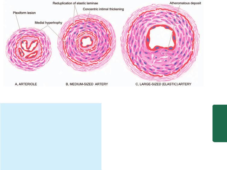
MORPHOLOGIC FEATURES. Irrespective of the type of pulmonary hypertension (primary or secondary), chronic cases invariably lead to cor pulmonale (Chapter 16). The pathologic changes are confined to the right side of the heart and pulmonary arterial tree in the lungs. There is hypertrophy of the right ventricle and dilatation of the right atrium. The vascular changes are similar in primary and secondary types and involve the entire arterial tree from the main pulmonary arteries down to the arterioles. These changes are as under (Fig. 17.5):
1. Arterioles and small pulmonary arteries: These branches show most conspicuous changes. These are as follows:
i) Medial hypertrophy.
467
Figure 17.5 
 Histologic changes in the pulmonary arterial branches of different sizes in pulmonary hypertension.
ii)Thickening and reduplication of elastic laminae.
iii)Plexiform pulmonary arteriopathy in which there is intraluminal tuft of capillary formation in dilated thinwalled arterial branches. These lesions are not so marked in secondary pulmonary hypertension.
2. Medium-sized pulmonary arteries:
i)Medial hypertrophy, which is not so marked in secondary pulmonary hypertension.
ii)Concentric intimal thickening.
iii)Adventitial fibrosis.
iv)Thickening and reduplication of elastic laminae.
3. Large pulmonary arteries: i) Atheromatous deposits.
PULMONARY INFECTIONS
Acute and chronic pulmonary infections are common at all ages and are a frequent cause of death. They are generally caused by a wide variety of microorganisms such as bacteria, viruses, fungi and mycoplasma. Important and common examples of acute pulmonary infectious diseases discussed
here are pneumonias, lung abscess and fungal infections, while pulmonary tuberculosis, generally regarded as an example of chronic lung infections, is discussed in Chapter 6.
PNEUMONIAS
Pneumonia is defined as acute inflammation of the lung parenchyma distal to the terminal bronchioles (consisting of the respiratory bronchiole, alveolar ducts, alveolar sacs and alveoli). The terms ‘pneumonia’ and ‘pneumonitis’ are often used synonymously for inflammation of the lungs, while ‘consolidation’ (meaning solidification) is the term used for gross and radiologic appearance of the lungs in pneumonia.
PATHOGENESIS. The microorganisms gain entry into the lungs by one of the following four routes:
1. Inhalation of the microbes present in the air.
2.Aspiration of organisms from the nasopharynx or oropharynx.
3.Haematogenous spread from a distant focus of infection.
4.Direct spread from an adjoining site of infection.
The normal lung is free of bacteria because of the presence of a number of lung defense mechanisms at different levels such as nasopharyngeal filtering action, mucociliary action of the lower respiratory airways, the presence of phagocytosing alveolar macrophages and immunoglobulins. Failure of these defense mechanisms and presence of certain predisposing factors result in pneumonias. These conditions are as under:
1.Altered consciousness. The oropharyngeal contents may be aspirated in states causing unconsciousness e.g. in coma, cranial trauma, seizures, cerebrovascular accidents, drug overdose, alcoholism etc.
2.Depressed cough and glottic reflexes. Depression of effective cough may allow aspiration of gastric contents e.g. in old age, pain from trauma or thoracoabdominal surgery, neuromuscular disease, weakness due to malnutrition, kyphoscoliosis, severe obstructive pulmonary diseases, endotracheal intubation and tracheostomy.
3.Impaired mucociliary transport. The normal protection offered by mucus-covered ciliated epithelium in the airways from the larynx to the terminal bronchioles is impaired or destroyed in many conditions favouring passage of bacteria into the lung parenchyma. These conditions are cigarette smoking, viral respiratory infections, immotile cilia syndrome, inhalation of hot or corrosive gases and old age.
4.Impaired alveolar macrophage function. Pneumonias may occur when alveolar macrophage function is impaired e.g. by cigarette smoke, hypoxia, starvation, anaemia, pulmonary oedema and viral respiratory infections.
5.Endobronchial obstruction. The effective clearance mechanism is interfered in endobronchial obstruction from tumour, foreign body, cystic fibrosis and chronic bronchitis.
System Respiratory The 17 CHAPTER

468 TABLE 17.1: Etiologic Classification of Pneumonias.
A. BACTERIAL PNEUMONIA
I. Lobar pneumonia
II. Bronchopneumonia (Lobular pneumonia)
B.VIRAL AND MYCOPLASMAL PNEUMONIA (PRIMARY ATYPICAL PNEUMONIA)
C.OTHER TYPES OF PNEUMONIAS
I. Pneumocystis carinii pneumonia
II. Legionella pneumonia (Legionnaire’s disease)
III. Aspiration (inhalation) pneumonia
IV. Hypostatic pneumonia
V.Lipid pneumonia
6.Leucocyte dysfunctions. Disorders of lymphocytes including congenital and acquired immunodeficiencies (e.g. AIDS, immunosuppressive therapy) and granulocyte abnormalities may predispose to pneumonia.
|
CLASSIFICATION. On the basis of the anatomic part of the |
||
|
lung parenchyma involved, pneumonias are traditionally |
||
|
classified into 3 main types: |
||
SECTION |
1. |
Lobar pneumonia |
|
2. |
Bronchopneumonia (or Lobular pneumonia) |
||
|
|||
|
3. |
Interstitial pneumonia. |
|
|
|
However, now that much is known about etiology and |
|
|
pathogenesis of pneumonias, current practice is to follow the |
||
III |
etiologic classification (Table 17.1) which divides pneu- |
||
monias into following 3 main groups: |
|||
|
|||
|
A. Bacterial pneumonia |
||
Systemic |
B. |
Viral pneumonia |
|
C. Pneumonias from other etiologies. |
|||
|
|||
|
|
In the present discussion, a combined approach of |
|
Pathology |
etiologic and morphologic classification will be followed. |
||
the lungs. Two types of acute bacterial pneumonias are |
|||
|
A. BACTERIAL PNEUMONIA |
||
Bacterial infection of the lung parenchyma is the most common cause of pneumonia or consolidation of one or both
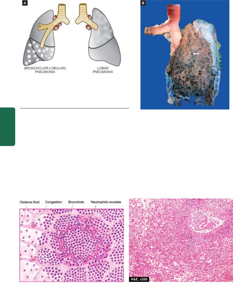
distinguished—lobar pneumonia and broncho-(lobular-) pneumonia, each with distinct etiologic agent and morphologic changes. Another type distinguished by some workers separately is confluent pneumonia which combines the features of both lobar and bronchopneumonia and involves larger (confluent) areas in both the lungs irregularly, while others consider this as a variant of bronchopneumonia.
Lobar Pneumonia
Lobar pneumonia is an acute bacterial infection of a part of a lobe, the entire lobe, or even two lobes of one or both the lungs.
ETIOLOGY. Based on the etiologic microbial agent causing lobar pneumonia, following types of lobar pneumonia are described:
1. Pneumococcal pneumonia. More than 90% of all lobar pneumonias are caused by Streptococcus pneumoniae, a lancet-shaped diplococcus. Out of various types,
type 3-S. pneumoniae causes particularly virulent form of lobar pneumonia. Pneumococcal pneumonia in majority of cases is community-acquired infection.
2.Staphylococcal pneumonia. Staphylococcus aureus causes pneumonia by haematogenous spread of infection from another focus or after viral infections.
3.Streptococcal pneumonia. β-haemolytic streptococci may rarely cause pneumonia such as in children after measles or influenza, in severely debilitated elderly patients and in diabetics.
4.Pneumonia by gram-negative aerobic bacteria. Less common causes of lobar pneumonia are gram-negative
bacteria like Haemophilus influenzae, Klebsiella pneumoniae
(Friedlander’s bacillus), Pseudomonas, Proteus and Escherichia coli, H. influenzae commonly causes pneumonia in children below 3 years of age after a preceding viral infection.
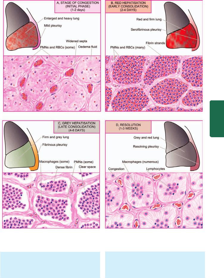
MORPHOLOGIC FEATURES. Laennec’s original description divides lobar pneumonia into 4 sequential pathologic phases: stage of congestion (initial phase), red hepatisation (early consolidation), grey hepatisation (late consolidation) and resolution. However, these classic stages seen in untreated cases are found much less often nowadays due to early institution of antibiotic therapy and improved medical care.
In lobar pneumonia, as the name suggests, part of a lobe, a whole lobe, or two lobes are involved, sometimes bilaterally. The lower lobes are affected most commonly. The sequence of pathologic changes described below represents the inflammatory response of lungs in bacterial infection.
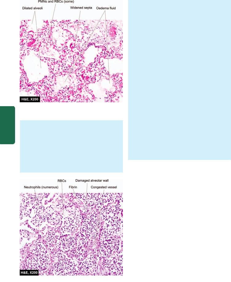
1. STAGE OF CONGESTION: INITIAL PHASE (Fig. 17.6,A). The initial phase represents the early acute inflammatory response to bacterial infection and lasts for 1 to 2 days.
Grossly, the affected lobe is enlarged, heavy, dark red and congested. Cut surface exudes blood-stained frothy fluid. Histologically, typical features of acute inflammatory response to the organisms are seen. These are as under
(Fig. 17.7):
i)Dilatation and congestion of the capillaries in the alveolar walls.
ii)Pale eosinophilic oedema fluid in the air spaces.
iii)A few red cells and neutrophils in the intra-alveolar fluid.
iv)Numerous bacteria demonstrated in the alveolar fluid by Gram’s staining.
2. RED HEPATISATION: EARLY CONSOLIDATION (Fig. 17.6,B). This phase lasts for 2 to 4 days. The term hepatisation in pneumonia refers to liver-like consistency of the affected lobe on cut section.
Grossly, the affected lobe is red, firm and consolidated. The cut surface of the involved lobe is airless, red-pink, dry, granular and has liver-like consistency. The stage of red hepatisation is accompanied by serofibrinous pleurisy. Histologically, the following features are observed
(Fig. 17.8):
469
System Respiratory The 17 CHAPTER
Figure 17.6 
 The four stages of lobar pneumonia, showing correlation of gross appearance of the lung with microscopic appearance in each stage. For details consult the text.
i)The oedema fluid of the preceding stage is replaced by strands of fibrin.
ii)There is marked cellular exudate of neutrophils and extravasation of red cells.
iii)Many neutrophils show ingested bacteria.
iv) The alveolar septa are less prominent than in the first stage due to cellular exudation.
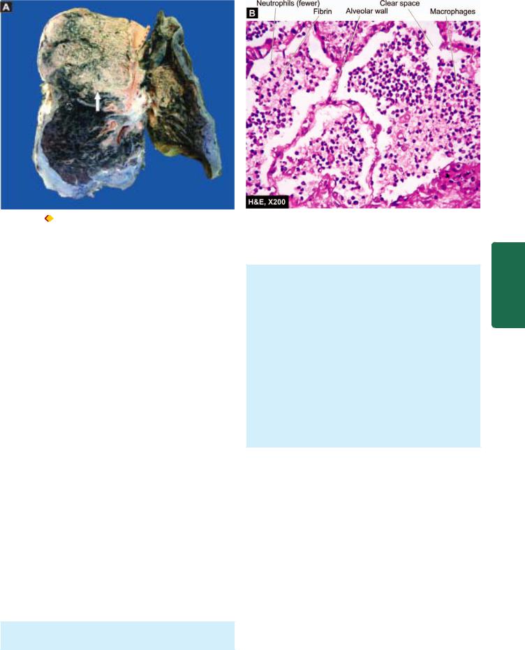
3. GREY HEPATISATION: LATE CONSOLIDATION (Fig. 17.6,C). This phase lasts for 4 to 8 days.
470
Pathology Systemic III SECTION
Figure 17.7 
 Lobar pneumonia, acute congestion stage. There is congestion of septal walls while the air spaces contain pale oedema fluid and a few red cells.
Grossly, the affected lobe is firm and heavy. The cut surface is dry, granular and grey in appearance with liverlike consistency (Fig. 17.9, A). The change in colour from red to grey begins at the hilum and spreads towards the periphery. Fibrinous pleurisy is prominent.
Histologically, the following changes are present
(Fig. 17.9,B):
i)The fibrin strands are dense and more numerous.
ii)The cellular exudate of neutrophils is reduced due to disintegration of many inflammatory cells as evidenced
Figure 17.8 
 Lobar pneumonia, red hepatisation stage. The alveoli are filled with cellular exudates composed of neutrophils admixed with some red cells.
by their pyknotic nuclei. The red cells are also fewer. The macrophages begin to appear in the exudate.
iii)The cellular exudate is often separated from the septal walls by a thin clear space.
iv)The organisms are less numerous and appear as degenerated forms.
4. RESOLUTION (Fig. 17.6,D). This stage begins by 8th to 9th day if no chemotherapy is administered and is completed in 1 to 3 weeks. However, antibiotic therapy induces resolution on about 3rd day. Resolution proceeds in a progressive manner.
Grossly, the previously solid fibrinous constituent is liquefied by enzymatic action, eventually restoring the normal aeration in the affected lobe. The process of softening begins centrally and spreads to the periphery. The cut surface is grey-red or dirty brown and frothy, yellow, creamy fluid can be expressed on pressing. The pleural reaction may also show resolution but may undergo organisation leading to fibrous obliteration of pleural cavity.
Histologically, the following features are noted:
i)Macrophages are the predominant cells in the alveolar spaces, while neutrophils diminish in number. Many of the macrophages contain engulfed neutrophils and debris.
ii)Granular and fragmented strands of fibrin in the alveolar spaces are seen due to progressive enzymatic digestion.
iii)Alveolar capillaries are engorged.
iv)There is progressive removal of fluid content as well as cellular exudate from the air spaces, partly by expectoration but mainly by lymphatics, resulting in restoration of normal lung parenchyma with aeration.
COMPLICATIONS. Since the advent of antibiotics, serious complications of lobar pneumonia are uncommon. However, they may develop in neglected cases and in patients with impaired immunologic defenses. These are as under:
1.Organisation. In about 3% of cases, resolution of the exudate does not occur but instead it undergoes organisation. There is ingrowth of fibroblasts from the alveolar septa resulting in fibrosed, tough, airless leathery lung tissue. This type of post-pneumonic fibrosis is called carnification.
2.Pleural effusion. About 5% of treated cases of lobar pneumonia develop inflammation of the pleura with effusion. The pleural effusion usually resolves but sometimes may undergo organisation with fibrous adhesions between visceral and parietal pleura.
3.Empyema. Less than 1% of treated cases of lobar pneumonia develop encysted pus in the pleural cavity termed empyema.
4.Lung abscess. A rare complication of lobar pneumonia is formation of lung abscess, especially when there is secondary infection by other organisms.
5.Metastatic infection. Occasionally, infection in the lungs and pleural cavity in lobar pneumonia may extend into the pericardium and the heart causing purulent pericarditis, bacterial endocarditis and myocarditis. Other forms of metastatic infection encountered rarely in lobar pneumonias
471
Figure 17.9 |
Lobar pneumonia, grey hepatisation stage. A, The sectioned surface of the lung shows grey-brown, firm area of consolidation |
(liver-like) affecting a lobe (arrow). B, The cellular exudates in the alveolar lumina is lying separated from the septal walls by a clear space. The infiltrate in the lumina is composed of neutrophils and macrophages.
are otitis media, mastoiditis, meningitis, brain abscess and purulent arthritis.
CLINICAL FEATURES. Classically, the onset of lobar pneumonia is sudden. The major symptoms are: shaking chills, fever, malaise with pleuritic chest pain, dyspnoea and cough with expectoration which may be mucoid, purulent or even bloody. The common physical findings are fever, tachycardia, and tachypnoea, and sometimes cyanosis if the patient is severely hypoxaemic. There is generally a marked neutrophilic leucocytosis. Blood cultures are positive in about 30% of cases. Chest radiograph may reveal consolidation. Culture of the organisms in the sputum and antibiotic sensitivity are most significant investigations for institution of specific antibiotics. The response to antibiotics is usually rapid with clinical improvement in 48 to 72 hours after the initiation of antibiotics.
Bronchopneumonia (Lobular Pneumonia)
Bronchopneumonia or lobular pneumonia is infection of the terminal bronchioles that extends into the surrounding alveoli resulting in patchy consolidation of the lung. The condition is particularly frequent at the extremes of life (i.e. in infancy and old age), as a terminal event in chronic debilitating diseases and as a secondary infection following viral respiratory infections such as influenza, measles etc.
ETIOLOGY. The common organisms responsible for bronchopneumonia are staphylococci, streptococci, pneumococci, Klebsiella pneumoniae, Haemophilus influenzae, and gram-negative bacilli like Pseudomonas and coliform bacteria.
MORPHOLOGIC FEATURES. Grossly, bronchopneumonia is identified by patchy areas of red or grey
consolidation affecting one or more lobes, frequently found bilaterally and more often involving the lower zones of the lungs due to gravitation of the secretions. On cut surface, these patchy consolidated lesions are dry, granular, firm, red or grey in colour, 3 to 4 cm in diameter, slightly elevated over the surface and are often centred around a bronchiole (Fig. 17.10). These patchy areas are best picked up by passing the fingertips on the cut surface. Histologically, the following features are observed
(Fig. 17.11):
i)Acute bronchiolitis.
ii)Suppurative exudate, consisting chiefly of neutrophils, in the peribronchiolar alveoli.
iii)Thickening of the alveolar septa by congested capillaries and leucocytic infiltration.
iv)Less involved alveoli contain oedema fluid.
COMPLICATIONS. The complications of lobar pneumonia may occur in bronchopneumonia as well. However, complete resolution of bronchopneumonia is uncommon. There is generally some degree of destruction of the bronchioles resulting in foci of bronchiolar fibrosis that may eventually cause bronchiectasis.
CLINICAL FEATURES. The patients of bronchopneumonia are generally infants or elderly individuals. There may be history of preceding bed-ridden illness, chronic debility, aspiration of gastric contents or upper respiratory infection. For initial 2 to 3 days, there are features of acute bronchitis but subsequently signs and symptoms similar to those of lobar pneumonia appear. Blood examination usually shows a neutrophilic leucocytosis. Chest radiograph shows mottled, focal opacities in both the lungs, chiefly in the lower zones.
The salient features of the two main types of bacterial pneumonias are contrasted in Table 17.2.
System Respiratory The 17 CHAPTER
472
Figure 17.10 
 A, Gross appearance of bronchopneumonia contrasted with that of lobar pneumonia. B, The pleural surface of the specimen of the lung shows serofibrinous exudate. The sectioned surface shows multiple, small, grey-brown, firm, patchy areas of consolidation around bronchioles
(arrow). while the intervening lung is spongy.
Pathology Systemic III SECTION
B.VIRAL AND MYCOPLASMAL PNEUMONIA (PRIMARY ATYPICAL PNEUMONIA)
Viral and mycoplasmal pneumonia is characterised by patchy inflammatory changes, largely confined to interstitial tissue of the lungs, without any alveolar exudate. Other terms used for these respiratory tract infections are interstitial pneumonitis, reflecting the interstitial location of the inflammation, and primary atypical pneumonia, atypicality being the absence of alveolar exudate commonly present in other pneumonias. Interstitial pneumonitis may occur in all ages. Most of the cases are mild and transient; exceptionally it may be severe and fulminant.
ETIOLOGY. Interstitial pneumonitis is caused by a wide variety of agents, the most common being respiratory syncytial virus (RSV). Others are Mycoplasma pneumoniae and many viruses such as influenza and parainfluenza viruses, adenoviruses, rhinoviruses, coxsackieviruses and cytomegaloviruses (CMV). Occasionally, psittacosis (Chlamydia) and Q fever (Coxiella) are associated with interstitial pneumonitis.
Infections of the respiratory tract with these organisms are quite common. In most cases, the infection remains confined to the upper respiratory tract presenting as common cold. Occasionally, it may extend lower down to involve the interstitium of the lungs. The circumstances favouring such
Figure 17.11 
 Microscopic appearance of bronchopneumonia. The bronchioles as well as the adjacent alveoli are filled with exudate consisting chiefly of neutrophils. The alveolar septa are thickened due to congested capillaries and neutrophilic infiltrate.
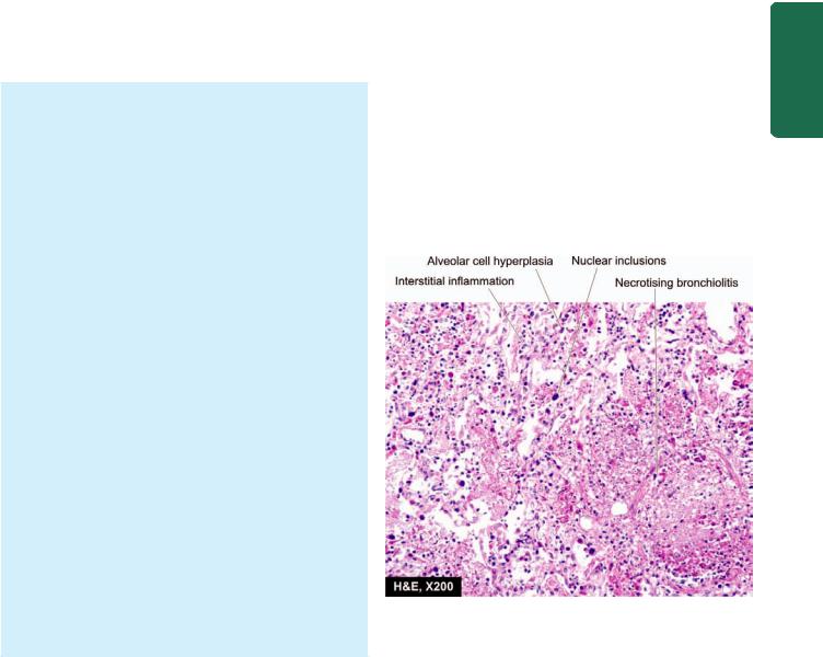
TABLE 17.2: Contrasting Features of Lobar Pneumonia and Bronchopneumonia. |
|
473 |
|||
|
Feature |
Lobar Pneumonia |
Bronchopneumonia |
|
|
1. |
Definition |
Acute bacterial infection of a part of a lobe |
Acute bacterial infection of the terminal |
||
|
|
of one or both lungs, or the entire lobe/s |
bronchioles extending into adjoining alveoli |
||
2. |
Age group |
More common in adults |
Commoner at extremes of age–infants and old age |
||
3. |
Predisposing factors |
More often affects healthy individuals |
Preexisting |
diseases e.g. chronic debility, terminal |
|
|
|
|
illness, flu, measles |
||
4. |
Common etiologic agents |
Pneumococci, Klebsiella pneumoniae, |
Staphylococci, streptococci, Pseudomonas, |
||
|
|
staphylococci, streptococci |
Haemophilus influenzae |
||
5. |
Pathologic features |
Typical case passes through stages of |
Patchy consolidation with central |
||
|
|
congestion (1-2 days) , early (2-4 days) and |
granularity, alveolar exudation, |
||
|
|
late consolidation (4-8 days), followed |
thickened septa |
||
|
|
by resolution (1-3 weeks) |
|
|
|
6. |
Investigations |
Neutrophilic leucocytosis, positive blood |
Neutrophilic leucocytosis, positive blood culture, |
||
|
|
culture, X-ray shows consolidation |
X-ray shows mottled focal opacities |
||
7. |
Prognosis |
Better response to treatment, resolution |
Response to treatment variable, |
||
|
|
common, prognosis good |
organisation may occur, prognosis poor |
||
8. |
Complications |
Less common; pleural effusion, empyema, |
Bronchiectasis may occur; other complications |
||
|
|
lung abscess, organisation |
same as for lobar pneumonia |
||
|
|
|
|
|
|
extension of infection are malnutrition, chronic debilitating diseases and alcoholism.
MORPHOLOGIC FEATURES. Irrespective of the etiologic agent, the pathologic changes are similar in all cases. Grossly, depending upon the severity of infection, the involvement may be patchy to massive and widespread consolidation of one or both the lungs. The lungs are heavy, congested and subcrepitant. Sectioned surface of the lung exudes small amount of frothy or bloody fluid. The pleural reaction is usually infrequent and mild.
Histologically, hallmark of viral pneumonias is the interstitial nature of the inflammatory reaction. The microscopic features are as under (Fig. 17.12):
i)Interstitial inflammation: There is thickening of alveolar walls due to congestion, oedema and mononuclear inflammatory infiltrate comprised by lymphocytes, macrophages and some plasma cells.
ii)Necrotising bronchiolitis: This is characterised by foci of necrosis of the bronchiolar epithelium, inspissated secretions in the lumina and mononuclear infiltrate in the walls and lumina.
iii)Reactive changes: The lining epithelial cells of the bronchioles and alveoli proliferate in the presence of virus and may form multinucleate giant cells and syncytia in the bronchiolar and alveolar walls. Occasionally, viral inclusions (intranuclear and/or intracytoplasmic) are found, especially in pneumonitis caused by CMV.
iv)Alveolar changes: In severe cases, the alveolar lumina may contain oedema fluid, fibrin, scanty inflammatory exudate and coating of alveolar walls by pink, hyaline membrane similar to the one seen in respiratory distress syndrome (page 462). Alveolar changes are prominent if bacterial infection supervenes.
COMPLICATIONS. The major complication of interstitial pneumonitis is superimposed bacterial infection and its complications. Most cases of interstitial pneumonitis recover completely. In more severe cases, there may be interstitial fibrosis and permanent damage.
CLINICAL FEATURES. Majority of cases of interstitial pneumonitis initially have upper respiratory symptoms with fever, headache and muscle-aches. A few days later appears dry, hacking, non-productive cough with retrosternal burning due to tracheitis and bronchitis. Blood film shows
Figure 17.12 
 Microscopic appearance of interstitial pneumonitis (viral pneumonia). There is necrotising bronchiolitis, reactive hyperplasia of alveolar epithelial cells, some having nuclear inclusions, and there is interstitial inflammation.
System Respiratory The 17 CHAPTER

474characteristic neutrophilic leucocytosis. Chest radiograph may show patchy or diffuse consolidation. Cold agglutinin titres in the serum are elevated in almost half the cases of mycoplasmal pneumonia, 20% cases of adenovirus infection but absent in other forms of viral pneumonia. Isolation of the etiologic agent, otherwise, is difficult.
C. OTHER TYPES OF PNEUMONIAS
Some other types of pneumonias caused by infective agents
(such as Pneumocystis carinii pneumonia and Legionella pneumonia) and certain non-infective varieties (e.g. aspiration pneumonia, hypostatic pneumonia and lipid pneumonia) are described here.
|
Pneumocystis carinii Pneumonia |
|
|
Pneumocystis carinii, a protozoon widespread in the |
|
|
environment, causes pneumonia by inhalation of the |
|
|
organisms as an opportunistic infection in neonates and |
|
|
immunosuppressed people. Almost 100% cases of HIV/ |
|
|
AIDS develop opportunistic infection during the course of |
|
|
disease, most commonly Pneumocystis carinii pneumonia. |
|
III SECTION |
Table 17.3 lists the various etiologic types of pneumonias |
|
associated with HIV infection due to profound immuno- |
||
suppression. Other immunosuppressed groups are patients |
||
on chemotherapy for organ transplant and tumours, |
||
malnutrition, agammaglobulinaemia etc. |
||
|
||
MORPHOLOGIC FEATURES. Grossly, the affected parts |
||
|
of the lung are consolidated, dry and grey. |
|
Systemic |
Microscopically, the features are as under: |
|
the organisms. |
||
|
i) |
Interstitial pneumonitis with thickening and |
|
mononuclear infiltration of the alveolar walls. |
|
|
ii) Alveolar lumina contain pink frothy fluid containing |
|
Pathology |
iii) By Gomori’s methenamine-silver (GMS) stain, the |
|
fluid. |
||
|
characteristic oval or crescentic cysts, about 5 μm in |
|
|
diameter and surrounded by numerous tiny black dot- |
|
|
like trophozoites of P. carinii are demonstrable in the frothy |
|
|
iv) No significant inflammatory exudate is seen in the air |
|
|
spaces. |
|
|
|
|
CLINICAL FEATURES. There is rapid onset of dyspnoea, tachycardia, cyanosis and non-productive cough. If untreated, it causes death in one or two weeks. Chest radiograph shows diffuse alveolar and interstitial infiltrate.
TABLE 17.3: Etiologic Types of HIV-Infection Associated Pneumonias.
1.Pneumocystis carinii
2.Cytomegalovirus
3.Mycobacterium avium-intracellulare
4.Mycobacterium tuberculosis
5.Streptococcus pneumoniae
6.Haemophilus influenzae
7.Invasive aspergillosis
8.Invasive candidiasis
Legionella Pneumonia
Legionella pneumonia or Legionnaire’s disease is an epidemic illness caused by gram-negative bacilli, Legionella pneumophila that thrives in aquatic environment. It was first recognised following investigation into high mortality among those attending American Legion Convention in Philadelphia in July 1976. The epidemic occurs in summer months by spread of organisms through contaminated drinking water or in airconditioning cooling towers. Impaired host defenses in the form of immunodeficiency, corticosteroid therapy, old age and cigarette smoking play important roles.
MORPHOLOGIC FEATURES. Grossly, there are changes of widespread bronchopneumonia involving many lobes and there may be consolidation of the entire lung. Pleural effusion is frequently present.
Histologically, the changes are not distinctive. Common features are as under:
i)Intra-alveolar exudate, initially of neutrophils but later composed mainly of macrophages.
ii)Alveolar septa show foci of hyperplasia of the lining epithelium and thrombosis of vessels in the septa.
iii)The organisms may be demonstrated in the macrophages by special stains or by immunofluorescent techniques.
CLINICAL FEATURES. The disease begins with malaise, headache and muscle-aches followed by high fever, chills, cough and tachypnoea. Systemic manifestations unrelated to pathologic changes in the lungs are seen due to bacteraemia and include abdominal pain, watery diarrhoea, proteinuria and mild hepatic dysfunction.
Aspiration (Inhalation) Pneumonia
Aspiration or inhalation pneumonia results from inhalation of different agents into the lungs. These substances include food, gastric contents, foreign body and infected material from oral cavity. A number of factors predispose to inhalation pneumonia which include: unconsciousness, drunkenness, neurological disorders affecting swallowing, drowning, necrotic oropharyngeal tumours, in premature infants and congenital tracheo-oesophageal fistula. Some patients die immediately from asphyxiation or laryngospasm without developing pneumonia.
MORPHOLOGIC FEATURES. Pathologic changes vary depending upon the particulate matter aspirated but in general right lung is affected more often due to direct path from the main bronchus:
1. Aspiration of small amount of sterile foreign matter such as acidic gastric contents produce chemical pneumonitis. It is characterised by haemorrhagic pulmonary oedema with presence of particles in the bronchioles. Patients rapidly develop cyanosis, dyspnoea, shock and bloody sputum and are often likely to die of cardiac failure. If the patient survives the acute episode, secondary bacterial infection is likely to occur.
2. Non-sterile aspirate causes widespread bronchopneumonia with multiple areas of necrosis and suppuration. A granulomatous reaction with foreign body giant cells may surround the aspirated vegetable matter.
Hypostatic Pneumonia
Hypostatic pneumonia is the term used for collection of oedema fluid and secretions in the dependent parts of the lungs in severely debilitated, bed-ridden patients. The accumulated fluid in the basal zone and posterior part of lungs gets infected by bacteria from the upper respiratory tract and sets in bacterial pneumonia. Hypostatic pneumonia is a common terminal event in the old, feeble, comatose patients.
Lipid Pneumonia
Another variety of non-infective pneumonia is lipid pneumonia which is of 2 types: exogenous and endogenous.
1.Exogenous lipid pneumonia. This is caused by aspiration of a variety of oily materials. These are: inhalation of oily nasal drops, regurgitation of oily medicines from stomach (e.g. liquid paraffin), administration of oily vitamin preparation to reluctant children or to debilitated old patients.
2.Endogenous lipid pneumonia. Endogenous origin of lipids causing pneumonic consolidation is more common. The sources of origin are tissue breakdown following obstruction to airways e.g. obstruction by bronchogenic cancer, tuberculosis and bronchiectasis.
MORPHOLOGIC FEATURES. Grossly, the exogenous lipid pneumonia affects the right lung more frequently due to direct path from the main bronchus. Quite often, the lesions are bilateral. The affected part of the lungs is consolidated. Cut surface is characteristically ‘golden yellow’.
Microscopically, the features are as under:
i)Lipid is finely dispersed in the cytoplasm of macrophages forming foamy macrophages within the alveolar spaces.
ii)There may be formation of cholesterol clefts due to liberation of cholesterol and other lipids.
iii)Formation of granulomas with foreign body giant cells may be seen around the large lipid droplets.
LUNG ABSCESS
Lung abscess is a localised area of necrosis of lung tissue with suppuration. It is of 2 types (Fig. 17.13):
 Primary lung abscess that develops in an otherwise normal lung. The commonest cause is aspiration of infected material.

 Secondary lung abscess that develops as a complication of some other disease of the lung or from another site.
ETIOPATHOGENESIS. The microorganisms commonly isolated from the lungs in lung abscess are streptococci, staphylococci and various gram-negative organisms. These
475
Figure 17.13 |
Common locations of lung abscess. A, Primary lung |
|
|
abscess—mostly single, large, commonly due to aspiration, located most |
|
||
frequently in the lower part of right upper lobe or apex of right lower lobe. |
|
||
B, Secondary lung abscesses—mostly multiple, small, most commonly |
|
||
post-pneumonic or following septic embolism. |
|
||
are introduced into the lungs from one of the following |
CHAPTER |
||
mechanisms: |
|
||
1. Aspiration of infected foreign material. A number of |
|||
|
|||
foreign materials such as food, decaying teeth, gastric |
|
||
contents, severely infected gingivae and teeth, and necrotic |
|
||
tissue from lesions in the mouth, upper respiratory tract or |
|
||
nasopharynx may be aspirated. This occurs particularly in |
17 |
||
favourable circumstances such as during sleep, |
|||
unconsciousness, anaesthesia, general debility and acute |
|
||
alcoholism. |
|
The |
|
2. Preceding bacterial infection. Preceding broncho- |
|||
pneumonia in a debilitated patient may develop into lung |
Respiratory |
||
abscess. Other infective conditions like tuberculosis, |
|||
|
|||
bronchiectasis and mycotic infections may occasionally result |
|
||
in formation of lung abscess. |
|
||
3. Bronchial obstruction. An abscess may form distal to an |
|
||
obstructed bronchus such as from bronchial tumour or from |
System |
||
impacted foreign body. |
|||
4. Septic embolism. Infected emboli originating from |
|||
pyaemia, thrombophlebitis or from vegetative bacterial |
|||
endocarditis may be disseminated in the venous circulation and reach the right side of the heart from where they are lodged in the lung and result in multiple abscesses.
5. Miscellaneous. Rarely lung abscesses may occur from following causes:
i)Infection in pulmonary infarcts.
ii)Amoebic abscesses due to infection with Entamoeba
histolytica.
iii)Trauma to the lungs.
iv)Direct extension from a suppurative focus in the mediastinum, oesophagus, subphrenic area or spine.
MORPHOLOGIC FEATURES. Abscesses due to aspiration are more likely to be in right lung due to more vertical main bronchus and are frequently single. They are commonly located in the lower part of the right upper lobe or apex of right lower lobe. Abscesses developing
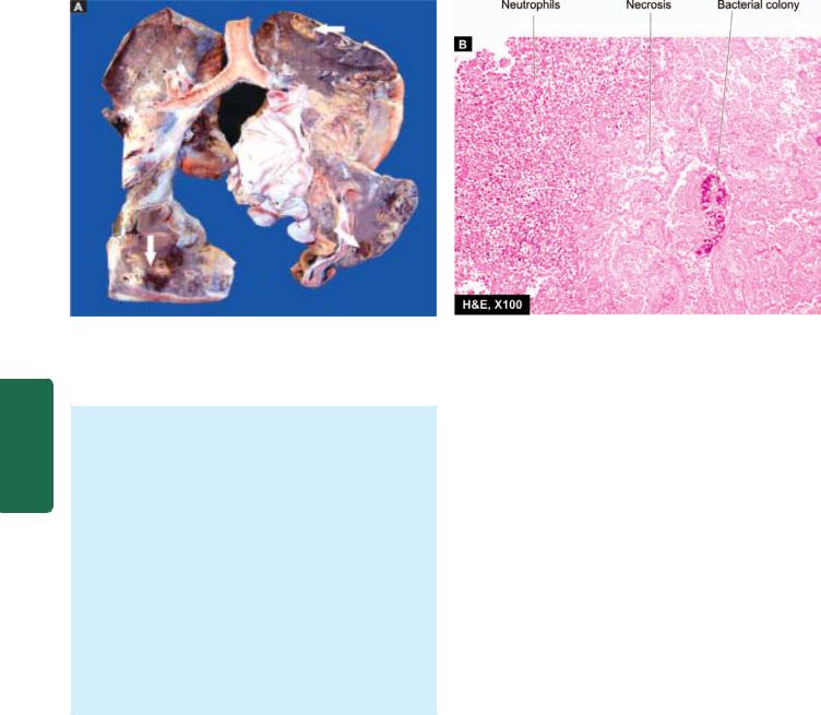
476
Pathology Systemic III SECTION
Figure 17.14 
 Lung abscess. A, The pleura is thickened. Cut surface of the lung shows multiple cavities 1-4 cm in diameter, having irregular and ragged inner walls (arrow). The lumina contain necrotic debris. The surrounding lung parenchyma is consolidated. B, The photomicrograph shows abscess formed by necrosed alveoli and dense acute and chronic inflammatory cells.
from preceding pneumonia and septic or pyaemic abscesses are often multiple and scattered throughout the lung.
Grossly, abscesses may be of variable size from a few millimeters to large cavities, 5 to 6 cm in diameter. The cavity often contains exudate. An acute lung abscess is initially surrounded by acute pneumonia and has poorlydefined ragged wall. With passage of time, the abscess becomes chronic and develops fibrous wall (Fig. 17.14,A).
Histologically, the characteristic feature is the destruction of lung parenchyma with suppurative exudate in the lung cavity. The cavity is initially surrounded by acute inflammation in the wall but later there is replacement by chronic inflammatory cell infiltrate composed of lymphocytes, plasma cells and macrophages. In more chronic cases, there is considerable fibroblastic proliferation forming a fibrocollagenic wall (Fig. 17.14,B).
CLINICAL FEATURES. The clinical manifestations are fever, malaise, loss of weight, cough, purulent expectoration and haemoptysis in half the cases. Clubbing of the fingers and toes appears in about 20% of patients. Secondary amyloidosis may occur in chronic long-standing cases.
FUNGAL INFECTIONS OF LUNG
Fungal infections of the lung are more common than tuberculosis in the US. These infections in healthy individuals are rarely serious but in immunosuppressed individuals may prove fatal. General aspects of mycotic infections are covered in Chapter 7. Here, some common examples of fungal infections of the lung are briefly given:
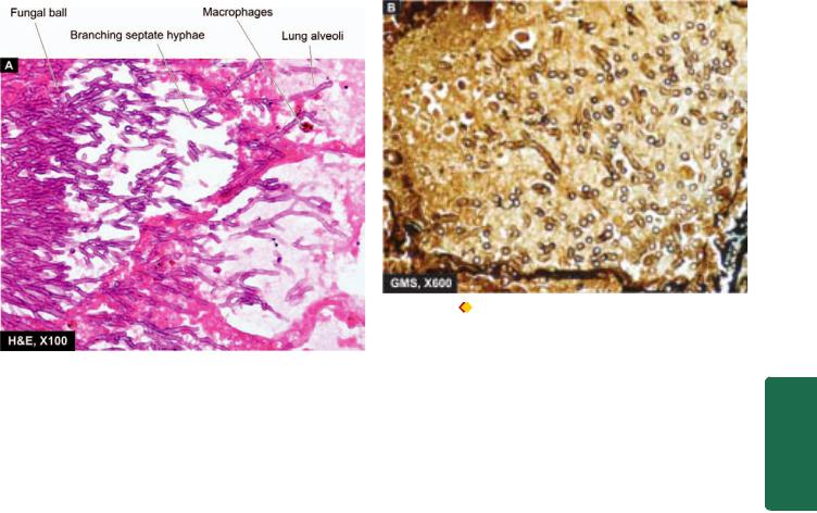
1. Aspergillosis. Aspergillosis is the most common fungal infection of the lung caused by Aspergillus fumigatus that grows best in cool, wet climate. The infection may result in
allergic bronchopulmonary aspergillosis, aspergilloma and necrotising bronchitis. Immunocompromised persons develop more serious manifestations of aspergillus infection, especially in leukaemic patients on cytotoxic drug therapy and HIV/AIDS. Extensive haematogenous spread of aspergillus infection may result in widespread changes in lung tissue due to arterial occlusion, thrombosis and infarction.
Grossly, pulmonary aspergillosis may occur within preexisting pulmonary cavities or in bronchiectasis as fungal ball.
Microscopically, the fungus may appear as a tangled mass within the cavity. The organisms are identified by their characteristic morphology— thin septate hyphae with dichotomous branching at acute angles which stain positive for fungal stains such as PAS and silver impregnation technique (Fig. 17.15). The wall of the cavity shows chronic inflammatory cells.
2.Mucormycosis. Mucormycosis or phycomycosis is caused by Mucor and Rhizopus. The infection in the lung occurs in a similar way as in aspergillosis. The pulmonary lesions are especially common in patients of diabetic ketoacidosis. Mucor is distinguished by its broad, non-parallel, nonseptate hyphae which branch at an obtuse angle. Mucormycosis is more often angioinvasive, and disseminates; hence it is more destructive than aspergillosis.
3.Candidiasis. Candidiasis or moniliasis caused by Candida albicans is a normal commensal in oral cavity, gut and vagina but attains pathologic form in immunocompromised host. Angioinvasive growth of the organism may occur in the airways.
4.Histoplasmosis. It is caused by oval organism, Histoplasma capsulatum, by inhalation of infected dust or bird droppings. The condition may remain asymptomatic or may produce lesions similar to the Ghon’s complex.
5.Cryptococcosis. It is caused by Cryptococcus neoformans which is round yeast having a halo around it due to shrinkage in tissue sections. The infection occurs from infection by inhalation of pigeon droppings. The lesions in the body may range from a small parenchymal granuloma in the lung to cryptococcal meningitis.
6.Coccidioidomycosis. Coccidioidomycosis is caused by Coccidioides immitis which are spherical spores. The infection in human beings is acquired by close contact with infected dogs. The lesions consist of peripheral parenchymal granuloma in the lung.
7.Blastomycosis. It is an uncommon condition caused by Blastomyces dermatitidis. The lesions result from inhalation of spores in the ground. Pathological features may present as Ghon’s complex-like lesion, as a pneumonic consolidation, and as multiple skin nodules.
PULMONARY TUBERCULOSIS
The classical and most common example of chronic infection of the lungs is pulmonary tuberculosis. Pulmonary lesions caused by Mycobacterium tuberculosis and other mycobacteria have already been discussed along with general aspects of tuberculosis and other granulomatous inflammations in Chapter 6.
CHRONIC OBSTRUCTIVE PULMONARY DISEASE
Chronic obstructive pulmonary disease (COPD) or chronic obstructive airway disease (COAD) are commonly used clinical terms for a group of pathological conditions in which there is chronic, partial or complete, obstruction to the airflow at any level from trachea to the smallest airways resulting in functional disability of the lungs i.e. they are diffuse lung diseases. The following 4 entities are included in COPD:
I. Chronic bronchitis
477
Figure 17.15 |
Aspergillosis lung. A, Acute angled septate hyphae |
|
||
lying in necrotic debris and acute inflammatory exudates in lung abscess. |
|
|||
B, Organisms, Apergillus flavus, are best identified with a special stain |
|
|||
for fungi, Gomory’s methenamine silver (GMS). |
|
|
||
Chronic bronchitis and emphysema are quite common |
CHAPTER |
|||
II. Emphysema |
|
|||
III. Bronchial asthma |
|
|||
IV. Bronchiectasis |
|
|||
and often occur together. Now, small airways disease |
17 |
|||
involving inflammation of small bronchi and bronchioles |
||||
|
||||
(bronchiolitis) has also been added to the group of COPD. |
The |
|||
CHRONIC BRONCHITIS |
||||
Chronic bronchitis is a common condition defined clinically |
Respiratory |
|||
as persistent cough with expectoration on most days for at |
||||
|
||||
least three months of the year for two or more consecutive |
|
|||
years. The cough is caused by oversecretion of mucus. In |
|
|||
spite of its name, chronic inflammation of the bronchi is not |
|
|||
a prominent feature. The condition is more common in |
System |
|||
middle-aged males than females; approximately 20% of adult men and 5% of adult women have chronic bronchitis, but only a minority of them develop serious disabling COPD or cor pulmonale. Quite frequently, chronic bronchitis is associated with emphysema.
ETIOPATHOGENESIS. The two most important etiologic factors responsible for majority of cases of chronic bronchitis are: cigarette smoking and atmospheric pollution. Other contributory factors are occupation, infection, familial and genetic factors.
1. Smoking. The most commonly identified factor implicated in causation of chronic bronchitis and in emphysema is heavy smoking. Heavy cigarette smokers have 4 to 10 times higher proneness to develop chronic bronchitis. Prolonged cigarette smoking appears to act on the lungs in a number of ways:
i)It impairs ciliary movement.
ii)It inhibits the function of alveolar macrophages.
iii)It leads to hypertrophy and hyperplasia of mucussecreting glands.
478iv) It causes considerable obstruction of small airways.
v)It stimulates the vagus and causes bronchoconstriction.
2. Atmospheric pollution. The incidence of chronic bronchitis is higher in industrialised urban areas where air is polluted. Some of the atmospheric pollutants which increase the risk of developing chronic bronchitis are sulfur dioxide, nitrogen dioxide, particulate dust and toxic fumes.
3. Occupation. Workers engaged in certain occupations such as in cotton mills (byssinosis), plastic factories etc. are exposed to various organic or inorganic dusts which contribute to disabling chronic bronchitis in such individuals.
4. Infection. Bacterial, viral and mycoplasmal infections do not initiate chronic bronchitis but usually occur secondary to bronchitis. Cigarette smoke, however, predisposes to infection responsible for acute exacerbation in chronic bronchitis.
|
5. Familial and genetic factors. There appears to be a |
|
|
poorly-defined familial tendency and genetic predisposition |
|
|
to develop disabling chronic bronchitis. However, it is more |
|
|
likely that nonsmoker family members who remain in the |
|
|
air-pollution of home are significantly exposed to smoke |
|
SECTION |
(passive smoking) and hence have increased blood levels of |
|
of the bronchi and bronchioles may contain mucus plugs |
||
|
carbon monoxide. |
|
|
MORPHOLOGIC FEATURES. Grossly, the bronchial |
|
|
wall is thickened, hyperaemic and oedematous. Lumina |
|
III |
and purulent exudate. |
|
Microscopically, just as there is clinical definition, there |
||
|
||
Systemic |
is histologic definition of chronic bronchitis by increased |
|
Reid index. Reid index is the ratio between thickness of |
||
|
||
|
the submucosal mucus glands (i.e. hypertrophy and |
|
|
hyperplasia) in the cartilage-containing large airways to |
|
|
that of the total bronchial wall (Fig. 17.16). The increase |
|
Pathology |
in thickness can be quantitatively assessed by micrometer |
|
lens. The bronchial epithelium may show squamous |
||
|
||
|
metaplasia and dysplasia. There is little chronic |
|
|
inflammatory cell infiltrate. The non-cartilage containing |
|
|
small airways show goblet cell hyperplasia and intra- |
|
|
luminal and peribronchial fibrosis. |
|
|
|
CLINICAL FEATURES. There is considerable overlap of clinical features of chronic bronchitis and pulmonary emphysema (discussed below) as quite often the two coexist. The contrasting features of ‘predominant emphysema’ and ‘predominant bronchitis’ are presented in Table 17.5. Some important features of ‘predominant bronchitis’ are as under:
1.Persistent cough with copious expectoration of long duration; initially beginning in a heavy smoker with ‘morning catarrh’ or ‘throat clearing’ which worsens in winter.
2.Recurrent respiratory infections are common.
3.Dyspnoea is generally not prominent at rest but is more on exertion.
4.Patients are called ‘blue bloaters’ due to cyanosis and oedema.
5.Features of right heart failure (cor pulmonale) are common.
6.Chest X-ray shows enlarged heart with prominent vessels.
Figure 17.16 
 Diagrammatic representation of increased Reid’s index in chronic bronchitis.
EMPHYSEMA
The WHO has defined pulmonary emphysema as combination of permanent dilatation of air spaces distal to the terminal bronchioles and the destruction of the walls of
dilated air spaces. Thus, emphysema is defined morphologically,
while chronic bronchitis is defined clinically. Since the two conditions coexist frequently and show considerable overlap in their clinical features, it is usual to label patients as ‘predominant emphysema’ and ‘predominant bronchitis’.
CLASSIFICATION. As mentioned in the beginning of this chapter, a lobule is composed of about 5 acini distal to a terminal bronchiole and that an acinus consists of 3 to 5 generations of respiratory bronchioles and a variable number of alveolar ducts and alveolar sacs (page 461). As per WHO definition of pulmonary emphysema, it is classified according to the portion of the acinus involved, into 5 types: centriacinar, panacinar (panlobular), para-septal (distal acinar), irregular (para-cicatricial) and mixed (unclassified) emphysema. A number of other conditions to which the term ‘emphysema’ is loosely applied are, in fact, examples of ‘overinflation’. A classification based on these principles is outlined in Table 17.4.
TABLE 17.4: Classification of ‘True Emphysema’ and ‘Overinflation’.
A.TRUE EMPHYSEMA
1.Centriacinar (centrilobular) emphysema
2.Panacinar (panlobular) emphysema
3.Paraseptal (distal acinar) emphysema
4.Irregular (para-cicatricial) emphysema
5.Mixed (unclassified) emphysema
B. OVERINFLATION
1.Compensatory overinflation (compensatory emphysema)
2.Senile hyperinflation (aging lung, senile emphysema)
3.Obstructive overinflation (infantile lobar emphysema)
4.Unilateral translucent lung (unilateral emphysema)
5.Interstitial emphysema (surgical emphysema)

TABLE 17.5: Contrasting Salient Features of ‘Predominant Bronchitis’ and ‘Predominant Emphysema’. |
479 |
|||
|
Feature |
Predominant Bronchitis |
Predominant Emphysema |
|
|
|
|
|
|
1. |
Age at diagnosis |
About 50 years |
About 60 years |
|
2. |
Underlying pathology |
Hypertrophy of mucus-producing |
Inflammatory narrowing of bronchioles and |
|
|
|
cells |
destruction of septal walls |
|
3. |
Dyspnoea |
Late, mild |
Early, severe |
|
4. |
Cough |
Before dyspnoea starts |
After dyspnoea starts |
|
5. |
Sputum |
Copious, purulent |
Scanty, mucoid, less frequent |
|
6. |
Bronchial infections |
More frequent |
Less frequent |
|
7. |
Respiratory insufficiency |
Repeated |
Terminal |
|
8. |
Cyanosis |
Common (‘blue-bloaters’) |
Rare (‘pink-puffers’) |
|
9. |
Lung capacity |
Normal |
Increased (barrel-chest) |
|
10. |
Blood gas values |
↓ pO2, ↓ pCO2, no compensatory |
pO2 and pCO2 usually within normal limits due |
|
|
|
hyperventilation |
to compensatory hyperventilation |
|
11. |
Cor pulmonale |
Frequent |
Rare and terminal |
|
12. |
Chest X-ray |
Large heart, prominent vessels |
Small heart, hyperinflated lungs |
|
|
|
|
|
|
ETIOPATHOGENESIS. The commonest form of COPD is the combination of chronic bronchitis and pulmonary emphysema. Chronic bronchitis, however, does not always lead to emphysema nor all cases of emphysema have changes of chronic bronchitis. The association of the two conditions is principally linked to the common etiologic factors— most importantly tobacco smoke and air pollutants. Other less significant contributory factors are occupational exposure, infection and somewhat poorly-understood familial and genetic influences. All these factors have already been discussed above.
However, pathogenesis of the most significant event in emphysema, the destruction of the alveolar walls, is not linked to bronchial changes but is closely related to deficiency of serum alpha-1-antitrypsin (α1-protease inhibitor) commonly termed protease-antiprotease hypothesis detailed below.
Protease-antiprotease hypothesis. Alpha-1-antitrypsin (α- 1-AT), also called α1-protease inhibitor (α-1-Pi), is a glycoprotein that forms the normal constituent of the α1- globulin fraction of the plasma proteins on serum electrophoresis. The single gene locus that codes for α-1-AT is located on the long arm of chromosome 15. It is normally synthesised in the liver and is distributed in the circulating blood, tissue fluids and macrophages. The normal function of α1-AT is to inhibit proteases and hence its name α1- protease inhibitor. The proteases (mainly elastases) are derived from neutrophils. Neutrophil elastase has the capability of digesting lung parenchyma but is inhibited from doing so by anti-elastase effect of α1-AT.
There are several known alleles of α1-AT which have an autosomal codominant inheritance pattern and are classified as normal (PiMM), deficient (PiZZ), null type (Pi null null) having no detectable level, and dysfunctional (PiSS) type having about half the normal level.

 The most common abnormal phenotype in classic α1-AT deficiency is homozygous state PiZZ resulting from a single amino acid substitution Glu→Lys which causes spontaneous polymerisation of α1-AT and inhibits its release from the liver. The remaining material of α1-AT in the liver causes hepatic cirrhosis (Chapter 22).

 Clinically significant deficiency is also associated with
homozygous Pi null null and heterozygous Pi nullZ.
The heterozygote pattern of PiMZ has intermediate levels which is not sufficient to produce clinical deficiency, but heterozygote individuals who smoke heavily have higher risk of developing emphysema.
The α1-AT deficiency develops in adults and causes pulmonary emphysema in smokers as well as in nonsmokers, though the smokers become symptomatic about 15 years earlier than non-smokers. The other organ showing effects of α1-AT deficiency is the liver which may develop obstructive jaundice early in infancy, and cirrhosis and hepatoma late in adulthood (Chapter 22).
The mechanism of alveolar wall destruction in emphysema by elastolytic action is based on the imbalance between proteases (chiefly elastase) and anti-proteases (chiefly
anti-elastase):

 By decreased anti-elastase activity i.e. deficiency of α-1 antitrypsin.

 By increased activity of elastase i.e. increased neutrophilic infiltration in the lungs causing excessive elaboration of neutrophil elastase.
There are enough evidences to suggest that smoking promotes emphysema by both decreasing the amount of antielastase as well as by increasing the elastolytic protease in the lungs. These are as under:
1. Oxidant in cigarette smoke has inhibitory influence on α-1-antitrypsin, thus lowering the level of anti-elastase activity.
System Respiratory The 17 CHAPTER

480
|
Figure 17.17 |
Pathogenesis of alveolar wall destruction in |
|
|
emphysema by protease-antiprotease mechanism. |
||
SECTION |
2. Smokers have up to ten times more phagocytes and |
||
neutrophils in their lungs than nonsmokers; thus they have |
|||
very high elastase activity. |
|||
Pathogenesis of emphysema by protease-antiprotease |
|||
mechanism is diagrammatically illustrated in Fig. 17.17. |
|||
III |
|
||
PATHOLOGIC FEATURES. Emphysema can be |
|||
|
diagnosed with certainty only by gross and histologic |
||
PathologySystemic |
examination of sections of whole lung. The lungs should |
||
be perfused with formalin under pressure in inflated state |
|||
|
|||
|
to grade the severity of emphysema with naked eye. |
||
|
|
|
|
Grossly, the lungs are voluminous, pale with little blood. The edges of the lungs are rounded. Mild cases show dilatation of air spaces visible with hand lens. Advanced cases show subpleural bullae and blebs bulging outwards from the surface of the lungs with rib markings between them. The bullae are air-filled cyst-like or bubble-like structures, larger than 1 cm in diameter (Fig. 17.18). They are formed by the rupture of adjacent air spaces while blebs are the result of rupture of alveoli directly into the subpleural interstitial tissue and are the common cause of spontaneous pneumothorax.
Microscopically, depending upon the type of emphysema, there is dilatation of air spaces and destruction of septal walls of part of acinus involved i.e. respiratory bronchioles, alveolar ducts and alveolar sacs. Changes of bronchitis may be present. Bullae and blebs when present show fibrosis and chronic inflammation of the walls.
CLINICAL FEATURES. Cases of ‘predominant emphysema’ develop clinical features after about one-third of the pulmonary parenchyma is damaged which occurs most severely in panacinar emphysema. The age at the time of diagnosis is often a decade later (about 60 years) than the age for predominant bronchitis (about 50 years). Though there is considerable overlap between the clinical features of chronic bronchitis and emphysema, the following features generally characterise ‘predominant emphysema’ (Table 17.5):
1.There is long history of slowly increasing severe exertional dyspnoea.
2.Patient is quite distressed with obvious use of accessory muscles of respiration.
Figure 17.18 |
Bullous emphysema. A, Diagrammatic view as seen |
on the external surface of the lung. B, Sectioned lung showing air-filled sacs.
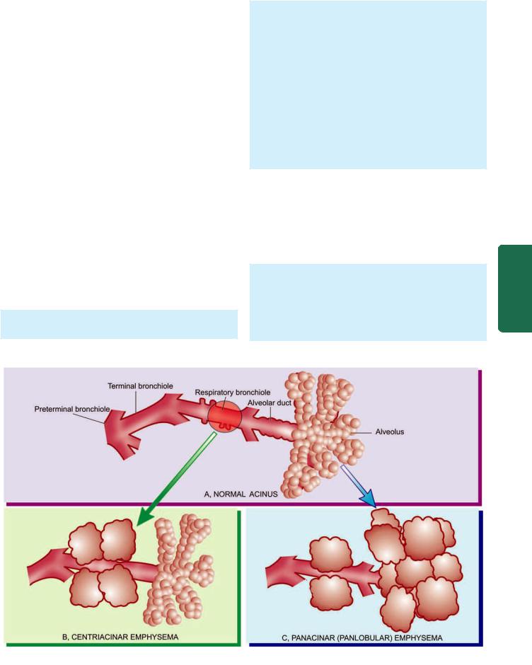
3.Chest is barrel-shaped and hyperresonant.
4.Cough occurs late after dyspnoea starts and is associated with scanty mucoid sputum.
5.Recurrent respiratory infections are not frequent.
6.Patients are called ‘pink puffers’ as they remain well oxygenated and have tachypnoea.
7.Weight loss is common.
8.Features of right heart failure (cor pulmonale) and hypercapneic respiratory failure are the usual terminal events.
9.Chest X-ray shows small heart with hyperinflated lungs.
After these general comments about morphologic and clinical features of emphysema, the specific pathologic changes in individual types of ‘emphysema’ and ‘overinflation’ as classified in Table 17.4 are described below.
Morphology of Individual Types of Emphysema
1. CENTRIACINAR (CENTRILOBULAR) EMPHYSEMA.
Centriacinar or centrilobular emphysema is one of the common types. It is characterised by initial involvement of respiratory bronchioles i.e. the central or proximal part of the acinus (Fig. 17.19,B). This is the type of emphysema that usually coexists with chronic bronchitis and occurs predominantly in smokers and in coal miners’ pneumoconiosis (page 488).
Grossly, the lesions are more common and more severe in the upper lobes of the lungs. The characteristic
appearance is obvious in cut surface of the lung. It shows distended air spaces in the centre of the lobules surrounded by a rim of normal lung parenchyma in the same lobule. The lobules are separated from each other by fine fibrous tissue septa. Large amount of black pigment is often present in the walls of emphysematous spaces. In more severe cases, distal parts of acini are also involved and the appearance may closely resemble panacinar emphysema.
Microscopically, there is distension and destruction of the respiratory bronchiole in the centre of lobules, surrounded peripherally by normal uninvolved alveoli. The terminal bronchioles supplying the acini show chronic inflammation and are narrowed.
2. PANACINAR (PANLOBULAR) EMPHYSEMA.
Panacinar or panlobular emphysema is the other common type. In this type, all portions of the acinus are affected but not of the entire lung (Fig. 17.19,C). Panacinar emphysema is most often associated with α1-antitrypsin deficiency in middle-aged smokers and is the one that produces the most characteristic anatomical changes in the lung in emphysema.
Grossly, in contrast to centriacinar emphysema, the panacinar emphysema involves lower zone of lungs more frequently and more severely than the upper zone. The involvement may be confined to a few lobules, or may be more widespread affecting a lobe or part of a lobe of the lung. The lungs are enlarged and overinflated.
481
System Respiratory The 17 CHAPTER
Figure 17.19 
 The anatomic regions of involvement in an acinus in major forms of emphysema.
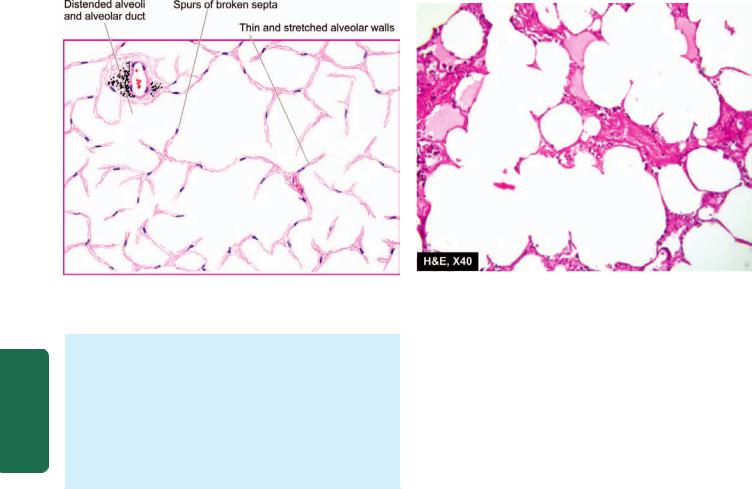
482
Figure 17.20 
 Panacinar (Panlobular) emphysema showing involvement of the entire lobules and whole of acinus.
Pathology Systemic III SECTION
Microscopically, usually all the alveoli within a lobule are affected to the same degree. All portions of acini are distended—respiratory bronchioles, alveolar ducts and alveoli, are all dilated and their walls stretched and thin. Ruptured alveolar walls and spurs of broken septa are seen between the adjacent alveoli. The capillaries are stretched and thinned. Special stains show loss of elastic tissue. Inflammatory changes are usually absent
(Fig. 17.20).
3.PARASEPTAL (DISTAL ACINAR) EMPHYSEMA. This type of emphysema involves distal part of acinus while the proximal part is normal. Paraseptal or distal acinar emphysema is localised along the pleura and along the perilobular septa. The involvement is seen adjacent to the areas of fibrosis and atelectasis and involves upper part of lungs more severely than the lower. This form of emphysema is seldom associated with COPD but is the common cause of spontaneous pneumothorax in young adults. Grossly, the subpleural portion of the lung shows air-filled cysts, 0.5 to
2cm in diameter.
4.IRREGULAR (PARA-CICATRICIAL) EMPHYSEMA.
This is the most common form of emphysema, seen surrounding scars from any cause. The involvement is irregular as regards the portion of acinus involved as well as within the lung as a whole. During life, irregular emphysema is often asymptomatic and may be only an incidental autopsy finding.
5.MIXED (UNCLASSIFIED) EMPHYSEMA. Quite often, the same lung may show more than one type of emphysema. It is usually due to more severe involvement resulting in loss of clearcut distinction between one type of emphysema and the other. Thus, the lungs of an elderly smoker at autopsy may show continuation of centriacinar emphysema in the upper lobes, panacinar in the lower lobes, and paraseptal emphysema in the subpleural region.
Morphology of Types of Overinflation
Under this heading are covered a group of lung conditions of heterogeneous etiology characterised by overinflation of the parts of acini but without significant destruction of the walls and are sometimes loosely termed emphysema.
1.COMPENSATORY OVERINFLATION (COMPENSATORY EMPHYSEMA). When part of a lung or a lobe of lung is surgically removed, the residual lung parenchyma undergoes compensatory hyperinflation so as to fill the pleural cavity. Histologic examination shows dilatation of alveoli but no destruction of septal walls and hence the term compensatory overinflation is preferable over ‘compensatory emphysema’.
2.SENILE HYPERINFLATION (AGING LUNG, SENILE EMPHYSEMA). In old people, the lungs become voluminous due to loss of elastic tissue, thinning and atrophy of the alveolar ducts and alveoli. The alveoli are thin-walled and distended throughout the lungs but there is no significant destruction of the septal walls and, therefore, preferable designation is ‘senile hyperinflation’ over ‘senile emphysema.’
3.OBSTRUCTIVE OVERINFLATION (INFANTILE LOBAR EMPHYSEMA). Partial obstruction to the bronchial tree such as by a tumour or a foreign body causes overinflation of the region supplied by obstructed bronchus. Infantile lobar emphysema is a variant of obstructive overinflation occurring in infants in the first few days of life who develop respiratory distress or who have congenital hypoplasia of bronchial cartilage. In all such cases, air enters the lungs during inspiration but cannot leave on expiration resulting in ballooning up of the affected part of the lung.
4.UNILATERAL TRANSLUCENT LUNG (UNILATERAL EMPHYSEMA). This is a form of overinflation in which one lung or one of its lobes or segments of a lobe are radiolucent. The condition occurs in adults and there is generally a history of serious pulmonary infection in childhood, probably bronchiolitis obliterans. The affected

lung is grossly overinflated. Microscopy shows overinflated alveoli and there is histologic evidence of preceding widespread bronchiolitis obliterans.
5. INTERSTITIAL EMPHYSEMA (SURGICAL EMPHYSEMA). The entry of air into the connective tissue framework of the lung is called interstitial or surgical emphysema. The usual sources of entry of air into stroma of the lung are rupture of alveoli or of larger airways. The causes are as under:
i)Violent coughing with bronchiolar obstruction e.g. in children with whooping cough, bronchitis, in patients with obstruction to the airways by foreign bodies, blood clots and exposure to irritant gases.
ii)Rupture of the oesophagus, trauma to the lung, or major bronchus and trachea.
iii)Entry of air through surgical incision.
iv)Fractured rib puncturing the lung parenchyma.
v)Sudden change in atmospheric pressure e.g. in decompression sickness.
The condition may affect patients of all ages. On rupture of alveoli, the leaked air enters the fibrous connective tissue of the alveolar walls from where it extends into the fibrous septa of the lung, into the mediastinum, the pleura, and even the subcutaneous tissues. Escape of air into the pleural cavity may cause pneumothorax. Collection of small quantities of air is generally harmless and is resorbed. However, extensive accumulation of air in surgical emphysema may produce impaired blood flow in the lungs. Pneumo-mediastinum may produce symptoms resembling myocardial infarction.
Histologically, the alveoli are distended but septal walls are not damaged; therefore it is not true emphysema. There are clear spaces of leaked out air in connective tissue septa.
BRONCHIAL ASTHMA
Asthma is a disease of airways that is characterised by increased responsiveness of the tracheobronchial tree to a variety of stimuli resulting in widespread spasmodic narrowing of the air passages which may be relieved spontaneously or by therapy. Asthma is an episodic disease manifested clinically by paroxysms of dyspnoea, cough and
wheezing. However, a severe and unremitting form of the disease termed status asthmaticus may prove fatal.
Bronchial asthma is common and prevalent worldwide; in the United States about 4% of population is reported to suffer from this disease. It occurs at all ages but nearly 50% of cases develop it before the age of 10 years. In adults, both sexes are affected equally but in children there is 2:1 malefemale ratio.
ETIOPATHOGENESIS AND TYPES. Based on the stimuli initiating bronchial asthma, two broad etiologic types are traditionally described: extrinsic (allergic, atopic) and intrinsic
(idiosyncratic, non-atopic) asthma. A third type is a mixed pattern in which the features do not fit clearly into either of the two main types. The contrasting features of the two main types are summed up in Table 17.6.
1. Extrinsic (atopic, allergic) asthma. This is the most common type of asthma. It usually begins in childhood or in early adult life. Most patients of this type of asthma have personal and/or family history of preceding allergic diseases such as rhinitis, urticaria or infantile eczema. Hypersensitivity to various extrinsic antigenic substances or ‘allergens’ is usually present in these cases. Most of these allergens cause ill-effects by inhalation e.g. house dust, pollens, animal danders, moulds etc. Occupational asthma stimulated by fumes, gases and organic and chemical dusts is a variant of extrinsic asthma. There are increased levels of IgE in the serum and positive skin test with the specific offending inhaled antigen representing an IgE-mediated type I hypersensitivity reaction which includes an ‘acute immediate response’ and a ‘late phase reaction’:
Acute immediate response is initiated by IgE-sensitised mast cells (tissue counterparts of circulating basophils) on the mucosal surface. Mast cells on degranulation release mediators like histamine, leukotrienes, prostaglandins, platelet activating factor and chemotactic factors for eosinophils and neutrophils. The net effects of these mediators are bronchoconstriction, oedema, mucus hypersecretion and accumulation of eosinophils and neutrophils.
Late phase reaction follows the acute immediate response and is responsible for the prolonged manifestations of asthma. It is caused by excessive mobilisation of blood leucocytes that include basophils besides eosinophils and
483
System Respiratory The 17 CHAPTER
TABLE 17.6: Contrasting Features of the Two Major Types of Asthma.
|
Feature |
Extrinsic Asthma |
Intrinsic Asthma |
|
|
|
|
1. |
Age at onset |
In childhood |
In adult |
2. |
Personal/family history |
Commonly present |
Absent |
3. |
Preceding allergic illness (atopy) |
Present (e.g. rhinitis, urticaria, eczema) |
Absent |
4. |
Allergens |
Present (dust, pollens, danders etc) |
None |
5. |
Drug hypersensitivity |
None |
Present (usually to aspirin) |
6. |
Serum IgE levels |
Elevated |
Normal |
7. |
Associated chronic bronchitis, nasal polyps |
Absent |
Present |
8. |
Emphysema |
Unusual |
Common |
|
|
|
|

484neutrophils. These result in further release of mediators which accentuate the above-mentioned effects. In addition, inflammatory injury is caused by neutrophils and by major basic protein (MBP) of eosinophils.
2.Intrinsic (idiosyncratic, non-atopic) asthma. This type of asthma develops later in adult life with negative personal or family history of allergy, negative skin test and normal serum levels of IgE. Most of these patients develop typical symptom-complex after an upper respiratory tract infection by viruses. Associated nasal polypi and chronic bronchitis are commonly present. There are no recognisable allergens but about 10% of patients become hypersensitive to drugs, most notably to small doses of aspirin (aspirin-sensitive asthma).
3.Mixed type. Many patients do not clearly fit into either of the above two categories and have mixed features of both. Those patients who develop asthma in early life have strong allergic component, while those who develop the disease late tend to be non-allergic. Either type of asthma can be precipitated by cold, exercise and emotional stress.
SECTION |
MORPHOLOGIC FEATURES. The pathologic changes |
|
are similar in both major types of asthma. The pathologic |
||
|
||
|
material examined is generally autopsy of lungs in |
|
|
patients dying of status asthmaticus but the changes are |
|
|
expected to be similar in non-fatal cases. |
|
III |
Grossly, the lungs are overdistended due to over-inflation. |
|
The cut surface shows characteristic occlusion of the |
||
|
bronchi and bronchioles by viscid mucus plugs. |
|
Systemic |
Microscopically, the following changes are observed |
|
Curschmann’s spirals. |
||
|
(Fig. 17.21): |
|
|
1. The mucus plugs contain normal or degenerated |
|
|
respiratory epithelium forming twisted strips called |
|
Pathology |
2. The sputum usually contains numerous eosinophils |
|
membrane of the bronchial epithelium, submucosal |
||
|
and diamond-shaped crystals derived from eosinophils |
|
|
called Charcot-Leyden crystals. |
|
|
3. The bronchial wall shows thickened basement |
|
|
|
Figure 17.21 
 Diagrammatic appearance of Curschmann’s spiral and
Charcot-Leyden crystals found in mucus plugs in patients with bronchial asthma.
oedema and inflammatory infiltrate consisting of lymphocytes and plasma cells with prominence of eosinophils. There is hypertrophy of submucosal glands as well as of the bronchial smooth muscle.
4. Changes of bronchitis and emphysema may supervene, especially in intrinsic asthma.
CLINICAL FEATURES. Asthmatic patients suffer from episodes of acute exacerbations interspersed with symptomfree periods. Characteristic clinical features are paroxysms of dyspnoea, cough and wheezing. Most attacks typically last for a few minutes to hours. When attacks occur continuously it may result in more serious condition called status asthmaticus. The clinical diagnosis is supported by demonstration of circulation eosinophilia and sputum demonstration of Curschmann’s spirals and Charcot-Leyden crystals. More chronic cases may develop cor pulmonale.
BRONCHIECTASIS
Bronchiectasis is defined as abnormal and irreversible dilatation of the bronchi and bronchioles (greater than 2 mm in diameter) developing secondary to inflammatory weakening of the bronchial walls. The most characteristic clinical manifestation of bronchiectasis is persistent cough with expectoration of copious amounts of foul-smelling, purulent sputum. Post-infectious cases commonly develop in childhood and in early adult life.
ETIOPATHOGENESIS. The origin of inflammatory destructive process of bronchial walls is nearly always a result of two basic mechanisms: endobronchial obstruction and infection.
Endobronchial obstruction by foreign body, neoplastic growth or enlarged lymph nodes causes resorption of air distal to the obstruction with consequent atelectasis and retention of secretions.
Infection may be secondary to local obstruction and impaired systemic defense mechanism promoting bacterial growth, or infection may be a primary event i.e. bronchiectasis developing in suppurative necrotising pneumonia.
These 2 mechanisms—endobronchial obstruction and infection, are seen in a number of clinical settings as under:
1. Hereditary and congenital factors. Several hereditary and congenital factors may result secondarily in diffuse bronchiectasis:
i)Congenital bronchiectasis caused by developmental defect of the bronchial system.
ii)Cystic fibrosis, a generalised defect of exocrine gland secretions, results in obstruction, infection and bronchiectasis (Chapter 22).
iii)Hereditary immune deficiency diseases are often associated with high incidence of bronchiectasis.
iv)Immotile cilia syndrome that includes Kartagener’s syndrome (bronchiectasis, situs inversus and sinusitis) is characterised by ultrastructural changes in the microtubules causing immotility of cilia of the respiratory tract epithelium, sperms and other cells. Males in this syndrome are often infertile (Chapter 23).
Figure 17.22 
 Types of bronchial dilatations in bronchiectasis.
v) Atopic bronchial asthma patients have often positive family history of allergic diseases and may rarely develop diffuse bronchiectasis.
2.Obstruction. Post-obstructive bronchiectasis, unlike the congenital-hereditary forms, is of the localised variety, usually confined to one part of the bronchial system. The causes of endobronchial obstruction include foreign bodies, endobronchial tumours, compression by enlarged hilar lymph nodes and post-inflammatory scarring (e.g. in healed tuberculosis) all of which favour the development of postobstructive bronchiectasis.
3.As secondary complication. Necrotising pneumonias such as in staphylococcal suppurative pneumonia and tuberculosis may develop bronchiectasis as a complication.
MORPHOLOGIC FEATURES. The disease characteristically affects distal bronchi and bronchioles beyond the segmental bronchi.
Grossly, the lungs may be involved diffusely or segmentally. Bilateral involvement of lower lobes occurs most frequently. More vertical air passages of left lower lobe are more often involved than the right. The pleura is usually fibrotic and thickened with adhesions to the chest wall. The dilated airways, depending upon their gross or bronchographic appearance, have been subclassified into the following different types (Fig. 17.22):
i)Cylindrical: the most common type characterised by tube-like bronchial dilatation.
ii)Fusiform: having spindle-shaped bronchial dilatation.
iii)Saccular: having rounded sac-like bronchial distension.
iv)Varicose: having irregular bronchial enlargements.
Cut surface of the affected lobes, generally the lower zones, shows characteristic honey-combed appearance. The bronchi are extensively dilated nearly to the pleura, their walls are thickened and the lumina are filled with mucus or muco-pus. The intervening lung parenchyma is reduced and fibrotic (Fig. 17.23).
Microscopically, fully-developed cases show the following histologic features (Fig. 17.24):
i)The bronchial epithelium may be normal, ulcerated or may show squamous metaplasia.
ii)The bronchial wall shows infiltration by acute and chronic inflammatory cells and destruction of normal muscle and elastic tissue with replacement by fibrosis.
iii)The intervening lung parenchyma shows fibrosis, while the surrounding lung tissue shows changes of interstitial pneumonia.
iv)The pleura in the affected area is adherent and shows bands of fibrous tissue between the bronchus and the pleura.
485
System Respiratory The 17 CHAPTER
Figure 17.23 
 Bronchiectasis of the lung. Sectioned surface shows honey-combed appearance of the lung in the lower lobe where many thickwalled dilated cavities with cartilaginous wall are seen (arrow).
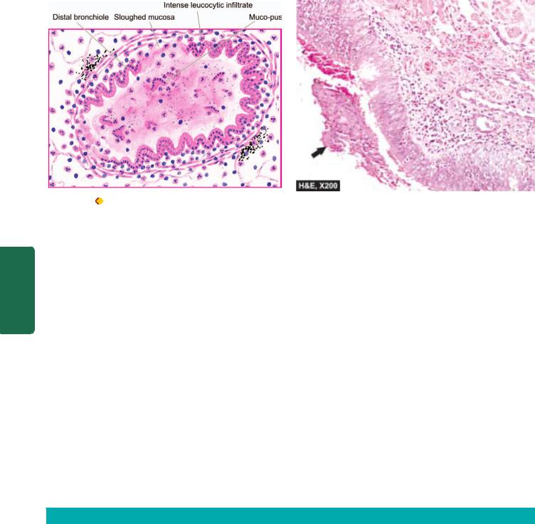
486
Pathology Systemic III SECTION
Figure 17.24 |
Microscopic appearance of a dilated distal bronchiole in bronchiectasis. The bronchial wall is thickened and infiltrated by acute |
and chronic inflammatory cells. The mucosa is sloughed off at places with exudate of muco-pus in the lumen (arrow).
CLINICAL FEATURES. The clinical manifestations of bronchiectasis typically consist of chronic cough with foulsmelling sputum production, haemoptysis and recurrent pneumonia. Sinusitis is a common accompaniment of diffuse bronchiectasis. Late complications occurring in cases uncontrolled for years include development of clubbing of the fingers, metastatic abscesses (often to the brain), amyloidosis and cor pulmonale.
CHRONIC RESTRICTIVE PULMONARY DISEASE
The second large group of diffuse lung disease is ‘chronic restrictive pulmonary disease’ characterised by reduced expansion of lung parenchyma with decreased total lung capacity. This group of diseases must be distinguished from the foregoing COPD (Table 17.7).
Restrictive lung disease includes 2 types of conditions:
A. Restriction due to chest wall disorder: It includes following conditions:
1.Kyphoscoliosis
2.Poliomyelitis
3.Severe obesity
4.Pleural diseases.
As a group, these conditions cause restriction to expansion of lungs due to alterations in chest wall, pleura and neuromuscular apparatus, but are primarily not lung parenchymal diseases.
B. Restriction due to interstitial and infiltrative diseases.
Commonly called as ‘interstitial lung diseases (ILDs)’, these are diseases characterised by non-infectious diffuse parenchymal involvement of the lung i.e. the alveolar lumina and alveolar epithelium, capillary basement membrane, the intervening space, perivascular tissue and lymphatic tissue. Diffuse lung parenchymal involvement may be primary, or it may be involved secondarily as a part of some other multiorgan disease process. The term ‘infiltrative’ is used here to denote the radiologic appearance of lungs in chest radiographs which show characteristic diffuse interstitial ground-glass opacities. The ILDs consist of more than 200 heterogeneous conditions which have common clinical, radiologic, and functional manifestations but diverse pathological features. As a group, ILDs have high mortality
TABLE 17.7: Obstructive versus Restrictive Pulmonary Diseases.
|
Feature |
Obstructive |
Restrictive |
|
|
|
|
|
|
1. |
Airways |
Obstructed at any level from |
Reduced expansion of lung parenchyma |
|
|
|
trachea to respiratory bronchiole |
|
|
2. |
Pulmonary |
Increased pulmonary resistance and |
Decreased total lung capacity |
|
|
function test |
obstruction of maximal expiratory airflow |
|
|
3. |
X-ray chest |
Variable appearance depending |
Typically bilateral infiltrates giving ground-glass |
|
|
|
upon the cause |
shadows |
|
4. |
Examples |
• |
Chronic bronchitis |
• Chest cage disorders (e.g. kyphoscoliosis, poliomyelitis, severe |
|
|
• |
Emphysema |
obesity and pleural disease) |
|
|
• |
Bronchial asthma |
• Interstitial lung diseases (ILDs) (e.g. pneumoconioses, |
|
|
• |
Bronchiectasis |
idiopathic pulmonary fibrosis, immunologic lung diseases, |
|
|
|
|
collagen-vascular disease and sarcoidosis) |
|
|
|
|
|
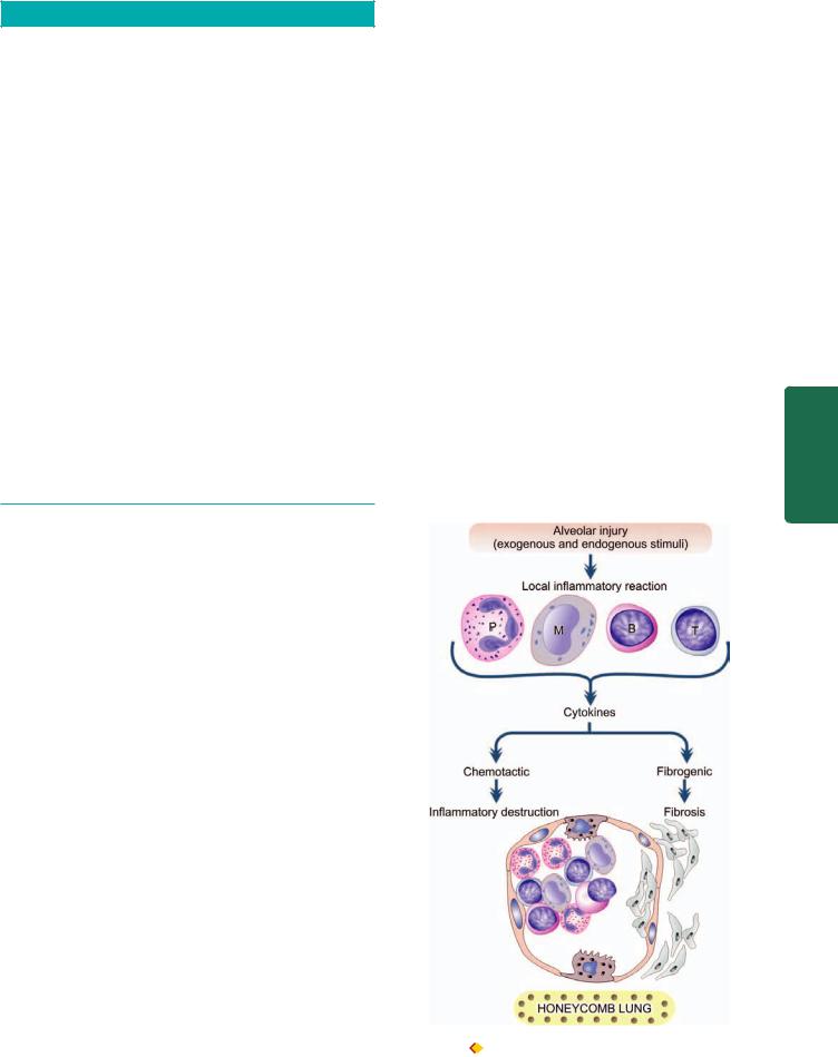
TABLE 17.8. Classification of Interstitial Lung Diseases (ILDs).
I.WITH PREDOMINANT FIBROSIS
1.Pneumoconiosis with inorganic minerals: coal, asbestos, fumes, gases
2.Idiopathic pulmonary fibrosis (Idiopathic interstitial pneumonias)
3.Nonspecific interstitial pneumonia
4.Acute interstitial pneumonia (Hamman-Rich syndrome)
5.Cryptogenic organising pneumonia
6.Therapy related-ILD (radiation, antibiotics, chemotherapy)
7.Connective tissue diseases
8.Residual effects of ARDS
II.WITH PREDOMINANT GRANULOMATOUS REACTION
1.Sarcoidosis (Chapter 6)
2.Pneumoconiosis with inorganic dusts: silica, beryllium
3.Granulomatous vasculitis
4.Wegener’s granulomatosis
III.IMMUNOLOGIC LUNG DISEASES (EOSINOPHILIC PNEUMONIAS)
1.Hypersensitivity pneumonitis: with organic dusts
2.Pulmonary infiltrates with eosinophilia (PIE)
3.Pulmonary haemorrhage syndromes (Goodpasture’s syndrome)
4.Pulmonary alveolar proteinosis
IV. SMOKINGASSOCIATED ILDs
1.Desquamative interstitial pneumonia (DIP)
2.Respiratory bronchiolitis-associated ILD
3.Pulmonary Langerhans cell histiocytosis (eosinophilic granuloma of the lung)
and morbidity. Depending upon the underlying pathologic findings, ILDs have been broadly classified into 2 groups, each further subdivided into those with known and unknown causes:

 Conditions with predominant non-specific inflammation (alveolitis, interstitial inflammation, and fibrosis).

 Conditions with predominant granulomatous inflammation.
Based on this, an abbreviated classification of ILDs is given in Table 17.8.
The exact pathogenesis of ILDs from injury to fibrosis is not known. However, it can be explained on immune basis as under (Fig. 17.25):
i)There is local inflammatory reaction in the alveoli in response to various exogenous and endogenous stimuli in the form of lymphocytes (both B and T) and macrophages.
ii)Activated macrophages cause recruitment of neutrophils and also produce fibrogenic and chemotactic cytokines. Neutrophils liberate proteases and oxidants which injure the type I pneumocytes resulting in initial microscopic alveolitis, while cytokines cause subsequent proliferation of type II pneumocytes and fibrosis.
iii)The result is inflammatory destruction of the pulmonary parenchyma followed by fibrosis. Eventually, there is widespread destruction of alveolar capillary walls resulting in end-stage lung or ‘honeycomb lung’.
The major common clinical manifestations of restrictive 487 lung diseases are exertional dyspnoea, non-persistent productive cough, tachypnoea, cyanosis and sometimes haemoptysis but no wheezing so characteristic of COPD.
Important and common examples of ILDs are discussed below.
PNEUMOCONIOSES
Pneumoconiosis is the term used for lung diseases caused by inhalation of dust, mostly at work (pneumo = lung; conis = dust in Greek). These diseases are, therefore, also called ‘dust diseases’ or ‘occupational lung diseases’.
The type of lung disease varies according to the nature of inhaled dust. Some dusts are inert and cause no reaction and no damage at all, while others cause immunologic damage and predispose to tuberculosis or to neoplasia. The factors which determine the extent of damage caused by inhaled dusts are as under:
1. |
size and shape of the particles; |
|
2. |
their solubility and physicochemical composition; |
|
3. |
the amount of dust retained in the lungs; |
|
4. |
the additional effect of other irritants such as tobacco |
CHAPTER |
5 μm reach the terminal airways where they are ingested by |
||
smoke; and |
|
|
5. |
host factors such as efficiency of clearance mechanism |
|
and immune status of the host. |
|
|
|
In general, most of the inhaled dust particles larger than |
|
|
|
17 |
|
|
System Respiratory The |
Figure 17.25 Schematic evolution of interstitial lung disease (ILD).

488
Pathology Systemic III SECTION
TABLE 17.9: Classification of Pneumoconioses.
|
Agent |
Diseases |
|
|
|
A. INORGANIC (MINERAL) DUSTS |
|
|
1. |
Coal dust |
Simple coal-workers’ |
|
|
pneumoconiosis |
|
|
Progressive massive |
|
|
fibrosis |
|
|
Caplan’s syndrome |
2. |
Silica |
Silicosis |
|
|
Caplan’s syndrome |
3. |
Asbestos |
Asbestosis |
|
|
Pleural diseases |
|
|
Tumours |
4. |
Beryllium |
Acute berylliosis |
|
|
Chronic berylliosis |
5. |
Iron oxide |
Pulmonary siderosis |
B. |
ORGANIC (BIOLOGIC) DUSTS |
|
1. |
Mouldy hay |
Farmer’s lungs |
2. |
Bagasse |
Bagassosis |
3. |
Cotton, flax, hemp dust |
Byssinosis |
4. |
Bird droppings |
Bird-breeders’ (bird |
|
|
fancier’s) lung |
5. |
Mushroom compost dust |
Mushroom-workers’ lung |
6. |
Mouldy barley, malt dust |
Malt-workers’ lung |
7. |
Mouldy maple bark |
Maple-bark disease |
8. |
Silage fermentation |
Silo-fillers’ disease |
|
|
|
alveolar macrophages. Most of these too are eliminated by expectoration but the remaining accumulate in alveolar tissue. Of particular interest are the particles smaller than 1 μm which are deposited in the alveoli most efficiently. Most of the dust-laden macrophages accumulated in the alveoli die leaving the dust, around which fibrous tissue is formed. Some macrophages enter the lymphatics and reach regional lymph nodes. The tissue response to inhaled dust may be one of the following three types:
 Fibrous nodules e.g. in coal-workers’ pneumoconiosis and silicosis.

 Interstitial fibrosis e.g. in asbestosis.

 Hypersensitivity reaction e.g. in berylliosis.
A comprehensive list of various types of occupational lung diseases caused by inorganic (mineral) dusts and organic dusts is presented in Table 17.9. The more common examples of pneumoconioses are described here.
Coal-Workers’ Pneumoconiosis
This is the commonest form of pneumoconiosis and is defined as the lung disease resulting from inhalation of coal dust particles, especially in coal-miners engaged in handling soft bituminous coal for a number of years, often 20 to 30 years. It exists in 2 forms—a milder form of the disease called simple coal workers’ pneumoconiosis and an advanced form termed progressive massive fibrosis (complicated coal-miners’ pneumoconiosis). Anthracosis, on the other hand, is not a lung disease in true sense but is the common, benign and
asymptomatic accumulation of carbon dust in the lungs of most urban dwellers due to atmospheric pollution and cigarette smoke (anthacite refers to coal). Anthracotic pigment is deposited in the macrophages in the alveoli and around the respiratory bronchioles and into the draining lymph nodes but does not produce any respiratory difficulty or radiologic changes.
PATHOGENESIS. Pathogenetically, it appears that anthracosis, simple coal-workers’ pneumoconiosis and progressive massive fibrosis are different stages in the evolution of fully-developed coal-workers’ pneumoconiosis. However, progressive massive fibrosis develops in a small proportion of cases (2-8%) of simple coal-workers’ pneumoconiosis. A number of predisposing factors have been implicated in this transformation as follows:
1.Older age of the miners.
2.Severity of coal dust burden engulfed by macrophages.
3.Prolonged exposure (20 to 30 years) to coal dust.
4.Concomitant tuberculosis.
5.Additional role of silica dust.
Activation of alveolar macrophage plays the most
significant role in the pathogenesis of progressive massive fibrosis by release of various mediators (Fig. 17.26,A):
i)Free radicals which are reactive oxygen species which damage the lung parenchyma.
ii)Chemotactic factors for various leucocytes (leukotrienes, TNF, IL-8 and IL-6) resulting in infiltration into pulmonary tissues by these inflammatory cells which on activation cause further damage.
iii)Fibrogenic cytokines such as IL-1, TNF and platelet derived growth factor (PDGF) which stimulate healing by fibrosis due to proliferation of fibroblasts at the damaged tissue site.
MORPHOLOGIC FEATURES. In life, the pathologic changes in lung in coal-workers’ pneumoconiosis are graded by radiologic appearance according to the size and extent of opacities. The pathologic findings at autopsy of lungs in the major forms of coal-workers’ pneumoconiosis are considered below under 3 headings: simple coalworkers’ pneumoconiosis, progressive massive fibrosis and rheumatoid pneumoconiosis (Caplan’s syndrome).
SIMPLE COAL-WORKERS’ PNEUMOCONIOSIS.
Grossly, the lung parenchyma shows small, black focal lesions, measuring less than 5 mm in diameter and evenly distributed throughout the lung but have a tendency to be more numerous in the upper lobes. These are termed coal macules, and if palpable are called nodules. The air spaces around coal macules are dilated with little destruction of alveolar walls (Fig. 17.26,A). Though some workers have called it centrilobular emphysema of coalminers (page 481), others prefer not to consider it emphysema because there is no significant destruction of alveolar walls. Similar blackish pigmentations are found on the pleural surface and in the regional lymph nodes
(Fig. 17.27).
Histologically, the following features are seen (Fig. 17.28): 1. Coal macules are composed of aggregates of dustladen macrophages. These are present in the alveoli and in the bronchiolar and alveolar walls.
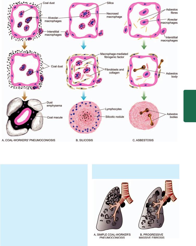
Figure 17.26 
 Pathogenesis of three common forms of pneumoconiosis. A, Coal-workers’ pneumoconiosis. The macrophages phagocytose large amount of coal dust particles which are then passed into the interstitial tissue of the lung and aggregate around respiratory bronchiole and cause focal dust emphysema. B, Silicosis. The tiny silica particles are toxic to macrophages. The dead macrophages release fibrogenic factor and eventually result in silicotic nodule. C, Asbestosis. Asbestos fibres initiate lot of interstitial fibrosis and also form asbestos bodies.
2.There is some increase in the network of reticulin and collagen in the coal macules.
3.Respiratory bronchioles and alveoli surrounding the macules are distended without significant destruction of the alveolar walls.
PROGRESSIVE MASSIVE FIBROSIS. Grossly, besides the coal macules and nodules of simple pneumoconiosis, there are larger, hard, black scattered areas measuring more than 2 cm in diameter and sometimes massive. They are usually bilateral and located more often in the upper parts of the lungs posteriorly. Sometimes, these masses break down centrally due to ischaemic necrosis or due to tuberculosis forming cavities filled with black semifluid resembling India ink. The pleura and the regional lymph nodes are also blackened and fibrotic (Fig. 17.27).
Histologically, the following features are present:
1. The fibrous lesions are composed almost entirely of dense collagen and carbon pigment.
2. The wall of respiratory bronchioles and pulmonary vessels included in the massive scars are thickened and their lumina obliterated.
Figure 17.27 
 Gross appearance of the lungs in simple coal-workers’ pneumoconiosis (A) and progressive massive fibrosis (B).
489
System Respiratory The 17 CHAPTER

490
Pathology Systemic III SECTION
Figure 17.28 
 Histologic appearance of the lung in coal-workers’ pneumoconiosis. Coal macules composed of aggregates of dust-laden macrophages and collagens are seen surrounding respiratory bronchioles. The alveoli and respiratory bronchioles surrounding the coal macule are distended.
3.There is scanty inflammatory infiltrate of lymphocytes and plasma cells around the areas of massive scars.
4.The alveoli surrounding the scars are markedly dilated.
Progressive massive fibrosis probably has immunological pathogenetic basis as described above.
RHEUMATOID PNEUMOCONIOSIS (CAPLAN’S SYNDROME). The development of rheumatoid arthritis in a few cases of coal-workers’ pneumoconiosis, silicosis or asbestosis is termed rheumatoid pneumoconiosis or Caplan’s syndrome.
Grossly, the lungs have rounded, firm nodules with central necrosis, cavitation or calcification. Histologically, the lung lesions are modified rheumatoid nodules with central zone of dust-laden fibrinoid necrosis enclosed by palisading fibroblasts and mononuclear cells.
The lung lesions in Caplan’s syndrome have immunological basis for their origin as evidenced by detection of rheumatoid factor and antinuclear antibodies.
CLINICAL FEATURES. Simple coal-workers’ pneumoconiosis is the mild form of disease characterised by chronic cough with black expectoration. The radiological findings of nodularities in the lungs appear after working for several years in coal-mines. Progressive massive fibrosis is, however, a serious disabling condition manifested by progressive dyspnoea and chronic cough with jet-black sputum. Recurrent bacterial infections may produce purulent sputum. More advanced cases develop pulmonary hypertension and right ventricular hypertrophy (cor pulmonale). The radiological appearance may suggest tuberculosis or cancer. Tuberculosis and rheumatoid arthritis are more common in coal miners than the general population. Coal workers have increased risk of developing carcinomas of the stomach,
probably due to swallowing of coal dust containing carcinogens. But bronchogenic carcinoma does not appear to be more common in coal-miners than in other groups.
Silicosis
Historically, silicosis used to be called ‘knife grinders’ lung. Silicosis is caused by prolonged inhalation of silicon dioxide, commonly called silica. Silica constitutes about one-fourth of earth’s crust. Therefore, a number of occupations engaged in silceous rocks or sand and products manufactured from them are at increased risk. These include miners (e.g. of granite, sandstone, slate, coal, gold, tin and copper), quarry workers, tunnellers, sandblasters, grinders, ceramic workers, foundry workers and those involved in the manufacture of abrasives containing silica. Peculiar to India are the occupational exposure to pencil, slate and agate-grinding industry carrying high risk of silicosis (agate = very hard stone containing silica). According to an Indian Council of Medical Research report, it is estimated that about 3 million workers in India are at high potential risk of silica exposure employed in a variety of occupations including construction workers. An infrequent acute form of silicosis called accelerated silicosis produces irregular fibrosis adjoining the alveoli which is filled with lipoproteinaceous exudate and resembles alveolar proteinosis (page 494). However, if not specified, silicosis refers to the common chronic form of the disease characterised by formation of small collagenous silicotic nodules.
PATHOGENESIS. Silicosis appears after prolonged exposure to silica dust, often a few decades. Besides, it depends upon a number of other factors such as total dose, duration of exposure, the type of silica inhaled and individual host factors. The mechanisms involved in the formation of silicotic nodules are not clearly understood. The following sequence of events has been proposed and schematically illustrated in Fig. 17.26, B:
1.Silica particles between 0.5 to 5 μm size on reaching the alveoli are taken by the macrophages which undergo necrosis. New macrophages engulf the debris and thus a repetitive cycle of phagocytosis and necrosis is set up.
2.Some silica-laden macrophages are carried to the respiratory bronchioles, alveoli and in the interstitial tissue. Some of the silica dust is transported to the subpleural and interlobar lymphatics and into the regional lymph nodes. The cellular aggregates containing silica become associated with lymphocytes, plasma cells, mast cells and fibroblasts.
3.Silica dust is fibrogenic. Crystalline form, particularly quartz, is more fibrogenic than non-crystalline form of silica.
4.Simultaneously, there is activation of T and B lymphocytes.
This results in increased serum levels of immunoglobulins (IgG and IgM), antinuclear antibodies, rheumatoid factor and circulating immune complexes as well as proliferation of T cells.
5.As noted above, silica is cytotoxic and kills the macrophages which engulf it. The released silica dust activates viable macrophages leading to secretion of macrophagederived growth factors such as interleukin-1 that favour fibroblast proliferation and collagen synthesis.

Figure 17.29 Gross appearance of the lung in silicosis, diagram-
matic appearance.
MORPHOLOGIC FEATURES. Grossly, the chronic silicotic lung is studded with well-circumscribed, hard, fibrotic nodules, 1 to 5 mm in diameters. They are scattered throughout the lung parenchyma but are initially more often located in the upper zones of the lungs. These nodular lesions frequently have simultaneous deposition of coal-dust and may develop calcification. The pleura is grossly thickened and adherent to the chest wall. There may be similar fibrotic nodules on the pleura and within the regional lymph nodes. The nodular lesions are detectable as egg-shell shadows in chest X-rays. The lesions may undergo ischaemic necrosis and develop cavitation, or be complicated by tuberculosis and rheumatoid pneumoconiosis (Fig. 17.29).
Histologically, the following features are observed
(Fig. 17.30):
1.The silicotic nodules are located in the region of respiratory bronchioles, adjacent alveoli, pulmonary arteries, in the pleura and the regional lymph nodes.
2.The silicotic nodules consist of central hyalinised material with scanty cellularity and some amount of dust. The hyalinised centre is surrounded by concentric laminations of collagen which is further enclosed by more cellular connective tissue, dust-filled macrophages and a few lymphocytes and plasma cells. Some of these nodules may have calcium deposits.
3.The collagenous nodules have cleft-like spaces between the lamellae of collagen which when examined polariscopically may demonstrate numerous birefringent particles of silica.
4.The severe and progressive form of the disease may result in coalescence of adjacent nodules and cause complicated silicosis similar to progressive massive fibrosis of coal-workers’ pneumoconiosis (described above).
5.The intervening lung parenchyma may show hyperinflation or emphysema.
6.Cavitation when present may be due to ischaemic necrosis in the nodules, or may reveal changes of tuberculosis or rheumatoid pneumoconiosis (Caplan’s syndrome), discussed already.
CLINICAL FEATURES. The functional effects of silicosis develop slowly and insidiously. The main presenting complaint is dyspnoea. In time, the patient may develop features of obstructive or restrictive pattern of disease. Other complications such as pulmonary tuberculosis, rheumatoid arthritis (Caplan’s syndrome) and cor pulmonale may occur. The chest radiograph initially shows fine nodularity, while later there are larger and coalescent nodules. Silicosis does not carry increased risk of developing bronchogenic carcinoma.
491
System Respiratory The 17 CHAPTER
Figure 17.30 |
Microscopic picture of the lung in silicosis. The silicotic nodule consists of hyaline centre surrounded by concentric layers of |
collagen which are further enclosed by fibroblasts and dust-laden macrophages. Polarising microscopy in photomicrograph on right shows bright fibres of silica.
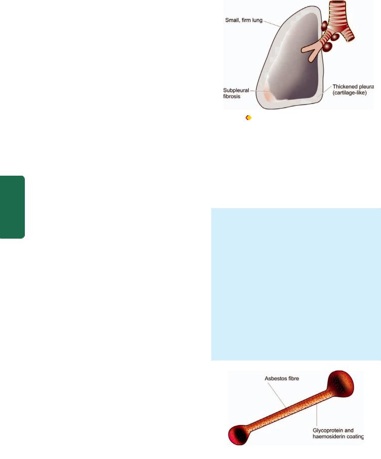
492 Asbestos Disease
|
Asbestos as a mineral is known to mankind for more than |
|
|
4000 years but its harmful effects have come to light during |
|
|
the last few decades. Asbestos is a Greek word meaning |
|
|
‘unquenchable’. In general, if coal is lot of dust and little fibrosis, |
|
|
asbestos is little dust and a lot of fibrosis. Prolonged exposure |
|
|
for a number of years to asbestos dust produces three types |
|
|
of severe diseases: asbestosis of lungs, pleural disease and |
|
|
tumours. In nature, asbestos exists as long thin fibrils which |
|
|
are fire-resistant and can be spun into yarns and fabrics |
|
|
suitable for thermal and electrical insulation and has many |
|
|
applications in industries. Particularly at risk are workers |
|
|
engaged in mining, fabrication and manufacture of a number |
|
|
of products from asbestos such as asbestos pipes, tiles, roofs, |
|
|
textiles, insulating boards, sewer and water conduits, brake |
|
|
lining, clutch castings etc. |
|
|
There are two major geometric forms of asbestos: |
|
|
Serpentine consisting of curly and flexible fibres. It |
|
|
includes the most common chemical form chrysotile (white |
|
|
asbestos) comprising more than 90% of commercially used |
|
|
asbestos. |
|
SECTION |
Amphibole consists of straight, stiff and rigid fibres. It |
|
includes the less common chemical forms crocidolite (blue |
||
asbestos), amosite (brown asbestos), tremolite, anthophyllite and |
||
actinolyte. However, the group of amphibole, though less |
||
common, is more important since it is associated with |
||
induction of malignant pleural tumours, particularly in |
||
III |
association with crocidolite. |
|
However, in view of long term harmful effects of asbestos |
||
|
||
Systemic |
exposure, it has been mostly replaced with synthetic mineral |
|
fibres such as fiberglass in developed countries since 1975 |
||
|
||
|
but it continues to be used in developing countries of the |
|
|
world. |
|
Pathology |
PATHOGENESIS. Overexposure to asbestos for more than |
|
a decade may produce asbestosis of the lung, pleural lesions |
||
|
||
|
and certain tumours. How asbestos causes all these lesions |
|
|
is not clearly understood but the following mechanisms have |
|
|
been suggested (Fig. 17.26,C): |
|
|
1. The inhaled asbestos fibres are phagocytosed by alveolar |
|
|
macrophages from where they reach the interstitium. Some of |
|
|
the engulfed dust is transported via lymphatics to the pleura |
|
|
and regional lymph nodes. |
|
|
2. The asbestos-laden macrophages release chemo-attractants |
|
|
for neutrophils and for more macrophages, thus inciting |
|
|
cellular reaction around them. |
|
|
3. Asbestos fibres are coated with glycoprotein and |
|
|
endogenous haemosiderin to produce characteristic beaded |
|
|
or dumbbell-shaped asbestos bodies. |
|
|
4. All types of asbestos are fibrogenic and result in interstitial |
|
|
fibrosis. Fibroblastic proliferation may occur via macrophage- |
|
|
derived growth factor such as interleukin-1. Alternatively, |
|
|
fibrosis may occur as a reparative response to tissue injury |
|
|
by lysosomal enzymes released from macrophages and |
|
|
neutrophils or by toxic free radicals. |
|
|
5. A few immunological abnormalities such as antinuclear |
|
|
antibodies and rheumatoid factor have been found in cases |
|
|
of asbestosis but their role in the genesis of disease is not |
|
|
clear. |
Figure 17.31 Gross appearance of the lung in asbestosis, diagram-
matic appearance.
6. Asbestos fibres are carcinogenic, the most carcinogenic being crocidolite. There is high incidence of bronchogenic carcinoma in asbestosis which is explained on the basis of the role of asbestos fibres as tumour promoters or by causing cell death of the airways so that it is exposed to the carcinogenic effect of cigarette smoke. The development of pleural mesothelioma in these cases is probably by carrying of asbestos fibres via lymphatics to the pleura.
MORPHOLOGIC FEATURES. As stated already, overexposure to asbestos is associated with 3 types of lesions: asbestosis, pleural disease and certain tumours.
A. ASBESTOSIS. The gross pulmonary fibrosis caused by asbestos exposure and histologic demonstration of asbestos bodies on asbestos fibres is termed asbestosis.
Grossly, the affected lungs are small and firm with cartilage-like thickening of the pleura. The sectioned surface shows variable degree of pulmonary fibrosis, especially in the subpleural areas and in the bases of lungs (Fig. 17.31). The advanced cases may show cystic changes. Histologically, the following changes are observed:
1.There is non-specific interstitial fibrosis.
2.There is presence of characteristic asbestos bodies in the involved areas (Fig. 17.32). These are asbestos fibres coated with glycoprotein and haemosiderin and appear beaded or dumbbell-shaped. The coating stains positively for Prussian blue reaction.
Figure 17.32 
 Microscopic appearance of asbestos body. An asbestos body is an asbestos fibre coated with glycoprotein and haemosiderin giving it beaded or dumbbell-shaped appearance with bulbous ends.

3.There may be changes of emphysema in the pulmonary parenchyma between the areas of interstitial fibrosis.
4.The involvement of hilar lymph nodes in asbestosis is not as significant as in silicosis.
B. PLEURAL DISEASE. Pleural disease in asbestos exposure may produce one of the following 3 types of lesions:
1.Pleural effusion. It develops in about 5% of asbestos workers and is usually serious type. Pleural effusion is generally accompanied by subpleural asbestosis.
2.Visceral pleural fibrosis. Quite often, asbestosis is associated with dense fibrous thickening of the visceral pleura encasing the lung.
3.Pleural plaques. Fibrocalcific pleural plaques are the most common lesions associated with asbestos exposure. Grossly, the lesions appear as circumscribed, flat, small (upto 1 cm in diameter), firm or hard, bilateral nodules. They are seen more often on the posterolateral part of parietal pleura and on the pleural surface of the diaphragm.
Microscopically, they consist of hyalinised collagenous tissue which may be calcified so that they are visible on chest X-ray. Asbestos bodies are generally not found within the plaques.
C. TUMOURS. Asbestos exposure predisposes to a number of cancers, most importantly bronchogenic carcinoma (page 496) and malignant mesothelioma (page 505). A few others are: carcinomas of oesophagus, stomach, colon, kidneys and larynx and various lymphoid malignancies.
1.Bronchogenic carcinoma is the most common malignancy in asbestos workers. Its incidence is 5 times higher in non-smoker asbestos workers than the nonsmoker general population and 10 times higher in smoker asbestos workers than the other smokers.
2.Malignant mesothelioma is an uncommon tumour but association with asbestos exposure is present in 30 to 80% of cases with mesothelioma. The exposure need not be heavy because mesothelioma is known to develop in people living near asbestos plants or in wives of asbestos workers.
CLINICAL FEATURES. Asbestosis is a slow and insidious illness. The patient may remain asymptomatic for a number of years in spite of radiological evidence of calcific pleural plaques and parenchymatous changes. However, onset of interstitial fibrosis brings about dyspnoea with dry or productive cough. More advanced cases show development of Caplan’s syndrome, pulmonary hypertension, cor pulmonale and various forms of cancers.
Berylliosis
Berylliosis is caused by heavy exposure to dust or fumes of metallic beryllium or its salts. Beryllium was used in the past in fluorescent tubes and light bulbs but currently it is
principally used in nuclear and aerospace industries and in 493 the manufacture of electrical and electronic equipments. Two forms of pulmonary berylliosis are recognised—acute and
chronic.
ACUTE BERYLLIOSIS. Acute berylliosis occurs in individuals who are unusually sensitive to it and are heavily exposed to it for 2 to 4 weeks. The pulmonary reaction is in the form of an exudative chemical pneumonitis in which the alveoli are filled with protein-rich fluid with formation of hyaline membrane. The patient develops sudden dyspnoea, hyperapnoea and substernal pain. Most patients recover completely.
CHRONIC BERYLLIOSIS. Chronic berylliosis develops in individuals who are sensitised to it for a number of years, often after a delay of 20 or more years. The disease is a cellmediated hypersensitivity reaction in which the metal beryllium acts as a hapten. The condition is characterised by development of non-caseating epithelioid granulomas like those of sarcoidosis. These granulomas are diffusely scattered throughout the lung parenchyma. The granulomas have
giant cells which frequently contain 3 types of inclusions: |
CHAPTER |
|
1. Birefringent crystals. |
||
2. Concentrically-laminated haematoxyphilic Schaumann or |
||
|
||
conchoid bodies. |
|
|
3. Acidophilic stellate-shaped asteroid bodies. |
|
|
These inclusions are described in giant cells of |
|
|
granulomas in sarcoidosis too (Chapter 6). Similar sarcoid- |
17 |
|
like granulomas can occur in other organs such as in the liver, |
||
kidneys, spleen or lymph nodes in chronic berylliosis. |
|
|
ILD ASSOCIATED WITH IMMUNOLOGIC LUNG DISEASES |
The |
|
Hypersensitivity (Allergic) Pneumonitis |
Respiratory |
|
|
||
Hypersensitivity pneumonitis is a group of immunologically- |
|
|
mediated ILDs occurring in workers inhaling a variety of |
|
|
organic (biologic) antigenic materials. The condition may |
|
|
have an acute onset due to isolated exposure or may be chronic |
System |
|
due to repeated low-dose exposure. |
||
|
||
ETIOPATHOGENESIS. A list of important organic |
|
|
(biologic) dusts which may be inhaled to produce hyper- |
|
|
sensitivity pneumonitis is already given in Table 17.9. The |
|
|
immunologic mechanisms underlying hypersensitivity |
|
|
pneumonitis from any of these causes appear to be either |
|
|
type III immune-complex disease or type IV delayed- |
|
|
hypersensitivity reaction. |
|
1.Farmers’ lung is the classic example resulting from exposure to thermophilic actinomycetes generated by humid and warm mouldy hay.
2.Bagassosis occurs in individuals engaged in manufacture of paper and cardboard from sugarcane bagasse. Spores of thermophilic actinomycetes grow rapidly in mouldy sugarcane bagasse which are inhaled.
3.Byssinosis is an occupational lung disease occurring in workers exposed to fibres of cotton, flex and hemp for a number of years. The role of immunologic mechanisms in byssinosis is not as clear as in exposure to other organic dusts.

4944. Bird-breeders’ (Bird-fanciers’) lung occurs in pigeon breeders, parrot breeders, chicken farmers and bird-fanciers who are exposed to bird-droppings and danders from their feathers.
5.Mushroom-workers’ lung is found in mushroom cultivators exposed to mushroom compost dust.
6.Malt-workers’ lung is seen in distillery and brewery workers who are exposed to mouldy barley and malt dust.
7.Maple-bark disease occurs in those involved in stripping of maple bark and inhale mouldy maple bark (maple tree is grown in northern hemisphere for timber and its leaf is the national emblem of Canada).
8.Silo-fillers’ disease occurs in individuals who enter the silo (silo is an airtight store-house of fodder for farm animals) in which toxic fumes of nitric oxide and nitrogen dioxide are formed due to fermentation of silage. The condition is generally rapidly fatal; less often it may lead to ILD.
|
MORPHOLOGIC FEATURES. The pathologic changes |
|
|
primarily involve the alveoli in contrast to bronchiolar |
|
|
involvement in asthma. The changes vary depending upon |
|
SECTION |
whether the biopsy is examined in early stage or in late |
|
stage. |
||
In early stage, the alveolar walls are diffusely infiltrated |
||
|
||
|
with lymphocytes, plasma cells and macrophages. A |
|
|
proportion of cases show granulomas consisting of |
|
III |
histiocytes and giant cells of foreign body or Langhans’ |
|
type. |
||
|
In chronic cases, the lungs show interstitial fibrosis with |
|
Systemic |
some inflammatory infiltrate. Honeycombing of the lung |
|
to the stage. In acute cases, there is generally sudden attack |
||
|
may be present. |
|
|
CLINICAL FEATURES. The clinical features vary according |
|
Pathology |
of fever, myalgia, dyspnoea, cough and leucocytosis. In more |
|
chronic cases, there are signs of slowly progressive |
||
|
||
|
respiratory failure, dyspnoea and cyanosis as seen in other |
|
|
interstitial lung diseases. |
|
|
Pulmonary Infiltrates with Eosinophilia |
|
|
Pulmonary eosinophilia, eosinophilic pneumonias or |
|
|
pulmonary infiltration with eosinophilia (PIE) syndrome are |
|
|
a group of immunologically-mediated lung diseases |
|
|
characterised by combination of 2 features: |
|
|
Infiltration of the lungs in chest radiographs. |
|
|
Elevated eosinophil count in the peripheral blood. |
|
|
ETIOPATHOGENESIS. PIE syndrome has a number of |
|
|
diverse causes and pathogenesis. These are as under: |
|
|
1. Löeffler’s syndrome is characterised by eosinophilia in |
|
|
the blood and typical wandering radiologic shadows, |
|
|
appearing in some part of the lung for a few days, and then |
|
|
disappearing to appear again somewhere else in the lung. |
|
|
The condition is generally self-limiting and mild, associated |
|
|
with slight fever and a few respiratory symptoms. The |
|
|
etiology is unknown. |
|
|
2. Tropical pulmonary eosinophilia is caused by the |
|
|
passage of larvae of worms through the lungs e.g. in filariasis, |
|
|
ascariasis, strongyloidosis, toxocariasis and ancylostomiasis. |
3.Secondary chronic pulmonary eosinophilia occurs secondary to adverse drug reactions; infection with fungi, bacteria, and helminths; allergic bronchopulmonary aspergillosis and in association with asthma.
4.Idiopathic chronic eosinophilic pneumonia is characterised by prominent focal areas of consolidation of the lung. The condition is clinically diagnosed by excluding other known causes of pulmonary eosinophilia.
5.Hypereosinophilic syndrome is occurrence of eosinophilia of over 1500/μl for more than 6 months without any identifiable cause and without eosinophilic infiltrates in the lungs and other organs.
MORPHOLOGIC FEATURES. The lesions in the lungs are similar in all cases of hypersensitivity pneumonitis. Grossly, the lungs usually show patchy consolidation.
Microscopically, there is thickening of the alveolar walls by oedema and exudate, chiefly of eosinophils, and some lymphocytes and plasma cells. The alveolar lumina also contain eosinophils. Occasionally, small granulomas may be present.
Goodpasture’s Syndrome
Goodpasture’s syndrome or pulmonary haemorrhage syndrome is combination of necrotising haemorrhagic interstitial pneumonitis and rapidly progressive glomerulonephritis. The renal lesions of Goodpasture’s syndrome are described in Chapter 22.
ETIOPATHOGENESIS. The condition results from immunologic damage produced by anti-basement membrane antibodies formed against antigens common to the glomerular and pulmonary basement membranes. The trigger for initiation of this autoimmune response is not clear; it could be virus infection, exposure to hydrocarbons and smoking.
MORPHOLOGIC FEATURES. Grossly, the lungs are heavy with red-brown areas of consolidation. Microscopically, the features vary according to the stage of the disease:
 In acute stage, there are focal areas of haemorrhages in the alveoli and focal necrosis in the alveolar walls.
 In more chronic cases, there is organisation of the haemorrhage leading to interstitial fibrosis and filling of alveoli with haemosiderin-laden macrophages.
CLINICAL FEATURES. The condition occurs commonly in 2nd or 3rd decades of life with preponderance in males. The pulmonary manifestations generally precede the renal disease. Most cases present with haemoptysis accompanied with dyspnoea, fatigue, weakness and anaemia. Renal manifestations soon appear which include haematuria, proteinuria, uraemia and progressive renal failure.
Pulmonary Alveolar Proteinosis
Pulmonary alveolar proteinosis is a rare chronic disease in which the distal airspaces of the lungs are filled with granular, PAS-positive, eosinophilic material with abundant
lipid in it. The condition can occur at any age from infancy to old age.
ETIOPATHOGENESIS. The etiology and pathogenesis of alveolar proteinosis are unknown. A number of possibilities have been suggested:
 Since the alveolar material is combination of lipid and protein, it is not simply an overproduction of surfactant.
 Alveolar proteinosis may have an occupational etiology as seen in patients heavily exposed to silica.
 It may have an etiologic association with haematologic malignancies.

 There may be defective alveolar clearance of debris.
MORPHOLOGIC FEATURES. Grossly, usually both lungs are involved, particularly the lower lobes. The lungs are heavier with areas of consolidation. Sectioned surface exudes abundant turbid fluid.
Histologically, the hallmark of the condition is presence of homogeneous, granular, eosinophilic material which stains brightly with PAS. Often, the material contains cholesterol clefts. There is no significant inflammatory infiltrate in the affected alveoli. Biochemically, the material consists of serum proteins of low molecular weight, cholesterol and phospholipids similar to surfactant. Electron microscopy reveals that the material consists of necrotic alveolar macrophages and desquamated alveolar epithelial cells.
CLINICAL FEATURES. The condition is manifested clinically by dyspnoea, cough, chest pain, pyrexia, fatigue and loss of weight. Chest X-ray shows confluent areas of consolidation. Occasionally, alveolar proteinosis may recover spontaneously but more often it is a fatal condition.
ILD ASSOCIATED WITH
CONNECTIVE TISSUE DISEASES
A number of connective tissue diseases or collagen diseases may result in chronic interstitial fibrosis and destruction of blood vessels. These diseases are described in detail in Chapter 4 but the lung involvement in important forms of collagen diseases is briefly considered here.
1.SCLERODERMA (PROGRESSIVE SYSTEMIC SCLEROSIS). The lungs are involved in 80% cases of scleroderma. Interstitial pulmonary fibrosis is the most common form of pulmonary involvement. The disease usually involves the lower lobes and subpleural regions of the lungs and may lead to honeycombing of the lung. There is increased risk of development of cancer of the lung in pulmonary fibrosis in scleroderma.
2.RHEUMATOID ARTHRITIS. Pulmonary involvement in rheumatoid arthritis may result in pleural effusion, interstitial pneumonitis, necrobiotic nodules and rheumatoid pneumoconiosis (Caplan’s syndrome, page 490). The parenchymatous lesions in rheumatoid arthritis are most commonly seen in the lower lobe. Necrobiotic nodules are the most specific manifestations of rheumatoid disease and closely resemble the subcutaneous nodules commonly found in rheumatoid arthritis.
3.SYSTEMIC LUPUS ERYTHEMATOSUS. Patients with systemic lupus erythematosus (SLE) commonly develop some form of lung disease during the course. The most common manifestation of SLE is pleurisy with small amount of pleural effusion that may contain LE cells. Other pulmonary lesions in SLE are interstitial pneumonitis, pulmonary haemorrhage and vasculitis.
4.SJÖGREN’S SYNDROME. Patients with Sjögren’s syndrome often have rheumatoid arthritis and associated pulmonary changes. Involvement of the bronchial mucous gland by a process similar to that in the salivary glands can lead to inadequate bronchial clearance and repeated infections.
5.DERMATOMYOSITIS AND POLYMYOSITIS.
Interstitial pneumonitis and interstitial fibrosis commonly accompany dermatomyositis and polymyositis.
6.WEGENER’S GRANULOMATOSIS. Wegener’s granulomatosis is an necro-inflammatory lesion having
4components—granulomas of the upper respiratory tract, granulomas of the lungs, systemic vasculitis (page 403) and focal necrotising glomerulonephritis. Localised or limited form of the disease occurs in the lungs without involvement of other organs. Pulmonary involvement is in the form of single or multiple granulomas.
Microscopically, these granulomas have foci of fibrinoid necrosis and intense infiltrate of lymphocytes, plasma cells and macrophages with scattered multinucleate giant cells. Besides necrotising granulomas, there is associated vasculitis.
IDIOPATHIC PULMONARY FIBROSIS
Idiopathic pulmonary fibrosis is the most common form of diffuse interstitial pneumonia and has bad prognosis compared with other forms of lung fibrosis. Diffuse interstitial fibrosis can occur as a result of a number of pathologic entities such as pneumoconiosis, hypersensitivity pneumonitis and collagen-vascular disease. However, in half the cases of diffuse interstitial fibrosis, no apparent cause or underlying disease is identifiable. Such cases are included under the entity ‘idiopathic pulmonary fibrosis’ in the United
States and ‘cryptogenic fibrosing alveolitis’ in Britain.
PATHOGENESIS. The pathogenesis of idiopathic pulmonary fibrosis is unknown and the condition is diagnosed by excluding all known causes of interstitial fibrosis. However, a few evidences point toward immunologic mechanism:
1.High levels of autoantibodies such as rheumatoid factor and antinuclear antibodies.
2.Elevated titres of circulating immune complexes.
3.Immunofluorescent demonstration of the deposits of immunoglobulins and complement on the alveolar walls in biopsy specimens.
MORPHOLOGIC FEATURES. The lung involvement in idiopathic pulmonary fibrosis is often bilateral and widespread.
495
System Respiratory The 17 CHAPTER

496
Pathology Systemic III SECTION
Grossly, the lungs are firm, heavier with reduced volume. Honeycombing (i.e. enlarged, thick-walled air spaces) develops in parts of lung, particularly in the subpleural region.
Histologically, the changes vary according to the stage of the disease.
 In early stage, there is widening of the alveolar septa by oedema and cellular infiltrate by mononuclear inflammatory cells. The alveolar lining cells may show hyperplasia at places and are flattened at other places. There is often formation of hyaline membranes. The alveolar spaces contain exudate consisting of macrophages, lymphocytes and neutrophils. Many of the macrophages contain lamellar bodies derived from surfactant of the necrotic alveolar lining epithelial cells. Based on the observation of desquamative component in the cellular exudate, some authors label the early stage of idiopathic pulmonary fibrosis as ‘desquamative interstitial
pneumonitis’.
 In advanced stage, there is organisation of the alveolar exudate and replacement fibrosis in the alveoli as well as in the interstitial septal wall with variable amount of inflammation. Eventually, there are small cystic areas (honeycomb lung) with alternating areas of fibrosis containing thick-walled and narrowed vessels. This stage
is often referred to as ‘chronic interstitial pneumonitis’ or ‘usual interstitial pneumonitis’.
CLINICAL FEATURES. Middle-aged males are affected more frequently. The usual features are of respiratory difficulty beginning with dry cough and slowly progressing dyspnoea. More advanced cases may develop clubbing of fingers and cor pulmonale. A rapidly progressive form of the idiopathic pulmonary fibrosis with death within 6 weeks to 6 months is termed Hamman-Rich syndrome.
ILD ASSOCIATED WITH SMOKING
Long-term consequences of smoking is associated with following non-neoplastic respiratory insufficiency:

 Smoking-related COPD: due to chronic bronchitis and emphysema.

 Smoking-related ILD: i.e. chronic restrictive pulmonary disease due to desquamative interstitial pneumonia (DIP), respiratory bronchiolitis-associated ILD, and pulmonary Langerhans cell histiocytosis (eosinophilic granuloma of the lung).
DESQUAMATIVE INTERSTITIAL PNEUMONIA (DIP).
It is an uncommon condition occurring exclusively in smokers in 4th to 5th decades of life and is more common in males. Most patients present with dyspnoea and cough. Chest X-ray shows peculiar diffuse hazy opacities which characterize all ILDs. DIP was earlier thought to represent forerunner lesion in the sequence of development of idiopathic pulmonary fibrosis. However, DIP has minimal fibrosis and has a far better prognosis on cessation of smoking compared to idiopathic pulmonary fibrosis.
Microscopically, the features are as under:
i)Hallmark finding is collections of large number of intraalveolar macrophages having abundant cytoplasm and containing brown-black pigment and are termed as smokers’ macrophages.
ii)The intervening septa contain a few lymphocytes, plasma cells and an occasional eosinophil.
iii)Late cases show mild interstitial fibrosis.
RESPIRATORY BRONCHIOLITIS-ASSOCIATED ILD.
Respiratory bronchiolitis is a far more common lesion in chronic smokers than DIP and is considered a milder form of DIP having similar clinical presentation. Respiratory bronchiolitis-associated ILD is the term used for advanced cases who develop impaired pulmonary function and radiologic features.
The condition resolves following cessation of smoking.
Microscopically, it is characterised by following features:
i)Patchy and bronchiolocentric location of similar smokers’ macrophages as are seen in DIP.
ii)Peribronchial infiltrate of lymphocytes and histiocytes.
iii)There may be mild peribronchial fibrosis.
iv)Centriacinar emphysema may coexist.
PULMONARY LANGERHANS CELL HISTIOCYTOSIS.
This is an uncommon smoking-related ILD occurring in younger men (20-40 years). Clinically, the features may vary from an asymptomatic state to a rapidly progressive course. Symptomatic cases present with cough, dyspnoea, weight loss and fever. CT scan shows presence of ill-defined stellate nodules and thin-walled cysts.
Discontinuation of smoking leads to reversal of the condition.
Microscopically, the features are as under:
i)There is presence of poorly-defined nodules distributed in peribronchiolar location while intervening lung parenchyma is uninvolved.
ii)Characteristically, these nodules are sclerosing and contain Langerhans cells along with other inflammatory cells.
iii)Progressive cases have fibrosis with concomitant cystic change.
TUMOURS OF LUNGS
A number of benign and malignant tumours occur in the lungs but the primary lung cancer, commonly termed bronchogenic carcinoma, is the most common (95% of all primary lung tumours). The lung is also the commonest site for metastasis from carcinomas and sarcomas. A histologic classification of various benign and malignant tumours of lungs as recommended by the World Health Organisation is given in Table 17.10.
BRONCHOGENIC CARCINOMA
The term bronchogenic carcinoma is commonly used for cancer of the lungs which includes carcinomas arising from

TABLE 17.10: WHO Classification of Lung Tumours.
I. EPITHELIAL TUMOURS
A.Benign
1.Papilloma
2.Adenoma
B.Dysplasia and carcinoma in situ
C.Malignant Bronchogenic carcinoma
1.Squamous cell (epidermoid) carcinoma
2.Small cell carcinoma
i)Oat cell carcinoma
ii)Intermediate cell carcinoma
iii)Combined oat cell carcinoma
3.Adenocarcinoma
i)Acinar adenocarcinoma
ii)Papillary adenocarcinoma
iii)Bronchiolo-alveolar carcinoma
iv)Solid carcinoma with mucus formation
4.Large cell carcinoma
5.Adenosquamous carcinoma
Other carcinomas
1.Pulmonary neuroendocrine tumour (carcinoid tumour)
2.Bronchial gland carcinomas
i)Adenoid cystic carcinoma
ii)Mucoepidermoid carcinoma
II.SOFT TISSUE TUMOURS
(Fibroma, fibrosarcoma; leiomyoma, leiomyosarcoma; lipoma, chondroma, haemangioma, lymphangioma, granular cell myoblastoma)
III.PLEURAL TUMOURS
A.Benign mesothelioma
B.Malignant mesothelioma
IV. MISCELLANEOUS TUMOURS
1.Carcinosarcoma
2.Pulmonary blastoma
3.Malignant melanoma
4.Malignant lymphoma V. SECONDARY TUMOURS
the respiratory epithelium lining the bronchi, bronchioles and alveoli.
INCIDENCE AND CLASSIFICATION. Lung cancer is the most common primary malignant tumour in men and accounts for nearly 30% of all cancer deaths in both sexes in developing countries. Currently, the incidence of lung cancer in females in the United States has already exceeded breast cancer as a cause of death in women. Cancer of the lung is a disease of middle and late life with peak incidence in 55-65 years of age, after which there is gradual fall in its incidence. Of late, there has been slight decline in lung cancer deaths in males due to smoking cessation efforts which started in the West 4 decades back and has started yielding results. However, worldwide the scene on its incidence and prognosis are quite grim; data from International Agency for Research on Cancer estimate that worldwide by the year 2030 there would be about 10 million deaths per year from lung cancer.
There are 5 main histologic types of lung cancer:
i)Squamous cell or epidermoid carcinoma
ii)Small cell carcinoma
iii)Adenocarcinoma (including bronchioalveolar carcinoma)
iv)Large cell carcinoma
v)Combined squamous cell carcinoma and adenocarcinoma (adenosquamous carcinoma).
As per reports on international data for the last 25 years, while there has been decline in the incidence of small cell carcinoma, incidence of adenocarcinoma of the lung has risen and has surpassed squamous cell carcinoma as the most frequent histologic subtype of lung cancer.
However, for therapeutic purposes, bronchogenic carcinoma can be classified into 3 groups:
1. Small cell carcinomas, SCC (20-25%)
2. Non-small cell carcinomas, NSCC (70-75%) (includes squamous cell carcinoma, adenocarcinoma, and large cell carcinoma)
3. Combined/mixed patterns (5-10%).
ETIOLOGY. The high incidence of lung cancer is associated with a number of etiologic factors, most important of which
is cigarette smoking.
1. Smoking. The most important factor for high incidence of all forms of bronchogenic carcinoma is tobacco smoking. About 80% of the lung cancer occurs in active smokers. A number of evidences support the positive relationship of lung cancer with tobacco smoking (see page 237):
i) Total dose: There is a direct statistical correlation between death rate from lung cancer and the total amount of cigarettes smoked e.g.
 An average regular smoker has 10 times greater risk of developing lung cancer than a non-smoker.
The risk of smokers of more than 2 packs (40 cigarettes)
per day for 20 years is 60-70 times greater than a non-smoker.
 Cessation of smoking by a regular smoker results in gradual decline in the chances of developing lung cancer. After 10 years of abstinence from smoking, the risk declines but never returns to the non-smoker level.

 Pipe and cigar smokers, though have higher risk than non-smokers but are at lesser risk than cigarette smokers.
ii)Histologic alterations: The association of tobacco smoking is strongest for squamous cell carcinoma and small cell carcinoma of the lung. More than 90% of smokers have sequential epithelial changes in the respiratory tract in the form of squamous metaplasia, dysplasia and carcinoma in situ (Fig. 17.33).
iii)Mechanism: How tobacco smoking causes lung cancer is not quite clear. However, following facts have been observed:

 Analysis of the tar from cigarette smoke has revealed a number of known carcinogens (e.g. polycyclic aromatic hydrocarbons, nitrosamines) and tumour promoters (e.g. phenol derivatives).

 In experimental animal studies, it has been possible to induce cancer by skin painting experiments with smoke-tar.
497
System Respiratory The 17 CHAPTER
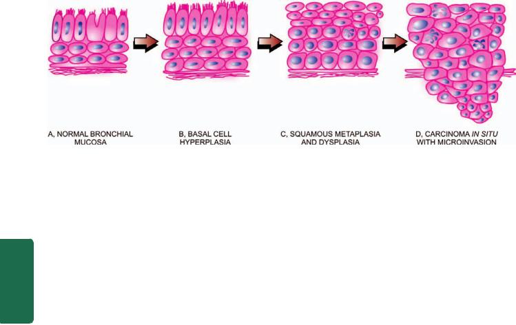
498
Figure 17.33 
 Schematic representation of sequential development of squamous cell carcinoma of the lung.
Pathology Systemic III SECTION
However, it has not been possible to reproduce pattern of human respiratory tract cancer, probably because of the difficulty in reproducing human smoking methods in animals.
2. Other factors. Although smoking is the dominant etiologic factor in lung cancer, 15% cases of lung cancer occur in non-smokers, more so in women probably related to hormonal factors. A few other factors implicated in lung cancer are as follows:
i)Atmospheric pollution. There is increased risk of developing bronchogenic carcinoma in non-smokers living in industrialised and smoky cities than in the less polluted rural areas. It is possible that specific industrial pollutants may be at fault as evidenced by high rates for lung cancer in people living in the neighbourhood of petrochemical industries.
ii)Occupational causes. There are a number of well-esta- blished occupational causes of lung cancer. These include workers exposed to asbestos, radiation of all types, bis-ethers, nickel, beryllium, arsenic, metallic iron and iron-oxide. Some industrial carcinogens and cigarette smoking have cocarcinogenic effect, particularly in uranium mines and asbestos workers.
iii)Dietary factors. Susceptibility to respiratory cancers is increased in vitamin A deficiency. Smokers with low vitamin A intake have a greater risk of lung cancer than those with vitamin A-rich diet. The incidence of lung cancer is inversely related to socioeconomic level reflecting their dietary pattern.
iv)Chronic scarring. Peripheral adenocarcinomas occur more frequently in areas of chronic scarring caused by chronic inflammatory changes, old tuberculosis, asbestosis, chronic interstitial fibrosis, old infarcts and in scleroderma.
MOLECULAR PATHOGENESIS. Molecular studies have revealed that there are several genetic alterations in cancer stem cells which produce clones of malignant cells to form tumour mass. Following genetic changes have been found:
1. Activation of growth-promoting oncogenes: Mutation in
K-RAS oncogene has been seen as the dominant change in lung cancer. Besides, there is mutation in tyrosine kinase domain of EGFR oncogene in cases of adenocarcinoma lung in non-smokers. Other mutations include BRAF, PIK3CA and
MYC family, and overexpression of bcl-2 and other antiapoptotic proteins.
2.Inactivation of tumour-suppressor genes. Inactivation of tumour suppressor genes has been found as another molecular mechanism in lung cancer. Many tumour suppressor genes have been found on chromosome 3p in lung cancer cases. These include inactivation of p53 and Rb gene. Besides, some tumour-acquired promoter genes have been identified in lung cancer e.g. p16, RASSFIA etc, which cause loss of normal function of growth-regulatory tumour suppressor genes.
3.Autocrine growth factors. Studies have shown that lung cancer is a multistep process—initiator carcinogen causing mutation, followed by action of tumour promoters. Nicotine acts as both initiator as well as promoter carcinogen. Derivatives of nicotine in smoke unmask and expresses nicotine acetylcholine receptors which activate the signaling pathway in tumour, blocking the apoptosis. Promoters also include several hormones which are elaborated by lung cancer by autocrine pathway as part of paraneoplastic syndrome.
4.Inherited predisposition. Although not common, but there are a few examples of inheritance of lung cancer as under: i) Patients of Li-Fraumeni syndrome who inherit p53 mutation may develop lung cancer.
ii) Clinical cases of retinoblastoma having mutation in Rb gene are predisposed to develop lung cancer if they live up to adulthood.
iii) First-degree relatives of lung cancer patients have a 2-3 fold higher risk of developing lung cancer in their lifetime. iv) Mutations of cytochrome P450 system have been identified in lung cancer patients; P450 metabolises chemical carcinogen in tobacco smoke (page 216).
5.Molecular targets for therapy and survival prediction.
Knowledge on the insight into molecular biology and pathogenesis of lung cancer has applications in discovering newer molecules for targeted therapy, predicting response to treatment and survival:
i) EGFR mutations and NSCC therapy: It has been reported that 70% cases of NSCC have overexpression of EGFR protein

or amplification of the EGFR gene. EGFR belonging to ERBB (HER) family of protooncogenes through mutation in its tyrosine kinase (TK) domain plays a role in both extracelluar and intracellular signaling resulting in tumour cell proliferation, metastasis and antiapoptotic action. Targeted molecular therapy against these mutations in EGFR include EGFR-TK inhibitor oral therapy.
ii)VEGF and monoclonal therapy: Although not mutated, VEGF is excessively produced in lung cancer and contributes to tumour angiogenesis. Monoclonal antibody therapy against EGFR in conjunction with chemotherapy has been used for curtailing tumour angiogenesis in lung caner.
iii)Molecular signature gene for prediction: Recent proteomic studies at research level have shown that each patient has unique protein pattern in the serum (i.e. molecular signatures) which may be used for early diagnosis, predict drug resistance, response to treatment and survival, but these are yet to be applied in clinical settings.
MORPHOLOGIC FEATURES. Bronchogenic carcinoma can occur anywhere in the lung but the most common location is hilar, followed in descending frequency by peripheral type.
Grossly, these 2 main types show variation in appearance:
1. Hilar type (Fig. 17.34,A): Most commonly, the lung cancer arises in the main bronchus or one of its segmental branches in the hilar parts of the lung, more often on the right side. The tumour begins as a small roughened area on the bronchial mucosa at the bifurcation. As the tumour enlarges, it thickens the bronchial mucosa producing nodular or ulcerated surface. As the nodules coalesce, the carcinoma grows into a friable spherical mass, 1 to 5 cm in diameter, narrowing and occluding the lumen. The cut surface of the tumour is yellowish-white with foci of necrosis and haemorrhages which may produce cavitary lesions (Table 17.11 sums up a list of common conditions having pulmonany cavitary lesions or ‘honey-comb lung’
TABLE 17.11: Conditions Producing Pulmonary Cavities (Honeycomb Lung).
A.INFECTIONS
1.Pulmonary tuberculosis
2.Primary lung abscess (e.g. due to aspiration)
3.Secondary lung abscess (e.g. preceding pneumonia, pyaemia, sepsis)
4.Bronchiectasis
5.Fungal infections (e.g. aspergillosis, mucormycosis)
6.Actinomycosis
7.Nocardiosis
B.NON-INFECTIOUS CAUSES
1.Pneumoconiosis (e.g. simple coal-workers’ pneumoconiosis, silicosis, asbestosis)
2.Bronchogenic carcinoma
3.Metastatic lung tumours
4.Wegener’s granulomatosis
5.Pulmonary infarction
6.Congenital cysts
7.Idiopathic pulmonary fibrosis
499
Figure 17.34 |
The two main gross patterns of bronchogenic |
|
|
carcinoma. |
|
|
|
|
|
||
during the course of different lung diseases). It is common |
|
||
to find secondary changes in bronchogenic carcinoma of |
|
||
lung such as bronchopneumonia, abscess formation and |
|
||
bronchiectasis as a result of obstruction and intercurrent |
CHAPTER |
||
infections. The tumour soon spreads within the lungs by |
|||
|
|||
direct extension or by lymphatics, and to distant sites by |
|
||
lymphatic or haematogenous routes, as described later. |
|
||
2. Peripheral type (Fig. 17.34,B): A small proportion of |
|
||
lung cancers, chiefly adenocarcinomas including |
17 |
||
bronchioloalveolar carcinomas, originate from a small |
|||
peripheral bronchiole but the exact site of origin may not |
|
||
be discernible. The tumour may be a single nodule or |
The |
||
multiple nodules in the periphery of the lung producing |
|||
|
|||
pneumonia-like consolidation of a large part of the lung. |
Respiratory |
||
The cut surface of the tumour is greyish and mucoid. |
|||
|
|||
Histologically, as per the WHO classification outlined in |
|
||
Table 17.10, five main histologic types of bronchogenic |
|
||
carcinoma are distinguished which is important because |
|
||
of prognostic and therapeutic considerations. However, |
System |
||
from clinical point of view, distinction between small cell |
|||
(SCC) and non-small cell carcinomas (NSCC) is important |
|||
|
|||
because the two not only differ in morphology, but there |
|
||
are major differences in immunophenotyping and |
|
||
response to treatment discussed above. The major |
|
||
differences between SCC and NSCC of the lung are |
|
||
summed up in Table 17.12. |
|
||
1. Squamous cell (epidermoid) carcinoma: This has been |
|
||
the most common histologic subtype of bronchogenic |
|
||
carcinoma until recently and is found more commonly in |
|
||
men, often with history of tobacco smoking. These |
|
||
tumours usually arise in a large bronchus and are prone |
|
||
to massive necrosis and cavitation (Fig. 17.35). The tumour |
|
||
is diagnosed microscopically by identification of either |
|
||
intercellular bridges or keratinisation. The tumour may |
|
||
show varying histologic grades of differentiation such as |
|
||
well-differentiated, moderately-differentiated and poorly- |
|
||
differentiated (Fig. 17.36). Occasionally, a variant of |
|
||
squamous cell carcinoma, spindle cell carcinoma, having |
|
||
biphasic pattern of growth due to the presence of a |
|
||
|
|
|
|

500 TABLE 17.12: Comparison of Features of Small Cell and Non-small Cell Carcinoma of the Lung.
|
Feature |
Small Cell Carcinoma |
Non-small Cell Carcinoma |
|
|
|
|
1. |
Etiologic relationship |
Strongly related to tobacco smoking |
Smoking implicated, other factors: |
|
|
|
pollution, chronic scars, asbestos exposure |
2. |
Morphology |
|
|
|
i) Pattern |
Diffuse sheets |
Squamous or glandular pattern |
|
ii) Nuclei |
Hyperchromatic, fine chromatin |
Pleomorphic, coarse chromatin |
|
iii) Nucleoli |
Indistinct |
Prominent |
|
iv) Cytoplasm |
Scanty |
Abundant |
3. |
Neuroendocrine markers |
Present |
Absent |
|
(e.g. dense-core granules on EM, |
|
|
|
chromogranin, synaptophysin, |
|
|
|
neuron-specific enolase, CD56, CD57) |
|
|
4. |
Epithelial markers |
Present |
Present |
|
(e.g. epithelial membrane antigen, |
|
|
|
carcinoembryonic antigen, cytokeratin) |
|
|
Pathology Systemic III SECTION
5. |
Mucin |
Absent |
Present in adenocarcinoma |
6. |
HLA, β2 microglobulin |
Absent to low |
Present |
7. |
Peptide hormone production |
Gastrin, ACTH, ADH, calcitonin |
Parathormone |
8. |
Genetic abnormalities |
3p allele loss, RB and |
3p allele loss, EGFR and K-RAS |
|
|
p53 mutations |
mutations |
9. |
Treatment type |
Radiotherapy and/or |
Surgical resection possible, limited |
|
|
chemotherapy |
response to radiotherapy and/or |
|
|
|
chemotherapy |
10. |
Prognosis |
Poor |
Better |
|
|
|
|
component of squamous cell carcinoma and the other sarcoma-like spindle cell component, is found.
Usually the spread of squamous cell carcinoma is more rapid than the other histologic types of NSCC. Frequently, the edge of the growth and the adjoining uninvolved
Figure 17.35 
 Squamous cell carcinoma lung, hilar type. Sectioned surface shows grey-white fleshy tumour in the bronchus at its bifurcation and occluding the lumen partly (arrow). The tumour is seen extending directly into adjacent lung parenchyma and hilar nodes.
bronchi show squamous metaplasia, epithelial dysplasia and carcinoma in situ.
2. Small cell carcinoma: Small cell carcinomas are frequently hilar or central in location, have strong relationship to cigarette smoking and are highly malignant tumours. They are most often associated with ectopic hormone production because of the presence of neurosecretory granules in majority of tumour cells which are similar to those found in argentaffin or Kulchitsky cells normally found in bronchial epithelium. By immunohistochemistry, these tumour cells are positive for neuroendocrine markers: chromogranin, neuron-specific enolase (NSE) and synaptophysin. Small cell carcinomas have 3 subtypes:
i)Oat cell carcinoma is composed of uniform, small cells, larger than lymphocytes with dense, round or oval nuclei having diffuse chromatin, inconspicuous nucleoli and very sparse cytoplasm (oat = a form of grain). These cells are organised into cords, aggregates and ribbons or around small blood vessels forming pseudorosettes (Fig. 17.37).
ii)Small cell carcinoma, intermediate cell type is composed of cells slightly larger than those of oat cell carcinoma and have similar nuclear characteristics but have more abundant cytoplasm. These cells are organised into lobules.
iii)Combined oat cell carcinoma is a tumour in which there is a definite component of oat cell carcinoma with squamous cell and/or adenocarcinoma.
3.Adenocarcinoma: Adenocarcinoma, also called peripheral carcinoma due to its location and scar carcinoma due to its association with areas of chronic scarring, is the most common bronchogenic carcinoma in women and is
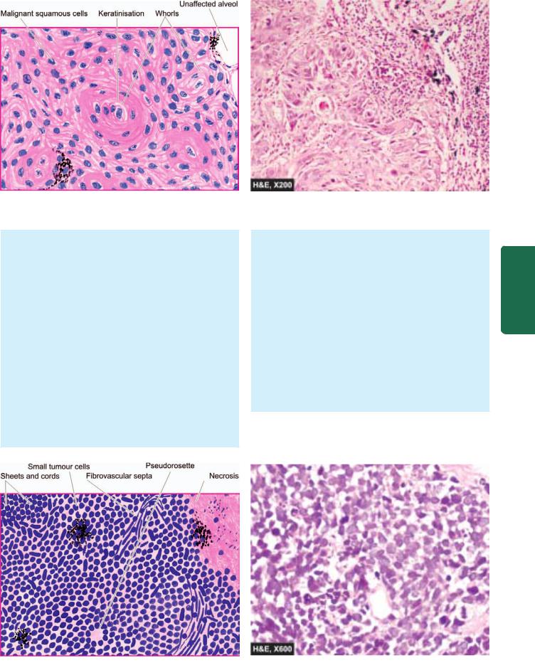
501
Figure 17.36 
 Squamous cell carcinoma of the lung. Islands of invading malignant squamous cells are seen. A few well-developed cell nests with keratinisation are evident.
slow-growing. Recent estimates on adenocarcinoma place this as the most frequent histologic subtype of lung cancer. Adenocarcinoma is further subclassified into 4 types:
i)Acinar adenocarcinoma which has predominance of glandular structure and often occurs in the larger bronchi.
ii)Papillary adenocarcinoma which has a pronounced papillary configuration and is frequently peripherally located in the lungs and is found in relation to pulmonary scars (scar carcinoma).
iii)Bronchiolo-alveolar carcinoma is characterised by cuboidal to tall columnar and mucus-secreting epithelial cells growing along the existing alveoli and forming numerous papillary structures (Fig. 17.38). Ultrastructurally, these tumour cells resemble Clara cells or less often type II pneumocytes.
iv)Solid carcinoma is a poorly-differentiated adenocarcinoma lacking acini, tubules or papillae but having mucuscontaining vacuoles in many tumour cells.
4.Large cell carcinoma: These are undifferentiated carcinomas which lack the specific features by which they could be assigned into squamous cell carcinoma or adenocarcinoma. Large cell carcinomas are more common in men, have strong association with cigarette smoking and are highly malignant tumours. The tumour cells have large nuclei, prominent nucleoli, abundant cytoplasm and well-defined cell borders. Variants of large cell undifferentiated carcinomas include giant cell carcinoma with prominence of highly pleomorphic multinucleate cells and clear cell carcinoma composed of cells with clear or foamy cytoplasm without mucin.
5.Adenosquamous carcinoma: These are a small proportion of peripheral scar carcinomas having clear evidence of both keratinisation and glandular differentiation.
SPREAD. Bronchogenic carcinoma can invade the adjoining structures directly, or may spread by lymphatic and haematogenous routes.
System Respiratory The 17 CHAPTER
Figure 17.37 
 Oat cell carcinoma of the lung. The tumour cells are arranged in sheets, cords or aggregates and at places form pseudorosettes.
The individual tumour cells are small, uniform, lymphocyte-like with scanty cytoplasm.
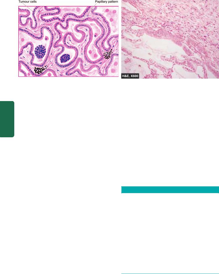
502
Pathology Systemic III SECTION
Figure 17.38 
 Bronchioloalveolar carcinoma. The alveolar walls are lined by cuboidal to tall columnar and mucin-secreting tumour cells with papillary growth pattern.
1. Direct spread. The tumour extends directly by invading through the wall of the bronchus and destroys and replaces the peribronchial lung tissue. As it grows further, it spreads to the opposite bronchus and lung, into the pleural cavity, the pericardium and the myocardium and along the great vessels of the heart causing their constriction. Extension of the cancer located at the apex of the lung into the thoracic cage may involve brachial plexus and the sympathetic chain causing pain and sensory disturbances, so called Pancoast’s
syndrome.
2.Lymphatic spread. Initially, hilar lymph nodes are affected. Later, lymphatic metastases occur to the other groups leading to spread to mediastinal, cervical, supraclavicular and para-aortic lymph nodes. Invasion of the thoracic duct may produce chylous ascites.
3.Haematogenous spread. Distant metastases via blood stream are widespread and early. The sites affected, in descending order of involvement, are: the liver, adrenals, bones, pancreas, brain, opposite lung, kidneys and thyroid.
CLINICAL FEATURES. Symptoms of lung cancer are quite variable and result from local effects, effects due to occlusion of a bronchus, direct and distant metastases, and paraneoplastic syndromes. Diagnostic aids include radiologic examination and CT scan of the chest, cytologic examination of the sputum, bronchial washings and bronchioalveolar lavage.
1.Local symptoms. Most common local complaints are cough, chest pain, dyspnoea and haemoptysis. (A list of various causes of haemoptysis is summed up in Table 17.13).
2.Bronchial obstructive symptoms. Occlusion of a bronchus may result in bronchopneumonia, lung abscess and bronchiectasis in the lung tissue distal to the site of obstruction and cause their attendant symptoms like fever, productive cough, pleural effusion and weight loss.
3.Symptoms due to metastases. Distant spread may produce varying features and sometimes these are the first manifestation of lung cancer. These include: superior vena
caval syndrome, painful bony lesions, paralysis of recurrent nerve and other neurologic manifestations resulting from brain metastases.
4. Paraneoplastic syndromes. A number of paraneoplastic syndromes (page 231) are associated with lung cancer. These include the following:
i) Ectopic hormone production: Different hormonal syndromes are characteristic of different histologic types of lung cancer. Small cell carcinomas are associated most often with ectopic hormone production. The various hormones elaborated by lung cancer are as follows:
a)ACTH, producing Cushing’s syndrome.
b)ADH, inducing hyponatraemia.
c)Parathormone, causing hypercalcaemia.
d)Calcitonin, producing hypocalcaemia.
e)Gonadotropins, causing gynaecomastia.
f)Serotonin, associated with carcinoid syndrome.
TABLE 17.13: Causes of Haemoptysis.
A.INFLAMMATORY
1.Bronchitis
2.Bronchiectasis
3.Tuberculosis
4.Lung abscess
5.Pneumonias
B.NEOPLASTIC
1.Primary and metastatic lung cancer
2.Bronchial adenoma
C.OTHERS
1.Pulmonary thromboembolism
2.Left ventricular failure
3.Mitral stenosis
4.Trauma
5.Foreign bodies
6.Primary pulmonary hypertension
7.Haemorrhagic diathesis
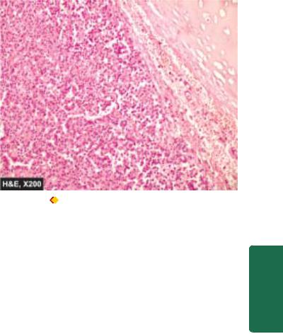
ii) Other systemic manifestations: These include the following:
a)Neuromuscular e.g. polymyositis, myopathy, peripheral neuropathy and subacute cerebellar degeneration.
b)Skeletal e.g. clubbing and hypertrophic osteoarthropathy.
c)Cutaneous e.g. acanthosis nigricans and dermatomyopathy.
d)Cardiovascular e.g. migratory thrombophlebitis (Trousseau’s syndrome), nonbacterial thrombotic endocarditis.
e)Haematologic e.g. abnormalities in coagulation and leukaemoid reaction.
STAGING AND PROGNOSIS. The widely accepted clinical staging of lung cancer is according to the TNM classification, combining features of primary Tumours, Nodal involvement and distant Metastases. TNM staging divides all lung cancers into the following 4 stages:
Occult: Malignant cells in the bronchopulmonary secretions but no evidence of primary tumour or metastasis.
Stage I: Tumour less than 3 cm, with or without ipsilateral nodal involvement, no distant metastasis.
Stage II: Tumour larger than 3 cm, with ipsilateral hilar lymph node involvement, no distant metastasis.
Stage III: Tumour of any size, involving adjacent structures, involving contralateral lymph nodes or distant metastasis.
In general, tumour size larger than 5 cm has worse prognosis. Symptomatic patients, particularly with systemic symptoms, fare far badly than the nonsymptomatic patients. The overall prognosis of bronchogenic carcinoma is dismal. However, 5-year survival rate with surgery combined with radiotherapy or chemotherapy in the last 30 years has doubled from its earlier rate of about 9% to present rate of 15%. Adenocarcinoma and squamous cell carcinoma which are localised, are resectable and have a slightly better prognosis. Small cell carcinoma has the worst prognosis since surgical treatment is ineffective though the tumour is sensitive to radiotherapy and chemotherapy.
BRONCHIAL CARCINOID AND
OTHER NEUROENDOCRINE TUMOURS
Neuroendocrine tumours of the lung represent a continuum spectrum of lung tumours with progressively increasing aggressiveness which include: typical carcinoid (least aggressive), atypical carcinoid, and large cell endocrine carcinoma, and also small cell carcinoma (most aggressive). All these tumours arise from neuroendocrine (Kulchitsky) cells of bronchial mucosa. Formerly, bronchial carcinoids used to be classified as ‘bronchial adenomas’ but now it is known that they are locally invasive and have the capacity to metastasise. Bronchial carcinoids tend to occur at a younger age than bronchogenic carcinoma, often appearing below the age of 40 years, and are not related to cigarette smoking.
503
Figure 17.39 |
Central carcinoid of the lung. The tumour shows a |
characteristic nested of cells separated by fibrovascular septa. These |
|
nests are composed of uniform cuboidal cells having granular cytoplasm. |
|
MORPHOLOGIC FEATURES. Bronchial carcinoids |
|
CHAPTER |
resemble their intestinal counterparts described in |
|
|
|
|
|
Chapter 20. |
|
|
Grossly, bronchial carcinoids most commonly arise from |
|
|
a major bronchus and project into the bronchial lumen as |
|
|
a spherical polypoid mass, 3-4 cm in diameter. Less |
|
17 |
commonly, the tumour may grow into the bronchial wall |
|
|
|
|
|
and produce collar-button like lesion. The overlying |
|
|
bronchial mucosa is usually intact. Cut surface of the |
|
The |
tumour is yellow-tan in colour. |
|
|
|
|
|
Histologically, the tumour is composed of uniform |
|
Respiratory |
cuboidal cells forming aggregates, trabeculae or ribbons |
|
|
|
|
|
separated by fine fibrous septa. The tumour cells have |
|
|
abundant, finely granular cytoplasm and oval central |
|
|
nuclei with clumped nuclear chromatin. Mitoses are rare |
|
|
and necrosis is uncommon (Fig. 17.39). The secretory |
|
|
granules of bronchial carcinoids resemble those of other |
|
System |
foregut carcinoids and stain positively with argyrophilic |
|
|
stains in which exogenous reducing agent is added for |
|
|
the reaction. Immunohistochemically, markers for |
|
|
neuroendocrine stain positive and include NSE, |
|
|
chromogranin, synaptophysin and neurofilaments. |
|
|
CLINICAL FEATURES. Bronchial carcinoids occur at a |
|
|
relatively early age and have equal sex incidence. Most of |
|
|
the symptoms in bronchial carcinoids occur as a result of |
|
|
bronchial obstruction such as cough, haemoptysis, atelectasis |
|
|
and secondary infection. About 5-10% of bronchial carcinoids |
|
|
metastasise to the liver and these cases are capable of |
|
|
producing carcinoid syndrome (Chapter 20). |
|
|
HAMARTOMA |
|
|
Hamartoma is a tumour-like lesion composed of an abnormal |
|
|
admixture of pulmonary tissue components and is |
|
|
discovered incidentally as a coin-lesion in the chest-X-ray. |
|
|

504 Pulmonary hamartomas are of 2 types: chondromatous and leiomyomatous.

 Chondromatous hamartoma is more common and usually asymptomatic. It forms a solitary, spherical mass, 2-5 cm in diameter, usually at the periphery of the lung. Typically, it shows nodules of cartilage associated with fibrous and adipose tissue admixed with bronchial epithelium.

 Leiomyomatous hamartoma has a prominent smooth muscle component and bronchiolar structures. They are frequently multiple, 1-2 mm in diameter and are more
|
commonly located near the pleura. |
|
|
METASTATIC LUNG TUMOURS |
|
|
Secondary tumours of the lungs are more common than the |
|
|
primary pulmonary tumours. Metastases from carcinomas |
|
|
as well as sarcomas arising from anywhere in the body may |
|
|
spread to the lung by haematogenous or lymphatic routes, |
|
|
or by direct extension. Blood-borne metastases are the most |
|
|
common since emboli of tumour cells from any malignant |
|
SECTION |
tumour entering the systemic venous circulation are likely |
|
to be lodged in the lungs. Metastases are most common in |
||
|
||
|
the peripheral part of the lung forming single or multiple, |
|
|
discrete nodular lesions which appear radiologically as |
|
|
‘cannon-ball secondaries’ (Fig. 17.40). Less frequently, the |
|
III |
metastatic growth is confined to peribronchiolar and |
|
perivascular locations which is due to spread via lymphatics. |
||
|
Most common sources of metastases in the lungs are: |
|
Systemic |
carcinomas of the bowel, breast, thyroid, kidney, pancreas, |
|
lung (ipsilateral or contralateral) and liver. Other tumours |
||
which frequently metastasise to the lungs are osteogenic |
||
sarcoma, neuroblastoma, Wilms’ tumour, melanoma, |
||
lymphomas and leukaemias. |
||
Pathology |
|
Figure 17.40 |
Metastatic deposits in the lung. Large parts of the |
lung parenchyma are replaced by multiple, variable-sized, circumscribed nodular masses which are grey-white in colour. Some of these show areas of haemorrhage and necrosis.
PLEURA
NORMAL STRUCTURE
Visceral pleura covers the lungs and extends into the fissures while the parietal pleura limits the mediastinum and covers the dome of the diaphragm and inner aspect of the chest wall. The two layers between them enclose pleural cavity which contains less than 15 ml of clear serous fluid.
Microscopically, both the pleural layers are lined by a single layer of flattened mesothelial cells facing each other. Underneath the lining cells is a thin layer of connective tissue.
Diseases affecting the pleura are nearly always secondary to some other underlying disease. Broadly, they fall into inflammations, non-inflammatory pleural effusions, pneumothorax, and tumours.
INFLAMMATIONS
Inflammatory involvement of the pleura is commonly termed pleuritis or pleurisy. Depending upon the character of resultant exudate, it can be divided into serous, fibrinous and serofibrinous, suppurative or empyema, and haemorrhagic pleuritis.
1.SEROUS, FIBRINOUS AND SEROFIBRINOUS PLEURITIS. Acute inflammation of the pleural sac (acute pleuritis) can result in serous, serofibrinous and fibrinous exudate. Most of the causes of such pleuritis are infective in origin, particularly within the lungs, such as tuberculosis, pneumonias, pulmonary infarcts, lung abscess and bronchiectasis. Other causes include a few collagen diseases (e.g. rheumatoid arthritis and disseminated lupus erythematosus), uraemia, metastatic involvement of the pleura, irradiation of lung tumours and diffuse systemic infections (e.g. typhoid fever, tularaemia, blastomycosis and coccidioidomycosis).
Pleurisy causes pain in the chest on breathing and a friction rub is audible on auscultation. In most patients, the exudate is minimal and is resorbed resulting in resolution. Repeated attacks of pleurisy may result in organisation leading to fibrous adhesions and obliteration of the pleural cavity.
2.SUPPURATIVE PLEURITIS (EMPYEMA THORACIS).
Bacterial or mycotic infection of the pleural cavity that converts a serofibrinous effusion into purulent exudate is termed suppurative pleuritis or empyema thoracis. The most common cause is direct spread of pyogenic infection from the lung. Other causes are direct extension from subdiaphragmatic abscess or liver abscess and penetrating injuries to the chest wall. Occasionally, the spread may occur by haematogenous or lymphatic routes.
In empyema, the exudate is yellow-green, creamy pus that accumulates in large volumes. Empyema is eventually replaced by granulation tissue and fibrous tissue. In time, tough fibrocollagenic adhesions develop which obliterate the cavity, and with passage of years, calcification may occur. The effect of these is serious respiratory difficulty due to inadequate pulmonary expansion.
3. HAEMORRHAGIC PLEURITIS. Haemorrhagic pleuritis differs from haemothorax in having inflammatory cells or exfoliated tumour cells in the exudate. The causes of haemorrhagic pleuritis are metastatic involvement of the pleura, bleeding disorders and rickettsial diseases.
NON-INFLAMMATORY PLEURAL EFFUSIONS
These include fluid collections in the pleural cavity such as hydrothorax, haemothorax and chylothorax.
1.HYDROTHORAX. Hydrothorax is non-inflammatory accumulation of serous fluid within the pleural cavities. Hydrothorax may be unilateral or bilateral depending upon the underlying cause. Occasionally, an effusion is limited to part of a pleural cavity by preexisting pleural adhesions.
The most common cause of hydrothorax, often bilateral, is congestive heart failure. Other causes are renal failure, cirrhosis of liver, Meig’s syndrome (Chapter 24), pulmonary oedema and primary and secondary tumours of the lungs.
The non-inflammatory serous effusion in hydrothorax is clear and straw-coloured and has the characteristics of transudate with a specific gravity of under 1.012, protein content below 1 gm/dl and little cellular content.
If the fluid collection in pleural cavity is less than 300 ml (normal is less than 15 ml), no signs or symptoms are produced and may be apparent in chest X-ray in standing posture as obliterated costodiaphragmatic angle. If the pleural cavity contains abundant fluid, it imparts a characteristic opaque radiographic appearance to the affected side with deviation of trachea to the opposite side. In such cases, symptoms such as respiratory embarrassment and dyspnoea are produced which are promptly relieved on withdrawal of fluid.
2.HAEMOTHORAX. Accumulation of pure blood in the pleural cavity is termed as haemothorax. The most common causes of haemothorax are trauma to the chest wall or to the thoracic viscera and rupture of aortic aneurysm. It is important to remove the blood from the pleural cavity as early as possible. Otherwise the blood will clot and organise, resulting in fibrous adhesions and obliteration of the pleural cavity.
3.CHYLOTHORAX. Chylothorax is an uncommon condition in which there is accumulation of milky fluid of lymphatic origin into the pleural cavity. Chylothorax results most commonly from rupture of the thoracic duct by trauma or obstruction of the thoracic duct such as by malignant tumours, most often malignant lymphomas. Chylothorax is more often confined to the left side. Chylous effusion is milky due to high content of finely emulsified fats in the chyle.
PNEUMOTHORAX
An accumulation of air in the pleural cavity is called pneumothorax. It may occur in one of the three circumstances: spontaneous, traumatic and therapeutic.
i) Spontaneous pneumothorax occurs due to spontaneous rupture of alveoli in any form of pulmonary disease. Most commonly, spontaneous pneumothorax occurs in association
with emphysema, asthma and tuberculosis. Other causes include chronic bronchitis in an old patient, bronchiectasis, pulmonary infarction and bronchial cancer. In young patients, recurrent spontaneous rupture of peripheral subpleural blebs may occur without any cause resulting in disabling condition termed spontaneous idiopathic
pneumothorax.
ii)Traumatic pneumothorax is caused by trauma to the chest wall or lungs, ruptured oesophagus or stomach, and surgical operations of the thorax.
iii)Therapeutic (artificial) pneumothorax used to be employed formerly in the treatment of chronic pulmonary tuberculosis in which air was introduced into the pleural sac so as to collapse the lung and limit its respiratory movements.
The effects of pneumothorax due to any cause depend upon the amount of air collected in the pleural cavity. If the quantity of air in the pleura is small, it is resorbed. Larger volume of air collection in the pleural cavity causes dyspnoea and pain in the chest. Pneumothorax causes lung collapse and pulls the mediastinum to the unaffected side. Occasionally, the defect in the lungs is such that it acts as flap-valve and allows entry of air during inspiration but does not permit its escape during expiration, creating tension pneumothorax which requires urgent relief of pressure so as to relieve severe dyspnoea and circulatory failure.
TUMOURS OF PLEURA
Pleural tumours may be primary or secondary. In line with pulmonary tumours, the secondary tumours in the pleura are more common. The only important primary tumour of pleura is mesothelioma.
MESOTHELIOMA
Mesothelioma is an uncommon tumour arising from mesothelial lining of serous cavities, most often in pleural cavity, and rarely in peritoneal cavity and pericardial sac. Mesotheliomas are of 2 types—benign (solitary) and malignant (diffuse). The biologic behaviour of pleural mesotheliomas is usually predicted by their gross appearance—those forming solitary, discrete masses are generally benign, whereas those which grow diffusely are usually malignant.
Benign (Solitary) Mesothelioma
Benign or solitary mesothelioma is also called as pleural fibroma. Asbestos exposure plays no role in etiology of benign
mesothelioma.
Grossly, it consists of a solitary, circumscribed, small, firm mass, generally less than 3 cm in diameter. Cut surface shows whorls of dense fibrous tissue.
Microscopically, the tumour is predominantly composed of whorls of collagen fibres and reticulin with interspersed fibroblasts. Rarely, mesothelial-lined clefts are seen in the tumour.
Benign mesothelioma causes no symptoms and is detected as an incidental radiologic finding. Sometimes the tumour is associated with systemic syndrome of
505
System Respiratory The 17 CHAPTER
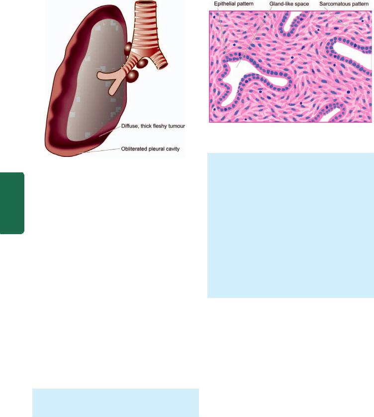
506
Pathology Systemic III SECTION
Figure 17.41 
 Malignant (diffuse) mesothelioma, gross appearance. The tumour is seen to form a thick, white, fleshy coat over the parietal and visceral surfaces.
osteoarthropathy or hypoglycaemia. Removal of the tumour is generally curative.
Malignant (Diffuse) Mesothelioma
Malignant or diffuse mesothelioma is rare. It is a highly malignant tumour associated with high mortality. The tumour is significant in view of its recognised association with occupational exposure to asbestos (particularly crocidolite) for a number of years, usually 20 to 40 years (page 492). About 90% of malignant mesotheliomas are asbestos-related. Mechanism of carcinogenicity by asbestos is not quite clear but it appears that prolonged exposure of amphibole type of asbestos is capable of inducing oncogenic mutation in the mesothelium. However, prolonged asbestos-exposure is considered more significant rather than heavy exposure as documented by occurrence of malignant mesothelioma in the family members of asbestos workers. Although combination of cigarette smoking and asbestos exposure greatly increases risk to develop bronchogenic carcinoma, there is no such extra increased risk of developing mesothelioma in asbestos workers who smoke. Recently, SV40 (simian vacuolating virus) antigen has also been implicated in the etiology of mesothelioma.
Grossly, the tumour is characteristically diffuse, forming a thick, white, fleshy coating over the parietal and visceral surfaces (Fig. 17.41).
Figure 17.42 
 Malignant mesothelioma, showing biphasic pattern of growth.
Microscopically, malignant mesothelioma may have epithelial, sarcomatoid or biphasic patterns.
i)Epithelial pattern resembles an adenocarcinoma, consisting of tubular and tubulo-papillary formations. The tumour cells are usually well-differentiated, cuboidal, flattened or columnar cells.
ii)Sarcomatoid pattern consists of spindle cell sarcoma resembling fibrosarcoma. The tumour cells are arranged in a storiform pattern with abundant collagen between them.
iii)Biphasic pattern shows mixed growth having epithelial as well as sarcomatoid pattern. Usually, there are slit-like or gland-like spaces lined by neoplastic mesothelial cells separated by proliferating spindleshaped tumour cells (Fig. 17.42).
Asbestos bodies are found in the lungs of most patients with malignant mesothelioma of any histologic type.
Clinical manifestations include chest pain, dyspnoea, pleural effusion and infections. The tumour spreads rapidly by direct invasion into lung and by lymphatic spread into hilar lymph nodes and pericardium. Sometimes distant metastases, particularly to the liver, occur. The prognosis is poor; 50% of patients die within one year of diagnosis.
SECONDARY PLEURAL TUMOURS
Metastatic malignancies in the pleura are more common than the primary tumours and appear as small nodules scattered over the lung surface. The most frequent primary malignant tumours metastasising to the pleura are of the lung and breast through lymphatics, and ovarian cancers via haematogenous route.