

- •Foreword
- •Preface
- •Contents
- •1. Introduction to Pathology
- •2. Techniques for the Study of Pathology
- •6. Inflammation and Healing
- •8. Neoplasia
- •16. The Heart
- •17. The Respiratory System
- •18. The Eye, ENT and Neck
- •20. The Gastrointestinal Tract
- •24. The Female Genital Tract
- •25. The Breast
- •26. The Skin
- •27. The Endocrine System
- •28. The Musculoskeletal System
- •29. Soft Tissue Tumours
- •30. The Nervous System
- •Appendix
- •Further Readings
- •Index
192
Techniques Basic and Pathology General I SECTION
Chapter 8 |
Neoplasia |
NOMENCLATURE AND CLASSIFICATION
INTRODUCTION. The term ‘neoplasia’ means new growth; the new growth produced is called ‘neoplasm’ or ‘tumour’. However, all ‘new growths’ are not neoplasms since examples of new growth of tissues and cells also exist in the processes of embryogenesis, regeneration and repair, hyperplasia and hormonal stimulation. The proliferation and maturation of cells in normal adults is controlled as a result of which some cells proliferate throughout life (labile cells), some have limited proliferation (stable cells), while others do not replicate (permanent cells). On the other hand, neoplastic cells lose control and regulation of replication and form an abnormal mass of tissue.
Therefore, satisfactory definition of a neoplasm or tumour is ‘a mass of tissue formed as a result of abnormal, excessive, uncoordinated, autonomous and purposeless proliferation of cells
even after cessation of stimulus for growth which caused it’. The branch of science dealing with the study of neoplasms or tumours is called oncology (oncos=tumour, logos=study). Neoplasms may be ‘benign’ when they are slow-growing and localised without causing much difficulty to the host, or ‘malignant’ when they proliferate rapidly, spread throughout the body and may eventually cause death of the host. The common term used for all malignant tumours is cancer. Hippocrates (460-377 BC) coined the term karkinos for cancer of the breast. The word ‘cancer’ means crab, thus reflecting the true character of cancer since ‘it sticks to the part stubbornly like a crab’.
All tumours, benign as well as malignant, have 2 basic components:
 ‘Parenchyma’ comprised by proliferating tumour cells; parenchyma determines the nature and evolution of the tumour.

 ‘Supportive stroma’ composed of fibrous connective tissue and blood vessels; it provides the framework on which the parenchymal tumour cells grow.
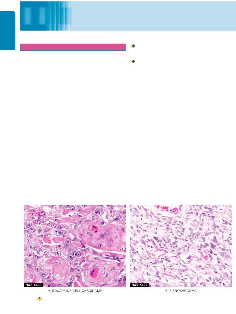
The tumours derive their nomenclature on the basis of the parenchymal component comprising them. The suffix ‘-oma’ is added to denote benign tumours. Malignant tumours of epithelial origin are called carcinomas, while malignant mesenchymal tumours are named sarcomas (sarcos = fleshy) (Fig. 8.1). However, some cancers are composed of highly undifferentiated cells and are referred to as undifferentiated
malignant tumours.
Although, this broad generalisation regarding nomenclature of tumours usually holds true in majority of instances, some examples contrary to this concept are: melanoma for carcinoma of the melanocytes, hepatoma for carcinoma of the hepatocytes, lymphoma for malignant tumour of the lymphoid tissue, and seminoma for malignant tumour of the testis. Leukaemia is the term used for cancer of blood forming cells.
SPECIAL CATEGORIES OF TUMOURS. Following categories of tumours are examples which defy the generalisation in nomenclature given above:
1. Mixed tumours. When two types of tumours are combined in the same tumour, it is called a mixed tumour. For example:
i) Adenosquamous carcinoma is the combination of adenocarcinoma and squamous cell carcinoma in the endometrium.
Figure 8.1 Examples of carcinoma (epithelial malignant tumour) (A) and sarcoma (mesenchymal malignant tumour) (B).

ii)Adenoacanthoma is the mixture of adenocarcinoma and benign squamous elements in the endometrium.
iii)Carcinosarcoma is the rare combination of malignant tumour of the epithelium (carcinoma) and of mesenchymal tissue (sarcoma) such as in thyroid.
iv)Collision tumour is the term used for morphologically two different cancers in the same organ which do not mix with each other.
v)Mixed tumour of the salivary gland (or pleomorphic adenoma) is the term used for benign tumour having combination of both epithelial and mesenchymal tissue elements.
2. Teratomas. These tumours are made up of a mixture of various tissue types arising from totipotent cells derived from the three germ cell layers—ectoderm, mesoderm and endoderm. Most common sites for teratomas are ovaries and testis (gonadal teratomas). But they occur at extra-gonadal sites as well, mainly in the midline of the body such as in the head and neck region, mediastinum, retroperitoneum, sacrococcygeal region etc. Teratomas may be benign or mature (most of the ovarian teratomas) or malignant or immature (most of the testicular teratomas).
3.Blastomas (Embryomas). Blastomas or embryomas are a group of malignant tumours which arise from embryonal or partially differentiated cells which would normally form blastema of the organs and tissue during embryogenesis. These tumours occur more frequently in infants and children (under 5 years of age) and include some examples of tumours in this age group: neuroblastoma, nephroblastoma (Wilms’ tumour), hepatoblastoma, retinoblastoma, medulloblastoma, pulmonary blastoma.
4.Hamartoma. Hamartoma is benign tumour which is made of mature but disorganised cells of tissues indigenous to the particular organ e.g. hamartoma of the lung consists of mature cartilage, mature smooth muscle and epithelium. Thus, all mature differentiated tissue elements which comprise the bronchus are present in it but are jumbled up as a mass.
5.Choristoma. Choristoma is the name given to the ectopic islands of normal tissue. Thus, choristoma is heterotopia but is not a true tumour, though it sounds like one.
CLASSIFICATION. Currently, classification of tumours is based on the histogenesis (i.e. cell of origin) and on the anticipated behaviour (Table 8.1). However, it must be
TABLE 8.1: Classification of Tumours.
Tissue of Origin |
Benign |
Malignant |
|
|
|
I. TUMOURS OF ONE PARENCHYMAL CELL TYPE
A.Epithelial Tumours
1. |
Squamous epithelium |
Squamous cell papilloma |
Squamous cell (Epidermoid) carcinoma |
2. |
Transitional epithelium |
Transitional cell papilloma |
Transitional cell carcinoma |
3. |
Glandular epithelium |
Adenoma |
Adenocarcinoma |
4. |
Basal cell layer skin |
— |
Basal cell carcinoma |
5. |
Neuroectoderm |
Naevus |
Melanoma (Melanocarcinoma) |
6. |
Hepatocytes |
Liver cell adenoma |
Hepatoma (Hepatocellular carcinoma) |
7. |
Placenta (Chorionic epithelium) |
Hydatidiform mole |
Choriocarcinoma |
B.Non-epithelial (Mesenchymal) Tumours
1. |
Adipose tissue |
Lipoma |
Liposarcoma |
2. |
Adult fibrous tissue |
Fibroma |
Fibrosarcoma |
3. |
Embryonic fibrous tissue |
Myxoma |
Myxosarcoma |
4. |
Cartilage |
Chondroma |
Chondrosarcoma |
5. |
Bone |
Osteoma |
Osteosarcoma |
6. |
Synovium |
Benign synovioma |
Synovial sarcoma |
7. |
Smooth muscle |
Leiomyoma |
Leiomyosarcoma |
8. |
Skeletal muscle |
Rhabdomyoma |
Rhabdomyosarcoma |
9. |
Mesothelium |
— |
Mesothelioma |
10. |
Blood vessels |
Haemangioma |
Angiosarcoma |
11. |
Lymph vessels |
Lymphangioma |
Lymphangiosarcoma |
12. |
Glomus |
Glomus tumour |
— |
13. |
Meninges |
Meningioma |
Invasive meningioma |
14. |
Haematopoietic cells |
— |
Leukaemias |
15. |
Lymphoid tissue |
Pseudolymphoma |
Malignant lymphomas |
16. |
Nerve sheath |
Neurilemmoma, Neurofibroma |
Neurogenic sarcoma |
17. |
Nerve cells |
Ganglioneuroma |
Neuroblastoma |
II. MIXED TUMOURS |
|
|
|
|
Salivary glands |
Pleomorphic adenoma |
Malignant mixed salivary tumour |
|
|
(mixed salivary tumour) |
|
III. TUMOURS OF MORE THAN ONE GERM CELL LAYER |
|
|
|
|
Totipotent cells in gonads or in embryonal rests |
Mature teratoma |
Immature teratoma |
|
|
|
|
193
Neoplasia 8 CHAPTER

194
Techniques Basic and Pathology General I SECTION
mentioned here that the classification described here is only a summary. Detailed classifications of benign and malignant tumours pertaining to different tissues and body systems along with morphologic features of specific tumours appear in the specific chapters of Systemic Pathology later.
CHARACTERISTICS OF TUMOURS
Majority of neoplasms can be categorised clinically and morphologically into benign and malignant on the basis of certain characteristics listed below. However, there are exceptions—a small proportion of tumours have some features suggesting innocent growth while other features point towards a more ominous behaviour. Therefore, it must be borne in mind that based characteristics of neoplasms, there is a wide variation in the degree of deviation from the normal in all the tumours.
The characteristics of tumours are described under the following headings:
I. Rate of growth
II. Cancer phenotype and stem cells III. Clinical and gross features
IV. Microscopic features
V. Local invasion (Direct spread) VI. Metastasis (Distant spread).
Based on these characteristics, contrasting features of benign and malignant tumours are summarised in Table 8.2 and illustrated in Fig. 8.2.
I. RATE OF GROWTH
The tumour cells generally proliferate more rapidly than the normal cells. In general, benign tumours grow slowly and malignant tumours rapidly. However, there are exceptions to this generalisation. The rate at which the tumour enlarges depends upon 2 main factors:
1.Rate of cell production, growth fraction and rate of cell loss
2.Degree of differentiation of the tumour.
1.Rate of cell production, growth fraction and rate of cell loss. Rate of growth of a tumour depends upon 3 important parameters:
i)doubling time of tumour cells,
ii)number of cells remaining in proliferative pool (growth fraction), and
iii)rate of loss of tumour cells by cell shedding.
In general, malignant tumour cells have increased mitotic rate (doubling time) and slower death rate i.e. the cancer cells do not follow normal controls in cell cycle and are immortal. If the rate of cell division is high, it is likely that tumour cells in the centre of the tumour do not receive adequate nourishment and undergo ischaemic necrosis. At a stage when malignant tumours grow relentlessly, they do so because a larger proportion of tumour cells remain in replicative pool but due to lack of availability of adequate nourishment, these tumour cells are either lost by shedding or leave the cell cycle to enter into G0 (resting phase) or G1
TABLE 8.2: Contrasting Features of Benign and Malignant Tumours.
Feature |
Benign |
Malignant |
|
|
|
||
I. CLINICAL AND GROSS FEATURES |
|
||
1. |
Boundaries |
Encapsulated or well-circumscribed |
Poorly-circumscribed and irregular |
2. |
Surrounding tissue |
Often compressed |
Usually invaded |
3. |
Size |
Usually small |
Often larger |
4. |
Secondary changes |
Occur less often |
Occur more often |
II. MICROSCOPIC FEATURES |
|
|
|
1. |
Pattern |
Usually resembles the tissue of origin closely |
Often poor resemblance to tissue of origin |
2. |
Basal polarity |
Retained |
Often lost |
3. |
Pleomorphism |
Usually not present |
Often present |
4. |
Nucleo-cytoplasmic ratio |
Normal |
Increased |
5. |
Anisonucleosis |
Absent |
Generally present |
6. |
Hyperchromatism |
Absent |
Often present |
7. |
Mitoses |
May be present but are always |
Mitotic figures increased and are generally |
|
|
typical mitoses |
atypical and abnormal |
8. |
Tumour giant cells |
May be present but without nuclear atypia |
Present with nuclear atypia |
9. |
Chromosomal abnormalities |
Infrequent |
Invariably present |
10. |
Function |
Usually well maintained |
May be retained, lost or become abnormal |
III. GROWTH RATE |
Usually slow |
Usually rapid |
|
IV. LOCAL INVASION |
Often compresses the surrounding tissues |
Usually infiltrates and invades the adjacent |
|
|
|
without invading or infiltrating them |
tissues |
V. METASTASIS |
Absent |
Frequently present |
|
VI. PROGNOSIS |
Local complications |
Death by local and metastatic complications |
|
|
|
|
|
195
Neoplasia 8 CHAPTER
Figure 8.2 
 Salient gross and microscopic features of prototypes of benign (left) and malignant (right) tumours.
phase. While dead tumour cells appear as ‘apoptotic figures’ (Chapter 3), the dividing cells of tumours are seen as normal and abnormal ‘mitotic figures’ (discussed later). Ultimately, malignant tumours grow in size because the cell production exceeds the cell loss.
2. Degree of differentiation. Secondly, the rate of growth of malignant tumour is directly proportionate to the degree
of differentiation. Poorly differentiated tumours show aggressive growth pattern as compared to better differentiated tumours. Some tumours, after a period of slow growth, may suddenly show spurt in their growth due to development of an aggressive clone of malignant cells. On the other hand, some tumours may cease to grow after sometime. Rarely, a malignant tumour may disappear spontaneously from the primary site, possibly due to necrosis caused by

196
Techniques Basic and Pathology General I SECTION
good host immune attack, only to reappear as secondaries elsewhere in the body e.g. choriocarcinoma, malignant melanoma.
The regulation of tumour growth is under the control of growth factors secreted by the tumour cells. Out of various growth factors, important ones modulating tumour biology are listed below and discussed later:
i)Epidermal growth factor (EGF)
ii)Fibroblast growth factor (FGF)
iii)Platelet-derived growth factor (PDGF)
iv)Colony stimulating factor (CSF)
v)Transforming growth factors-β (TGF-β)
vi)Interleukins (IL)
vii)Vascular endothelial growth factor (VEGF)
II. CANCER PHENOTYPE AND STEM CELLS
Normally growing cells in an organ are related to the neighbouring cells—they grow under normal growth controls, perform their assigned function and there is a balance between the rate of cell proliferation and the rate of cell death including cell suicide (i.e. apoptosis). Thus normal cells are socially desirable. However, cancer cells exhibit antisocial behaviour as under:
i) Cancer cells disobey the growth controlling signals in the body and thus proliferate rapidly.
ii)Cancer cells escape death signals and achieve immortality.
iii)Imbalance between cell proliferation and cell death in cancer causes excessive growth.
iv)Cancer cells lose properties of differentiation and thus perform little or no function.
v)Due to loss of growth controls, cancer cells are genetically unstable and develop newer mutations.
vi)Cancer cells overrun their neighbouring tissue and invade
locally.
vii) Cancer cells have the ability to travel from the site of origin to other sites in the body where they colonise and establish distant metastasis.
Cancer cells originate by clonal prolferation of a single progeny of a cell (monoclonality). Cancer cells arise from stem cells normally present in the tissues in small number and are not readily identifiable. These stem cells have the properties of prolonged self-renewal, asymmetric replication and transdifferentiation (i.e. plasticity). These cancer stem cells are called tumour-initiating cells. Their definite existence in acute leukaemias has been known for sometime and have now been found to be present in some other malignant tumours.
III. CLINICAL AND GROSS FEATURES
Clinically, benign tumours are generally slow growing, and depending upon the location, may remain asymptomatic (e.g. subcutaneous lipoma), or may produce serious symptoms (e.g. meningioma in the nervous system). On the other hand, malignant tumours grow rapidly, may ulcerate on the surface, invade locally into deeper tissues, may spread to distant sites (metastasis), and also produce systemic features such as weight loss, anorexia and anemia. In fact, two of the
cardinal clinical features of malignant tumours are: invasiveness and metastasis (discussed later).
Gross appearance of benign and malignant tumours may be quite variable and the features may not be diagnostic on the basis of gross appearance alone. However, certain distinctive features characterise almost all tumours compared to neighbouring normal tissue of origin—they have a different colour, texture and consistency. Gross terms such as papillary, fungating, infiltrating, haemorrhagic, ulcerative and cystic are used to describe the macroscopic appearance of the tumours. General gross features of benign and malignant tumours are as under (Figs. 8.2 and 8.3):
 Benign tumours are generally spherical or ovoid in shape. They are encapsulated or well-circumscribed, freely movable, more often firm and uniform, unless secondary changes like haemorrhage or infarction supervene (Fig. 8.2,A, E).

 Malignant tumours, on the other hand, are usually irregular in shape, poorly-circumscribed and extend into the adjacent tissues. Secondary changes like haemorrhage, infarction and ulceration are seen more often. Sarcomas typically have fish-flesh like consistency while carcinomas are generally firm (Fig. 8.2,C, G).
IV. MICROSCOPIC FEATURES
For recognising and classifying the tumours, the microscopic characteristics of tumour cells are of greatest importance. These features which are appreciated in histologic sections are as under:
1.microscopic pattern;
2.cytomorphology of neoplastic cells (differentiation and anaplasia);
3.tumour angiogenesis and stroma; and
4.inflammatory reaction.
1. Microscopic Pattern
The tumour cells may be arranged in a variety of patterns in different tumours as under:

 The epithelial tumours generally consist of acini, sheets, columns or cords of epithelial tumour cells that may be arranged in solid or papillary pattern (Fig. 8.2,B, D).
 The mesenchymal tumours have mesenchymal tumour cells arranged as interlacing bundles, fasicles or whorls, lying separated from each other usually by the intercellular matrix substance such as hyaline material in leiomyoma (Fig. 8.2,E), cartilaginous matrix in chondroma, osteoid in osteosarcoma, reticulin network in soft tissue sarcomas etc (Fig. 8.2,H).

 Certain tumours have mixed patterns e.g. teratoma arising from totipotent cells, pleomorphic adenoma of salivary gland (mixed salivary tumour), fibroadenoma of the breast, carcinosarcoma of the uterus and various other combinations of tumour types.

 Haematopoietic tumours such as leukaemias and lymphomas often have none or little stromal support.

 Generally, most benign tumours and low grade malignant tumours reduplicate the normal structure of origin more closely so that there is little difficulty in identifying and

197
Neoplasia 8 CHAPTER
Figure 8.3 
 Gross appearance of a prototype of benign and malignant tumour.
classifying such tumours (Fig. 8.2,B, F). However, anaplastic tumours differ greatly from the arrangement in normal tissue of origin of the tumour and may occasionally pose problems in classifying the tumour.
2.Cytomorphology of Neoplastic Cells (Differentiation and Anaplasia)
The neoplastic cell is characterised by morphologic and functional alterations, the most significant of which are
‘differentiation’ and ‘anaplasia’.

 Differentiation is defined as the extent of morphological and functional resemblance of parenchymal tumour cells to corresponding normal cells. If the deviation of neoplastic cell in structure and function is minimal as compared to normal cell, the tumour is described as ‘well-differentiated’ such as most benign and low-grade malignant tumours. ‘Poorly
differentiated’, ‘undifferentiated’ or ‘dedifferentiated’ are synonymous terms for poor structural and functional resemblance to corresponding normal cell.

 Anaplasia is lack of differentiation and is a characteristic feature of most malignant tumours. Depending upon the
degree of differentiation, the extent of anaplasia is also variable i.e. poorly differentiated malignant tumours have high degree of anaplasia.
As a result of anaplasia, noticeable morphological and functional alterations in the neoplastic cells are observed. These are considered below and are diagrammatically illustrated in Fig. 8.4:
i)Loss of polarity. Normally, the nuclei of epithelial cells are oriented along the basement membrane which is termed as basal polarity. This property is based on cell adhesion molecules, particularly selectins. Early in malignancy, tumour cells lose their basal polarity so that the nuclei tend to lie away from the basement membrane (Fig. 8.5).
ii)Pleomorphism. The term pleomorphism means variation in size and shape of the tumour cells. The extent of cellular pleomorphism generally correlates with the degree of anaplasia. Tumour cells are often bigger than normal but in some tumours they can be of normal size or smaller than normal (Fig. 8.6).
iii)N:C ratio. Generally, the nuclei of malignant tumour cells show more conspicuous changes. Nuclei are enlarged
Figure 8.4 
 Diagrammatic representation of cytomorphologic features of neoplastic cells. Characteristics of cancer (B) in a gland are contrasted with the appearance of an acinus (A).
198
Techniques Basic and Pathology General I SECTION
Figure 8.5 
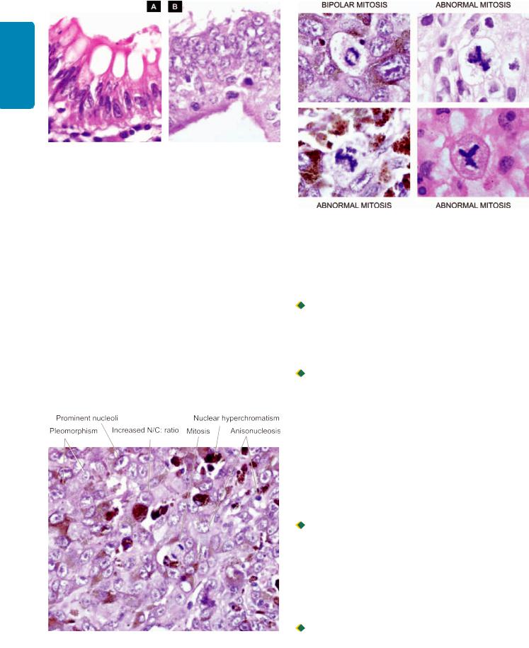
 Microscopic appearance of loss of nuclear polarity (B) contrasted with normal basal polarity in columnar epithelium (A). The basement membrane is intact in both.
disproportionate to the cell size so that the nucleocytoplasmic ratio is increased from normal 1:5 to 1:1 (Fig. 8.6).
iv)Anisonucleosis. Just like cellular pleomorphism, the nuclei too, show variation in size and shape in malignant tumour cells (Fig. 8.6).
v)Hyperchromatism. Characteristically, the nuclear chromatin of malignant cell is increased and coarsely clumped. This is due to increase in the amount of nucleoprotein resulting in dark-staining nuclei, referred to as hyperchromatism (Fig. 8.6). Nuclear shape may vary, nuclear membrane may be irregular and nuclear chromatin is clumped along the nuclear membrane.
vi)Nucleolar changes. Malignant cells frequently have a prominent nucleolus or nucleoli in the nucleus reflecting increased nucleoprotein synthesis (Fig. 8.6). This may be demonstrated as Nucleolar Organiser Region (NOR) by silver (Ag) staining called AgNOR material.
Figure 8.6 
 Nuclear features of malignant cells in malignant melanoma—pleomorphism, anisonucleosis, increased N/C: ratio, nuclear hyperchromatism and prominent nucleoli.
Figure 8.7 
 Normal and abnormal (atypical) mitotic figures.
vii)Mitotic figures. The parenchymal cells of poorlydifferentiated tumours often show large number of mitoses as compared with benign tumours and well-differentiated malignant tumours. As stated above, these appear as either normal or abnormal mitotic figures (Fig. 8.7):

 Normal mitotic figures may be seen in some non-neoplastic proliferating cells (e.g. haematopoietic cells of the bone marrow, intestinal epithelium, hepatocytes etc), in certain benign tumours and some low grade malignant tumours; in sections they are seen as a dark band of dividing chromatin at two poles of the nuclear spindle.

 Abnormal or atypical mitotic figures are more important in malignant tumours and are identified as tripolar, quadripolar and multipolar spindles in malignant tumour cells.
viii)Tumour giant cells. Multinucleate tumour giant cells or giant cells containing a single large and bizarre nucleus, possessing nuclear characters of the adjacent tumour cells, are another important feature of anaplasia in malignant tumours (Fig. 8.8).
ix)Functional (Cytoplasmic) changes. Structural anaplasia in tumours is accompanied with functional anaplasia as appreciated from the cytoplasmic constituents of the tumour cells. The functional abnormality in neoplasms may be quantitative, qualitative, or both.
 Generally, benign tumours and better-differentiated malignant tumours continue to function well qualitatively, though there may be quantitative abnormality in the product e.g. large or small amount of collagen produced by benign tumours of fibrous tissue, keratin formation in welldifferentiated squamous cell carcinoma. In more anaplastic tumours, there is usually quantitative fall in the product made by the tumour cells e.g. absence of keratin in anaplastic squamous cell carcinoma.

 There may be both qualitative and quantitative abnormality of the cellular function in some anaplastic tumours e.g. multiple myeloma producing abnormal immunoglobulin in large quantities.
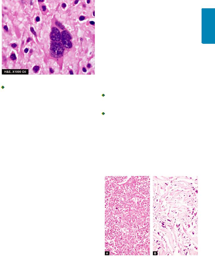
Figure 8.8 
 A multinulceate tumour giant cell in osteosarcoma.

 Endocrine tumours may cause excessive hormone production leading to characteristic clinical syndromes. Besides the production of hormones by endocrine tumours, hormones or hormone-like substances may be produced by certain tumours quite unrelated to the endocrine glands. This property of tumours is called ectopic hormone production e.g. oat cell carcinoma of the lung can secrete ACTH and ADH; less often it may produce gonadotropin, thyrotropin, parathormone, calcitonin and growth hormone. Ectopic erythropoietin may be produced by carcinoma of kidneys, hepatocellular carcinoma and cerebellar haemangioblastoma.
x) Chromosomal abnormalities. All tumour cells have abnormal genetic composition and on division they transmit the genetic abnormality to their progeny. The chromosomal abnormalities are more marked in more malignant tumours which include deviations in both morphology and number of chromosomes. Most malignant tumours show DNA aneuploidy, often in the form of an increase in the number of chromosomes, reflected morphologically by the increase in the size of nuclei.
One of the most important examples of a consistent chromosomal abnormality in human malignancy is the presence of Philadelphia chromosome (named after the city in which it was first described) in 95% cases of chronic myeloid leukaemia. In this, part of the long arm of chromosome 9 is translocated to part of the long arm of chromosome 22 (t 9; 22). Other examples of neoplasms showing chromosomal abnormalities are Burkitt’s lymphoma, acute lymphoid leukaemia, multiple myeloma, retinoblastoma, oat cell carcinoma, Wilms’ tumour etc.
3. Tumour Angiogenesis and Stroma
The connective tissue alongwith its vascular network forms the supportive framework on which the parenchymal tumour cells grow and receive nourishment. In addition to variable amount of connective tissue and vascularity, the stroma may have nerves and metaplastic bone or cartilage but no lymphatics.
TUMOUR ANGIOGENESIS. In order to provide nourishment to growing tumour, new blood vessels are formed from
pre-existing ones (angiogenesis). How this takes place under |
199 |
the influence of angiogenic factors elaborated by tumour cells |
|
such as vascular endothelium growth factor (VEGF) is |
CHAPTER |
marker to assess the rate of growth of tumours and hence |
|
discussed later under molecular pathogenesis of cancer. |
|
However, related morphologic features are as under: |
|
i) Microvascular density. The new capillaries add to the |
|
vascular density of the tumour which has been used as a |
|
grade the tumours. This is done by counting microvascular |
8 |
density in the section of the tumour. |
|
ii) Central necrosis. However, if the tumour outgrows its blood |
Neoplasia |
may be scanty or excessive. In the former case, the tumour is |
|
supply as occurs in rapidly growing tumours or tumour |
|
angiogenesis fails, its core undergoes ischaemic necrosis. |
|
TUMOUR STROMA. The collagenous tissue in the stroma |
|
soft and fleshy (e.g. in sarcomas, lymphomas), while in the |
|
latter case the tumour is hard and gritty (e.g. infiltrating duct |
|
carcinoma breast). Growth of fibrous tissue in tumour is |
|
stimulated by basic fibroblast growth factor (bFGF) |
|
elaborated by tumour cells. |
|
If the epithelial tumour is almost entirely composed of |
|
parenchymal cells, it is called medullary e.g. medullary |
|
carcinoma of the breast (Fig. 8.9, A), medullary carcinoma |
|
of the thyroid. |
|
If there is excessive connective tissue stroma in the |
|
epithelial tumour, it is referred to as desmoplasia and the |
|
tumour is hard or scirrhous e.g. infiltrating duct carcinoma |
|
breast (Fig. 8.9, B), linitis plastica of the stomach. |
|
4. Inflammatory Reaction |
|
At times, prominent inflammatory reaction is present in and |
|
around the tumours. It could be the result of ulceration in |
|
the cancer when there is secondary infection. The |
|
inflammatory reaction in such instances may be acute or |
|
chronic. However, some tumours show chronic inflammatory |
|
reaction, chiefly of lymphocytes, plasma cells and |
|
Figure 8.9 
 Tumour stroma. A, Medullary carcinoma of breast, rich in parenchymal cells. B, Scirrhous carcinoma of breast having abundant collagenised (desmopastic) stroma.
200
Techniques Basic and Pathology General I SECTION
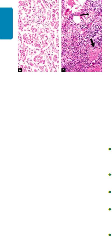
Figure 8.10 
 Inflammatory reaction in the stroma of the tumour. A,
Lymphocytic reaction in seminoma testis. B, Granulomatous reaction (thick arrow) in Hodgkin’s lymphoma (thin arrow for RS cell).
macrophages, and in some instances granulomatous reaction, in the absence of ulceration. This is due to cell-mediated immunologic response by the host in an attempt to destroy the tumour. In some cases, such an immune response improves the prognosis.
The examples of such reaction are: seminoma testis (Fig. 8.10), malignant melanoma of the skin, lymphoepithelioma of the throat, medullary carcinoma of the breast, choriocarcinoma, Warthin’s tumour of salivary glands etc.
V. LOCAL INVASION (DIRECT SPREAD)
BENIGN TUMOURS. Most benign tumours form encapsulated or circumscribed masses that expand and push aside the surrounding normal tissues without actually invading, infiltrating or metastasising.
MALIGNANT TUMOURS. Malignant tumours also enlarge by expansion and some well-differentiated tumours may be partially encapsulated as well e.g. follicular carcinoma thyroid. But characteristically, they are distinguished from benign tumours by invasion, infiltration and destruction of the surrounding tissue, besides distant metastasis (described below). In general, tumours invade via the route of least resistance, though eventually most cancers recognise no anatomic boundaries. Often, cancers extend through tissue spaces, permeate lymphatics, blood vessels, perineural spaces and may penetrate a bone by growing through nutrient foramina. More commonly, the tumours invade thinwalled capillaries and veins than thick-walled arteries. Dense compact collagen, elastic tissue and cartilage are some of the tissues which are sufficiently resistant to invasion by tumours.
Mechanism of invasion of malignant tumours is discussed together with that of metastasis below.
VI. METASTASIS (DISTANT SPREAD)
Metastasis (meta = transformation, stasis = residence) is defined as spread of tumour by invasion in such a way that
discontinuous secondary tumour mass/masses are formed at the site of lodgement. Metastasis and invasiveness are the
two most important features to distinguish malignant from benign tumours: benign tumours do not metastasise while all the malignant tumours with a few exceptions like gliomas of the central nervous system and basal cell carcinoma of the skin, can metastasise. Generally, larger, more aggressive and rapidly-growing tumours are more likely to metastasise but there are numerous exceptions. About one-third of malignant tumours at presentation have evident metastatic deposits while another 20% have occult metastasis.
Routes of Metastasis
Cancers may spread to distant sites by following pathways:
1.Lymphatic spread
2.Haematogenous spread
3.Spread along body cavities and natural passages (Transcoelomic spread, along epithelium-lined surfaces, spread via cerebrospinal fluid, implantation).
1.LYMPHATIC SPREAD. In general, carcinomas metastasise
by lymphatic route while sarcomas favour haematogenous route.
However, sarcomas may also spread by lymphatic pathway. The involvement of lymph nodes by malignant cells may be of two forms:
i)Lymphatic permeation. The walls of lymphatics are readily invaded by cancer cells and may form a continuous growth in the lymphatic channels called lymphatic permeation.
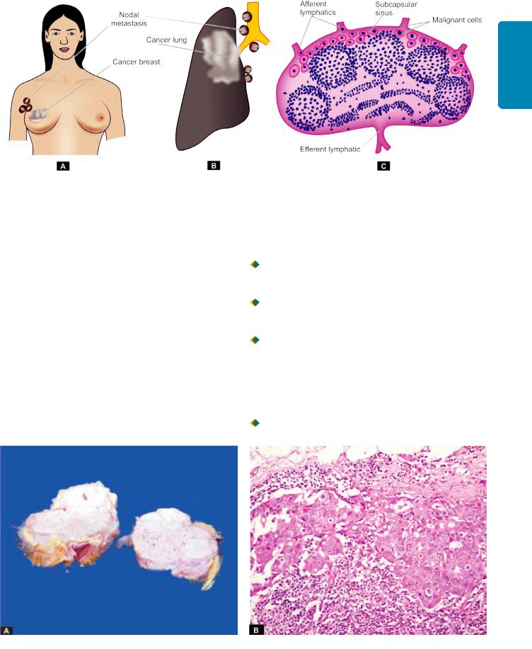
ii)Lymphatic emboli. Alternatively, the malignant cells may detach to form tumour emboli so as to be carried along the lymph to the next draining lymph node. The tumour emboli enter the lymph node at its convex surface and are lodged in the subcapsular sinus where they start growing (Fig. 8.11). Later, of course, the whole lymph node may be replaced and enlarged by the metastatic tumour (Fig. 8.12).
 Generally, regional lymph nodes draining the tumour are invariably involved producing regional nodal metastasis e.g. from carcinoma breast to axillary lymph nodes, from carcinoma thyroid to lateral cervical lymph nodes, bronchogenic carcinoma to hilar and para-tracheal lymph nodes etc.

 However, all regional nodal enlargements are not due to nodal metastasis because necrotic products of tumour and antigens may also incite regional lymphadenitis of sinus
histiocytosis.

 Sometimes lymphatic metastases do not develop first in the lymph node nearest to the tumour because of venouslymphatic anastomoses or due to obliteration of lymphatics by inflammation or radiation, so called skip metastasis.
 Other times, due to obstruction of the lymphatics by tumour cells, the lymph flow is disturbed and tumour cells spread against the flow of lymph causing retrograde metastases at unusual sites e.g. metastasis of carcinoma prostate to the supraclavicular lymph nodes, metastatic deposits from bronchogenic carcinoma to the axillary lymph nodes.

 Virchow’s lymph node is nodal metastasis preferentially to supraclavicular lymph node from cancers of abdominal organs e.g. cancer stomach, colon, and gall bladder.
201
Neoplasia 8 CHAPTER
Figure 8.11 
 Regional nodal metastasis. A, Axillary nodes involved by carcinoma breast. B, Hilar and para-tracheal lymph nodes involved by bronchogenic carcinoma. C, Lymphatic spread begins by lodgement of tumour cells in subcapsular sinus via afferent lymphatics entering at the convex surface of the lymph node.
It is believed that lymph nodes in the vicinity of tumour perform multiple roles—as initial barrier filter, and in destruction of tumour cells, while later provide fertile soil for growth of tumour cells.
Mechanism of lymphatic route of metastasis is discussed later under biology of invasion and metastasis.
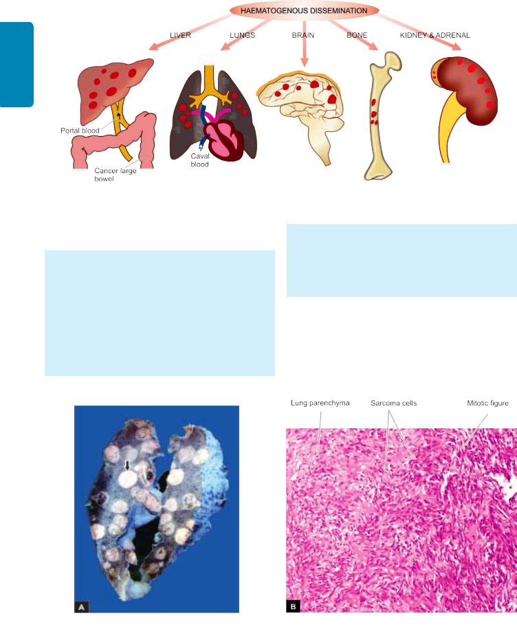
2. HAEMATOGENOUS SPREAD. Blood-borne metastasis is
the common route for sarcomas but certain carcinomas also frequently metastasise by this mode, especially those of the lung, breast, thyroid, kidney, liver, prostate and ovary. The sites where blood-borne metastasis commonly occurs are: the liver, lungs, brain, bones, kidney and adrenals, all of which provide ‘good soil’ for the growth of ‘good seeds’ (seed-soil theory). However, a few organs such as spleen, heart, and skeletal muscle generally do not allow tumour metastasis to grow. Spleen is unfavourable site due to open sinusoidal pattern which does not permit tumour cells to stay there long enough to produce metastasis. In general, only a proportion
of cancer cells are capable of clonal proliferation in the proper environment; others die without establishing a metastasis.
 Systemic veins drain blood into vena cavae from limbs, head and neck and pelvis. Therefore, cancers of these sites more often metastasise to the lungs.

 Portal veins drain blood from the bowel, spleen and pancreas into the liver. Thus, tumours of these organs frequently have secondaries in the liver.
 Arterial spread of tumours is less likely because they are thick-walled and contain elastic tissue which is resistant to invasion. Nevertheless, arterial spread may occur when tumour cells pass through pulmonary capillary bed or through pulmonary arterial branches which have thin walls. Cancer of the lung may, however, metastasise by pulmonary arterial route to kidneys, adrenals, bones, brain etc.

 Retrograde spread by blood route may occur at unusual sites due to retrograde spread after venous obstruction, just
Figure 8.12 
 Metastatic carcinoma in lymph nodes. A, Matted mass of lymph nodes is surrounded by increased fat. Sectioned surface shows merging capsules of lymph nodes and replacement of grey brown tissue of nodes by large grey white areas of tumour. B, Masses of malignant cells are seen in the subcapsular sinus and extending into the underlying nodal tissue.
202
Techniques Basic and Pathology General I SECTION
Figure 8.13 
 Gross appearance of haematogenous metastases at common sites.
as with lymphatic metastases. Important examples are vertebral metastases in cancers of the thyroid and prostate.
Grossly, blood-borne metastases in an organ appear as multiple, rounded nodules of varying size, scattered throughout the organ (Fig. 8.13). Sometimes, the metastasis may grow bigger than the primary tumour. At times, metastatic deposits may come to attention first without an evident primary tumour. In such cases search for primary tumour may be rewarding, but rarely the primary tumour may remain undetected or occult. Metastatic deposits just like primary tumour may cause further dissemination via lymphatics and blood vessels
(Fig. 8.14, A).
Microscopically, the secondary deposits generally reproduce the structure of primary tumour (Fig. 8.14, B). However, the same primary tumour on metastasis at different sites may show varying grades of differentiation, apparently due to the influence of local environment surrounding the tumour for its growth.
3. SPREAD ALONG BODY CAVITIES AND NATURAL PASSAGES. Uncommonly, some cancers may spread by seeding across body cavities and natural passages. These routes of distant spread are as under:
i) Transcoelomic spread. Certain cancers invade through the serosal wall of the coelomic cavity so that tumour
Figure 8.14 
 Metastatic sarcoma lung. A, Sectioned surface of the lung shows replacement of slaty-grey spongy parenchyma with multiple, firm, grey-white nodular masses, some having areas of haemorhages and necrosis. B, Microscopic appearance of pulmonary metastatic deposits from sarcoma.
fragments or clusters of tumour cells break off to be carried in the coelomic fluid and are implanted elsewhere in the body cavity. Peritoneal cavity is involved most often, but occasionally pleural and pericardial cavities are also affected. A few examples of transcoelomic spread are as follows:
a)Carcinoma of the stomach seeding to both ovaries (Krukenberg tumour).
b)Carcinoma of the ovary spreading to the entire peritoneal cavity without infiltrating the underlying organs.
c)Pseudomyxoma peritonei is the gelatinous coating of the peritoneum from mucin-secreting carcinoma of the ovary or apppendix.
d)Carcinoma of the bronchus and breast seeding to the pleura and pericardium.
ii) Spread along epithelium-lined surfaces. It is unusual for a malignant tumour to spread along the epithelium-lined surfaces because intact epithelium and mucus coat are quite resistant to penetration by tumour cells. However, exceptionally a malignant tumour may spread through:
a) the fallopian tube from the endometrium to the ovaries or vice-versa;
b)through the bronchus into alveoli; and
c)through the ureters from the kidneys into lower urinary tract.
iii)Spread via cerebrospinal fluid. Malignant tumour of the ependyma and leptomeninges may spread by release of tumour fragments and tumour cells into the CSF and produce metastases at other sites in the central nervous system.
iv)Implantation. Rarely, a tumour may spread by implantation by surgeon’s scalpel, needles, sutures, or may be implanted by direct contact such as transfer of cancer of the lower lip to the apposing upper lip.
MECHANISM AND BIOLOGY OF
INVASION AND METASTASIS
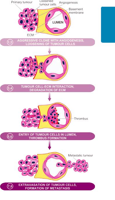
The process of local invasion and distant spread by lymphatic and haematogenous routes discussed above involves passage through barriers before gaining access to the vascular lumen. This includes making the passage by the cancer cells by dissolution of extracellular matrix (ECM) at three levels— at the basement membrane of tumour itself, at the level of interstitial connective tissue, and at the basement membrane of microvasculature. The following steps are involved at the molecular level which are schematically illustrated in
Fig. 8.15.
1. Aggressive clonal proliferation and angiogenesis. The first step in the spread of cancer cells is the development of rapidly proliferating clone of cancer cells. This is explained on the basis of tumour heterogeneity, i.e. in the population of monoclonal tumour cells, a subpopulation or clone of tumour cells has the right biologic characteristics to complete the steps involved in the development of metastasis. Tumour angiogenesis plays a very significant role in metastasis since the new vessels formed as part of growing tumour are more vulnerable to invasion as these evolving vessels are directly in contact with cancer cells.
203
Neoplasia 8 CHAPTER
Figure 8.15 
 Mechanism and biology of local invasion and metastasis. The serial numbers in the figure correspond to their description in the text.
2.Tumour cell loosening. Normal cells remain glued to each other due to presence of cell adhesion molecules (CAMs) i.e.E (epithelial)-cadherin. In epithelial cancers, there is either loss or inactivation of E-cadherin and also other CAMs of immunoglobulin superfamily, all of which results in loosening of cancer cells.
3.Tumour cell-ECM interaction. Loosened cancer cells are now attached to ECM proteins, mainly laminin and fibronectin. This attachment is facilitated due to profoundness of receptors on the cancer cells for both these proteins. There is also loss of integrins, the transmembrane receptors, further favouring invasion.

204
Techniques Basic and Pathology General I SECTION
4.Degradation of ECM. Tumour cells overexpress proteases and matrix-degrading enzymes, metalloproteinases, that includes collagenases and gelatinase, while the inhibitors of metalloproteinases are decreased. Another protease, cathepsin D, is also increased in certain cancers. These enzymes bring about dissolution of ECM—firstly basement membrane of tumour itself, then make way for tumour cells through the interstitial matrix, and finally dissolve the basement membrane of the vessel wall.
5.Entry of tumour cells into capillary lumen. The tumour cells after degrading the basement membrane are ready to migrate into lumen of capillaries or venules for which the following mechanisms play a role:
i)Autocrine motility factor (AMF) is a cytokine derived from tumour cells and stimulates receptor-mediated motility of tumour cells.
ii)Cleavage products of matrix components which are formed following degradation of ECM have properties of tumour cell chemotaxis, growth promotion and angiogenesis in the cancer.
After the malignant cells have migrated through the breached basement membrane, these cells enter the lumen of lymphatic and capillary channels.
6.Thrombus formation. The tumour cells protruding in the lumen of the capillary are now covered with constituents of the circulating blood and form the thrombus. Thrombus provides nourishment to the tumour cells and also protects them from the immune attack by the circulating host cells. In fact, normally a large number of tumour cells are released into circulation but they are attacked by the host immune cells. Actually a very small proportion of malignant cells (less than 0.1%) in the blood stream survive to develop into metastasis.
7.Extravasation of tumour cells. Tumour cells in the circulation (capillaries, venules, lymphatics) may mechanically block these vascular channels and attach to vascular endothelium. In this way, the sequence similar to local invasion is repeated and the basement membrane in exposed.
8.Survival and growth of metastatic deposit. The extravasated malignant cells on lodgement in the right environment grow further under the influence of growth factors produced by host tissues, tumour cells and by cleavage products of matrix components. These growth factors in particular include: PDGF, FGF, TGF-β and VEGF. The metastatic deposits grow further if the host immune defense mechanism fails to eliminate it. Metastatic deposits may further metastasise to the same organ or to other sites by forming emboli.
PROGNOSTIC MARKERS
Metastasis is a common event in malignant tumours which greatly reduces the survival of patient. In the biology of tumour, metastasis is a form of unusual cell differentiation in which the tumour cells form disorderly masses at ectopic sites and start growing there. This random phenomenon takes place in a stepwise manner involving only a
subpopulation of tumour cells selectively. The process is
governed by inappropriate expression of genes which normally partake in physiologic processes i.e. it is a genetically programmed phenomenon.
Recent evidence has shown that in metastatic tumours, survival of host is correlated with some clinical and molecular features of tumours which act as prognostic markers. These are as under:
i)Clinical prognostic markers: Size, grade, vascular invasion and nodal involvement by the tumour.
ii)Molecular prognostic markers: Molecular markers indicative of poor prognosis in certain specific tumours are:
a)expression of an oncogene by tumour cells (C-met);
b)CD 44 molecule;
c)oestrogen receptors;
d)epidermal growth factor receptor;
e)angiogenesis factors and degree of neovascularisation; and
f)expression of metastasis associated gene or nucleic acid (MAGNA) in the DNA fragment in metastasising tumour.
GRADING AND STAGING OF CANCER
‘Grading’ and ‘staging’ are the two systems to predict tumour behaviour and guide therapy after a malignant tumour is detected. Grading is defined as the gross and microscopic degree
of differentiation of the tumour, while staging means extent of spread of the tumour within the patient. Thus, grading is histologic while staging is clinical.
Grading
Cancers may be graded grossly and microscopically. Gross features like exophytic or fungating appearance are indicative of less malignant growth than diffusely infiltrating tumours. However, grading is largely based on 2 important histologic features: the degree of anaplasia, and the rate of growth.
Based on these features, cancers are categorised from grade I as the most differentiated, to grade III or IV as the most undifferentiated or anaplastic. Many systems of grading have been proposed but the one described by Broders for dividing squamous cell carcinoma into 4 grades depending upon the degree of differentiation is followed for other malignant tumours as well. Broders’ grading is as under:
Grade I: Well-differentiated (less than 25% anaplastic cells). Grade II: Moderately-differentiated (25-50% anaplastic cells). Grade III: Moderately-differentiated (50-75% anaplastic cells).
Grade IV: Poorly-differentiated or anaplastic (more than 75% anaplastic cells).
However, grading of tumours has several shortcomings. It is subjective and the degree of differentiation may vary from one area of tumour to the other. Therefore, it is common practice with pathologists to grade cancers in descriptive terms (e.g. well-differentiated, undifferentiated, keratinising, non-keratinising etc) rather than giving the tumours grade numbers.
More objective criteria for histologic grading include use of flow cytometry for mitotic cell counts, cell proliferation
markers by immunohistochemistry, and by applying image morphometry for cancer cell and nuclear parameters.
Staging
The extent of spread of cancers can be assessed by 3 ways— by clinical examination, by investigations, and by pathologic examination of the tissue removed. Two important staging systems currently followed are: TNM staging and AJC staging.
TNM staging. (T for primary tumour, N for regional nodal involvement, and M for distant metastases) was developed by the UICC (Union Internationale Contre Cancer, Geneva). For each of the 3 components namely T, N and M, numbers are added to indicate the extent of involvement, as under:
T0 to T4: In situ lesion to largest and most extensive primary tumour.
N0 to N3: No nodal involvement to widespread lymph node involvement.
M0 to M2: No metastasis to disseminated haematogenous metastases.
AJC staging. American Joint Committee staging divides all cancers into stage 0 to IV, and takes into account all the 3 components of the preceding system (primary tumour, nodal involvement and distant metastases) in each stage.
TNM and AJC staging systems can be applied for staging most malignant tumours.
Currently, clinical staging of tumours does not rest on routine radiography (X-ray, ultrasound) and exploratory surgery but more modern techniques are available by which it is possible to ‘stage’ a malignant tumour by non-invasive techniques. These include use of computed tomography (CT) and magnetic resonance imaging (MRI) scan based on tissue density for locating the local extent of tumour and its spread to other organs. More recently, availability of positron emission tomography (PET) scan has overcome the limitation of CT and MRI scan because PET scan facilitates distinction of benign and malignant tumour on the basis of biochemical and molecular processes in tumours. Radioactive tracer studies in vivo such as use of iodine isotope 125 bound to specific tumour antibodies is another method by which small number of tumour cells in the body can be detected by imaging of tracer substance bound to specific tumour antigen.
EPIDEMIOLOGY AND
PREDISPOSITION TO NEOPLASIA
CANCER INCIDENCE
The overall incidence of cancer in a population or a country is known by registration of all cancer cases (cancer registry) and by rate of death from cancer. Worldwide, it is estimated that about 20% of all deaths are cancer-related; in US, cancer is the second most common cause of deaths, next to heart disease. There have been changing patterns in incidence of cancers in both the sexes and in different geographic locations as outlined below. Table 8.3 shows worldwide incidence (in descending order) of different forms of cancer in men,
TABLE 8.3. Five Most Common Primary Cancers in the World.
|
Men |
Women |
Children (Under 20) |
|
|
|
|
1. |
Lung |
Breast |
Acute leukaemia |
|
(oral cavity in India) |
(cervix in India) |
|
2. |
Prostate |
Lung |
CNS tumour |
3. |
Colorectal |
Colorectal |
Bone sarcoma |
4. |
Urinary bladder |
Endometrial |
Endocrine |
5. |
Lymphoma |
Lymphoma |
Soft tissue sarcoma |
|
|
|
|
women, and children. As evident from the Table, some types of cancers are more common in India while others are commoner in the Western populations since etiologic factors are different.
In general, most common cancers in the developed and developing countries are as under:

 Developed world: lung, breast, prostate and colorectal.
 Developing world: liver, cervical and oesophageal. About one-third of all cancers worldwide are attributed
to 9 modifiable life-style factors: tobacco use, alcohol consumption, obesity, physical inactivity, low fiber diet, unprotected sex, polluted air, indoor household smoke, and contaminated injections. Overall, there has been a declining trend in incidence of some of the cancers due to cancer screening programmes for cervical, breast, colorectal and prostate cancer.
EPIDEMIOLOGIC FACTORS
A lot of clinical and experimental research and epidemiological studies have been carried out in the field of oncology so as to know the possible causes of cancer and mechanisms involved in transformation of a normal cell into a neoplastic cell. It is widely known that no single factor is responsible for development of tumours. The role of some factors in causation of neoplasia is established while that of others is epidemiological and many others are still unknown.
Besides the etiologic role of some agents discussed later, the pattern and incidence of cancer depends upon the following:
A)A large number of predisposing epidemiologic factors or cofactors which include a number of endogenous host factors and exogenous environmental factors.
B)Chronic non-neoplastic (pre-malignant) conditions.
C)Role of hormones in cancer.
A. Predisposing Factors
1. FAMILIAL AND GENETIC FACTORS. It has long been suspected that familial predisposition and heredity play a role in the development of cancers. In general, the risk of developing cancer in relatives of a known cancer patient is almost three times higher as compared to control subjects. Some of the cancers with familial occurrence are colon, breast, ovary, brain and melanoma. Familial cancers occur at a relatively early age, appear at multiple sites and occur in 2 or more close relatives. The overall estimates suggest that genetic cancers comprise not greater than 5% of all cancers. Some of the common examples are as under:
i) Retinoblastoma. About 40% of retinoblastomas are familial and show an autosomal dominant inheritance.
205
Neoplasia 8 CHAPTER

206
Techniques Basic and Pathology General I SECTION
Carriers of such genetic composition have 10,000 times higher risk of developing retinoblastoma which is often bilateral. Such patients are predisposed to develop another primary malignant tumour, notably osteogenic sarcoma.
Familial form of retinoblastoma is due to missing of a portion of chromosome 13 where RB gene is normally located. This results in a genetic defect of absence of RB gene, the first ever tumour suppressor gene identified. An absent RB gene predisposes an individual to retinoblastoma but cancer develops when other copy of RB gene from the other parent is also defective.
ii)Familial polyposis coli. This condition has autosomal dominant inheritance. The polypoid adenomas may be seen at birth or in early age. By the age of 50 years, almost 100% cases of familial polyposis coli develop cancer of the colon.
iii)Multiple endocrine neoplasia (MEN). A combination of adenomas of pituitary, parathyroid and pancreatic islets (MEN-I) or syndrome of medullary carcinoma thyroid, pheochromocytoma and parathyroid tumour (MEN-II) are encountered in families.
iv)Neurofibromatosis or von Recklinghausen’s disease.
This condition is characterised by multiple neurofibromas and pigmented skin spots (cafe au lait spots). These patients have family history consistent with autosomal dominant inheritance in 50% of patients.
v)Cancer of the breast. Female relatives of breast cancer patients have 2 to 6 times higher risk of developing breast cancer. Inherited breast cancer comprises about 5-10% of all breast cancers. As discussed later, there are two breast cancer susceptibility genes, BRCA-1 and BRCA-2. Mutations in these genes appear in about 3% cases and these patients have about 85% risk of development of breast cancer.
vi)DNA-chromosomal instability syndromes. These are a group of pre-neoplastic conditions having defect in DNA repair mechanism. A classical example is xeroderma pigmentosum, an autosomal recessive disorder, characterised by extreme sensitivity to ultraviolet radiation. The patients may develop various types of skin cancers such as basal cell carcinoma, squamous cell carcinoma and malignant melanoma.
2.RACIAL AND GEOGRAPHIC FACTORS. Differences in racial incidence of some cancers may be partly attributed to the role of genetic composition but are largely due to influence of the environment and geographic differences affecting the whole population such as climate, soil, water, diet, habits, customs etc. Some of the examples of racial and geographic variations in various cancers are as under:
i)White Europeans and Americans develop most commonly malignancies of the lung, breast, and colon. Liver cancer is uncommon in these races. Breast cancer is uncommon in Japanese women but is more common in American women.
ii)Black Africans, on the other hand, have more commonly cancers of the skin, penis, cervix and liver.
iii)Japanese have five times higher incidence of carcinoma of the stomach than the Americans.
iv)South-East Asians, especially of Chinese origin develop nasopharyngeal cancer more commonly.
v)Indians of both sexes have higher incidence of carcinoma of the oral cavity and upper aerodigestive tract, while in females carcinoma of uterine cervix and of the breast run parallel in incidence. Cancer of the liver in India is more often due to viral hepatitis (HBV and HCV) and subsequent cirrhosis, while in western populations it is more often due to alcoholic cirrhosis.
3. ENVIRONMENTAL AND CULTURAL FACTORS.
Surprising as it may seem, we are surrounded by an environment of carcinogens which we eat, drink, inhale and touch. Some of the examples are given below:
i)Cigarette smoking is the single most important environmental factor implicated in the etiology of cancer of the oral cavity, pharynx, larynx, oesophagus, lungs, pancreas and urinary bladder.
ii)Alcohol abuse predisposes to the development of cancer of oropharynx, larynx, oesophagus and liver.
iii)Alcohol and tobacco together further accentuate the risk of developing cancer of the upper aerodigestive tract.
iv)Cancer of the cervix is linked to a number of factors such as age at first coitus, frequency of coitus, multiplicity of partners, parity etc. Sexual partners of circumcised males have lower incidence of cervical cancer than the partners of uncircumcised males.
v)Penile cancer is rare in the Jews and Muslims as they are customarily circumcised. Carcinogenic component of smegma appears to play a role in the etiology of penile cancer.
vi)Betel nut cancer of the cheek and tongue is quite common in some parts of India due to habitual practice of keeping the bolus of paan in a particular place in mouth for a long time.
vii)A large number of industrial and environmental substances are carcinogenic and are occupational hazard for some populations. These include exposure to substances like arsenic, asbestos, benzene, vinyl chloride, naphthylamine etc.
viii)Certain constituents of diet have also been implicated in the causation of cancer. Overweight individuals, deficiency of vitamin A and people consuming diet rich in animal fats and low in fibre content are more at risk of developing certain cancers such as colonic cancer. Diet rich in vitamin E, on the other hand, possibly has some protective influence by its antioxidant action.
4. AGE. The most significant risk factor for cancer is age. Generally, cancers occur in older individuals past 5th decade of life (two-third of all cancers occur above 65 years of age), though there are variations in age incidence in different forms of cancers. It is not clear whether higher incidence of cancer in advanced age is due to alteration in the cells of the host, longer exposure to the effect of carcinogen, or decreased ability of the host immune response. Some tumours have two peaks of incidence e.g. acute leukaemias occur in children and in older age group. The biologic behaviour of tumours in children does not always correlate with histologic features.
Besides acute leukaemias, other tumours in infancy and childhood are: neuroblastoma, nephroblastoma (Wilms’ tumour), retinoblastoma, hepatoblastoma, rhabdomyosarcoma, Ewing’s sarcoma, teratoma and CNS tumours.
5. SEX. Apart from the malignant tumours of organs peculiar to each sex, most tumours are generally more common in men than in women except cancer of the breast, gall bladder, thyroid and hypopharynx. Although there are geographic and racial variations, cancer of the breast is the commonest cancer in women throughout the world while lung cancer is the commonest cancer in men. The differences in incidence of certain cancers in the two sexes may be related to the presence of specific sex hormones.
B. Chronic Non-neoplastic (Pre-malignant) Conditions
Premalignant lesions are a group of conditions which predispose to the subsequent development of cancer. Such conditions are important to recognise so as to prevent the subsequent occurrence of an invasive cancer. Many of these conditions are characterised by morphologic changes in the cells such as increased nuclear-cytoplasmic ratio, pleomorphism of cells and nuclei, increased mitotic activity, poor differentiation, and sometimes accompanied by chronic inflammatory cells.
Some examples of premalignant lesions are given below:
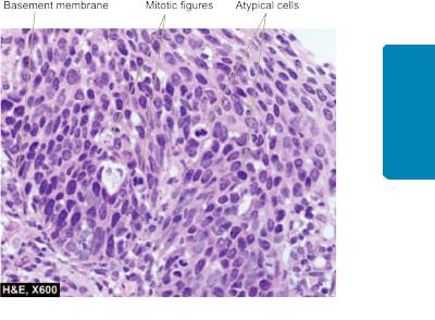
1. Carcinoma in situ (intraepithelial neoplasia). When the cytological features of malignancy are present but the malignant cells are confined to epithelium without invasion across the basement membrane, it is called as carcinoma in situ or intraepithelial neoplasia (CIN). The common sites are as under:
i)Uterine cervix at the junction of ectoand endocervix
(Fig. 8.16)
ii)Bowen’s disease of the skin
iii)Actinic or solar keratosis
iv)Oral leukoplakia
v)Intralobular and intraductal carcinoma of the breast. The area involved in carcinoma in situ may be single and
small, or multifocal. As regards the behaviour of CIN, it may regress and return to normal or may develop into invasive cancer. In some instances such as in cervical cancer, there is a sequential transformation from squamous metaplasia, to epithelial dysplasia, to carcinoma in situ, and eventually to invasive cancer.
2. Some benign tumours. Commonly, benign tumours do not become malignant. However, there are some exceptions e.g.
i)Multiple villous adenomas of the large intestine have high incidence of developing adenocarcinoma.
ii)Neurofibromatosis (von Recklinghausen’s disease) may develop into sarcoma.
3. Miscellaneous conditions. Certain inflammatory and hyperplastic conditions are prone to development of cancer, e.g.
i) Patients of long-standing ulcerative colitis are predisposed to develop colorectal cancer.
207
Neoplasia 8 CHAPTER
Figure 8.16 
 Carcinoma in situ of uterine cervix. The atypical dysplastic squamous cells are confined to all the layers of the mucosa but the basement membrane on which these layers rest is intact.
ii)Cirrhosis of the liver has predisposition to develop hepatocellular carcinoma.
iii)Chronic bronchitis in heavy cigarette smokers may develop cancer of the bronchus.
iv)Chronic irritation from jagged tooth or ill-fitting denture may lead to cancer of the oral cavity.
v)Squamous cell carcinoma developing in an old burn scar (Marjolin’s ulcer).
C. Hormones and Cancer
Cancer is more likely to develop in organs and tissues which undergo proliferation under the influence of excessive hormonal stimulation. On cessation of hormonal stimulation, such tissues become atrophic. Hormone-sensitive tissues developing tumours are the breast, endometrium, myometrium, vagina, thyroid, liver, prostate and testis. Some examples of hormones influencing carcinogenesis in experimental animals and humans are given below:
1. OESTROGEN. Examples of oestrogen-induced cancers are as under:
i)In experimental animals. Induction of breast cancer in mice by administration of high-dose of oestrogen and reduction of the tumour development following oophorectomy is the most important example. It has been known that associated infection with mouse mammary tumour virus (MMTV, Bittner milk factor) has an added influence on the development of breast cancer in mice. Other cancers which can be experimentally induced in mice by oestrogens are squamous cell carcinoma of the cervix, connective tissue tumour of the myometrium, Leydig cell tumour of the testis in male mice, tumour of the kidney in hamsters, and benign as well as malignant tumours of the liver in rats.
ii)In humans. Women receiving oestrogen therapy and women with oestrogen-secreting granulosa cell tumour of the ovary have increased risk of developing endometrial carcinoma. Adenocarcinoma of the vagina is seen with increased frequency in adolescent daughters of mothers who
had received oestrogen therapy during pregnancy.
208
Techniques Basic and Pathology General I SECTION
2.CONTRACEPTIVE HORMONES. The sequential types of oral contraceptives increase the risk of developing breast cancer. Other tumours showing a slightly increased frequency in women receiving contraceptive pills for long durations are benign tumours of the liver, and a few patients have been reported to have developed hepatocellular carcinoma.
3.ANABOLIC STEROIDS. Consumption of anabolic steroids by athletes to increase the muscle mass is not only unethical athletic practice but also increases the risk of developing benign and malignant tumours of the liver.
4.HORMONE-DEPENDENT TUMOURS. It has been shown in experimental animals that induction of hyperfunction of adenohypophysis is associated with increased risk of developing neoplasia of the target organs following preceding functional hyperplasia. There is tumour regression on removal of the stimulus for excessive hormonal secretion. A few examples of such phenomena are seen in humans:
i)Prostatic cancer usually responds to the administration of oestrogens.
ii)Breast cancer may regress with oophorectomy, hypophysectomy or on administration of male hormones.
iii)Thyroid cancer may slow down in growth with administration of thyroxine that suppresses the secretion of TSH by the pituitary.
CARCINOGENESIS: ETIOLOGY AND
PATHOGENESIS OF CANCER
Carcinogenesis or oncogenesis or tumorigenesis means mechanism of induction of tumours (pathogenesis of cancer); agents which can induce tumours are called carcinogens (etiology of cancer). Since the time first ever carcinogen was identified, there has been ever-increasing list of agents implicated in etiology of cancer. There has been still greater accumulation in volumes of knowledge on pathogenesis of cancer, especially due to tremendous strides made in the field of molecular biology and genetics in recent times.
The subject of etiology and pathogenesis of cancer is discussed under the following 4 broad headings:
A.Molecular pathogenesis of cancer (genes and cancer)
B.Chemical carcinogens and chemical carcinogenesis
C.Physical carcinogens and radiation carcinogenesis
D.Biologic carcinogens and viral oncogenesis.
A. MOLECULAR PATHOGENESIS OF CANCER (GENETIC MECHANISMS OF CANCER)
Basic Concept of Molecular Pathogenesis
The mechanism as to how a normal cell is transformed to a cancer cell is complex. At different times, attempts have been made to unravel this mystery by various mechanisms. Currently, a lot of literature has accumulated to explain the pathogenesis of cancer at molecular level. The general concept of molecular mechanisms of cancer is briefly outlined below and diagrammatically shown in Fig. 8.17.
1. Monoclonality of tumours. There is strong evidence to support that most human cancers arise from a single clone of cells by genetic transformation or mutation. For example:
i)In a case of multiple myeloma (a malignant disorder of plasma cells), there is production of a single type of immunoglobulin or its chain as seen by monoclonal spike in serum electrophoresis.
ii)Due to inactivation of one of the two X-chromosomes in females (paternal or maternal derived), women are mosaics with two types of cell populations for glucose-6-phosphatase dehydrogenase (G6PD) isoenzyme A and B. It is observed that all the tumour cells in benign uterine tumours (leiomyoma) contain either A or B genotype of G6PD (i.e. the tumour cells are derived from a single progenitor clone of cell), while the normal myometrial cells are mosaic of both types of cells derived from A as well as B isoenzyme
(Fig. 8.18).
2.Field theory of cancer. In an organ developing cancer, in the backdrop of normal cells, limited number of cells only grow in to cancer after undergoing sequence of changes under the influence of etiologic agents. This is termed as ‘field effect’ and the concept called as field theory of cancer.
3.Multi-step process of cancer growth and progression.
Carcinogenesis is a gradual multi-step process involving many generations of cells. The various causes may act on the cell one after another (multi-hit process). The same process is also involved in further progression of the tumour.
Figure 8.17 Schematic illustration to show molecular basis of cancer.

Figure 8.18 
 The monoclonal origin of tumour cells in uterine leiomyoma.
Ultimately, the cells so formed are genetically and phenotypically transformed cells having phenotypic features of malignancy—excessive growth, invasiveness and distant metastasis.
4.Genetic theory of cancer. Cell growth of normal as well as abnormal types is under genetic control. In cancer, there are either genetic abnormalities in the cell, or there are normal genes with abnormal expression. The abnormalities in genetic composition may be from inherited or induced mutations (induced by etiologic carcinogenic agents namely: chemicals, viruses, radiation). The mutated cells transmit their characters to the next progeny of cells and result in cancer.
5.Genetic regulators of normal and abnormal mitosis. In normal cell growth, regulatory genes control mitosis as well as cell aging, terminating in cell death by apoptosis.
In normal cell growth, there are 4 regulatory genes:
i)Proto-oncogenes are growth-promoting genes i.e. they encode for cell proliferation pathway.
ii)Anti-oncogenes are growth-inhibiting or growth suppressor genes.
iii)Apoptosis regulatory genes control the programmed cell death.
iv)DNA repair genes are those normal genes which regulate the repair of DNA damage that has occurred during mitosis and also control the damage to proto-oncogenes and antioncogenes.

 In cancer, the transformed cells are produced by abnormal cell growth due to genetic damage to these normal controlling genes. Thus, corresponding abnormalities in these 4 cell regulatory genes are as under:
i)Activation of growth-promoting oncogenes causing transformation of cell (mutant form of normal protooncogene in cancer is termed oncogene). Many of these cancer associated genes, oncogenes, were first discovered in viruses, and hence named as v-onc. Gene products of oncogenes are called oncoproteins. Oncogenes are considered dominant since they appear in spite of presence of normal proto-oncogenes.
ii)Inactivation of cancer-suppressor genes (i.e. inactivation of anti-oncogenes) permitting the cellular proliferation of transformed cells. Anti-oncogenes are active in recessive form i.e. they are active only if both alleles are damaged.
iii)Abnormal apoptosis regulatory genes which may act as oncogenes or anti-oncogenes. Accordingly, these genes may be active in dominant or recessive form.
iv)Failure of DNA repair genes and thus inability to repair the DNA damage resulting in mutations.
Cancer-related Genes and Cell Growth
(Hallmarks of Cancer)
It is apparent from the above discussion that genes control the normal cellular growth, while in cancer these controlling genes are altered, typically by mutations. A large number of such cancer-associated genes have been described, each with a specific function in cell growth. Some of these genes are commonly associated in many tumours (e.g. p53 or TP53), while others are specific to particular tumours. Therefore, it is considered appropriate to discuss the role of cancer-related genes with regard to their functions in cellular growth. Following are the major genetic properties or hallmarks of cancer:
1.Excessive and autonomous growth: Growth-promoting oncogenes.
2.Refractoriness to growth inhibition: Growth suppressing anti-oncogenes.
3.Escaping cell death by apoptosis: Genes regulating apoptosis and cancer.
4.Avoiding cellular aging: Telomeres and telomerase in cancer.
5.Continued perfusion of cancer: Cancer angiogenesis.
6.Invasion and distant metastasis: Cancer dissemination.
7.DNA damage and repair system: Mutator genes and cancer.
8.Cancer progression and tumour heterogeneity: Clonal aggressiveness.
9.Cancer a sequential multistep molecular phenomenon: Multistep theory.
10.MicroRNAs in cancer: OncomiRs.
These properties of cancer cells are described below in terms of molecular genetics and schematically illustrated in
Fig. 8.19.
1. EXCESSIVE AND AUTONOMOUS GROWTH: GROWTH PROMOTING ONCOGENES
Mutated form of normal protooncogenes in cancer is called oncogenes. Protooncogenes become activated oncogenes by following mechanisms as under:
209
Neoplasia 8 CHAPTER
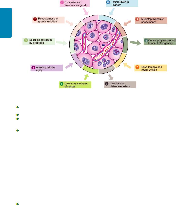
210
Techniques Basic and Pathology General I SECTION
Figure 8.19 
 Schematic representation of major properties of cancer in terms of molecular carcinogenesis.

 By mutation in the protooncogene which alters its structure and function.

 By retroviral insertion in the host cell.

 By damage to the DNA sequence that normally regulates growth-promoting signals of protooncogenes resulting in its abnormal activation.
 By erroneous formation of extra copies of protooncogene causing gene amplification and hence its overexpression or overproduction that promotes autonomous and excessive cellular proliferation.
In general, overactivity of oncogenes enhances cell proliferation and promotes development of human cancer. About 100 different oncogenes have been described in various cancers. Transformation of proto-oncogene (i.e. normal cell proliferation gene) to oncogenes (i.e. cancer cell proliferation gene) may occur by three mechanisms:
i)Point mutations i.e. an alteration of a single base in the DNA chain. The most important example is RAS oncogene carried in many human tumours such as bladder cancer, pancreatic adenocarcinoma, cholangiocarcinoma.
ii)Chromosomal translocations i.e. transfer of a portion of one chromosome carrying protooncogene to another chromosome and making it independent of growth controls. This is implicated in the pathogenesis of leukaemias and lymphomas e.g.

 Philadelphia chromosome seen in 95% cases of chronic myelogenous leukaemia in which c-ABL protooncogene on chromosome 9 is translocated to chromosome 22.
In 75% cases of Burkitt’s lymphoma, translocation of c- MYC proto-oncogene from its site on chromosome 8 to a portion on chromosome 14.
iii) Gene amplification i.e. increasing the number of copies of DNA sequence in protooncogene leading to increased mDNA and thus increased or overexpressed gene product. Examples of gene amplification are found in some solid human tumours e.g.
Neuroblastoma having n-MYC HSR region. ERB-B1 in breast and ovarian cancer.
Most of the oncogenes encode for components of cell signaling system for promoting cell proliferation. Possible effects of oncogenes in signal transduction for cell proliferation in human tumours are discussed below in relation to the role of protooncogenes in mitosis in normal cell cycle and are listed in Table 8.4 and schematically shown in Fig. 8.20:
i) Growth factors (GFs). GFs were the first protoonocgenes to be discovered which encode for cell proliferation cascade. They act by binding to cell surface receptors to activate cell proliferation cascade within the cell. GFs are small polypeptides elaborated by many cells and normally act on another cell than the one which synthesised it to stimulate its proliferation i.e. paracrine action.
However, a cancer cell may synthesise a GF and respond to it as well; this way cancer cells acquire growth selfsufficiency. Most often, growth factor genes are not altered or mutated but instead growth factor genes are overexpressed to stimulate large secretion of GFs which stimulate cell proliferation. The examples of such tumour secreted GFs are as under:
a) Platelet-derived growth factor- (PDGF-β): Overexpression of
SIS protooncogene that encodes for PDGF-β and thus there
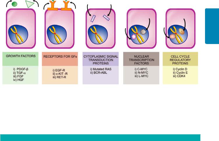
211
Neoplasia 8 CHAPTER
Figure 8.20 
 Mechanisms of activation of protooncogenes to form growth promoting oncogenes.
is increased secretion of PDGF-β e.g. in gliomas and sarcomas.
b) Transforming growth factor-α (TGF-α): Overexpression of
TGF-α gene occurs by stimulation of RAS protooncogene and induces cell proliferation by binding to epidermal growth factor (EGF) receptor e.g. in carcinoma and astrocytoma.
c) Fibroblast growth factor (FGF): Overexpression of HST-1 protoonogene and amplification of INT-2 protoonogene causes excess secretion of FGF e.g. in cancer of the bowel and breast.
d) Hepatocyte growth factor (HGF): Overexpression by binding to its receptor c-MET e.g. follicular carcinoma thyroid.
TABLE 8.4: Important Oncogenes, their Mechanism of Activation and Associated Human Tumours.
Type |
|
Proto-oncogene |
Mechanism |
Associated Human Tumours |
|
|
|
|
|
1. GROWTH FACTORS |
|
|
|
|
i) |
PDGF-β |
SIS |
Overexpression |
Gliomas, sarcoma |
ii) |
TGF-α |
RAS |
Overexpression |
Carcinomas, sarcomas |
iii) |
FGF |
HST-1 |
Overexpression |
Bowel cancers |
|
|
INT-2 |
Amplification |
Breast cancer |
iv) |
HGF |
HGF |
Overexpression |
Follicular carcinoma thyroid |
2. RECEPTORS FOR GROWTH FACTORS |
|
|
||
i) |
EGF receptors |
ERB B1(HER 1) |
Overexpression |
Squamous cell carcinoma lung, glioblastoma |
|
|
ERB B2 (HER 2/neu) |
Amplification |
Ca breast, ovary, stomach, lungs |
ii) |
c-KIT receptor |
c-KIT |
Point mutation |
GIST |
|
(Steel factor) |
|
|
|
iii) |
RET receptor |
RET |
Point mutation |
MEN type 2A and type 2B, medullary ca thyroid |
3. CYTOPLASMIC SIGNAL TRANSDUCTION PROTEINS |
|
|
||
|
GTP-bound |
RAS |
Point mutation |
Common in 1/3rd human tumours, Ca lung, colon, |
|
|
|
|
pancreas |
|
Non-receptor tyrosine |
BCR-ABL |
Translocation |
CML, acute leukaemias |
|
kinase |
|
|
|
4. NUCLEAR TRANSCRIPTION FACTORS |
|
|
||
|
C-MYC |
MYC |
Translocation |
Burkitt’s lymphoma |
|
N-MYC |
MYC |
Amplification |
Neuroblastoma, small cell Ca lung |
|
L-MYC |
MYC |
Amplification |
Small cell Ca lung |
5. CELL CYCLE REGULATORY PROTEINS |
|
|
||
|
Cyclins |
Cyclin D |
Translocation |
Ca breast, liver, mantle cell lymphoma |
|
|
Cyclin E |
Overexpression |
Ca breast |
|
CDKs |
CDK4 |
Amplification |
Glioblastoma, melanoma, sarcomas |
|
|
|
|
|

212
Techniques Basic and Pathology General I SECTION
ii) Receptors for GFs. Growth factors cannot penetrate the cell directly and require to be transported intracellularly by GF-specific cell surface receptors. These receptors are transmembrane proteins and thus have two surfaces: the outer surface of the membrane has an area for binding growth factor, and the inner surface of the membrane has enzymeactivating area which eventually activates cell proliferation pathway.
Most often, mutated form of growth factor receptors stimulate cell proliferation even without binding to growth factors i.e. with little or no growth factor bound to them. Various forms of oncogenes encoding for GF receptors include other mechanisms: overexpression, mutation and gene rearrangement. Examples of tumours by mutated receptors for growth factors are as under:
a)EGF receptors: Normal EGF receptor gene is ERB B1, and hence this receptor is termed as EGFR or HER1 (i.e. human epidermal growth factor receptor type 1). EGFR (or HER1) acts by overexpression of normal GF receptor e.g. in 80% of squamous cell carcinoma of lung and 50% cases of glioblastomas.
Another EGF receptor gene called ERB B2 (or HER2/neu) acts by gene amplification e.g. in breast cancer (25% cases), carcinoma of lungs, ovary, stomach.
b)c-KIT receptor: The gene coding for receptor for stem cell factor (or steel factor) is c-KIT, that activates tyrosine kinase pathway in cell proliferation. Mutated form of c-KIT by point mutation activates receptor for tyrosine kinase e.g. in gastrointestinal stromal tumours (GIST).
c)RET receptor: RET (abbreviation of ‘rearranged during transfection’) protooncogene is a receptor for tyrosine kinase normally expressed in neuroendocrine cells of different tissues. Mutated form by point mutation is seen in MEN type 2A and 2B and in medullary carcinoma thyroid.
iii) Cytoplasmic signal transduction proteins. The normal signal transduction proteins in the cytoplasm transduce signal from the GF receptors present on the cell surface, to the nucleus of the cell, to activate intracellular growth signaling pathways.
There are examples of oncogenes having mutated forms of cytoplasmic signaling pathways located in the inner surface of cell membrane in some cancers. These are as under:
a)Mutated RAS gene. This is the most common form of oncogene in human tumours, the abnormality being induced by point mutation in RAS gene. About a third of all human tumours carry mutated RAS gene (RAS for Rat Sarcoma gene where it was first described), seen particularly in carcinoma colon, lung and pancreas. Normally, the inactive form of RAS protein is GDP (guanosine diphosphate)-bound while the activated form is bound to guanosine triphosphate (GTP). GDP/GTP are homologous to G proteins and take part in signal transduction in a similar way just as G proteins act as ‘on-off switch’ for signal transduction. Normally, active RAS protein is inactivated by GTPase activity, while mutated RAS gene remains unaffected by GTPase, and therefore, continues to signal the cell proliferation.
b)BCR-ABL hybrid gene. ABL gene is a non-GF receptor protooncogene having tyrosine kinase activity. ABL gene
from its normal location on chromosome 9 is translocated to chromosome 22 where it fuses with BCR (breakpoint cluster region) gene and forms an ABL-BCR hybrid gene which is more potent in signal transduction pathway. ABL-BCR hybrid gene is seen in chronic myeloid leukaemia and some acute leukaemias.
iv)Nuclear transcription factors. The signal transduction pathway that started with GFs ultimately reaches the nucleus where it regulates DNA transcription and induces the cell to enter into S phase. Out of various nuclear regulatory transcription proteins described, the most important is MYC gene located on long arm of chromosome 8. Normally MYC protein binds to the DNA and regulates the cell cycle by transcriptional activation and its levels fall immediately after cell enters the cell cycle.
MYC oncogene (originally isolated from myelocytomatosis virus and accordingly abbreviated) is seen most commonly in human tumours. It is associated with persistence of or overexpression of MYC oncoproteins which, in turn, causes autonomous cell proliferation. The examples of tumours carrying MYC oncogene are as under:
a)C-MYC oncogene: Mutated MYC gene due to translocation t(8;14) seen in Burkitt’s lymphoma.
b)N-MYC oncogene: Mutated MYC gene due to amplification seen in neuroblastoma, small cell carcinoma lung.
c)L-MYC oncogene: Mutated MYC gene due to amplification seen in small cell carcinoma lung.
v)Cell cycle regulatory proteins. As discussed in Chapter 3, normally the cell cycle is under regulatory control of cyclins and cyclin-dependent kinases (CDKs) A, B, E and D. Cyclins are so named since they are cyclically synthesised during different phases of the cell cycle and their degradation is also cyclic. Cyclins activate as well as work together with CDKs, while many inhibitors of CDKs (CDKIs) are also known.
Although all steps in the cell cycle are under regulatory controls, G1 → S phase is the most important checkpoint for regulation by oncogenes as well as anti-oncogenes (discussed below). Mutations in cyclins (in particular cyclin D) and CDKs (in particular CDK4) are most important growth promoting signals in cancers. The examples of tumours having such oncogenes are as under:
a)Mutated form of cyclin D protooncogene by translocation seen in mantle cell lymphoma.
b)Mutated form of cyclin E by overexpression seen in breast cancer.
b)Mutated from of CDK4 by gene amplification seen in malignant melanoma, glioblastoma and sarcomas.
2. REFRACTORINESS TO GROWTH INHIBITION: GROWTH SUPPRESSING ANTI-ONCOGENES
The mutation of normal growth suppressor anti-oncogenes results in removal of the brakes for growth; thus the inhibitory effect to cell growth is removed and the abnormal growth continues unchecked. In other words, mutated antioncogenes behave like growth-promoting oncogenes.
As compared to the signals and signal transduction pathways for oncogenes described above, the steps in mechanisms of action by growth suppressors are not so well

TABLE 8.5: Important Tumour-suppressor Anti-oncogenes and Associated Human Tumours.
|
Gene |
Location |
Associated Human Tumours |
|
|
|
|
1. |
RB |
Nucleus (13q) |
Retinoblastoma, osteosarcoma |
2. |
p53 (TP53) |
Nucleus (17p) |
Most human cancers, common in |
|
|
|
Ca lung, head and neck, colon, breast |
3. |
TGF–β and its receptor |
Extracellular |
Ca pancreas, colon, stomach |
4. |
APC and β-catenin proteins |
Cytosol |
Ca colon |
5.Others
i) |
BRCA 1 and 2 |
Nucleus (BRCA1 17q21, |
Ca breast, ovary |
|
|
BRCA2 13q12-13) |
|
ii) |
VHL |
Nucleus (3p) |
Renal cell carcinoma |
iii) WT 1 and 2 |
Nucleus (11p) |
Wilms’ tumour |
|
iv) |
NF 1 and 2 |
Plasma membrane |
Neurofibromatosis type 1 and 2 |
|
|
|
|
understood. In general, the point of action by anti-oncogenes is also G1 → S phase transition and probably act either by inducing the dividing cell from the cell cycle to enter into G0 (resting) phase, or by acting in a way that the cell lies in the post-mitotic pool losing its dividing capability. Just as with activation of protooncogenes to become oncogenes, the mechanisms of loss of tumour suppressor actions of genes are due to chromosomal deletions, point mutations and loss of portions of chromosomes.
Major anti-oncogenes implicated in human cancers are as under (Table 8.5):
i) RB gene. RB gene is located on long arm (q) of chromosome 13. This is the first ever tumour suppressor gene identified and thus has been amply studied. RB gene codes for a nuclear transcription protein pRB. RB gene is termed as master ‘break’ in the cell cycle and is virtually present in every human cell. It can exist in both an active and an inactive form: 
 The active form of RB gene, it blocks cell division by binding to transcription factor, E2F, and thus inhibits the cell from transcription of cell cycle-related genes, thereby inhibiting the cell cycle at G1 → S phase i.e. cell cycle is arrested at G1 phase.
 Inactive form of RB gene occurs when it is hyperphosphorylated by cyclin dependent kinases (CDKs) which occurs when growth factors bind to their receptors. This removes pRB function from the cell (i.e. the ‘break’ on cell division is removed) and thus cell proliferation pathway is stimulated by permitting the cell to cross G1 → S phase. Activity of CDKs is inhibited by activation of inhibitory signal, transforming growth factor- (TGF-β), on cell through activation of inhibitory protein p16.
The mutant form of RB gene (i.e. inactivating mutation of RB gene) is involved in several human tumours, most commonly in retinoblastoma, the most common intraocular tumour in young children. The tumour occurs in two forms: sporadic and inherited/familial. More than half the cases are sporadic affecting one eye; these cases have acquired simultaneous mutation in both the alleles in retinal cells after birth. In inherited cases, all somatic cells inherit one mutant RB gene from a carrier parent, while the other allele gets mutated later. The latter genetic explanation given by Knudson forms the basis of two hit hypothesis of inherited cancers. Besides retinoblastoma, children inheriting mutant
RB gene have 200 times greater risk of development of other cancers in early adult life, most notably osteosarcoma; others are cancers of breast, colon and lungs.
ii) p53 gene (TP53). Located on the short arm (p) of chromosome 17, p53 gene (also termed TP53 because of molecular weight of 53 kd for the protein) like pRB is inhibitory to cell cycle. However, p53 is normally present in very small amounts and accumulates only after DNA damage.
The two major functions of p53 in the normal cell cycle are as under:
a)In blocking mitotic activity: p53 inhibits the cyclins and CDKs and prevents the cell to enter G1 phase transiently. This breathing time in the cell cycle is utilised by the cell to repair the DNA damage.
b)In promoting apoptosis: p53 acts together with another antioncogene, RB gene, and identifies the genes that have damaged DNA which cannot be repaired by inbuilt system. p53 directs such cells to apoptosis by activating apoptosisinducing BAX gene, and thus bringing the defective cells to an end by apoptosis. This process operates in the cell cycle at G1 and G2 phases before the cell enters the S or M phase.
Because of these significant roles in cell cycle, p53 is called as ‘protector of the genome’.
In its mutated form, p53 ceases to act as protector or as growth suppressor but instead acts like a growth promoter or oncogene. Homozygous loss of p53 gene allows genetically damaged and unrepaired cells to survive and proliferate resulting in malignant transformation. More than 70% of human cancers have homozygous loss of p53 by acquired mutations in somatic cells; some common examples are cancers of the lung, head and neck, colon and breast. Besides, mutated p53 is also seen in the sequential development stages of cancer from hyperplasia to carcinoma in situ and into invasive carcinoma.
Less commonly, both alleles of p53 gene become defective by another way: one allele of p53 mutated by inheritance in germ cell lines rendering the individual to another hit of somatic mutation on the second allele. This defect like in RB gene predisposes the individual to develop cancers of multiple organs (breast, bone, brain, sarcomas etc), termed
Li-Fraumeni syndrome.
213
Neoplasia 8 CHAPTER

214 iii) Transforming growth factor-β (TGF-β) and its receptor.
|
Normally, TGF-β is significant inhibitor of cell proliferation, |
||
|
especially in epithelial, endothelial and haematopoieitc cells |
||
ISECTION |
It acts by binding to TGF-β receptor and then the complex so |
||
CDKs and cyclins. |
|||
|
formed acts in G1 phase of cell cycle at two levels: |
||
|
|
It activates CDK inhibitors (CDKIs) with growth |
|
|
inhibitory effect. |
||
|
|
It suppresses the growth prmoter genes such as MYC, |
|
|
|
Mutant form of TGF-β gene or its receptor impairs the |
|
General |
growth inhibiting effect and thus permits cell proliferation. |
||
Examples of mutated form of TGF-β are seen in cancers of |
|||
pancreas, colon, stomach and endometrium. |
|||
|
|||
|
iv) Adenomatous polyposis coli (APC) gene and β-catenin |
||
Pathology |
protein. The APC gene is normally inhibitory to mitosis, |
||
which takes place by a cytoplasmic protein, β-catenin. β- |
|||
|
|||
|
catenin normally has dual functions: firstly, it binds to |
||
|
cytoplasmic E-cadherin that is involved in intercellular |
||
|
interactions, and secondly it can activate cell proliferation |
||
and |
signaling pathway. In colon cancer cells, APC gene is lost |
||
and thus β-catenin fails to get degraded, allowing the cancer |
|||
|
|||
Basic |
cells to undergo mitosis without the inhibitory influence of |
||
β-catenin. |
|||
|
|||
|
|
Patients born with one mutant APC gene allele develop |
|
Techniques |
large number of polyps in the colon early in life, while after |
||
the age of 20 years these cases start developing loss of second |
|||
|
|||
|
APC gene allele. It is then that almost all these patients |
||
|
invariably develop malignant transformation of one or more |
||
|
polyps. |
||
|
v) |
Other antioncogenes. A few other tumour-suppressor |
|
|
genes having mutated germline in various tumours are as |
||
|
under: |
||
|
a) |
BRCA 1 and BRCA 2 genes: These are two breast (BR) |
|
|
cancer (CA) susceptibility genes: BRCA1 located on |
||
|
chromosoe 17q21 and BRCA2 on chromosome 13q12-13. |
||
Women with inherited defect in BRCA1 gene have very high risk (85%) of developing breast cancer and ovarian cancer (40%). Inherited breast cancer constitutes about 5-10% cases, it tends to occur at a relatively younger age and more often tends to be bilateral.
b) VHL gene. von-Hippel-Lindau (VHL) disease is a rare autosomal dominant disease characterised by benign and malignant tumours of multiple tissues. The disease is
TABLE 8.6: Oncogenes versus Antioncogenes.
inherited as a mutation in VHL tumour suppressor gene located on chromosome 3p. This results in activation of genes that promote angiogenesis, survival and proliferation; VHL gene is found inactivated in 60% cases of renal cell carcinoma.
c)Wilms’ tumour (WT) gene: Both WT1 an WT2 genes are located on chromosome 11 and normally prevent neoplastic proliferation of cells in embryonic kidney. Mutant form of WT-1 and 2 are seen in hereditary Wilms’ tumour.
d)Neurofibroma (NF) gene: NF genes normally prevent proliferation of Schwann cells. Two mutant forms are described: NF1 and NF2 seen in neurofibromatosis type 1 and type 2.
The contrasting features of growth-promoting oncogenes and growth-suppressing anti-oncogenes are summarised in
Table 8.6.
3. ESCAPING CELL DEATH BY APOPTOSIS: GENES REGULATING APOPTOSIS AND CANCER
Besides the role of mutant forms of growth-promoting oncogenes and growth-suppressing anti-oncogenes, another mechanism of tumour growth is by escaping cell death by apoptosis. Apoptosis in normal cell is guided by cell death receptor, CD95, resulting in DNA damage. Besides, there is role of some other pro-apoptotic factors (BAD, BAX, BID and p53) and apoptosis-inhibitors (BCL2, BCL-X).
In cancer cells, the function of apoptosis is interfered due to mutations in the above genes which regulate apoptosis in the normal cell. The examples of tumours by this mechanism are as under:
a)BCL2 gene is seen in normal lymphocytes, but its mutant form with characteristic translocation (t14;18) (q32;q21) was first described in B-cell lymphoma and hence the name BCL. It is also seen in many other human cancers such as that of breast, thyroid and prostate. Mutation in BCL2 gene removes the apoptosis-inhibitory control on cancer cells, thus more live cells undergoing mitosis contributing to tumour growth. Besides, MYC oncogene and p53 tumour suppressor gene are also connected to apoptosis. While MYC allows cell growth BCL2 inhibits cell death; thus MYC and BCL2 together allow cell proliferation. Normally, p53 activates proapoptotic gene BAX but mutated p53 (i.e. absence of p53) reduces apoptotic activity and thus allows cell proliferation.
b)CD95 receptors are depleted in hepatocellular carcinoma and hence the tumour cells escape apoptosis.
|
Feature |
Oncogene |
Antioncogene |
|
|
|
|
|
|
1. |
Derived from |
Mutated form of normal protooncogenes |
Mutated form of normal growth |
|
|
|
|
suppressor genes |
|
2. |
Genetic abnormality |
Mutations (point, translocation, amplification, |
Loss of genes by deletion, point mutation |
|
|
|
overexpression) retroviral insertion, DNA damage |
and loss of portion of chromosome |
|
3. |
Major action |
Allows cell proliferation by increased growth |
Allows cell proliferation by removal of cell |
|
|
|
promotion pathways |
growth suppressor pathway |
|
4. |
Level of action in cell |
At different levels (cell surface, cytoplasm, |
At different levels (cell surface, cytoplasm, |
|
|
|
mutations) |
nucleus) |
|
5. |
Major types |
i) GFs (PDGF-β, TGF-α, FGF, HGF) |
i) |
RB |
|
|
ii) GF receptors (EGFR, cKIT, RET) |
ii) |
p53 |
|
|
iii) Cytoplasmic signal proteins (RAS, BCR-ABL) |
iii) TGF-β and its receptor |
|
|
|
iv) Nuclear transcription proteins (MYC) |
iv) |
APC and β-catenin |
|
|
v) Cell cycle regular proteins (CDKs, cyclins) |
v) Others (BRCA 1 and 2, VHL, WT 1 and 2, |
|
|
|
|
|
NF 1 and 2) |
|
|
|
|
|
4.AVOIDING CELLULAR AGING: TELOMERES AND TELOMERASE IN CANCER
As discussed in pathology of aging in Chapter 3, after each mitosis (cell doubling) there is progressive shortening of telomeres which are the terminal tips of chromosomes. Telomerase is the RNA enzyme that helps in repair of such damage to DNA and maintains normal telomere length in successive cell divisions. However, it has been seen that after repetitive mitosis for a maximum of 60 to 70 times, telomeres are lost in normal cells and the cells cease to undergo mitosis. Telomerase is active in normal stem cells but not in normal somatic cells.
Cancer cells in most malignancies have markedly upregulated telomerase enzyme, and hence telomere length is maintained. Thus, cancer cells avoid aging, mitosis does not slow down or cease, thereby immortalising the cancer cells.
5.CONTINUED PERFUSION OF CANCER: TUMOUR ANGIOGENESIS
Cancers can only survive and thrive if the cancer cells are adequately nourished and perfused, as otherwise they cannot grow further. Neovascularisation in the cancers not only supplies the tumour with oxygen and nutrients, but the newly formed endothelial cells also elaborate a few growth factors for progression of primary as well as metastatic cancer. The stimulus for angiogenesis is provided by the release of various factors:
i) Promoters of tumour angiogenesis include the most important vascular endothelial growth factor (VEGF) (released from genes in the parenchymal tumour cells) and basic
fibroblast growth factor (bFGF).
ii) Anti-angiogenesis factors inhibiting angiogenesis include thrombospondin-1 (also produced by tumour cells themselves),
angiostatin, endostatin and vasculostatin. Mutated form of p53 gene in both alleles in various cancers results in removal of anti-angiogenic role of thrombospondin-1, thus favouring continued angiogenesis.
6.INVASION AND DISTANT METASTASIS: CANCER DISSEMINATION
One of the most important characteristic of cancers is invasiveness and metastasis. The mechanisms involved in the biology of invasion and metastasis are discussed already along with spread of tumours.
7.DNA DAMAGE AND REPAIR SYSTEM: MUTATOR GENES AND CANCER
Normal cells during complex mitosis suffer from minor damage to the DNA which is detected and repaired before mitosis is completed so that integrity of the genome is maintained. Similarly, small mutational damage to the dividing cell by exogenous factors (e.g. by radiation, chemical carcinogens etc) is also repaired. p53 gene is held responsible for detection and repair of DNA damage. However, if this system of DNA repair is defective as happens in some inherited mutations (mutator genes), the defect in unrepaired DNA is passed to the next progeny of cells and cancer results.
The examples of mutator genes exist in the following inherited disorders associated with increased propensity to cancer:
i)Hereditary non-polyposis colon cancer (Lynch syndrome) is characterised by hereditary predisposition to develop colorectal cancer. It is due to defect in genes involved in DNA mismatch repair which results in accumulation of errors in the form of mutations in many genes.
ii)Ataxia telangiectasia (AT) has ATM (M for mutated) gene. These patients have multiple cancers besides other features such as cerebellar degeneration, immunologic derangements and oculo-cutaneous manifestations.
iii)Xeroderma pigmentosum is an inherited disorder in which there is defect in DNA repair mechanism. Upon exposure to sunlight, the UV radiation damage to DNA cannot be repaired. Thus, such patients are more prone to various forms of skin cancers.
iv)Bloom syndrome is an example of damage by ionising radiation which cannot be repaired due to inherited defect and the patients have increased risk to develop cancers, particularly leukaemia.
v)Hereditary breast cancer patients having mutated BRCA1 and BRCA2 genes carry inherited defect in DNA repair mechanism. These patients are not only predisposed to develop breast cancer but also cancers of various other organs.
8.CANCER PROGRESSION AND HETEROGENEITY: CLONAL AGGRESSIVENESS
Another feature of note in biology of cancers is that with passage of time cancers become more aggressive; this property is termed tumour progression. Clinical parameters of cancer progression are: increasing size of the tumour, higher histologic grade (as seen by poorer differentiation and greater anaplasia), areas of tumour necrosis (i.e. tumour outgrows its blood supply), invasiveness and distant metastasis.
In terms of molecular biology, this attribute of cancer is due to the fact that with passage of time cancer cells acquire more and more heterogeneity. This means that though cancer cells remain monoclonal in origin, they acquire more and more mutations which, in turn, produce multiple-mutated subpopulations of more aggressive clones of cancer cells (i.e. heterogeneous cells) in the growth which have tendency to invade, metastasise and be refractory to hormonal influences. Some of these mutations in fact may kill the tumour cells as well.
9.CANCER A SEQUENTIAL MULTISTEP MOLECULAR PHENOMENON: MULTISTEP THEORY
It needs to be appreciated that cancer occurs following several sequential steps of abnormalities in the target cell e.g. initiation, promotion and progression in proper sequence. Similarly, multiple steps are involved at genetic level by which cell proliferation of cancer cells is activated: by activation of growth promoters, loss of growth suppressors, inactivation of intrinsic apoptotic mechanisms and escaping cellular aging. A classic example of this sequential genetic abnormalities in cancer is seen in adenoma-carcinoma
215
Neoplasia 8 CHAPTER
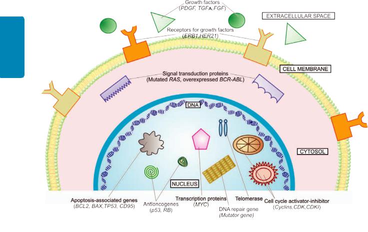
216
Techniques Basic and Pathology General I SECTION
Figure 8.21 
 Schematic representation of activation-inactivation of cancer-associated genes in cell cycle.
sequence in development of colorectal carcinoma. Recent studies on human genome in cancers of breast and colon have revealed that there is a multistep phenomenon of carcinogenesis at molecular level; on an average a malignant tumour has large number of genetic mutations in cancers.
10. MICRORNAs IN CANCER: ONCOMIRS
MicroRNAs (miRNAs) are evolutionally conserved, endogenous, noncoding single stranded RNA molecules with a length of 22 nucleotides only. Normally, miRNAs function as the posttranslational gene regulators of cell proliferation, differentiation and survival. More than 500 miRNAs have been identified. Recent evidence indicates that miRNAs have an oncogenic role in initiation and progression of cancer and are termed as oncogenic microRNAs, abbreviated as oncomiRs. In combination with other tumour associated genes, oncomiRs can perform various functions: as tumour suppressor, as tumour promoter, and as pro-apoptotic.
The above properties of cancer cells are schematically illustrated in Fig. 8.21.
B. CHEMICAL CARCINOGENESIS
The first ever evidence of any cause for neoplasia came from the observation of Sir Percival Pott in 1775 that there was higher incidence of cancer of the scrotum in chimney-sweeps in London than in the general population. This invoked wide interest in soot and coal tar as possible carcinogenic agent and the possibility of other occupational cancers. The first successful experimental induction of cancer was produced
by two Japanese workers (Yamagiwa and Ichikawa) in 1914 in the rabbit’s skin by repeatedly painting with coal tar. Since then the list of chemical carcinogens which can experimentally induce cancer in animals and have epidemiological evidence in causing human neoplasia, is ever increasing.
Stages in Chemical Carcinogenesis
The induction of cancer by chemical carcinogens occurs after a delay—weeks to months in the case of experimental animals, and often several years in man. Other factors that influence the induction of cancer are the dose and mode of administration of carcinogenic chemical, individual susceptibility and various predisposing factors.
Basic mechanism of chemical carcinogenesis is by induction of mutation in the proto-oncogenes and antioncogenes. The phenomena of cellular transformation by chemical carcinogens (as also other carcinogens) is a progressive process involving 3 sequential stages: initiation, promotion and progression (Fig. 8.22).
1. INITIATION OF CARCINOGENESIS
Initiation is the first stage in carcinogenesis induced by initiator chemical carcinogens. The change can be produced by a single dose of the initiating agent for a short time, though larger dose for longer duration is more effective. The change so induced is sudden, irreversible and permanent. Chemical carcinogens acting as initiators of carcinogenesis can be grouped into 2 categories (Table 8.7):
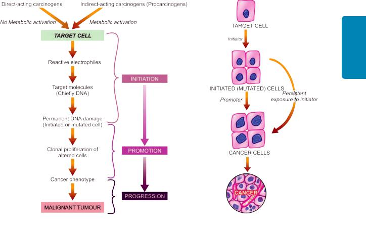
217
Neoplasia 8 CHAPTER
Figure 8.22 
 Sequential stages in chemical carcinogenesis (left) in evolution of cancer (right).
I. Direct-acting carcinogens. These are a few chemical substances (e.g. alkylating agents, acylating agents) which can induce cellular transformation without undergoing any prior metabolic activation.
II. Indirect-acting carcinogens or procarcinogens. These require metabolic conversion within the body so as to become ‘ultimate’ carcinogens having carcinogenicity e.g. polycyclic aromatic hydrocarbons, aromatic amines, azo dyes, naturally-occurring products and others.
In either case, the following steps are involved in transforming ‘the target cell’ into ‘the initiated cell’:
a) Metabolic activation. Vast majority of chemical carcinogens are indirect-acting or procarcinogens requiring metabolic activation, while direct-acting carcinogens do not require this activation. The indirect-acting carcinogens are activated in the liver by the mono-oxygenases of the cytochrome P-450 system in the endoplasmic reticulum. In some circumstances, the procarcinogen may be detoxified and rendered inactive metabolically.
In fact, following 2 requirements determine the carcinogenic potency of a chemical:
i)Balance between activation and inactivation reaction of the carcinogenic chemical.
ii)Genes that code for cytochrome P450-dependent enzymes involved in metabolic activation e.g a genotype carrying susceptibility gene CYP1A1 for the enzyme system has far higher incidence of lung cancer in light smokers as compared to those not having this permissive gene.
Besides these two, additional factors such as age, sex and nutritional status of the host also play some role in determining response of the individual to chemical carcinogen.
b)Reactive electrophiles. While direct-acting carcinogens are intrinsically electrophilic, indirect-acting substances become electron-deficient after metabolic activation i.e. they become reactive electrophiles. Following this step, both types of chemical carcinogens behave alike and their reactive electrophiles bind to electron-rich portions of other molecules of the cell such as DNA, RNA and other proteins.
c)Target molecules. The primary target of electrophiles is DNA, producing mutagenesis. The change in DNA may lead to ‘the initiated cell’ or some form of cellular enzymes may be able to repair the damage in DNA. The classic example of such a situation occurs in xeroderma pigmentosum, a precancerous condition, in which there is hereditary defect in DNA repair mechanism of the cell and thus such patients are prone to develop skin cancer. The carcinogenic potential of a chemical can be tested in vitro by Ames’ test for mutagenesis (described later).
Any gene may be the target molecule in the DNA for the chemical carcinogen. However, on the basis of chemically induced cancers in experimental animals and epidemiologic studies in human beings, it has been observed that most frequently affected growth promoter oncogene is RAS gene mutation and anti-oncogene (tumour suppressor) is p53 gene mutation.

218
Techniques Basic and Pathology General I SECTION
d) The initiated cell. The unrepaired damage produced in the DNA of the cell becomes permanent and fixed only if the altered cell undergoes at least one cycle of proliferation. This results in transferring the change to the next progeny of cells so that the DNA damage becomes permanent and irreversible, which are the characteristics of the initiated cell, vulnerable to the action of promoters of carcinogenesis.
The stimulus for proliferation may come from regeneration of surviving cells, dietary factors, hormoneinduced hyperplasia, viruses etc. A few examples are the occurrence of hepatocellular carcinoma in cases of viral hepatitis, association of endometrial hyperplasia with endometrial carcinoma, effect of oestrogen in breast cancer.
2. PROMOTION OF CARCINOGENESIS
Promotion is the next sequential stage in the chemical carcinogenesis. Promoters of carcinogenesis are substances such as phorbol esters, phenols, hormones, artificial sweeteners and drugs like phenobarbital. They differ from initiators in the following respects:
i)They do not produce sudden change.
ii)They require application or administration, as the case may be, following initiator exposure, for sufficient time and in sufficient dose.
iii)The change induced may be reversible.
iv)They do not damage the DNA per se and are thus not mutagenic but instead enhance the effect of direct-acting carcinogens or procarcinogens.
v)Tumour promoters act by further clonal proliferation and expansion of initiated (mutated) cells, and have reduced requirement of growth factor, especially after RAS gene mutation.
It may be mentioned here that persistent and sustained application/exposure of the cell to initiator alone unassociated with subsequent application of promoter may also result in cancer. But the vice versa does not hold true since neither application of promoter alone, nor its application prior to exposure to initiator carcinogen, would result in transformation of target cell.
3. PROGRESSION OF CARCINOGENESIS
Progression of cancer is the stage when mutated proliferated cell shows phenotypic features of malignancy. These features pertain to morphology, biochemical composition and molecular features of malignancy. Such phenotypic features appear only when the initiated cell starts to proliferate rapidly and in the process acquires more and more mutations. The new progeny of cells that develops after such repetitive proliferation inherits genetic and biochemical characteristics of malignancy.
Carcinogenic Chemicals in Humans
The list of diverse chemical compounds which can produce cancer in experimental animals is a long one but only some of them have sufficient epidemiological evidence in human neoplasia.
Depending upon the mode of action of carcinogenic chemicals, they are divided into 2 broad groups: initiators and promoters (Table 8.7).
1. INITIATOR CARCINOGENS
Chemical carcinogens which can initiate the process of neoplastic transformation are further categorised into 2 subgroups—direct-acting and indirect-acting carcinogens or procarcinogens.
I. DIRECT-ACTING CARCINOGENS. These chemical carcinogens do not require metabolic activation and fall into 2 classes:
a)Alkylating agents. This group includes mainly various anti-cancer drugs (e.g. cyclophosphamide, chlorambucil, busulfan, melphalan, nitrosourea etc), β-propiolactone and epoxides. They are weakly carcinogenic and are implicated in the etiology of the lymphomas and leukaemias in human beings.
b)Acylating agents. The examples are acetyl imidazole and dimethyl carbamyl chloride.
II.INDIRECT-ACTING CARCINOGENS (PROCARCINOGENS). These are chemical substances which require prior metabolic activation before becoming potent ‘ultimate’ carcinogens. This group includes vast majority of carcinogenic chemicals. It includes the following 4 categories:
a)Polycyclic aromatic hydrocarbons. They comprise the largest group of common procarcinogens which, after metabolic activation, can induce neoplasia in many tissues in experimental animals and are also implicated in a number of human neoplasms. They cause different effects by various modes of administration e.g. by topical application may induce skin cancer, by subcutaneous injection may cause sarcomas, inhalation produces lung cancer, when introduced in different organs by parenteral/metabolising routes may cause cancer of that organ.
Main sources of polycyclic aromatic hydrocarbons are: combustion and chewing of tobacco, smoke, fossil fuel (e.g. coal), soot, tar, mineral oil, smoked animal foods, industrial and atmospheric pollutants. Important chemical compounds included in this group are: anthracenes (benza-, dibenza-, dimethyl benza-), benzapyrene and methylcholanthrene. The following examples have evidence to support the etiologic role of these substances:

 Smoking and lung cancer: There is 20 times higher incidence of lung cancer in smokers of 2 packs (40 cigarettes) per day for 20 years.

 Skin cancer: Direct contact of polycyclic aromatic hydrocarbon compounds with skin is associated with higher incidence of skin cancer. For example, the natives of Kashmir carry an earthen pot containing embers, the kangri, under their clothes close to abdomen to keep themselves warm, and skin cancer of the abdominal wall termed kangri cancer is common among them.

 Tobacco and betel nut chewing and cancer oral cavity: Cancer of the oral cavity is more common in people chewing tobacco and betel nuts. The chutta is a cigar that is smoked in South India (in Andhra Pradesh) with the lighted end in the mouth (i.e. reversed smoking) and such individuals have higher incidence of cancer of the mouth.
b)Aromatic amines and azo-dyes. This category includes the following substances implicated in chemical carcinogenesis:

TABLE 8.7: Important Chemical Carcinogens.
Carcinogen Tumour
I.DIRECT-ACTING CARCINOGENS
a) |
Alkylating agents |
} |
|
|
|
|
• Anti-cancer drugs |
|
|
||
|
|
(e.g. cyclophosphamide, chlorambucil, busulfan, |
|
|
|
|
|
melphalan, nitrosourea etc) |
|
|
|
|
• |
β-propiolactone |
• |
Lymphomas |
|
|
• |
Epoxides |
• |
AML |
|
b) |
Acylating agents |
• |
Bladder cancer |
||
|
• |
Acetyl imidazole |
|
|
|
|
• |
Dimethyl carbamyl chloride |
|
|
|
II. INDIRECT-ACTING CARCINOGENS (PROCARCINOGENS) |
} |
|
|
||
a) |
Polycyclic, aromatic hydrocarbons |
|
|
||
|
(in tobacco, smoke, fossil fuel, soot, tar, |
|
|
||
|
minerals oil, smoked animal foods, industrial |
• |
Lung cancer |
||
|
and atmospheric pollutants) |
• |
Skin cancer |
||
|
• |
Anthracenes (benza-, dibenza-, dimethyl benza-) |
• Cancer of upper aerodigestive tract |
||
|
• |
Benzapyrene |
|
|
|
|
• |
Methylcholanthrene |
|
|
|
b)Aromatic amines and azo-dyes
•β-naphthylamine
•Benzidine
•Azo-dyes (e.g. butter yellow, scarlet red etc)
c)Naturally-occurring products
•Aflatoxin Bl
•Actinomycin D
•Mitomycin C
•Safrole
•Betel nuts
d)Miscellaneous
•Nitrosamines and nitrosamides
•Vinyl chloride monomer
•Asbestos
•Arsenical compounds
•Metals (e.g. nickel, lead, cobalt, chromium etc)
•Insecticides, fungicides (e.g. aldrin, dieldrin, chlordane etc)
•Saccharin and cyclomates
}
}
}
•Bladder cancer
•Hepatocellular carcinoma
•Hepatocellular carcinoma
•Gastric carcinoma
•Angiosarcoma of liver
•Bronchogenic carcinoma, mesothelioma
•Cancer, skin, lung
•Lung cancer
•Cancer in experimental animals
219
Neoplasia 8 CHAPTER

 β-naphthylamine in the causation of bladder cancer, especially in aniline dye and rubber industry workers.

 Benzidine in the induction of bladder cancer.

 Azo-dyes used for colouring foods (e.g. butter and margarine to give them yellow colour, scarlet red for colouring cherries etc) in the causation of hepatocellular carcinoma.
c)Naturally-occurring products. Some of the important chemical carcinogens derived from plant and microbial sources are aflatoxin B1, actinomycin D, mitomycin C, safrole and betel nuts. Out of these, aflatoxin B1 implicated in causing human hepatocellular carcinoma is the most important, especially when concomitant viral hepatitis B is present. It is derived from the fungus, Aspergillus flavus, that grows in stored grains and plants.
d)Miscellaneous. A variety of other chemical carcinogens having a role in the etiology of human cancer are as under:

 Nitrosamines and nitrosamides are involved in gastric carcinoma. These compounds are actually made in the stomach by nitrosylation of food preservatives.
 Vinyl chloride monomer derived from PVC (polyvinyl chloride) polymer in the causation of haemangiosarcoma of the liver.

 Asbestos in bronchogenic carcinoma and mesothelioma, especially in smokers.
 Arsenical compounds in causing epidermal hyperplasia and basal cell carcinoma.

 Metals like nickel, lead, cobalt, chromium etc in industrial workers causing lung cancer.
 Insecticides and fungicides (e.g. aldrin, dieldrin, chlordane) in carcinogenesis in experimental animals.

 Saccharin and cyclomates in cancer in experimental animals.
2. PROMOTER CARCINOGENS
Promoters are chemical substances which lack the intrinsic carcinogenic potential but their application subsequent to initiator exposure helps the initiated cell to proliferate further. These substances include phorbol esters, phenols, certain hormones and drugs.
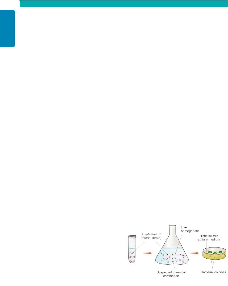
220
Techniques Basic and Pathology General I SECTION
TABLE 8.8: Contrasting Features of Initiator and Promoter Carcinogens.
|
Feature |
Initiator Carcinogens |
Promoter Carcinogens |
|
|
|
|
1. |
Mechanism |
Induction of mutation |
Not mutagenic |
2. |
Dose |
Single for a short time |
Repeated dose exposure, for a long time |
3. |
Response |
Sudden response |
Slow response |
4. |
Change |
Permanent, irreversible |
Change may be reversible |
5. |
Sequence |
Applied first, then followed by promoter |
Applied after prior exposure to initiator |
6. |
Effectivity |
Effective alone if exposed in large dose |
Not effective alone |
7. |
Molecular changes |
Most common mutation of |
Clonal expansion of mutated cells |
|
|
RAS oncogene, p53 anti-oncogene |
|
8. |
Examples |
Most chemical carcinogens, radiation |
Hormones, phorbol esters |
|
|
|
|
i)Phorbol esters. The best known promoter in experimental animals is TPA (tetradecanoyl phorbol acetate) which acts by signal induction protein activation pathway.
ii)Hormones. Endogenous or exogenous oestrogen excess in promotion of cancers of endometrium and breast, prolonged administration of diethylstilbestrol in the etiology of postmenopausal endometrial carcinoma and in vaginal cancer in adolescent girls born to mothers exposed to this hormone during their pregnancy.
iii)Miscellaneous e.g. dietary fat in cancer of colon, cigarette smoke and viral infections etc.
The feature of initiators and promoters are contrasted in
Table 8.8.
Tests for Chemical Carcinogenicity
There are 2 main methods of testing chemical compound for its carcinogenicity:
1.EXPERIMENTAL INDUCTION. The traditional method is to administer the chemical compound under test to a batch of experimental animals like mice or other rodents by an appropriate route e.g. painting on the skin, giving orally or parenterally, or by inhalation. The chemical is administered repeatedly, the dose varied, and promoting agents are administered subsequently. After many months, the animal is autopsied and results obtained. However, all positive or negative tests cannot be applied to humans since there is sufficient species variation in susceptibility to particular carcinogen. Besides, the test is rather prolonged and expensive.
2.TESTS FOR MUTAGENICITY (AMES’ TEST). A mutagen is a substance that can permanently alter the genetic composition of a cell. Ames’ test evaluates the ability of a chemical to induce mutation in the mutant strain of Salmonella typhimurium that cannot synthesise histidine. Such strains are incubated with the potential carcinogen to which liver homogenate is added to supply enzymes required to convert procarcinogen to ultimate carcinogen. If the chemical under test is mutagenic, it will induce mutation in the mutant strains of S. typhimurium in the form of functional histidine gene, which will be reflected by the number of bacterial colonies growing on histidine-free culture medium (Fig. 8.23). Most of the carcinogenic chemicals tested positive in Ames’ test are carcinogenic in vivo.
C. PHYSICAL CARCINOGENESIS
Physical agents in carcinogenesis are divided into 2 groups:
1.Radiation, both ultraviolet light and ionising radiation, is the most important physical agent. The role of radiation as carcinogenic agent is discussed below while its nonneoplastic complications are described in Chapter 3.
2.Non-radiation physical agents are the various forms of injury and are less important.
1. Radiation Carcinogenesis
Ultraviolet (UV) light and ionising radiation are the two main forms of radiation carcinogens which can induce cancer in experimental animals and are implicated in causation of some forms of human cancers. A property common between the two forms of radiation carcinogens is the appearance of mutations followed by a long period of latency after initial exposure, often 10-20 years or even later. Also, radiation carcinogens may act to enhance the effect of another carcinogen (co-carcinogens) and, like chemical carcinogens, may have sequential stages of initiation, promotion and progression in their evolution. Ultraviolet light and ionising radiation differ in their mode of action as described below:
i) ULTRAVIOLET LIGHT. The main source of UV radiation is the sunlight; others are UV lamps and welder’s arcs. UV light penetrates the skin for a few millimetres only so that its effect is limited to epidermis. The efficiency of UV light as carcinogen depends upon the extent of light-
Figure 8.23 
 Schematic representation of the Ames’ test.

absorbing protective melanin pigmentation of the skin. In humans, excessive exposure to UV rays can cause various forms of skin cancers—squamous cell carcinoma, basal cell carcinoma and malignant melanoma. In support of this is the epidemiological evidence of high incidence of these skin cancers in fair-skinned Europeans, albinos who do not tan readily, in inhabitants of Australia and New Zealand living close to the equator who receive more sunlight, and in farmers and outdoor workers due to the effect of actinic light radiation.
Mechanism. UV radiation may have various effects on the cells. The most important is induction of mutation; others are inhibition of cell division, inactivation of enzymes and sometimes causing cell death. The most important biochemical effect of UV radiation is the formation of pyrimidine dimers in DNA. Such UV-induced DNA damage in normal individuals is repaired, while in the predisposed persons who are excessively exposed to sunlight such damage remain unrepaired. The proof in favour of mutagenic effect of UV radiation comes from following recessive hereditary diseases characterised by a defect in DNA repair mechanism and associated with high incidence of cancers:
a)Xeroderma pigmentosum is predisposed to skin cancers at younger age (under 20 years of age).
b)Ataxia telangiectasia is predisposed to leukaemia.
c)Bloom’s syndrome is predisposed to all types of cancers.
d)Fanconi’s anaemia with increased risk to develop cancer. Besides, like with other carcinogens, UV radiation also
induces mutated forms of oncogenes (in particular RAS gene) and anti-oncogenes (p53 gene).
ii) IONISING RADIATION. Ionising radiation of all kinds like X-rays, α-, β- and γ-rays, radioactive isotopes, protons and neutrons can cause cancer in animals and in man. Most frequently, radiation-induced cancers are all forms of leukaemias (except chronic lymphocytic leukaemia); others are cancers of the thyroid (most commonly papillary carcinoma), skin, breast, ovary, uterus, lung, myeloma, and salivary glands (Fig. 8.24). The risk is increased by higher dose and with high LET (linear energy transfer) such as in neutrons and α-rays than with low LET as in X-rays and γ- rays. The evidence in support of carcinogenic role of ionising radiation is cited in the following examples:
a)Higher incidence of radiation dermatitis and subsequent malignant tumours of the skin was noted in X-ray workers and radiotherapists who did initial pioneering work in these fields before the advent of safety measures.
b)High incidence of osteosarcoma was observed in young American watch-working girls engaged in painting the dials with luminous radium who unknowingly ingested radium while using lips to point their brushes.
c)Miners in radioactive elements have higher incidence of cancers.
d)Japanese atom bomb survivors of the twin cities of Hiroshima and Nagasaki after World War II have increased frequency of malignant tumours, notably acute and chronic myeloid leukaemias, and various solid tumours of breast, colon, thyroid and lung.
221
Neoplasia 8 CHAPTER
Figure 8.24 
 Neoplastic (left) and non-neoplastic complications (right) of ionising radiation.
e)Accidental leakage at nuclear power plant in 1985 in Chernobyl (in former USSR, now in Ukraine) has caused long-term hazardous effects of radioactive material to the population living in the vicinity.
f)It has been observed that therapeutic X-ray irradiation results in increased frequency of cancers, e.g. in patients of ankylosing spondylitis, in children with enlarged thymus, and in children irradiated in utero during investigations on the mother.
Mechanism. Radiation damages the DNA of the cell by one of the 2 possible mechanisms:
a)It may directly alter the cellular DNA.
b)It may dislodge ions from water and other molecules of the cell and result in formation of highly reactive free radicals that may bring about the damage.
Damage to the DNA resulting in mutagenesis is the most important action of ionising radiation. It may cause chromosomal breakage, translocation, or point mutation. The effect depends upon a number of factors such as type of radiation, dose, dose-rate, frequency and various host factors such as age, individual susceptibility, immune competence, hormonal influences and type of cells irradiated.
2. Non-radiation Physical Carcinogenesis
Mechanical injury to the tissues such as from stones in the gallbladder, stones in the urinary tract, and healed scars following burns or trauma, has been suggested as the cause of increased risk of carcinoma in these tissues but the evidence is not convincing. Asbestosis and asbestosassociated tumours of the lung are discussed in Chapter 17,

222
Techniques Basic and Pathology General I SECTION
the characteristic tumour being malignant mesothelioma of the pleura. Other examples of physical agents in carcinogenesis are the implants of inert materials such as plastic, glass etc in prostheses or otherwise, and foreign bodies observed to cause tumour development in experimental animals. However, tumorigenesis by these materials in humans is rare.
D. BIOLOGIC CARCINOGENESIS
The epidemiological studies on different types of cancers indicate the involvement of transmissible biologic agents in their development, chiefly viruses. Other biologic agents implicated in carcinogenesis are as follows:
 Parasites. Schistosoma haematobium infection of the urinary bladder is associated with high incidence of squamous cell carcinoma of the urinary bladder in some parts of the world such as in Egypt. Clonorchis sinensis, the liver fluke, lives in the hepatic duct and is implicated in causation of cholangiocarcinoma.

 Fungus. Aspergillus flavus grows in stored grains and liberates aflatoxin; its human consumption, especially by those with HBV infection, is associated with development of hepatocellular carcinoma.
 Bacteria. Helicobacter pylori, a gram-positive spiral-shaped micro-organism, colonises the gastric mucosa and has been found in cases of chronic gastritis and peptic ulcer; its prolonged infection may lead to gastric lymphoma and gastric carcinoma, this subject is discussed in detail in Chapter 20.
However, the role of viruses in the causation of cancer is more significant. Therefore, biologic carcinogenesis is largely viral carcinogenesis, described below.
VIRAL CARCINOGENESIS
It has been estimated that about 20% of all cancers worldwide are due to persistent virus infection. The association of oncogenic viruses with neoplasia was first observed by an Italian physician Sanarelli in 1889 who noted association between myxomatosis of rabbits with poxvirus. The contagious nature of the common human wart was first established in 1907. Since then, a number of viruses capable of inducing tumours (oncogenic viruses) in experimental animals, and some implicated in humans, have been identified.
Oncogenic Viral Infections: General Aspects
Most of the common viral infections (including oncogenic viruses) can be transmitted by one of the 3 routes:
i)Horizontal transmission. Commonly, viral infection passes from one to another by direct contact, by ingestion of contaminated water or food, or by inhalation as occurs in most contagious diseases. Most of these infections begin on the epithelial surfaces, spread into deeper tissues, and then through haematogenous or lymphatic or neural route disseminate to other sites in the body.
ii)By parenteral route such by inoculation as happens in some viruses by inter-human spread and from animals and insects to humans.
iii) Vertical transmission, when the infection is genetically transmitted from infected parents to offsprings.
Based on their nucleic acid content, oncogenic viruses fall into 2 broad groups:
1. Those containing deoxyribonucleic acid are called DNA
oncogenic viruses.
2. Those containing ribonucleic acid are termed RNA
oncogenic viruse or retroviruses.
Both types of oncogenic viruses usually have 3 genes and are abbreviated according to the coding pattern by each gene:
i)gag gene: codes for group antigen.
ii)pol gene: codes for polymerase enzyme.
iii)env gene: codes fro envelope protein.

 Primary viral infections are majority of the common viral infections in which the infection lasts for a few days to a few weeks and produce clinical manifestations. Primary viral infections are generally cleared by body’s innate immunity and specific immune responses. Subsequently, an immunocompetent host is generally immune to the disease or reinfection by the same virus. However, body’s immune system is not effective against surface colonization or deep infection or persistence of viral infection.

 Persistence of viral infection or latent infection in some viruses may occur by acquiring mutations in viruses which resist immune attack by the host, or virus per se induces immunosuppression in the host such as HIV.
Viral Oncogenesis: General Aspects
In general, persistence of DNA or RNA viruses may induce mutation in the target host cell, although persistence of viral infection alone is not sufficient for oncogenesis but is one step in the multistep process of cancer development. Generally, RNA viruses have very high mutation rate (e.g. HIV, HCV) than DNA viruses. Mechanisms as to how specific DNA and RNA viruses cause mutation in the host cell are varied, but in general persistence of DNA or RNA viral infection causes activation of growth-promoting pathways or inhibition of tumour-suppressor products in the infected cells. Thus, such virus-infected host cells after having undergone genetic changes enter cell cycle and produce next progeny of transformed cells which have characteristics of autonomous growth and survival completing their role as oncogenic viruses.
General mode of oncogenesis by each group of DNA and RNA oncogenic viruses is briefly considered below:
1. Mode of DNA viral oncogenesis. Host cells infected by DNA oncogenic viruses may have one of the following 2 results (Fig. 8.25):
i)Replication. The virus may replicate in the host cell with consequent lysis of the infected cell and release of virions.
ii)Integration. The viral DNA may integrate into the host cell DNA.
The latter event (integration) results in inducing mutation and thus neoplastic transformation of the host cell, while the former (replication) brings about cell death but no neoplastic transformation. A feature essential for host cell transformation is the expression of virus-specific T-(transforming protein) antigens immediately after infection of the host cell by DNA oncogenic virus (discussed later).
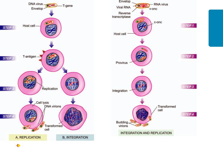
Figure 8.25 
 Replication and integration of DNA virus in the host cell.
A, Replication: Step 1. The DNA virus invades the host cell. Step 2. Viral
DNA is incorporated into the host nucleus and T-antigen is expressed immediately after infection. Step 3. Replication of viral DNA occurs and other components of virion are formed. The new virions are assembled in the cell nucleus. Step 4. The new virions are released, accompanied by host cell lysis. B, Integration : Steps 1 and 2 are similar as in replication.
Step 3. Integration of viral genome into the host cell genome occurs which requires essential presence of functional T-antigen. Step 4. A ‘transformed (neoplastic) cell’ is formed.
223
Neoplasia 8 CHAPTER
Figure 8.26 
 Integration and replication of RNA virus (retrovirus) in the host cell.
Step 1. The RNA virus invades the host cell. The viral envelope fuses with the plasma membrane of the host cell; viral RNA genome as well as reverse transcriptase are released into the cytosol. Step 2. Reverse transcriptase acts as template to synthesise single strand of matching viral DNA which is then copied to form complementary DNA resulting in double-stranded viral DNA (provirus). Step 3. The provirus is integrated into the host cell genome producing ‘transformed host cell.’ Step 4. Integration of the provirus brings about replication of viral components which are then assembled and released by budding.
2. Mode of RNA viral oncogenesis. RNA viruses or retroviruses contain two identical strands of RNA and the enzyme, reverse transcriptase (Fig. 8.26):
i)Reverse transcriptase is RNA-dependent DNA synthetase that acts as a template to synthesise a single strand of matching viral DNA i.e. reverse of the normal in which DNA is transcribed into messenger RNA.
ii)The single strand of viral DNA is then copied by DNAdependent DNA synthetase to form another strand of complementary DNA resulting in double-stranded viral DNA or provirus.
iii)The provirus is then integrated into the DNA of the host cell genome and may induce mutation and thus transform
the cell into neoplastic cell.
iv) Retroviruses are replication-competent. The host cells which allow replication of integrated retrovirus are called permissive cells. Non-permissible cells do not permit replication of the integrated retrovirus.
v) Viral replication begins after integration of the provirus into host cell genome. Integration results in transcription of proviral genes or progenes into messenger RNA which then forms components of the virus particle—virion core protein from gag gene, reverse transcriptase from pol gene, and envelope glycoprotein from env gene. The three components of virus particle are then assembled at the plasma membrane of the host cell and the virus particles released by budding off from the plasma membrane, thus completing the process of replication.
Support to the etiologic role of oncogenic viruses in causation of human cancers is based on the following:
1.Epidemiologic data.
2.Presence of viral DNA in the genome of host target cell.
3.Demonstration of virally induced transformation of human target cells in culture.
4.In vivo demonstration of expressed specific transforming viral genes in premalignant and malignant cells.

224
Techniques Basic and Pathology General I SECTION
5. |
In vitro assay of specific viral gene products which |
supports implication of low-risk HPV types 1,2, 4 and 7 in |
||
produce effects on cell proliferation and survival. |
common viral warts. |
|||
|
With these general comments, now we turn to specific |
Low-risk HPV types 6 and 11 are involved in the etiology |
||
DNA and RNA oncogenic viruses and oncogenesis by them. |
of genital warts (condyloma acuminata). |
|||
|
|
|
Viral DNA of high-risk HPV types 16, 18, 31, 33 and 45 |
|
Specific DNA Oncogenic Viruses |
|
has been seen in 75-100% cases of invasive cervical cancer and |
||
DNA oncogenic viruses have direct access to the host cell |
its precursor lesions (carcinoma in situ and dysplasia) and is |
|||
strongly implicated. |
||||
nucleus and are incorporated into the genome of the host |
||||
High-risk HPVs are also involved in causation of other |
||||
cell. DNA viruses are classified into 5 subgroups, each of |
||||
squamous cell carcinomas and dysplasias such as of anus, |
||||
which is capable of producing neoplasms in different hosts |
||||
perianal region, vagina, vulva, penis and oral cavity. |
||||
(Table 8.9). These are: Papovaviruses, Herpesviruses, |
||||
HPV types 5 and 8 are responsible for causing an |
||||
Adenoviruses, Poxviruses and Hepadna viruses. |
||||
uncommon condition, epidermodysplasia verruciformis. The |
||||
|
|
|
||
1. |
PAPOVAVIRUSES. This group consists of the papilloma |
condition is characterised by multiple skin warts and a |
||
genetic defect in the cell-mediated immunity. About one- |
||||
virus including the human papilloma virus (HPV), polyoma |
||||
third of cases develop squamous cell carcinoma in the sun- |
||||
virus and SV-40 (simian vacuolating) virus. These viruses |
||||
exposed warts. |
||||
have an etiologic role in following benign and malignant |
||||
Some strains of HPV are responsible for causing multiple |
||||
neoplasms in animals and in humans: |
|
|||
|
juvenile papillomas of the larynx. |
|||
i) Papilloma viruses. These viruses were the first to be |
||||
In animals— |
||||
implicated in the etiology of any human neoplasia. These |
||||
Benign warty lesions similar to those seen in humans are |
||||
viruses appear to replicate in the layers of stratified squamous |
||||
produced by different members of the papilloma virus family |
||||
epithelium. More than 100 HPV types have been identified; |
||||
in susceptible animals such as in rabbits by cottontail rabbit |
||||
the individual types are associated with different lesions. The |
||||
papilloma virus, and in cattle by bovine papilloma virus |
||||
following examples of benign and malignant tumours are |
||||
(BPV). |
||||
cited to demonstrate their role in oncogenesis: |
||||
There is evidence to suggest the association of BPV and |
||||
|
|
|
||
In humans— |
|
cancer of the alimentary tract in cattle. |
||
|
HPV was first detected as etiologic agent in common skin |
HPV ONCOGENESIS IN HUMAN CANCER— |
||
warts or verruca vulgaris (squamous cell papillomas) by |
Persistent infection with high-risk HPV types in target |
|||
Shope in 1933; the condition is infectious. Current evidence |
||||
epithelial cells drives the molecular hallmarks of cancer |
||||
|
|
|
||
|
|
|
||
TABLE 8.9: DNA Oncogenic Viruses. |
|
|
||
|
Virus |
Host |
Associated Tumour |
|
|
|
|
||
1. PAPOVAVIRUSES |
|
|
||
|
Human papilloma virus |
Humans |
Cervical cancer and its precursor lesions, squamous cell |
|
|
|
|
carcinoma at other sites |
|
|
|
|
Skin cancer in epidermodysplasia verruciformis |
|
|
|
|
Papillomas (warts) on skin, larynx, genitals (genital warts) |
|
|
Papilloma viruses |
Cotton-tail rabbits |
Papillomas (warts) |
|
|
|
Bovine |
Alimentary tract cancer |
|
|
Polyoma virus |
Mice |
Various carcinomas, sarcomas |
|
|
SV-40 virus |
Monkeys |
Harmless |
|
|
|
Hamsters |
Sarcoma |
|
|
|
Humans |
? Mesothelioma |
|
2. HERPESVIRUSES |
|
|
||
|
Epstein-Barr virus |
Humans |
Burkitt’s lymphoma |
|
|
|
|
Nasopharyngeal carcinoma |
|
|
Human herpesvirus 8 |
Humans |
Kaposi’s sarcoma |
|
|
(Kaposi's sarcoma herpesvirus) |
|
Pleural effusion lymphoma |
|
|
Lucke’ frog virus |
Frog |
Renal cell carcinoma |
|
|
Marek’s disease virus |
Chickens |
T-cell leukaemia-lymphoma |
|
3. |
ADENOVIRUSES |
Hamsters |
Sarcomas |
|
4. |
POXVIRUSES |
Rabbits |
Myxomatosis |
|
|
|
Humans |
Molluscum contagiosum, papilloma |
|
5. HEPADNAVIRUSES |
|
|
||
|
Hepatitis B virus |
Humans |
Hepatocellular carcinoma |
|
|
|
|
|
|
discussed earlier and directly affect cell growth by following mechanisms:
i)HPV integrates in to the host cell DNA which results in overexpression of viral proteins E6 and E7 from high risk HPV types. E6 and E7 from high-risk HPVs has high affinity for target host cells than these viral oncoproteins from lowrisk HPVs.
ii)E6 and E7 viral proteins cause loss of p53 and pRB, the two cell proteins with tumour-suppressor properties. Thus the breaks in cell proliferation are removed, permitting the uncontrolled proliferation.
iii)These viral proteins activate cyclin A and E, and inactivate CDKIs, thus permitting further cell proliferation.
iv)These viral proteins mediate and degrade BAX, a proapoptotic gene, thus inhibiting apoptosis.
v)These viral proteins activate telomerase, immortalising the transformed host target cells.
ii)Polyoma virus. Polyoma virus occurs as a natural infection in mice.

 In animals—Polyoma virus infection is responsible for various kinds of carcinomas and sarcomas in immunodeficient (nude) mice and other rodents. In view of its involvement in causation of several unrelated tumours in animals, it was named polyoma.

 In humans—Polyoma virus infection is not known to produce any human tumour. But it is involved in causation of polyomavirus nephropathy in renal allograft recipients and is also implicated in the etiology of progressive demyelinating leucoencephalopathy, a fatal demyelinating disease.
iii)SV-40 virus. As the name suggests, simian vacuolating virus exists in monkeys without causing any harm but can induce sarcoma in hamsters. There is some evidence of involvement of SV-40 infection in mesothelioma of the pleura.
2. HERPESVIRUSES. Primary infection of all the herpesviruses in man persists probably for life in a latent stage which can get reactivated later. Important members of herpesvirus family are Epstein-Barr virus, herpes simplex virus type 2 (HSV-2) and human herpesvirus 8 (HHV8), cytomegalovirus (CMV), Lucke’s frog virus and Marek’s disease virus. Out of these, Lucke’s frog virus and Marek’s disease virus are implicated in animal tumours only (renal cell carcinoma and T-cell leukaemia-lymphoma respectively). There is no oncogenic role of HSV-2 and CMV in human tumours. The other two—EBV and HHV are implicated in human tumours as under.
EPSTEIN-BARR VIRUS (EBV). EBV infects human B- lymphocytes and epithelial cells and long-term infection stimulates them to proliferate and development of malignancies. EBV is implicated in the following human tumours—Burkitt’s lymphoma, anaplastic nasopharyngeal carcinoma, post-transplant lymphoproliferative disease, primary CNS lymphoma in AIDS patients, and Hodgkin’s lymphoma. It is also shown to be causative for infectious mononucleosis, a self-limiting disease in humans. The role
of EBV in the first two human tumours is given below while 225 others have been discussed elsewhere in relevant chapters.
Burkitt’s lymphoma. Burkitt’s lymphoma was initially |
CHAPTER |
|
noticed in African children by Burkitt in 1958 but is now |
||
|
||
known to occur in 2 forms—African endemic form, and sporadic |
|
|
form seen elsewhere in the world. The morphological aspects |
|
|
of the tumour are explained in Chapter 14, while oncogenesis |
|
|
is described here. |
8 |
|
There is strong evidence linking Burkitt’s lymphoma, a |
||
|
||
B-lymphocyte neoplasm, with EBV as observed from the |
Neoplasia |
|
following features: |
||
a) Over 90% of Burkitt’s lymphomas are EBV-positive in |
||
|
||
which the tumour cells carry the viral DNA. |
|
|
b) 100% cases of Burkitt’s lymphoma show elevated levels |
|
|
of antibody titers to various EBV antigens. |
|
|
c) EBV has strong tropism for B lymphocytes. EBV-infected |
|
|
B cells grown in cultures are immortalised i.e. they continue |
|
|
to develop further along B cell-line to propagate their progeny |
|
|
in the altered form. |
|
|
d) Though EBV infection is almost worldwide in all adults |
|
|
and is also known to cause self-limiting infectious |
|
|
mononucleosis, but the fraction of EBV-infected circulating |
|
|
B cells in such individuals is extremely small. |
|
|
e) Linkage between Burkitt’s lymphoma and EBV infection |
|
|
is very high in African endemic form of the disease and |
|
|
probably in cases of AIDS than in sporadic form of the |
|
|
disease. |
|
|
However, a few observations, especially regarding spora- |
|
|
dic cases of Burkitt’s lymphoma, suggest that certain other |
|
|
supportive factors may be contributing. Immunosuppression |
|
|
appears to be one such most significant factor. The evidence |
|
|
in favour is as follows: |
|
|
Normal individuals harbouring EBV-infection as well as |
|
|
cases developing infectious mononucleosis are able to mount |
|
|
good immune response so that they do not develop Burkitt’s |
|
|
lymphoma. |
|
|
In immunosuppressed patients such as in HIV infection |
|
|
and organ transplant recipients, there is marked reduction |
|
|
in body’s T-cell immune response and higher incidence of |
|
|
this neoplasm. |
|
|
It is observed that malaria, which confers immuno- |
|
|
suppressive effect on the host, is prevalent in endemic |
|
|
proportions in regions where endemic form of Burkitt’s |
|
|
lymphoma is frequent. This supports the linkage of EBV |
|
|
infection and immunosuppression in the etiology of Burkitt’s |
|
|
lymphoma. |
|
Anaplastic nasopharyngeal carcinoma. This is the other tumour having close association with EBV infection. The tumour is prevalent in South-East Asia, especially in the Chinese, and in Eskimos. The morphology of nasopharyngeal carcinoma is described in Chapter 18. The evidence linking EBV infection with this tumour is as follows:
a)100% cases of nasopharyngeal carcinoma carry DNA of EBV in nuclei of tumour cells.
b)Individuals with this tumour have high titers of antibodies to various EBV antigens.

226 However, like in case of Burkitt’s lymphoma, there may be some co-factors such as genetic susceptibility that account for the unusual geographic distribution.
SECTION |
EBV ONCOGENESIS IN HUMAN CANCER— |
|
|
||
|
Persistent EBV infection is implicated in the causation of |
|
|
malignancies of B lymphocytes and epithelial cells. The |
|
|
mechanism of oncogenesis is as under: |
|
I |
i) Latently infected epithelial cells or B lymphocytes express |
|
viral oncogene LMP1 (latent membrane protein) which is |
||
|
||
|
most crucial step in evolution of EBV-associated malig- |
|
General |
nancies. Immunosuppressed individuals are unable to mount |
|
and survival of infected cells and acts like CD40 receptor |
||
|
attack against EBV infection and thus are more affected. |
|
|
ii) LMP1 viral protein dysregulates normal cell proliferation |
|
Pathology |
molecule on B cell surface. Thus, it stimulates B-cell |
|
prevents apoptosis. |
||
|
proliferation by activating growth signaling pathways via |
|
|
nuclear factor κB (NF-κB) and JAK/STAT pathway. |
|
|
iii) LMP1 viral oncoprotein also activates BCL2 and thereby |
|
and |
iv) Persistent EBV infection elaborates another viral protein |
|
EBNA-2 which activates cyclin D in the host cells and thus |
||
Basic |
promotes cell proliferation. |
|
v) In immunocompetent individuals, LMP1 is kept under |
||
|
||
Techniques |
contro by the body’s immune system and in these persons |
|
infection with HHV-8 or Kaposi’s sarcoma-associated |
||
|
therefore lymphoma cells appear only after another |
|
|
characteristic mutation t(8;14) activates growth promoting |
|
|
MYC oncogene. |
|
|
HUMAN HERPESVIRUS 8 (HHV-8). It has been shown that |
|
|
herpesvirus (KSHV) is associated with Kaposi’s sarcoma, a |
|
|
vascular neoplasm common in patients of AIDS. Compared |
|
|
to sporadic Kaposi’s sarcoma, the AIDS-associated tumour |
|
|
is multicentric and more aggressive. HHV-8 has lympho- |
|
|
tropism and is also implicated in causation of pleural effusion |
|
|
lymphoma and multicentric variant of Castleman’s disease. |
HHV-8 (KSHV) ONCOGENESIS IN HUMAN CANCER—
i)Viral DNA is seen in nuclei of all tumour cells in Kaposi’s sarcoma.
ii)There is overexpression of several KSHV oncoproteins by latently infected cells: v-cyclin, v-interferon regulatory factor (v-IRF) and LANA (latency-associated nuclear antigen).
iii)These viral proteins cause increased proliferation and survival of host cells and thus induce malignancy.
3.ADENOVIRUSES. The human adenoviruses cause upper respiratory infections and pharyngitis.

 In humans, they are not known to be involved in any tumour.

 In hamsters, they may induce sarcomas.
4.POXVIRUSES. This group of oncogenic viruses is involved in the etiology of following lesions:

 In rabbits—poxviruses cause myxomatosis.
 In humans—poxviruses cause molluscum contagiosum and may induce squamous cell papilloma.
5.HEPADNAVIRUSES. Hepatitis B virus (HBV) is a member of hepadnavirus (hepa- from hepatitis, -dna from DNA) family. HBV infection in man causes an acute hepatitis
and is responsible for a carrier state, which can result in some cases to chronic hepatitis progressing to hepatic cirrhosis, and onto hepatocellular carcinoma. These lesions and the structure of HBV are described in detail in Chapter 21. Suffice this to say here that there is strong epidemiological evidence linking HBV infection to development of hepatocellular carcinoma as evidenced by the following:
a)The geographic differences in the incidence of hepatocellular carcinoma closely match the variation in prevalence of HBV infection e.g. high incidence in Far-East and Africa.
b)Epidemiological studies in high incidence regions indicate about 200 times higher risk of developing hepatocellular carcinoma in HBV-infected cases as compared to uninfected population in the same area.
Posssible mechanism of hepatocellular carcinoma occurring in those harbouring long-standing infection with HBV is chronic destruction of HBV-infected hepatocytes followed by continued hepatocyte proliferation. This process renders the hepatocytes vulnerable to the action of other risk factors such as to aflatoxin causing mutation and neoplastic proliferation.
More recent evidence has linked an oncogenic role to another hepatotropic virus, hepatitis C virus (HCV) as well which is an RNA virus, while HBV is a DNA virus. HCV is implicated in about half the cases of hepatocellular carcinoma in much the same way as HBV.
HEPATITIS VIRUS ONCOGENESIS IN HUMAN CANCER—
Epidemiologic data firmly support that two hepatotropic viruses, HBV—a DNA virus, and HCV—an RNA virus, are currently involved in causation of 70-80% cases of hepatocellular carcinoma worldwide. Although HBV DNA has been found integrated in the genome of human hepatocytes in many cases of liver cancer which causes mutational changes but a definite pattern is lacking. Thus, exact molecular mechanism as to how HBV and HCV cause hepatocellular carcinoma is yet not quite clear. Probably, multiple factors are involved:
i)Chronic and persistent viral infection with HBV or HCV incites repetitive cycles of inflammation, immune response, cell degeneration/cell death, and regeneration of the hepatocytes which leads to DNA damage to liver cells of the host.
ii)It is possible that immune response by the host to persistent unresolved infection with these hepatitis viruses becomes defective which promotes tumour development.
iii)On regeneration, proliferation of hepatocytes is stimulated by several growth factors and cytokines elaborated by activated immune cells which contribute to tumour development e.g. factors for angiogenesis, cell survival etc.
iv)Activated immune cells produce nuclear factor κB (NF-κB) that inhibits apoptosis, thus allowing cell survival and growth.
v)HBV genome contains a gene HBx which activates growth signaling pathway.
vi)HBV and HCV do not encode for any specific viral oncoproteins.

Specific RNA Oncogenic Viruses |
|
of neoplastic transformation (‘slow’). Slow transforming |
||
RNA oncogenic viruses are retroviruses i.e. they contain the |
viruses cause neoplastic transformation by insertional |
|||
mutagenesis i.e. viral DNA synthesised by viral RNA via |
||||
enzyme reverse transcriptase (RT), though all retroviruses |
||||
reverse transcriptase is inserted or integrated near the |
||||
are not oncogenic (Table 8.10). The enzyme, reverse trans- |
||||
protooncogenes of the host cell resulting in damage to host |
||||
criptase, is required for reverse transcription of viral RNA |
||||
cell genome (mutagenesis) leading to neoplastic trans- |
||||
to synthesise viral DNA strands i.e. reverse of normal—rather |
||||
formation. |
|
|||
than DNA encoding for RNA synthesis, viral RNA transcripts |
|
|||
3. HUMAN T-CELL LYMPHOTROPIC VIRUSES |
||||
for the DNA by the enzyme RT present in the RNA viruses. |
||||
RT is a DNA polymerase and helps to form complementary |
(HTLV). HTLV is a form of slow transforming virus but is |
|||
DNA (cDNA) that moves in to host cell nucleus and gets |
described separately because of 2 reasons: |
|||
incorporated in to it. |
|
i) This is the only retrovirus implicated in human cancer. |
||
Based on their activity to transform target cells into |
ii) The mechanism of neoplastic transformation is different |
|||
neoplastic cells, RNA viruses are divided into 3 subgroups— |
from slow transforming as well as from acute transforming |
|||
acute transforming viruses, slow transforming viruses, and |
viruses. |
|
||
human T-cell lymphotropic viruses (HTLV). The former two |
Four types of HTLVs are recognised—HTLV-I, HTLV-II, |
|||
are implicated in inducing a variety of tumours in animals |
HTLV-III and HTLV-IV. It may be mentioned in passing here |
|||
only while HTLV is causative for human T-cell leukaemia |
that the etiologic agent for AIDS, HIV, is also an HTLV |
|||
and lymphoma. |
|
(HTLV-III) as described in Chapter 4. |
||
1. ACUTE TRANSFORMING VIRUSES. This group |
A link between HTLV-I infection and cutaneous adult T- |
|||
cell leukaemia-lymphoma (ATLL) has been identified while |
||||
includes retroviruses which transform all the cells infected |
||||
HTLV-II is implicated in causation of T-cell variant of hairy |
||||
by them into malignant cells rapidly (‘acute’). All the viruses |
||||
cell leukaemia. HTLV-I is transmitted through sexual contact, |
||||
in this group possess one or more viral oncogenes (v-oncs). |
||||
by blood, or to infants during breastfeeding. The highlights |
||||
All the members of acute transforming viruses discovered |
||||
of this association and mode of neoplastic transformation |
||||
so far are defective viruses in which the particular v-onc has |
||||
are as under: |
|
|||
substituted other essential genetic material such as gag, pol |
|
|||
i) Epidemiological studies by tests for antibodies have |
||||
and env. These defective viruses cannot replicate by |
||||
shown that HTLV-I infection is endemic in parts of Japan |
||||
themselves unless the host cell is infected by another ‘helper |
||||
and West Indies where the incidence of ATLL is high. The |
||||
virus’. Acute oncogenic viruses have been identified in |
||||
latent period after HTLV-I infection is, however, very long |
||||
tumours in different animals only e.g. |
|
|||
|
(20-30 years). |
|
||
a) Rous sarcoma virus in chickens. |
|
|
||
|
ii) The initiation of neoplastic process is similar to that for |
|||
b) Leukaemia-sarcoma viruses of various types such as |
||||
Burkitt’s lymphoma except that HTLV-I has tropism for |
||||
avian, feline, bovine and primate. |
|
CD4+T lymphocytes as in HIV infection, while EBV of |
||
2. SLOW TRANSFORMING VIRUSES. These oncogenic |
Burkitt’s lymphoma has tropism for B lymphocytes. |
|||
iii) As in Burkitt’s lymphoma, immunosuppression plays a |
||||
retroviruses cause development of leukaemias and |
||||
supportive role in the neoplastic transformation by HTLV-I |
||||
lymphomas in different species of animals (e.g. in mice, cats |
||||
infection. |
|
|||
and bovine) and include the mouse mammary tumour virus |
|
|||
|
|
|||
(MMTV) that causes breast cancer in the daughter-mice |
HTLV ONCOGENESIS IN HUMAN CANCER— |
|||
suckled by the MMTV-infected mother via the causal agent |
The molecular mechanism of ATLL leukaemogenesis by |
|||
in the mother’s milk (Bittner milk factor). These viruses have |
HTLV infection of CD4+ T lymphocytes is not clear. |
|||
long incubation period between infection and development |
Neoplastic transformation by HTLV-I infection differs from |
|||
|
|
|
|
|
TABLE 8.10: RNA Oncogenic Viruses. |
|
|
|
|
Virus |
Host |
|
Associated Tumour |
|
|
|
|
|
|
1. ACUTE TRANSFORMING VIRUSES |
|
|
|
|
Rous sarcoma virus |
Chickens |
|
Sarcoma |
|
Leukaemia-sarcoma virus |
Avian, feline, bovine, primate |
Leukaemias, sarcomas |
||
2. SLOW TRANSFORMING VIRUSES |
|
|
|
|
|
Mice, cats, bovine |
Leukaemias, lymphomas |
||
Mouse mammary tumour virus |
Daughter mice |
Breast cancer |
||
(Bittner milk factor) |
|
|
|
|
3. HUMAN T-CELL LYMPHOTROPIC VIRUS (HTLV) |
|
|
|
|
HTLV-I |
Human |
|
Adult T-cell leukaemia lymphoma (ATLL) |
|
HTLV-II |
Human |
|
T-cell variant of hairy cell leukaemia |
|
4. HEPATITIS C VIRUS |
|
|
|
|
HCV |
Human |
|
Hepatocellular carcinoma |
|
|
|
|
|
|
227
Neoplasia 8 CHAPTER
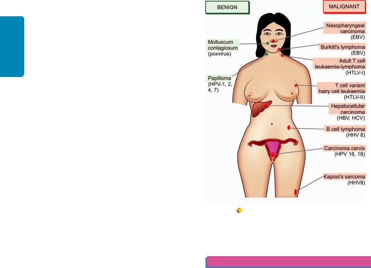
228
Techniques Basic and Pathology General I SECTION
acute transforming viruses because it does not contain v-onc, and from other slow transforming viruses because it does not have fixed site of insertion for insertional mutagenesis. Probably, the process is multifactorial:
i)HTLV-I genome has unique region called pX distinct from other retroviruses, which encodes for two essential viral oncoproteins— TAX and REX. TAX protein up-regulates the expression of cellular genes controlling T-cell replication, while REX gene product regulates viral protein production by affecting mRNA expression.
ii)TAX viral protein interacts with transcription factor, NF- κB, which stimulates genes for cytokines (interleukins) and their receptors in infected T cells which activates proliferation of T cells by autocrine pathway.
iii)The inappropriate gene expression activates pathway of the cell proliferation by activation of cyclins and inactivation of tumour suppressor genes CDKN2A/p16 and p53, stimulating cell cycle.
iv)Initially, proliferation of infected T cells is polyclonal but subsequently several mutations appear due to TAX-based genomic changes in the host cell and monoclonal proliferation of leukaemia occurs.
VIRUSES AND HUMAN CANCER: A SUMMARY
In man, epidemiological as well as circumstantial evidence has been accumulating since the discovery of contagious nature of common human wart (papilloma) in 1907 that cancer may have viral etiology. Presently, about 20% of all human cancers worldwide are virally induced. Aside from experimental evidence, the etiologic role of DNA and RNA viruses in a variety of human neoplasms has already been explained above. Here, a summary of different viruses implicated in human tumours is presented (Fig. 8.27):
Benign tumours. There are 2 conditions which are actually doubtful as tumours in which definite viral etiology is established. These are:
i)Human wart (papilloma) caused by human papilloma virus; and
ii)Molluscum contagiosum caused by poxvirus.
Malignant tumours. The following 8 human cancers have enough epidemiological, serological, and in some cases genomic evidence, that viruses are implicated in their etiology:
i)Burkitt’s lymphoma by Epstein-Barr virus.
ii)Nasopharyngeal carcinoma by Epstein-Barr virus.
iii)Primary hepatocellular carcinoma by hepatitis B virus and hepatitis C virus.
iv)Cervical cancer by high risk human papilloma virus types (HPV 16 and 18).
v)Kaposi’s sarcoma by human herpes virus type 8 (HHV-8).
vi)Pleural effusion B cell lymphoma by HHV8.
Figure 8.27  Viruses (in brackets) in human tumours.
hepatocellular carcinoma. HPV vaccine has been launched lately and is likely to lower the incidence of HPV-associated cervical cancer.
CLINICAL ASPECTS OF NEOPLASIA
Two major aspects of clinical significance in assessing the course and management of neoplasia are: tumour-host interrelationship (i.e. the effect of tumour on host and vice versa) and laboratory diagnosis of cancer.
TUMOUR-HOST INTER-RELATIONSHIP
The natural history of a neoplasm depends upon 2 features:
i)Host response against tumour (Immunology of cancer)
ii)Effect of tumour on host
HOST RESPONSE AGAINST TUMOUR (TUMOUR IMMUNOLOGY)
It has long been known that body’s immune system can recognise tumour cells as ‘non-self’ and attempt to destroy them and limit the spread of cancer. The following observations provide basis for this concept:
1. Certain cancers evoke significant lymphocytic infiltrates
vii)Adult T-cell leukaemia and lymphoma by HTLV-I. composed of immunocompetent cells and such tumours have
viii)T-cell variant of hairy cell leukaemia by HTLV-II. somewhat better prognosis e.g. medullary carcinoma breast
Current knowledge and understanding of viral |
(as compared with infiltrating ductal carcinoma), seminoma |
carcinogenesis has provided an opportunity to invent specific |
testis (as compared with other germ cell tumours of testis). |
vaccines and suggest appropriate specific therapy. For |
2. Rarely, a cancer may spontaneously regress partially or |
example, hepatitis B vaccines is being widely used to control |
completely, probably under the influence of host defense |
hepatitis B and is expected to lower incidence of HBV-related |
mechanism. For example, rare spontaneous disappearance |

of malignant melanoma temporarily from the primary site which may then reappear as metastasis.
3.It is highly unusual to have primary and secondary tumours in the spleen due to its ability to destroy the growth and proliferation of tumour cells.
4.Immune surveillance exists is substantiated by increased frequency of cancers in immunodeficient host e.g. in AIDS patients, or development of post-transplant lymphoproliferative disease.
In an attempt to substantiate the above observations and to understand the underlying host defense mechanisms, experimental animal studies involving tumour transplants were carried out. The findings of animal experiments coupled with research on human cancers has led to the concept of immunology of cancer discussed under the following headings:
1.Tumour antigens
2.Antitumour immune responses
3Immunotherapy.
1. TUMOUR ANTIGENS. Tumour cells express surface antigens which have been seen in animals and in some human tumours. Older classification of tumour antigens was based on their surface sharing characteristics on normal versus tumour cells and on their recognition by cytotoxic T lymphocytes CTL (CD8+T cells) on the basis of class I MHC molecules. Accordingly, tumour antigens were categorised into following two types:
i)Tumour-specific antigens (TSAs) located on tumour cells and are unique or specific antigens for particular tumour and not shared by normal cells.
ii)Tumour associated antigens (TAAs) are present on tumour cells as well as on some normal cells from where the tumour originated.
However, it is now known that TSAs and TAAs can both be present on normal cells and categorisation into TSA and TAA does not hold true. Thus, presently distinction of tumour antigens is based on their recognition by the host immune cells, i.e. CD8+ T cells (CTL), and by the molecular structure of the tumour antigens. Currently, various groups of tumour antigens are as follows:
1.Oncoproteins from mutated oncogenes: Protein products derived from mutated oncogenes result in expression of cell surface antigens on tumour cells. The examples include products of RAS, BCL/ABL and CDK4.
2.Protein products of tumour suppressor genes. In some tumours, protein products of mutated tumour suppressor genes cause expression of tumour antigens on the cell surface. The examples are mutated proteins p53 and β-catenin.
3.Overexpressed cellular proteins. Some tumours are associated with a normal cellular protein but is excessively expressed in tumour cells and incite host immune response. For example, in melanoma the tumour antigen is structurally normal melanocyte specific protein, tyrosinase, which is overexpressed compared with normal cells. Similarly, HER2/ neu protein is overexpressed in many cases of breast cancer.
4.Abnormally expressed cellular proteins. Sometimes, a cellular protein is present in some normal cells but is abnormally expressed on the surface of tumour cells of some cancers. The classic example is presence of MAGE gene silent in
normal adult tissues except in male germ line but MAGE genes are expressed on surface of many tumours such as melanoma (abbreviation MAGE from ‘melanoma antigen’ in which it was first found), cancers of liver, lung, stomach and oesophagus. Other examples of similar aberrantly expressed gene products in cancers are GAGE (G antigen), BAGE (B melanoma antigen) and RAGE (renal tumour antigen).
5.Tumour antigens from viral oncoproteins. As already discussed above, many oncogenic viruses express viral oncoproteins which result in expression of antigens on tumour cells e.g. viral oncoproteins of HPV (E6, E7) in cervical cancer and EBNA proteins of EBV in Burkitt’s lymphoma.
6.Tumour antigens from randomly mutated genes. Various other carcinogens such as chemicals and radiation induce random mutations in the target cells. These mutated cells elaborate protein products targeted by the CTL of the immune system causing expression of tumour antigens.
7.Cell specific differentiation antigens. Normally differentiated cells have cellular antigens which forms the basis of diagnostic immunohistochemistry. Cancers have varying degree of loss of differentiation but particular lineage of the tumour cells can be identified by tumour antigens. For example, various CD markers for various subtypes of lymphomas, prostate specific antigen (PSA) in carcinoma of prostate.
8.Oncofoetal antigens. Oncofoetal antigens such as α- foetoprotein (AFP) and carcinoembryonic antigen (CEA) are normally expressed in embryonic life. But these antigens appear in certain cancers—AFP in liver cancer and CEA in colon cancer which can be detected in serum as cancer markers.
9.Abnormal cell surface molecules. The normal cell expresses surface molecules of glycolipids, glycoproteins, mucins and blood group antigens. In some cancers, there is abnormally changed expression of these molecules. For example, there may be changed blood group antigen, or abnormal expression of mucin in ovarian cancer (CA-125) and in breast cancer (MUC-1).
2. ANTI-TUMOUR IMMUNE RESPONSES. Although the host immune response to tumour is by both cell-mediated and humoral immunity, the major anti-tumour effector mechanism is cell-mediated.
i) Cell-mediated mechanism. This is the main mechanism of destruction of tumour cells by the host. The following cellular responses can destroy the tumour cells and induce tumour immunity in humans:
a)Specifically sensitised cytotoxic T lymphocytes (CTL) i.e. CD8+ T cells are directly cytotoxic to the target cell and require contact between them and tumour cells. CTL have been found to be effective against virally-induced cancers e.g. in Burkitt’s lymphoma (EBV-induced), invasive squamous cell carcinoma of cervix (HPV-induced).
b)Natural killer (NK) cells are lymphocytes which after activation by IL-2, destroy tumour cells without sensitisation, either directly or by antibody-dependent cellular cytotoxicity (ADCC). NK cells together with T lymphocytes are the first line of defense against tumour cells and can lyse tumour cells.
c)Macrophages are activated by interferon-γ secreted by T-cells and NK-cells, and therefore there is close collaboration
229
Neoplasia 8 CHAPTER
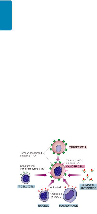
230 |
of these two subpopulation of lymphocytes and |
|
|
macrophages. Activated macrophages mediate cytotoxicity |
|
SECTION |
by production of oxygen free radicals or by tumour necrosis |
|
factor. |
||
tumour cells by complement activation or by antibody- |
||
|
ii) Humoral mechanism. As such there are no anti-tumour |
|
|
humoral antibodies which are effective against cancer cells |
|
|
in vivo. However, in vitro humoral antibodies may kill |
|
I |
dependent cytotoxicity. Based on this, monoclonal antibody |
|
|
treatment is offered to cases of some non-Hodgkin’s |
|
General |
lymphoma. |
|
iii) Immune regulatory mechanism. In spite of host immune |
||
responses, most cancers grow relentlessly. This is due to some |
||
of the following controlling mechanisms: |
||
Pathology |
a) During progression of the cancer, immunogenic cells may |
|
c) Immunosuppression mediated by various acquired |
||
|
disappear. |
|
|
b) Cytotoxic T-cells and NK-cells may play a self regulatory |
|
|
role. |
|
and |
carcinogenic agents (viruses, chemicals, radiation). |
|
d) Immunosuppressive role of factors secreted by tumour |
||
Basic |
cells e.g. transforming growth factor-β. |
|
The mechanisms of these immune responses are |
||
|
||
Techniques |
schematically illustrated in Fig. 8.28. |
|
rapidly than their rate of proliferation and hence the role of |
||
|
3. IMMUNOTHERAPY. Despite the existence of anti- |
|
|
tumour immune responses, the cancers still progress and |
|
|
eventually cause death of the host. The immune responses |
|
|
to be effective enough must eliminate the cancer cells more |
|
|
boosting the immune response or immunotherapy. |
|
|
i) Non-specific stimulation of the host immune response |
|
|
was initially attempted with BCG, Corynebacterium parvum |
|
|
and levamisole, but except slight effect in acute lymphoid |
|
|
leukaemia, it failed to have any significant influence in any |
|
|
other tumour. |
Figure 8.28 
 Schematic illustration of immune responses in cancer. For details see the text (CTL = cytotoxic T-lymphocyte; NK cell = natural killer cell; ADCC = antibody-dependent cellular cytotoxicity).
ii)Specific stimulation of the immune system was attempted next by immunising the host with irradiated tumour cells but failed to yield desired results because if the patient’s tumour within the body failed to stimulate effective immunity, the implanted cells of the same tumour are unlikely to do so.
iii)Current status of immunotherapy is focussed on following three main approaches:
a)Cellular immunotherapy consists of infusion of tumourspecific cytotoxic T cells which will increase the population of tumour-infiltrating lymphocytes (TIL). The patient’s peripheral blood lymphocytes are cultured with interleukin- 2 which generates lymphokine-activated killer cells having potent anti-tumour effect.
b)Cytokine therapy is used to build up specific and nonspecific host defenses. These include: interleukin-2, interferon-α and -γ , tumour necrosis factor-α , and granulocyte-monocyte colony stimulating factor (GM-CSF).
c)Monoclonal antibody therapy is currently being tried against CD20 molecule of B cells in certain B cell leukaemias and lymphomas.
EFFECT OF TUMOUR ON HOST
Malignant tumours produce more ill-effects than the benign tumours. The effects may be local, or generalised and more widespread.
1. LOCAL EFFECTS. Both benign and malignant tumours cause local effects on the host due to their size or location. Malignant tumours due to rapid and invasive growth potential have more serious effects. Some of the local effects of tumours are as under:
i)Compression. Many benign tumours pose only a cosmetic problem. Some benign tumours, however, due to their critical location, have more serious consequences e.g. pituitary adenoma may lead to serious endocrinopathy; a small benign tumour in ampulla of Vater may lead to biliary obstruction.
ii)Mechanical obstruction. Benign and malignant tumours in the gut may produce intestinal obstruction.
iii)Tissue destruction. Malignant tumours, both primary and metastatic, infiltrate and destroy the vital structures.
iv)Infarction, ulceration, haemorrhage. Cancers have a greater tendency to undergo infarction, surface ulceration and haemorrhage than the benign tumours. Secondary bacterial infection may supervene. Large tumours in mobile organs (e.g. an ovarian tumour) may undergo torsion and produce infarction and haemorrhage.
2.CANCER CACHEXIA. Patients with advanced and disseminated cancers terminally have asthenia (emaciation), and anorexia, together referred to as cancer cachexia (meaning wasting). Exact mechanism of cachexia is not clear but it does not occur due to increased nutritional demands of the tumour. Possibly, cachectin or tumour necrosis factor
α(TNF-α) and interleukin-1 derived from macrophages play a contributory role in cachexia. Various other causes include necrosis, ulceration, haemorrhage, infection, malabsorption, anxiety, pain, insomnia, hypermetabolism and pyrexia.

3.FEVER. Fever of unexplained origin may be presenting feature in some malignancies such as in Hodgkin’s disease, adenocarcinoma kidney, osteogenic sarcoma and many other tumours. The exact mechanism of tumour associated fever is not known but probably the tumour cells themselves elaborate pyrogens.
4.TUMOUR LYSIS SYNDROME. This is a condition caused by extensive destruction of a large number of rapidly proliferating tumour cells. The condition is seen more often in cases of lymphomas and leukaemias than solid tumours and may be due to large tumour burden (e.g. in Burkitt’s lymphoma), chemotherapy, administration of glucocorticoids or certain hormonal agents (e.g. tamoxifen). It is
characterised by hyperuricaemia, hyperkalaemia, hyperphosphataemia and hypocalcaemia, all of which may result in acidosis and renal failure.
5. PARANEOPLASTIC SYNDROMES. Paraneoplastic syndromes (PNS) are a group of conditions developing in patients with advanced cancer which are neither explained by direct and distant spread of the tumour, nor by the usual hormone elaboration by the tissue of origin of the tumour. About 10 to 15% of the patients with advanced cancer develop one or more of the syndromes included in the PNS. Rarely, PNS may be the earliest manifestation of a latent cancer.
The various clinical syndromes included in the PNS are as summarised in Table 8.11 and are briefly outlined below:
TABLE 8.11: Summary of Paraneoplastic Syndromes.
|
Clinical Syndrome |
Underlying Cancer |
Mechanism |
|
1. |
ENDOCRINE SYNDROME: |
|
|
|
|
i. |
Hypercalcaemia |
Lung (sq. cell Ca), kidney, breast, |
Parathormone-like protein |
|
|
|
Adult T-cell leukaemia-lymphoma |
vitamin D |
|
ii. |
Cushing’s syndrome |
Lung (small cell carcinoma), |
ACTH or ACTH-like |
|
|
|
pancreas, neural tumours |
substance |
|
iii. |
Inappropriate anti-diuresis |
Lung (small cell Ca), |
ADH or atrial natriuretic |
|
|
|
prostate, intracranial tumour |
factor |
|
iv. |
Hypoglycaemia |
Pancreas (islet cell tumour), |
Insulin or insulin-like |
|
|
|
mesothelioma, fibrosarcoma |
substance |
|
v. |
Carcinoid syndrome |
Bronchial carcinoid tumour, |
Serotonin, bradykinin |
|
|
|
carcinoma pancreas, stomach |
|
|
vi. |
Polycythaemia |
Kidney, liver, cerebellar |
Erythropoietin |
|
|
|
haemangioma |
|
2. |
NEUROMUSCULAR SYNDROMES: |
|
|
|
|
i. |
Myasthenia gravis |
Thymoma |
Immunologic |
|
ii. |
Neuromuscular disorders |
Lung (small cell Ca), breast |
Immunologic |
3. |
OSSEOUS, JOINT AND SOFT TISSUE: |
|
|
|
|
i. |
Hypertrophic osteoarthropathy |
Lung |
Not known |
|
ii. |
Clubbing of fingers |
Lung |
Not known |
4. |
HAEMATOLOGIC SYNDROMES: |
|
|
|
|
i. |
Thrombophlebitis |
Pancreas, lung, GIT |
Hypercoagulability |
|
|
(Trousseau’s phenomenon) |
|
|
|
ii. |
Non-bacterial thrombotic endocarditis |
Advanced cancers |
Hypercoagulability |
|
iii. |
Disseminated intravascular |
AML, adenocarcinoma |
Chronic thrombotic |
|
|
coagulation (DIC) |
|
phenomena |
|
iv. |
Anaemia |
Thymoma |
Unknown |
5. |
GASTROINTESTINAL SYNDROMES: |
|
|
|
|
i. |
Malabsorption |
Lymphoma of small bowel |
Hypoalbuminaemia |
6. |
RENAL SYNDROMES: |
|
|
|
|
i. |
Nephrotic syndrome |
Advanced cancers |
Renal vein thrombosis, |
|
|
|
|
systemic amyloidosis |
7. |
CUTANEOUS SYNDROMES: |
|
|
|
|
i. |
Acanthosis nigricans |
Stomach, large bowel |
Immunologic |
|
ii. |
Seborrheic dermatitis |
Bowel |
Immunologic |
|
iii. |
Exfoliative dermatitis |
Lymphoma |
Immunologic |
8. |
AMYLOIDOSIS: |
|
|
|
|
i. Primary |
Multiple myeloma |
Immunologic (AL protein) |
|
|
ii. Secondary |
Kidney, lymphoma, solid tumours |
AA protein |
|
|
|
|
|
|
231
Neoplasia 8 CHAPTER

232
Techniques Basic and Pathology General I SECTION
i) Endocrine syndrome. Elaboration of hormones or hormone-like substances by cancer cells of non-endocrine origin is called as ectopic hormone production. Some examples are given below:
a)Hypercalcaemia. Symptomatic hypercalcaemia unrelated to hyperparathyroidism is the most common syndrome in PNS. It occurs from elaboration of parathormone-like substance by tumours such as squamous cell carcinoma of the lung, carcinoma kidney, breast and adult T cell leukaemia lymphoma.
b)Cushing’s syndrome. About 10% patients of small cell carcinoma of the lung elaborate ACTH or ACTH-like substance producing Cushing’s syndrome. In addition, cases with pancreatic carcinoma and neurogenic tumours may be associated with Cushing’s syndrome.
c)Polycythaemia. Secretion of erythropoietin by certain tumours such as renal cell carcinoma, hepatocellular carcinoma and cerebellar haemangioma may cause polycythaemia.
d)Hypoglycaemia. Elaboration of insulin-like substance by fibrosarcomas, islet cell tumours of pancreas and mesothelioma may cause hypoglycaemia.
ii)Neuromyopathic syndromes. About 5% of cancers are associated with progressive destruction of neurons throughout the nervous system without evidence of metastasis in the brain and spinal cord. This is probably mediated by immunologic mechanisms. The changes in the neurons may affect the muscles as well. The changes are: peripheral neuropathy, cortical cerebellar degeneration, myasthenia gravis syndrome, polymyositis.
iii)Effects on osseous, joints and soft tissue. e.g. hypertrophic osteoarthropathy and clubbing of fingers in cases of bronchogenic carcinoma by unknown mechanism.
iv)Haematologic and vascular syndrome. e.g. venous thrombosis (Trousseau’s phenomenon), non-bacterial thrombotic endocarditis, disseminated intravascular coagulation (DIC), leukemoid reaction and normocytic normochromic anaemia occurring in advanced cancers. Autoimmune haemolytic anaemia may be associated with B-cell malignancies.
v)Gastrointestinal syndromes. Malabsorption of various dietary components as well as hypoalbuminaemia may be associated with a variety of cancers which do not directly involve small bowel.
vi)Renal syndromes. Renal vein thrombosis or systemic amyloidosis may produce nephrotic syndrome in patients with cancer.
vii)Cutaneous syndromes. Acanthosis nigricans characterised by the appearance of black warty lesions in the axillae and the groins may appear in the course of adenocarcinoma of gastrointestinal tract. Other cutaneous lesions in PNS include seborrheric dermatitis in advanced malignant tumours and exfoliative dermatitis in lymphomas and Hodgkin’s disease.
viii)Amyloidosis. Primary amyloid deposits may occur in multiple myeloma whereas renal cell carcinoma and other
solid tumours may be associated with secondary systemic amyloidosis.
PATHOLOGIC DIAGNOSIS OF CANCER
When the diagnosis of cancer is suspected on clinical examination and on other investigations, it must be confirmed. The most certain and reliable method which has stood the test of time is the histological examination of biopsy, though recently many other methods to arrive at the correct diagnosis or confirm the histological diagnosis are available which are discussed in Chapter 2.
1. Histological Methods
These methods are based on microscopic examination of properly fixed tissue (excised tumour mass or open/needle biopsy from the mass), supported with complete clinical and investigative data. These methods are most valuable in arriving at the accurate diagnosis. The tissue must be fixed in 10% formalin for light microscopic examination and in glutaraldehyde for electron microscopic studies, while quickfrozen section and hormonal analysis are carried out on fresh unfixed tissues.
The histological diagnosis by either of these methods is made on the basis that morphological features of benign tumours resemble those of normal tissue and that they are unable to invade and metastasise, while malignant tumours are identified by lack of differentiation in cancer cells termed ‘anaplasia’ or ‘cellular atypia’ and may invade as well as metastasise. The light microscopic and ultrastructural characteristics of neoplastic cell have been described in earlier part of this chapter.
2. Cytological Methods
These are discussed in detail in Chapter 11.
Cytological methods for diagnosis consist of study of cells shed off into body cavities (exfoliative cytology) and study of cells by putting a fine needle introduced under vacuum into the lesion (fine needle aspiration cytology, FNAC).
i)Exfoliative cytology. Cytologic smear (Papanicolaou or Pap smear) method was initially employed for detecting dysplasia, carcinoma in situ and invasive carcinoma of the uterine cervix. However, its use has now been widely extended to include examination of sputum and bronchial washings; pleural, peritoneal and pericardial effusions; urine, gastric secretions, and CSF. The method is based on microscopic identification of the characteristics of malignant cells which are incohesive and loose and are thus shed off or ‘exfoliated’ into the lumen. However, a ‘negative diagnosis’ does not altogether rule out malignancy due to possibility of sampling error.
ii)Fine needle aspiration cytology (FNAC). Currently, cytopathology includes not only study of exfoliated cells but also materials obtained from superficial and deep-seated lesions in the body which do not shed off cells freely. The latter method consists of study of cells obtained by a fine needle introduced under vacuum into the lesion, so called

fine needle aspiration cytology (FNAC). The superficial masses can be aspirated under direct vision while deep-seated masses such as intra-abdominal, pelvic organs and retroperitoneum are frequently investigated by ultrasound (US) or computed tomography (CT)-guided fine needle aspirations. The smears are fixed in 95% ethanol by wet fixation, or may be air-dried unfixed. While Papanicolaou method of staining is routinely employed in most laboratories for wet fixed smears, others prefer H and E due to similarity in staining characteristics in the sections obtained by paraffinembedding. Air-dried smears are stained by May-Grunwald- Giemsa or Leishman stain. FNAC has a diagnostic reliability between 80-97% but it must not be substituted for clinical judgement or compete with an indicated histopathologic biopsy.
3. Histochemistry and Cytochemistry
Histochemistry and cytochemistry are additional diagnostic tools which help the pathologist in identifying the chemical composition of cells, their constituents and their products by special staining methods.
Though immunohistochemical techniques are more useful for tumour diagnosis (see below), histochemical and cytochemical methods are still employed for this purpose.
Some of the common examples are summarised in Table 8.12, while the subject is discussed at length in Chapter 2.
4. Immunohistochemistry
This is an immunological method of recognising a cell by one or more of its specific components in the cell membrane, cytoplasm or nucleus. These cell components (called antigens) combine with specific antibodies on the formalin-
TABLE 8.12: Common Histochemical/Cytochemical Stains in Tumour Diagnosis.
|
Substance |
Stain |
|
|
|
|
|
1. |
Basement membrane/ |
• |
Periodic acid-Schiff (PAS) |
|
collagen |
• |
Reticulin |
|
|
• |
Van Gieson |
|
|
• |
Masson’s trichrome |
2. |
Glycogen |
• |
PAS with diastase loss |
3. |
Glycoproteins, |
• |
PAS with diastase |
|
glycolipids, glycomucins |
|
persistence |
|
(epithelial origin) |
|
|
4. |
Acid mucin |
• |
Alcian blue |
|
(mesenchymal origin) |
|
|
5. |
Mucin (in general) |
• |
Combined Alcian blue-PAS |
6. |
Argyrophilic/ |
• |
Silver stains |
|
argentaffin granules |
|
|
7. |
Cross striations |
• |
PTAH stain |
8. |
Enzymes |
• |
Myeloperoxidase |
|
|
• |
Acid phosphatase |
|
|
• |
Alkaline phosphatase |
9. |
Nucleolar organiser |
• |
Colloidal silver stain |
|
regions (NORs) |
|
|
|
|
|
|
fixed paraffin sections or cytological smears. The complex of antigen-antibody on slide is made visible for light microscopic identification by either fluorescent dyes (‘fluorochromes’) or by enzyme system (‘chromogens’). The specific antibody against a particular cellular antigen is obtained by hybridoma technique for monoclonal antibody production. These monoclonal antibodies, besides being specific against antigen, are highly sensitive in detection of antigenic component, and, therefore, impart objectivity to the subjective tumour diagnosis made by the surgical pathologist.
Though the list of immunohistochemical stains is ever increasing, an important group of antibody stains directed against various classes of intermediate filaments is useful in classification of poorly-differentiated tumours of epithelial or mesenchymal origin (Table 8.13). This subject is discussed already in Chapter 2 and an abbreviated list of antibody stains in some common cancers of unknown origin is given in Table 2.3.
5. Electron Microscopy
Ultrastructural examination of tumour cells offers selective role in diagnostic pathology. EM examination may be helpful in confirming or substantiating a tumour diagnosis arrived at by light microscopy and immunohistochemistry. A few general features of malignant tumour cells by EM examination can be appreciated:
i)Cell junctions, their presence and type.
ii)Cell surface, e.g. presence of microvilli.
iii)Cell shape and cytoplasmic extensions.
iv)Shape of the nucleus and features of nuclear membrane.
v)Nucleoli, their size and density.
vi)Cytoplasmic organelles—their number is generally reduced.
vii)Dense bodies in the cytoplasm.
viii)Any other secretory product in the cytoplasm e.g. melanosomes in melanoma and membrane-bound granules in endocrine tumours.
6. Tumour Markers (Biochemical Assays)
In order to distinguish from the preceding techniques of tumour diagnosis in which ‘stains’ are imparted on the tumour cells in section or smear, tumour markers are biochemical assays of products elaborated by the tumour cells in blood or other body fluids. It is, therefore, pertinent to keep in mind that many of these products are produced by normal body cells too, and thus the biochemical estimation of the product in blood or other fluid reflects the total substance and not by the tumour cells alone. These methods, therefore, lack sensitivity as well as specificity and can only be employed for the following:

 Firstly, as an adjunct to the pathologic diagnosis arrived at by other methods and not for primary diagnosis of cancer.
Secondly, it can be used for prognostic and therapeutic purposes.
Tumour markers include: cell surface antigens (or oncofoetal antigens), cytoplasmic proteins, enzymes,
233
Neoplasia 8 CHAPTER

234
Techniques Basic and Pathology General I SECTION
TABLE 8.13: Intermediate Filaments and their Significance in Tumour Diagnosis.
|
Intermediate |
Tumour |
|
Filament |
|
|
|
|
1. |
Keratins |
Carcinomas, mesotheliomas, |
|
|
some germ cells tumours |
2. |
Vimentin |
Sarcomas, melanomas, |
|
|
lymphomas |
3. |
Desmin |
Myogenic tumours |
4. |
Neurofilaments (NF) |
Neural tumours |
5. |
Glial fibrillary |
Glial tumours |
|
acidic protein (GFAP) |
|
|
|
|
hormones and cancer antigens; these are listed in Table 8.14. However, two of the best known examples of oncofoetal antigens secreted by foetal tissues as well as by tumours are alpha-foetoproteins (AFP) and carcinoembryonic antigens (CEA):
a)Alpha-foetoprotein (AFP): This is a glycoprotein synthesised normally by foetal liver cells. Their serum levels are elevated in hepatocellular carcinoma and nonseminomatous germ cell tumours of the testis. Certain nonneoplastic conditions also have increased serum levels of AFP e.g. in hepatitis, cirrhosis, toxic liver injury and pregnancy.
b)Carcino-embryonic antigen (CEA): CEA is also a glycoprotein normally synthesised in embryonic tissue of the gut, pancreas and liver. Their serum levels are high in cancers of the gastrointestinal tract, pancreas and breast. As in AFP, CEA levels are also elevated in certain non-neoplastic conditions e.g. in ulcerative colitis, Crohn’s disease, hepatitis and chronic bronchitis.
7.Other Modern Aids in Pathologic Diagnosis of Tumours
In addition to the methods described above, some other modern diagnostic techniques have emerged for tumour diagnostic pathology but their availability as well as applicability are limited. These methods are discussed in Chapter 2. Briefly, their role in tumour diagnosis is outlined below.
i)Flow cytometry. This is a computerised technique by which the detailed characteristics of individual tumour cells are recognised and quantified and the data can be stored for subsequent comparison too. Since for flow cytometry, single cell suspensions are required to ‘flow’ through the ‘cytometer’, it can be employed on blood cells and their precursors in bone marrow aspirates and body fluids, and sometimes on fresh-frozen unfixed tissue. The method employs either identification of cell surface antigen (e.g. in classification of leukaemias and lymphomas), or by the DNA content analysis (e.g. aneuploidy in various cancers).
ii)In situ hybridisation. This is a molecular technique by which nucleic acid sequences (cellular/viral DNA and RNA) can be localised by specifically-labelled nucleic acid probe directly in the intact cell (in situ) rather than by DNA extraction (see below). A modification of in situ hybridisation technique is fluorescence in situ hybridisation (FISH) in which fluorescence dyes applied and is used to detect microdeletions, subtelomere deletions and to look for alterations in chromosomal numbers. In situ hybridisation may be used for analysis of certain human tumours by the study of oncogenes aside from its use in diagnosis of viral infection.
TABLE 8.14: Important Tumour Markers.
Marker |
Cancer |
|
|
|
|
1. ONCOFOETAL ANTIGENS: |
|
|
i. |
Alpha-foetoprotein (AFP) |
Hepatocellular carcinoma, non-seminomatous germ cell tumours of testis |
ii. |
Carcinoembryonic antigen (CEA) |
Cancer of bowel, pancreas, breast |
2. ENZYMES: |
|
|
i. |
Prostate acid phosphatase (PAP) |
Prostatic carcinoma |
ii. |
Neuron-specific enolase (NSE) |
Neuroblastoma, oat cell carcinoma lung |
iii. |
Lactic dehydrogenase (LDH) |
Lymphoma, Ewing’s sarcoma |
3. HORMONES: |
|
|
i. |
Human chorionic gonadotropin (hCG) |
Trophoblastic tumours, non-seminomatous germ cell tumours of testis |
ii. |
Calcitonin |
Medullary carcinoma thyroid |
iii. Catecholamines and vanillylmandelic acid (VMA) |
Neuroblastoma, pheochromocytoma |
|
iv. |
Ectopic hormone production |
Paraneoplastic syndromes |
4. CANCER ASSOCIATED PROTEINS: |
|
|
i. |
CA-125 |
Ovary |
ii. |
CA 15-3 |
Breast |
iii. |
CA 19-9 |
Colon, pancreas, breast |
iv. |
CD30 |
Hodgkin’s disease, anaplastic large cell lymphoma (ALCL) |
v. |
CD25 |
Hairy cell leukaemia (HCL), adult T cell leukaemia lymphoma (ATLL) |
vi. |
Monoclonal immunoglobulins |
Multiple myeloma, other gammopathies |
vii. |
Prostate specific antigen (PSA) |
Prostate carcinoma |
|
|
|
iii) Molecular diagnostic techniques. The group of molecular biologic methods in the tumour diagnostic laboratory are a variety of DNA/RNA-based molecular techniques in which the DNA/RNA are extracted (compared from in situ above) from the cell and then analysed. These techniques are highly sensitive, specific and rapid and have revolutionised diagnostic pathology in neoplastic as well as non-neoplastic conditions (e.g. in infectious and inherited disorders, and in identity diagnosis). Molecular diagnostic techniques include: DNA analysis by Southern blot, RNA analysis by northern blot, and polymerase chain reaction (PCR). The following techniques of molecular methods in tumour diagnosis have applications in haematologic as well as non-haematologic malignancies:

 Analysis of molecular cytogenetic abnormalities

 Mutational analysis

 Antigen receptor gene rearrangement
Study of oncogenic viruses at molecular level. |
235 |
|
Besides the application of these molecular techniques for |
|
|
diagnosis of tumour, many of he newer molecular techniques |
CHAPTER |
|
are being applied for predicting prognosis, biologic |
||
|
||
behaviour of tumour, detection of minimal residual disease |
|
|
and for hereditary predisposition of other family members |
|
|
to develop a particular cancer. |
|
|
iv) DNA microarray analysis of tumours. Currently, it is |
8 |
|
possible to perform molecular profiling of a tumour by use |
||
|
||
of gene chip technology which allows measurement of levels |
|
|
of expression of several thousand genes (up-regulation or |
Neoplasia |
|
which contains the entire range of genes and high resolution |
||
down-regulation) simultaneously. Fluorescent labels are |
|
|
used to code the cDNA synthesised by trigger from mRNA. |
|
|
The conventional DNA probes are substituted by silicon chip |
|
|
scanners are used for the measurement. |
|

236
Techniques Basic and Pathology General I SECTION
Chapter 9
Environmental and
Nutritional Diseases
INTRODUCTION
The subject of environmental hazards to health has assumed great significance in the modern world. In olden times, the discipline of ‘tropical medicine’ was of interest to the physician, largely due to contamination of air, food and water by infectious and parasitic organisms. Subsequently, the interest got focussed on ‘geographic pathology’ due to occurrence of certain environment-related diseases confined to geographic boundaries. Then emerged the knowledge of ‘occupational diseases’ caused by overexposure to a pollutant by virtue of an individual’s occupation. Currently, the field of ‘environmental pathology’ encompasses all such diseases caused by progressive deterioration in the environment, most of which is man-made. In addition, is the related problem of overand undernutrition.
Some of the important factors which have led to the alarming environmental degradation are as under:
1.Population explosion
2.Urbanisation of rural and forest land to accommodate the increasing numbers
3.Accumulation of wastes
4.Unsatisfactory disposal of radioactive waste
5.Industrial effluents and automobile exhausts.
But the above atmospheric pollutants appear relatively
minor compared with voluntary intake of three pollutants—use of tobacco, consumption of alcohol and intoxicant drugs. The WHO estimates that 80% cases of cardiovascular disease and type 2 diabetes mellitus, and 40% of all cancers are preventable through ‘three pillars of prevention’: avoidance of tobacco, healthy diet and physical activity. The WHO has further determined that about a quarter of global burden of diseases and 23% of all deaths are related to modifiable environmental factors. Infant mortality related to environmental factors in developing countries is 12 times higher than in the developed countries.
Attempts at prohibition of alcohol in some states in India have not been quite effective due to difficulty in implementation. Instead, prohibition has only resulted in off and on catastrophe of ‘hooch tragedies’ in some parts of this country due to illicit liquor consumption.
The present discussion on environmental and nutritional diseases is covered under the following groups:
1. Environmental pollution:

 Air pollution

 Tobacco smoking
2. Chemical and drug injury:

 Therapeutic (iatrogenic) drug injury
 Non-therapeutic toxic agents (e.g. alcohol, lead, carbon monoxide, drug abuse)

 Environmental chemicals
3. Injury by physical agents:

 Thermal and electrical injury
 Injury by ionising radiation
4. Nutritional diseases:

 Overnutrition (obesity)

 Undernutrition (starvation, protein energy malnutrition, vitamin deficiencies).
ENVIRONMENTAL POLLUTION
Any agent—chemical, physical or microbial, that alters the composition of environment is called pollutant. For survival of mankind, it is important to prevent depletion of ozone layer (O3) in the outer space from pollutants such as chloroflurocarbons and nitrogen dioxide produced in abundance by day-to-day activities on our planet earth due to industrial effluent and automobile exhausts.
AIR POLLUTION
A vast variety of pollutants are inhaled daily, some of which may cause trivial irritation to the upper respiratory pathways, while others may lead to acute or chronic injury to the lungs, and some are implicated in causation of lung cancer. Whereas some pollutants are prevalent in certain industries (such as coal dust, silica, asbestos), others are general pollutants present widespread in the ambient atmosphere (e.g. sulphur dioxide, nitrogen dioxide, carbon monoxide). The latter group of environmental pollutants is acted upon by sunlight to produce secondary pollutants such as ozone and free radicals capable of oxidant cell injury to respiratory passages. In highly polluted cities where coal consumption and automobile exhaust accumulate in the atmosphere, the air pollutants become visible as ‘smog’. It has been reported that 6 out of 10 largest cities in India have such severe air pollution problem that the annual level of suspended particles is about three times higher than the WHO standards. An estimated 50,000 persons die prematurely every year due to high level of pollution in these cities.
The adverse effects of air pollutants on lung depend upon a few variables that include:

 longer duration of exposure;
 total dose of exposure;
 impaired ability of the host to clear inhaled particles; and
  particle size of 1-5 μm capable of getting impacted in the distal airways to produce tissue injury.

Pneumoconiosis—the group of lung diseases due to occupational over-exposure to pollutants is discussed in Chapter 17.
TOBACCO SMOKING
Habits
Tobacco smoking is the most prevalent and preventable cause of disease and death. The harmful effects of smoking pipe and cigar are somewhat less. Long-term smokers of filtertipped cigarettes appear to have 30-50% lower risk of development of cancer due to reduced inhalation of tobacco smoke constituents.
In India, a country of 1.2 billion people, a quarter (300 million) are tobacco users in one form or the other (Fig. 9.1). Smoking bidis and chewing pan masala, zarda and gutka are more widely practiced than cigarettes. Habit of smoking chutta (a kind of indigenous cigar) in which the lighted end is put in mouth is practiced in the Indian state of Andhra Pradesh and is associated with higher incidence of squamous cell carcinoma of hard palate. Another habit prevalent in Indian states of Uttar Pradesh and Bihar and in parts of Sri Lanka is chewing of tabacco alone or mixed with slaked lime as a bolus of paan kept in mouth for long hours which is the major cause of cancer of upper aerodigestive tract and oral cavity. Hookah smoking, in which tobacco smoke passes through a water-filled chamber which cools the smoke before it is inhaled by the smoker, is believed by some reports to deliver less tar and nicotine than cigarettes and hence fewer tobacco-related health consequences. In view of serious health hazards of tobacco, India has recently succeeded in enacting a law with effect from 2nd October 2008, Mahatma Gandhi’s birth anniversary, banning smoking at all public places, imposing world’s biggest smoking ban. If implementation of this ban is effective, it is likely to have a favourable impact in coming years on the public health in this populous country. In US, Canada and most European countries, health awareness by people has resulted in decline in tobacco smoking by about 20%.
Besides the harmful effects of smoking on active smokers themselves, involuntary exposure of smoke to bystanders (passive smoking) is also injurious to health, particularly to infants and children.
Figure 9.1 
 Consumption of tobacco in India as estimated by weight (Source: National Council of Applied Economic Research, New Delhi).
TABLE 9.1: Major Constituents of Tobacco Smoke with Adverse Effects.
|
Adverse Effect |
Constituents |
|
1. |
Carcinogenesis |
• |
Tar |
|
|
• |
Polycyclic aromatic |
|
|
|
hydrocarbons |
|
|
• |
Nitrosamines |
2. |
Tumour promoters |
• |
Nicotine |
|
|
• |
Phenol |
3. |
Irritation and toxicity |
• |
Formaldehyde |
|
to respiratory mucosa |
|
Nitrogen oxide |
4. |
Reduced oxygen transport |
• |
Carbon monoxide |
|
|
|
|
Dose and Duration
Tobacco contains several harmful constituents which include nicotine, many carcinogens, carbon monoxide and other toxins (Table 9.1).
The harmful effects of smoking are related to a variety of factors, the most important of which is dose of exposure expressed in terms of pack years. For example, one pack of cigarettes daily for 5 years means 5 pack years. It is estimated that a person who smokes 2 packs of cigarettes daily at the age of 30 years reduces his life by 8 years than a non-smoker. On cessation of smoking, the higher mortality slowly declines and the beneficial effect reaches the level of non-smokers after 20 or more of smoke-free years.
Tobacco-Related Diseases
Tobacco contains numerous toxic chemicals having adverse effects varying from minor throat irritation to carcinogenesis. Some of the important constituents of tobacco smoke with adverse effects are given in Table 9.1.
The major diseases accounting for higher mortality in tobacco smokers include the following (in descending order of frequency):
i)Coronary heart disease
ii)Cancer of the lung
iii)Chronic obstructive pulmonary disease (COPD).
Besides above, smokers suffer higher risk of development of a few other cancers and non-neoplastic conditions as illustrated in Fig. 9.2.
CORONARY HEART DISEASE. Cigarette smoking is one of the four major risk factors for myocardial infarction and acts synergistically with the other three—hypercholes- terolaemia, hypertension and diabetes mellitus (Chapter 15). There is more severe, extensive and accelerated atherosclerosis of coronary arteries and aorta in smokers, possibly due to increased platelet aggregation and impaired lung function that causes reduced myocardial oxygen supply. Besides, the smokers have higher risk of development of atherosclerotic aortic aneurysm and Buerger’s disease (thromboangiitis obliterans) affecting lower extremities (Chapter 15).
LUNG CANCER. This is the most common cancer in men throughout world and most frequent cancer in women too
237
Diseases Nutritional and Environmental 9 CHAPTER
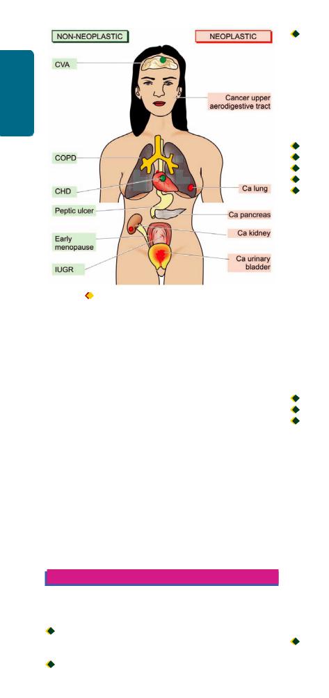
238
I SECTION |
|
Basic and Pathology General |
Major adverse effects of tobacco smoking. Right side |
Figure 9.2 |
|
Techniques |
|
shows smoking-related neoplastic diseases while left side indicates nonneoplastic diseases associated with smoking, numbered serially in order of frequency of occurrence.
in the United States exceeding in incidence beyond that of breast cancer in that country. Cigarette smoking is strongly implicated in evolution of lung cancer as described in Chapter 17.
OTHER CANCERS. Besides lung cancer, smokers have higher risk of development of cancer of upper aerodigestive tract (lips, oral cavity, larynx, oesophagus), pancreas, urinary bladder and kidney.
NON-NEOPLASTIC DISEASES. These include the following:
i)Chronic obstructive pulmonary disease (COPD) that includes chronic bronchitis and emphysema as the most common.
ii)Peptic ulcer disease with 70% higher risk in smokers.
iii)Early menopause in smoker women.
iv)In smoking pregnant women, higher risk of lower birth weight of foetus, higher perinatal mortality and intellectual deterioration of newborn.
CHEMICAL AND DRUG INJURY
During life, each one of us is exposed to a variety of chemicals and drugs. These are broadly divided into the following three categories:
 Therapeutic (iatrogenic) agents e.g. drugs, which when administered indiscriminately are associated with adverse effects.

 Non-therapeutic agents e.g. alcohol, lead, carbon monoxide, drug abuse.

 Environmental chemicals e.g. long-term or accidental exposure to certain man-made or naturally-occurring chemicals.
THERAPEUTIC (IATROGENIC) DRUG INJURY
Though the basis of patient management is rational drug therapy, nevertheless adverse drug reactions do occur in 2- 5% of patients. In general, the risk of adverse drug reaction increases with increasing number of drugs administered. Adverse effects of drugs may appear due to:

 overdose;
 genetic predisposition;
 exaggerated pharmacologic response;
 interaction with other drugs; and
 unknown factors.
It is beyond the scope of this book to delve into the list of drugs with their harmful effects. However, some of the common forms of iatrogenic drug injury and the offending drugs are listed in Table 9.2.
NON-THERAPEUTIC TOXIC AGENTS
ALCOHOLISM
Chronic alcoholism is defined as the regular imbibing of an amount of ethyl alcohol (ethanol) that is sufficient to harm an individual socially, psychologically or physically. It is difficult to give the number of ‘drinks’ after which the diagnosis of alcoholism can be made because of differences in individual susceptibility. However, adverse effects—acute as well as chronic, are related to the quantity of alcohol content imbibed and duration of consumption. Generally, 10 gm of ethanol is present in:

 a can of beer (or half a bottle of beer);

 120 ml of neat wine; or

 30 ml of 43% liquor (small peg).
A daily consumption of 40 gm of ethanol (4 small pegs or 2 large pegs) is likely to be harmful but intake of 100 gm or more daily is certainly dangerous. Daily and heavy consumption of alcohol is more harmful than moderate social drinking since the liver, where ethanol is metabolised, gets time to heal.
Metabolism
Absorption of alcohol begins in the stomach and small intestine and appears in blood shortly after ingestion. Alcohol is then distributed to different organs and body fluids proportionate to the blood levels of alcohol. About 2-10% of absorbed alcohol is excreted via urine, sweat and exhaled through breath, the last one being the basis of breath test employed by law-enforcement agencies for alcohol abuse. Metabolism of alcohol is discussed in detail in Chapter 21; in brief alcohol is metabolised in the liver by the following 3 pathways (Fig. 9.3):

 By the major rate-limiting pathway of alcohol dehydrogenase (ADH) in the cytosol, which is then quickly destroyed by aldehyde dehydrogenase (ALDH), especially with low blood alcohol levels.

TABLE 9.2: Iatrogenic Drug Injury.
|
Adverse Effect |
Offending Drug |
|
|
|
1. |
GASTROINTESTINAL TRACT |
|
|
Gastritis, peptic ulcer |
Aspirin, nonsteroidal |
|
|
anti-inflammatory drugs |
|
|
(NSAIDs) |
|
Jejunal ulcer |
Enteric-coated potassium tablets |
|
Pancreatitis |
Thiazide diuretics |
2. |
LIVER |
|
|
Cholestatic jaundice |
Phenothiazines, tranquillisers, |
|
|
oral contraceptives |
|
Hepatitis |
Halothane, isoniazid |
|
Fatty change |
Tetracycline |
3. |
NERVOUS SYSTEM |
|
|
Cerebrovascular accidents |
Anticoagulants, |
|
|
Oral contraceptives |
|
Peripheral neuropathy |
Vincristine, antimalarials |
|
8th nerve deafness |
Streptomycin |
4. |
SKIN |
|
|
Acne |
Corticosteroids |
|
Urticaria |
Penicillin, sulfonamides |
|
Exfoliative dermatitis, |
Penicillin, sulfonamides, |
|
Stevens-Johnson syndrome |
phenyl butazone |
|
Fixed drug eruptions |
Chemotherapeutic agents |
5. |
HEART |
|
|
Arrhythmias |
Digitalis, propranalol |
|
Congestive heart failure |
Corticosteroids |
|
Cardiomyopathy |
Adriamycin |
6. |
BLOOD |
|
|
Aplastic anaemia |
Chloramphenicol |
|
Agranulocytosis, |
Antineoplastic drugs |
|
thrombocytopenia |
|
|
Immune haemolytic |
Penicillin |
|
anaemia |
|
|
Megaloblastic anaemia |
Methotrexate |
7. |
LUNGS |
|
|
Alveolitis, interstitial |
Anti-neoplastic drugs |
|
pulmonary fibrosis |
|
|
Asthma |
Aspirin, indomethacin |
8. |
KIDNEYS |
|
|
Acute tubular necrosis |
Gentamycin, kanamycin |
|
Nephrotic syndrome |
Gold salts |
|
Chronic interstitial |
Phenacetin, salicylates |
|
nephritis, papillary necrosis |
|
9. |
METABOLIC EFFECTS |
|
|
Hypercalcaemia |
Hypervitaminosis D, |
|
|
thiazide diuretics |
|
Hepatic porphyria |
Barbiturates |
|
Hyperuricaemia |
Anti-cancer chemotherapy |
10. |
FEMALE REPRODUCTIVE TRACT |
|
|
Cholelithiasis, thrombo- |
Long-term use of oral |
|
phlebitis, thrombo- |
contraceptives |
|
embolism, benign liver |
|
|
cell adenomas |
|
|
Vaginal adenosis, adeno- |
Diethylstilbesterol by |
|
carcinoma in daughters |
pregnant women |
|
Foetal congenital anomalies |
Thalidomide in pregnancy |
|
|
|
 Via microsomal P-450 system (microsomal ethanol oxidising system, MEOS) when the blood alcohol level is high.

 Minor pathway via catalase from peroxisomes.
In any of the three pathways, ethanol is biotransformed to toxic acetaldehyde in the liver and finally to carbon dioxide and water by acetyl coenzyme A.
Ill-Effects of Alcoholism
Alcohol consumption in moderation and socially acceptable limits is practiced mainly for its mood-altering effects. Heavy alcohol consumption in unhabituated person is likely to cause acute ill-effects on different organs. Though the diseases associated with alcoholism are discussed in respective chapters later, the spectrum of ill-effects are outlined below.
A. ACUTE ALCOHOLISM. The acute effects of inebriation are most prominent on the central nervous system but it also injures the stomach and liver.
1. Central nervous system. Alcohol acts as a CNS depressant; the intensity of effects of alcohol on the CNS are related to the quantity consumed and duration over which consumed, which are reflected by the blood levels of alcohol: 
 Initial effect of alcohol is on subcortical structures which is followed by disordered cortical function, motor ataxia and behavioural changes. These changes are apparent when blood alcohol levels do not exceed 100 mg/dl which is the upper limit of sobriety in drinking as defined by lawenforcing agencies in most Western countries while dealing with cases of driving in drunken state.
 Blood levels of 100-200 mg/dl are associated with depression of cortical centres, lack of coordination, impaired judgement and drowsiness.

 Stupor and coma supervene when blood alcohol levels are about 300 mg/dl.
 Blood levels of alcohol above 400 mg/dl can cause anaesthesia, depression of medullary centre and death from respiratory arrest.
However, chronic alcoholics develop CNS tolerance and adaptation and, therefore, can withstand higher blood levels of alcohol without such serious effects.
2.Stomach. Acute alcohol intoxication may cause vomiting, acute gastritis and peptic ulceration.
3.Liver. Acute alcoholic injury to the liver is explained in Chapter 21.
B. CHRONIC ALCOHOLISM. Chronic alcoholism produces widespread injury to organs and systems. Contrary to the earlier belief that chronic alcoholic injury results from nutritional deficiencies, it is now known that most of the alcohol-related injury to different organs is due to toxic effects of alcohol and accumulation of its main toxic metabolite, acetaldehyde, in the blood. Other proposed mechanisms of tissue injury in chronic alcoholism is free-radical mediated injury and genetic susceptibility to alcohol-dependence and tissue damage.
Some of the more important organ effects in chronic alcoholism are as under (Fig. 9.4):
1. Liver. Alcoholic liver disease and cirrhosis are the most common and important effects of chronic alcoholism (Chapter 21).
239
Diseases Nutritional and Environmental 9 CHAPTER
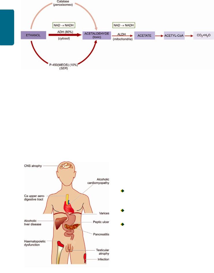
240
Techniques Basic and Pathology General I SECTION
Figure 9.3 
 Metabolism of ethanol in the liver. Thickness and intensity of colour of arrow on left side of figure corresponds to extent of metabolic pathway followed (MEOS = Microsomal ethanol oxidizing system; ADH-alcohol dehydrogenase; ALDH = aldehyde dehydrogenase; NAD=nicotinamide adenine dinucleotide; NADH = reduced NAD; NADP = nicotinamide adenine dinucleotide phosphate; NADPH = reduced NADP).
2.Pancreas. Chronic calcifying pancreatitis and acute pancreatitis are serious complications of chronic alcoholism.
3.Gastrointestinal tract. Gastritis, peptic ulcer and oesophageal varices associated with fatal massive bleeding may occur.
4.Central nervous system. Peripheral neuropathies and Wernicke-Korsakoff syndrome, cerebral atrophy, cerebellar degeneration and amblyopia (impaired vision) are seen in chronic alcoholics.
5.Cardiovascular system. Alcoholic cardiomyopathy and beer-drinkers’ myocardiosis with consequent dilated cardiomyopathy may occur. Level of HDL (atherosclerosis-
Figure 9.4 
 Complications of chronic alcoholism.
protective lipoprotein), however, has been shown to increase with moderate consumption of alcohol.
6.Endocrine system. In men, testicular atrophy, feminisation, loss of libido and potency, and gynaecomastia may develop. These effects appear to be due to lowering of testosterone levels.
7.Blood. Haematopoietic dysfunction with secondary megaloblastic anaemia and increased red blood cell volume may occur.
8.Immune system. Alcoholics are more susceptible to various infections.
9.Cancer. There is higher incidence of cancers of upper aerodigestive tract in chronic alcoholics but the mechanism is not clear.
LEAD POISONING
Lead poisoning may occur in children or adults due to accidental or occupational ingestion.
In children, following are the main sources of lead poisoning:
 Chewing of lead-containing furniture items, toys or pencils.

 Eating of lead paint flakes from walls.
In adults, the sources are as follows:

 Occupational exposure to lead during spray painting, recycling of automobile batteries (lead oxide fumes), mining, and extraction of lead.
 Accidental exposure by contaminated water supply, house freshly coated with lead paint, and sniffing of leadcontaining petrol (hence unleaded petrol introduced as fuel).
Lead is absorbed through the gastrointestinal tract or lungs. The absorbed lead is distributed in two types of tissues
(Fig. 9.5):
a) Bones, teeth, nails and hair representing relatively harmless pool of lead. About 90% of absorbed lead accumulates in the developing metaphysis of bones in children and appears as areas of increased bone densities (‘lead lines’) on X-ray. Lead lines are also seen in the gingiva.
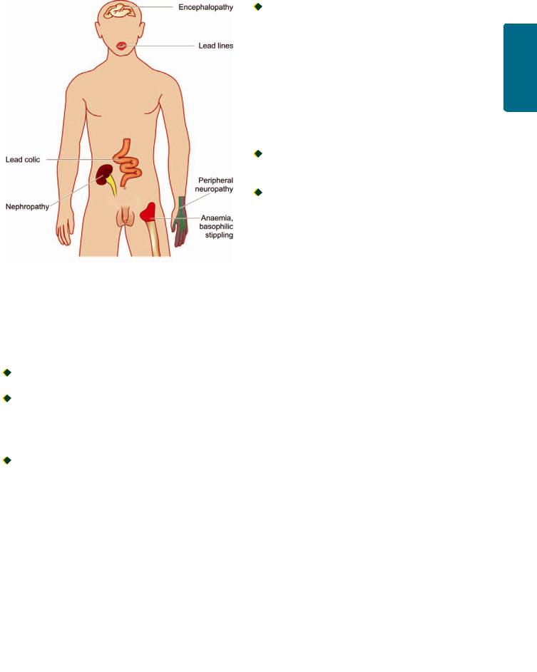
Figure 9.5 
 Complications of lead poisoning.
b) Brain, liver, kidneys and bone marrow accumulate the remaining 10% lead which is directly toxic to these organs. It is excreted via kidneys.
Lead toxicity occurs in the following organs predominantly:
1. Nervous system: The changes are as under:
 In children, lead encephalopathy; oedema of brain, flattening of gyri and compression of ventricles.
 In adults, demyelinating peripheral motor neuropathy which typically affects radial and peroneal nerves resulting in wristdrop and footdrop respectively.
2. Haematopoietic system: The changes in blood are quite characteristic:
 Microcytic hypochromic anaemia due to inhibition of two enzymes: delta-aminolevulinic acid dehydrogenase required for haem synthesis, and through inhibition of ferroketolase required for incorporation of ferrous iron into the porphyrin ring.

 Prominent basophilic stippling of erythrocytes.
3.Kidneys: Lead is toxic to proximal tubular cells of the kidney and produces lead nephropathy characterised by accumulation of intranuclear inclusion bodies consisting of lead-protein complex in the proximal tubular cells.
4.Gastrointestinal tract: Lead toxicity in the bowel manifests as acute abdomen presenting as lead colic.
CARBON MONOXIDE POISONING
Carbon monoxide (CO) is a colourless and odourless gas produced by incomplete combustion of carbon. Sources of CO gas are:

 automobile exhaust;

 burning of fossil fuel in industries or at home; and

 tobacco smoke.
CO is an important cause of accidental death due to systemic oxygen deprivation of tissues. This is because haemoglobin has about 200-times higher affinity for CO than for O2 and thus varying amount of carboxyhaemoglobin is formed depending upon the extent of CO poisoning. Besides, carboxyhaemoglobin interferes with the release of O2 from oxyhaemoglobin causing further aggravation of tissue hypoxia. Diagnosis of CO poisoning is, therefore, best confirmed by carboxyhaemoglobin levels in the blood.
CO poisoning may present in 2 ways:
 Acute CO poisoning in which there is sudden development of brain hypoxia characterised by oedema and petechial haemorrhages.

 Chronic CO poisoning presents with nonspecific changes of slowly developing hypoxia of the brain.
DRUG ABUSE
Drug abuse is defined as the use of certain drugs for the purpose of ‘mood alteration’ or ‘euphoria’ or ‘kick’ but subsequently leading to habit-forming, dependence and eventually addiction. Some of the commonly abused drugs and substances are as under:
1.Marijuana or ‘pot’ is psychoactive substance most widely used. It is obtained from the leaves of the plant Cannabis sativa and contains tetrahydrocannabinol (THC). It may be smoked or ingested.
2.Derivatives of opium that includes heroin and morphine. Opioids are derived from the poppy plant. Heroin and morphine are self-administered intravenously or subcutaneously.
3.CNS depressants include barbiturates, tranquilisers and alcohol.
4.CNS stimulants e.g. cocaine and amphetamines.
5.Psychedelic drugs (meaning enjoyable perception-giving) e.g. LSD.
6.lnhalants e.g. glue, paint thinner, nail polish remover, aerosols, amyl nitrite.
It is beyond the scope of the present discussion to go into the pharmacologic actions of all these substances. However, apart from pharmacologic and physiologic actions of these street drugs, the most common complication is introduction of infection by parenteral use of many of these drugs. Sharing of needles by the drug-addicts accounts for high risk of most feared viral infections in them, AIDS and viral hepatitis (both HBV and HCV). Following are a few common drug abuserelated infectious complications:
1.At the site of injection—cellulitis, abscesses, ulcers, thrombosed veins
2.Thrombophlebitis
3.Bacterial endocarditis
4.High risk for AIDS
5.Viral hepatitis and its complications
6.Focal glomerulonephritis
7.Talc (foreign body) granuloma formation in the lungs.
241
Diseases Nutritional and Environmental 9 CHAPTER

242 |
ENVIRONMENTAL CHEMICALS |
|
|
A large number of chemicals are found as contaminants in |
|
SECTION |
the ecosystem, food and water supply and find their way |
|
into the food chain of man. These substances exert their toxic |
||
|
||
|
effects depending upon their mode of absorption, |
|
|
distribution, metabolism and excretion. Some of the |
|
|
substances are directly toxic while others cause ill-effects via |
|
I |
their metabolites. Environmental chemicals may have slow |
|
damaging effect or there may be sudden accidental exposure |
||
|
||
|
such as the Bhopal gas tragedy in India due to accidental |
|
General |
leakage of methyl isocyanate (MIC) gas in December 1984. |
|
Some of the common examples of environmental |
||
|
||
|
chemicals are given below: |
|
Pathology |
1. Agriculture chemicals. Modern agriculture thrives on |
|
pesticides, fungicides, herbicides and organic fertilisers |
||
|
||
|
which may pose a potential acute poisoning as well as long- |
|
|
term hazard. The problem is particularly alarming in |
|
|
developing countries like India, China and Mexico where |
|
and |
farmers and their families are unknowingly exposed to these |
|
hazardous chemicals during aerial spraying of crops. |
||
|
||
Basic |
Acute poisoning by organophosphate insecticides is quite |
|
well known in India as accidental or suicidal poison by |
||
|
||
|
inhibiting acetyl cholinesterase and sudden death. |
|
Techniques |
Chronic human exposure to low level agricultural |
|
are taken ill every year worldwide with pesticide poisoning, |
||
|
chemicals is implicated in cancer, chronic degenerative |
|
|
diseases, congenital malformations and impotence but the |
|
|
exact cause-and-effect relationship is lacking. |
|
|
According to the WHO estimates, about 7.5 lakh people |
|
|
half of which occur in the developing countries due to ready |
|
|
availability and indiscriminate use of hazardous pesticides |
|
|
which are otherwise banned in advanced countries. Pesticide |
|
|
residues in food items such as in fruits, vegetables, cereals, |
|
|
grains, pulses etc is of greatest concern. |
|
|
2. Volatile organic solvents. Volatile organic solvents and |
|
|
vapours are used in industry quite commonly and their |
|
|
exposure may cause acute toxicity or chronic hazard, often |
|
|
by inhalation than by ingestion. Such substances include |
|
|
methanol, chloroform, petrol, kerosene, benzene, ethylene |
|
|
glycol, toluene etc. |
|
|
3. Metals. Pollution by occupational exposure to toxic |
|
|
metals such as mercury, arsenic, cadmium, iron, nickel and |
|
|
aluminium are important hazardous environmental |
|
|
chemicals. |
|
|
4. Aromatic hydrocarbons. The halogenated aromatic |
|
|
hydrocarbons containing polychlorinated biphenyl which are |
|
|
contaminant in several preservatives, herbicides and |
|
|
antibacterial agents are a chronic health hazard. |
|
|
5. Cyanide. Cyanide in the environment is released by |
|
|
combustion of plastic, silk and is also present in cassava and |
|
|
the seeds of apricots and wild cherries. Cyanide is a very |
|
|
toxic chemical and kills by blocking cellular respiration by |
|
|
binding to mitochondrial cytochrome oxidase. |
|
|
6. Environmental dusts. These substances causing |
|
|
pneumoconioses are discussed in chapter 17 while those |
|
|
implicated in cancer are discussed in Chapter 8. |
INJURY BY PHYSICAL AGENTS
THERMAL AND ELECTRICAL INJURY
Thermal and electrical burns, fall in body temperature below 35°C (hypothermia) and elevation of body temperature above 41°C (hyperthermia), are all associated with tissue injury.
 Hypothermia may cause focal injury as in frostbite, or systemic injury and death as occurs on immersion in cold water for varying time.

 Hyperthermia likewise, may be localised as in cutaneous burns, and systemic as occurs in fevers.
 Thermal burns depending upon severity are categorised into full thickness (third degree) and partial thickness (first and second degree). The most serious complications of burns are haemoconcentration, infections and contractures on healing.

 Electrical burns may cause damage firstly, by electrical dysfunction of the conduction system of the heart and death by ventricular fibrillation, and secondly by heat produced by electrical energy.
INJURY BY RADIATION
As discussed in the preceding chapter, the most important form of radiation injury is ionising radiation which has three types of effects on cells:
i)Somatic effects which cause acute cell killing.
ii)Genetic damage by mutations and therefore, passes genetic defects in the next progeny of cells.
iii)Malignant transformation of cells (Chapter 8).
Ionising radiation is widely employed for diagnostic
purpose as well as for radiotherapy of malignant tumours. Radiation-induced cell death is mediated by radiolysis of water in the cell with generation of toxic hydroxyl radicals (page 32). During radiotherapy, some normal cells coming in the field of radiation are also damaged. In general, radiation-induced tissue injury predominantly affects endothelial cells of small arteries and arterioles, causing necrosis and ischaemia.
Ionising radiation causes damage to the following major organs:
1.Skin: radiation dermatitis, cancer.
2.Lungs: interstitial pulmonary fibrosis.
3.Heart: myocardial fibrosis, constrictive pericarditis.
4.Kidney: radiation nephritis.
5.Gastrointestinal tract: strictures of small bowel and oesophagus.
6Gonads: testicular atrophy in males and destruction of ovaries.
7.Haematopoietic tissue: pancytopenia due to bone marrow depression.
8.Eyes: cataract.
Besides ionising radiation, other form of harmful radiation is solar (u.v.) radiation which may cause acute skin injury as sunburns, chronic conditions such as solar keratosis and early onset of cataracts in the eyes. It may, however, be mentioned in passing here that electromagnetic radiation

produced by microwaves (ovens, radars, diathermy) or ultrasound waves used for diagnostic purposes do not produce ionisation and thus are not known to cause any tissue injury.
NUTRITIONAL DISEASES
Nutritional status of a society varies according to the socioeconomic conditions. In the Western world, nutritional imbalance is more often a problem accounting for increased frequency of obesity, while in developing countries of Africa, Asia and South America, chronic malnutrition is a serious health problem, particularly in children.
Before describing the nutritional diseases, it is essential to know the components of normal and adequate nutrition. For good health, humans require energy-providing nutrients (proteins, fats and carbohydrates), vitamins, minerals, water and some non-essential nutrients.
1.Energy. The requirement of energy by the body is calculated in Kcal per day. In order to retain stable weight and undertake day-to-day activities, the energy intake must match the energy output. The average requirement of energy for an individual is estimated by the formula: 900+10w for males, and 700+7w for females (where w stands for the weight of the individual in kilograms). Since the requirement of energy varies according to the level of physical activities performed by the person, the figure arrived at by the above formula is multiplied by: 1.2 for sedentary person, 1.4 for moderately active person and 1.8 for very active person.
2.Proteins. Dietary proteins provide the body with amino acids for endogenous protein synthesis and are also a metabolic fuel for energy (1 g of protein provides 4 Kcal). Nine essential amino acids (histidine, isoleucine, leucine, lysine, methionine/cystine, phenylalanine/tyrosine, theonine, tryptophan and valine) must be supplied by dietary intake as these cannot be synthesised in the body. The recommended average requirement of proteins for an adult is 0.6 g/kg of the desired weight per day. For a healthy person, 10-14% of caloric requirement should come from proteins.
3.Fats. Fats and fatty acids (in particular linolenic, linoleic and arachidonic acid) should comprise about 35% of diet. In order to minimise the risk of atherosclerosis, polyunsaturated fats should be limited to <10% of calories and saturated fats and trans-fats should comprise <10% of calories while monounsaturated fats to constitute the remainder of fat intake (1 g of fat yields 9 Kcal).
4.Carbohydrates. Dietary carbohydrates, are the major source of dietary calories, especially for the brain, RBCs and muscles (1 g of carbohydrate provides 4 Kcal). At least 55% of total caloric requirement should be derived from carbohydrates.
5.Vitamins. These are mainly derived from exogenous dietary sources and are essential for maintaining the normal structure and function of cells. A healthy individual requires
4fat-soluble vitamins (A, D, E and K) and 11 water-soluble vitamins (C, B1/thiamine, B2/riboflavin, B3/niacin/nicotinic
acid, B5/pantothenic acid, B6/pyridoxine, folate/folic acid, B12/ cyanocobalamin, choline, biotin, and flavonoids). Vitamin deficiencies result in individual deficiency syndromes, or may be part of a multiple deficiency state.
6.Minerals. A number of minerals like iron, calcium, phosphorus and certain trace elements (e.g. zinc, copper, selenium, iodine, chlorine, sodium, potassium, magnesium, manganese, cobalt, molybdenum etc) are essential for health. Their deficiencies result in a variety of lesions and deficiency syndromes.
7.Water. Water intake is essential to cover the losses in faeces, urine, exhalation and insensible loss so as to avoid underor over-hydration. Although body’s water needs varies according to physical activities and weather conditions, average requirement of water is 1.0-1.5 ml water/ Kcal of energy spent. Infants and pregnant women have relatively higher requirements of water.
8.Non-essential nutrients. Dietary fibre composed of cellulose, hemicellulose and pectin, though considered nonessential, are important due to their beneficial effects in lowering the risk of colonic cancer, diabetes and coronary artery disease.
Pathogenesis of Deficiency Diseases
The nutritional deficiency disease develops when the essential nutrients are not provided to the cells adequately. The nutritional deficiency may be of 2 types:
1.Primary deficiency. This is due to either the lack or decreased amount of essential nutrients in diet.
2.Secondary or conditioned deficiency. Secondary or conditioned deficiency is malnutrition occurring as a result of the various factors. These are as under:
i) Interference with ingestion e.g. in gastrointestinal disorders such as malabsorption syndrome, chronic alcoholism, neuropsychiatric illness, anorexia, food allergy, pregnancy.
ii)Interference with absorption e.g. in hypermotility of the gut, achlorhydria, biliary disease.
iii)Interference with utilisation e.g. in liver dysfunction, malignancy, hypothyroidism.
iv)Increased excretion e.g. in lactation, perspiration, polyuria.
v)Increased nutritional demand e.g. in fever, pregnancy, lactation, hyperthyroidism.
Irrespective of the type of nutritional deficiency (primary or secondary), nutrient reserves in the tissues begin to get depleted, which initially result in biochemical alterations and eventually lead to functional and morphological changes in tissues and organs.
In the following pages, a brief account of nutritional imbalance (viz. obesity) is followed by description of multiple or mixed deficiencies (e.g. starvation, protein-energy malnutrition) and individual nutrient deficiencies (e.g. vitamin deficiencies).
OBESITY
Dietary imbalance and overnutrition may lead to diseases like obesity. Obesity is defined as an excess of adipose tissue that
imparts health risk; a body weight of 20% excess over ideal weight
243
Diseases Nutritional and Environmental 9 CHAPTER
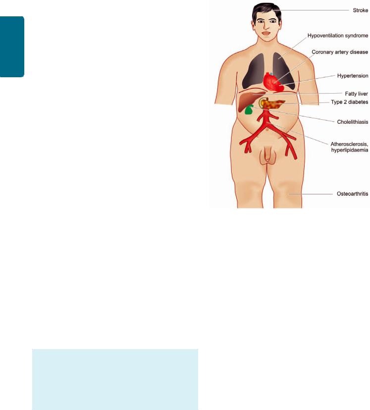
244
Techniques Basic and Pathology General I SECTION
for age, sex and height is considered a health risk. The most widely used method to gauge obesity is body mass index (BMI) which is equal to weight in kg/height in m2. A cut off BMI value of 30 is used for obesity in both men and women.
ETIOLOGY. Obesity results when caloric intake exceeds utilisation. The imbalance of these two components can occur in the following situations:
1.Inadequate pushing of oneself away from the dining table causing overeating.
2.Insufficient pushing of oneself out of the chair leading to inactivity and sedentary life style.
3.Genetic predisposition to develop obesity.
4.Diets largely derived from carbohydrates and fats than protein-rich diet.
5.Secondary obesity may result following a number of underlying diseases such as hypothyroidism, Cushing’s disease, insulinoma and hypothalamic disorders.
PATHOGENESIS. The lipid storing cells, adipocytes comprise the adipose tissue, and are present in vascular and stromal compartment in the body. Besides the generally accepted role of adipocytes for fat storage, these cells also release endocrine-regulating molecules. These molecules include: energy regulatory hormone (leptin), cytokines (TNF-α and interleukin-6), insulin sensitivity regulating agents (adiponectin, resistin and RBP4), prothrombotic factors (plasminogen activator inhibitor), and blood pressure regulating agent (angiotensingen).
Adipose mass is increased due to enlargement of adipose cells due to excess of intracellular lipid deposition as well as due to increase in the number of adipocytes. The most important environmental factor of excess consumption of nutrients can lead to obesity. However, underlying molecular mechanisms of obesity are beginning to unfold based on observations that obesity is familial and is seen in identical twins. Recently, two obesity genes have been found: ob gene and its protein product leptin, and db gene and its protein product leptin receptor.
SEQUELAE OF OBESITY. Marked obesity is a serious health hazard and may predispose to a number of clinical disorders and pathological changes described below and illustrated in
Fig. 9.6.
MORPHOLOGIC FEATURES. Obesity is associated with increased adipose stores in the subcutaneous tissues, skeletal muscles, internal organs such as the kidneys, heart, liver and omentum; fatty liver is also more common in obese individuals. There is increase in both size and number of adipocytes i.e. there is hypertrophy as well as hyperplasia.
METABOLIC CHANGES. These are as under:
1. Hyperinsulinaemia. Increased insulin secretion is a feature of obesity. Many obese individuals exhibit hyperglycaemia or frank diabetes despite hyperinsulinaemia. This is due to a state of insulin-resistance consequent to tissue insensitivity.
Figure 9.6 
 Major sequelae of obesity.
2.Type 2 diabetes mellitus. There is a strong association of type 2 diabetes mellitus with obesity. Obesity often exacerbates the diabetic state and in many cases weight reduction often leads to amelioration of diabetes.
3.Hypertension. A strong association between hypertension and obesity is observed which is perhaps due to increased blood volume. Weight reduction leads to significant reduction in systolic blood pressure.
4.Hyperlipoproteinaemia. The plasma cholesterol circulates in the blood as low-density lipoprotein (LDL) containing most of the circulating triglycerides. Obesity is strongly associated with VLDL and mildly with LDL. Total blood cholesterol levels are also elevated in obesity.
5.Atherosclerosis. Obesity predisposes to development of atherosclerosis. As a result of atherosclerosis and hypertension, there is increased risk of myocardial infarction and stroke in obese individuals.
6.Nonalcoholic fatty liver disease (NAFLD). Obesity contributes to development of NAFLD which may progress further to cirrhosis of the liver.
7.Cholelithiasis. There is six times higher incidence of gallstones in obese persons, mainly due to increased total body cholesterol.
8.Hypoventilation syndrome (Pickwickian syndrome).
This is characterised by hypersomnolence, both at night and during day in obese individuals along with carbon dioxide retention, hypoxia, polycythaemia and eventually right-sided heart failure. (Mr Pickwick was a character, the fat boy, in
Charles Dickens’ Pickwick Papers. The term pickwickian syndrome was first used by Sir William Osler for the sleepapnoea syndrome).
9.Osteoarthritis. These individuals are more prone to develop degenerative joint disease due to wear and tear following trauma to joints as a result of large body weight.
10.Cancer. Diet rich in fats, particularly derived from animal fats and meats, is associated with higher incidence of cancers of colon, breast, endometrium and prostate.
STARVATION
Starvation is a state of overall deprivation of nutrients. Its causes may be the following:
i)deliberate fasting—religious or political;
ii)famine conditions in a country or community; or
iii)secondary undernutrition such as due to chronic wasting diseases (infections, inflammatory conditions, liver disease), cancer etc. Cancer results in malignant cachexia as a result of which cytokines are elaborated e.g. tumour necrosis factor- α, elastases, proteases etc.
A starved individual has lax, dry skin, wasted muscles and atrophy of internal organs.
METABOLIC CHANGES. The following metabolic changes take place in starvation:
1.Glucose. Glucose stores of the body are sufficient for one day’s metabolic needs only. During fasting state, insulinindependent tissues such as the brain, blood cells and renal medulla continue to utilise glucose while insulin-dependent tissues like muscle stop taking up glucose. This results in release of glycogen stores of the liver to maintain normal blood glucose level. Subsequently, hepatic gluconeogenesis from other sources such as breakdown of proteins takes place.
2.Proteins. Protein stores and the triglycerides of adipose tissue have enough energy for about 3 months in an individual. Proteins breakdown to release amino acids which are used as fuel for hepatic gluconeogenesis so as to maintain
Figure 9.7 
 Two forms of PEM.
glucose needs of the brain. This results in nitrogen imbalance due to excretion of nitrogen compounds as urea.
3. Fats. After about one week of starvation, protein breakdown is decreased while triglycerides of adipose tissue breakdown to form glycerol and fatty acids. The fatty acids are converted into ketone bodies in the liver which are used by most organs including brain in place of glucose. Starvation can then continue till all the body fat stores are exhausted following which death occurs.
PROTEIN-ENERGY MALNUTRITION
The inadequate consumption of protein and energy as a result of primary dietary deficiency or conditioned deficiency may cause loss of body mass and adipose tissue, resulting in protein energy or protein calorie malnutrition (PEM or PCM). The primary deficiency is more frequent due to socioeconomic factors limiting the quantity and quality of dietary intake, particularly prevalent in the developing countries of Africa, Asia and South America. The impact of deficiency is marked in infants and children.
245
Diseases Nutritional and Environmental 9 CHAPTER
TABLE 9.3: Contrasting Features of Kwashiorkor and Marasmus.
Feature |
Kwashiorkor |
Marasmus |
|
|
|
Definition |
Protein deficiency with sufficient calorie intake |
Starvation in infants with overall lack of calories |
Clinical features |
Occurs in children between 6 months and 3 years |
Common in infants under 1 year of age |
(Fig. 9.7) |
of age |
|
|
Growth failure |
Growth failure |
|
Wasting of muscles but preserved adipose tissues |
Wasting of all tissues including muscles and adipose |
|
|
tissues |
|
Oedema, localised or generalised, present |
Oedema absent |
|
Enlarged fatty liver |
No hepatic enlargement |
|
Serum proteins low |
Serum proteins low |
|
Anaemia present |
Anaemia present |
|
‘Flag sign’—alternate bands of light (depigmented) |
Monkey-like face, protuberant abdomen, thin limbs |
|
and dark (pigmented) hair |
|
Morphology |
Enlarged fatty liver |
No fatty liver |
|
Atrophy of different tissues and organs but |
Atrophy of different tissues and organs including |
|
subcutaneous fat preserved |
subcutaneous fat |
|
|
|

246 The spectrum of clinical syndromes produced as a result of PEM includes the following (Fig. 9.7):
SECTION |
1. Kwashiorkor which is related to protein deficiency though |
|||
calorie intake may be sufficient. |
||||
|
||||
|
2. Marasmus is starvation in infants occurring due to overall |
|||
|
lack of calories. |
|
||
|
|
The salient features of the two conditions are contrasted |
||
I |
in Table 9.3. However, it must be remembered that mixed |
|||
forms of kwashiorkor-marasmus syndrome may also occur. |
||||
|
||||
|
|
|
||
General |
|
DISORDERS OF VITAMINS |
||
|
||||
Vitamins are organic substances which cannot be synthesised |
||||
|
||||
|
within the body and are essential for maintenance of normal |
|||
Pathology |
structure and function of cells. Thus, these substances must be |
|||
required in minute amounts in contrast to the relatively large |
||||
|
provided in the human diet. Most of the vitamins are of plant |
|||
|
or animal origin so that they normally enter the body as |
|||
|
constituents of ingested plant food or animal food. They are |
|||
and |
|
TABLE 9.4: Vitamin Deficiencies. |
||
Basic |
|
|||
|
|
|||
|
|
Vitamins |
|
|
Techniques |
|
I. FAT-SOLUBLE VITAMINS |
||
|
||||
|
|
|||
|
|
Vitamin A |
||
|
|
(Retinol) |
||
|
|
Vitamin D |
||
|
|
(Calcitriol) |
||
|
|
Vitamin E |
||
|
|
(α-Tocopherol) |
||
|
|
Vitamin K |
||
|
|
II. WATER-SOLUBLE VITAMINS |
||
|
|
Vitamin C |
||
|
|
(Ascorbic acid) |
||
|
|
Vitamin B Complex |
||
|
|
(i) |
Thiamine |
|
|
|
|
(Vitamin B1) |
|
|
|
(ii) |
Riboflavin |
|
|
|
|
(Vitamin B2) |
|
|
|
(iii) |
Niacin/Nicotinic acid |
|
|
|
|
(Vitamin B3) |
|
|
|
(iv) |
Pyridoxine |
|
|
|
|
(Vitamin B6) |
|
|
|
(v) |
Folate/Folic acid |
|
|
|
(vi) |
Cyanocobalamin |
|
|
|
|
(Vitamin B12) |
|
|
|
(vii) |
Biotin |
|
|
|
(viii) Choline |
||
|
|
(ix) |
Flavonoids |
|
amounts of essential amino acids and fatty acids. Vitamins do not play any part in production of energy.
ETIOLOGY OF VITAMIN DEFICIENCIES. In the developing countries, multiple deficiencies of vitamins and other nutrients are common due to generalised malnutrition of dietary origin. In the developed countries, individual vitamin deficiencies are noted more often, particularly in children, adolescent, pregnant and lactating women, and in some due to poverty. General secondary causes of conditioned nutritional deficiencies listed already above (i.e. interference in ingestion, absorption, utilization, excretion) can result in vitamin deficinecy in either case. Chronic alcoholism is a common denominator in many of vitamin deficiencies. A few other noteworthy features about vitamins are as under:
1.While both vitamin deficiency and excess may occur from another disease, the states of excess and deficiency themselves also cause disease.
2.Vitamins in high dose can be used as drugs.
Deficiency Disorders
Ocular lesions (night blindness, xerophthalmia, keratomalacia,
Bitot’s spots, blindness) Cutaneous lesions (xeroderma)
Other lesions (squamous metaplasia of respiratory epithelium, urothelium and pancreatic ductal epithelium, subsequent anaplasia; retarded bone growth)
Rickets in growing children
Osteomalacia in adults
Hypocalcaemic tetany
Degeneration of neurons, retinal pigments, axons of peripheral nerves; denervation of muscles
Reduced red cell lifespan
Sterility in male and female animals
Hypoprothrombinaemia (in haemorrhagic disease of newborn, biliary obstruction, malabsorption, anticoagulant therapy, antibiotic therapy, diffuse liver disease)
Scurvy (haemorrhagic diathesis, skeletal lesions, delayed wound healing, anaemia, lesions in teeth and gums)
Beriberi (‘dry’ or peripheral neuritis, ‘wet’ or cardiac manifestations, ‘cerebral’ or Wernicke-Korsakoff’s syndrome)
Ariboflavinosis (ocular lesions, cheilosis, glossitis, dermatitis)
Pellagra (dermatitis, diarrhoea, dementia)
Vague lesions (convulsions in infants, dermatitis, cheilosis, glossitis, sideroblastic anaemia)
Megaloblastic anaemia
Megaloblastic anaemia
Pernicious anaemia
Mental and neurological symptoms
Fatty liver, muscle damage
Preventive of neurodegenerative disease, osteoporosis, diabetes
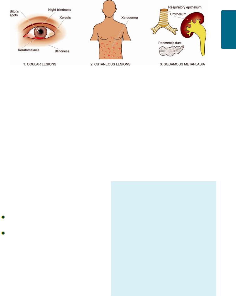
Figure 9.8 
 Lesions resulting from vitamin A deficiency.
CLASSIFICATION OF VITAMINS. Vitamins are conventionally divided into 2 groups: fat-soluble and water-soluble.
1.Fat-soluble vitamins. There are 4 fat-soluble vitamins: A, D, E and K. They are absorbed from intestine in the presence of bile salts and intact pancreatic function. Their deficiencies occur more readily due to conditioning factors (secondary deficiency). Beside the deficiency syndromes of these vitamins, a state of hypervitaminosis due to excess of vitamin A and D also occurs.
2.Water-soluble vitamins. This group conventionally consists of vitamin C and members of B complex group. Besides, choline, biotin and flavonoids are new additions to this group. Water-soluble vitamins are more readily absorbed from small intestine. Deficiency of these vitamins is mainly due to primary (dietary) factors. Being water soluble, these vitamins are more easily lost due to cooking or processing of food.
Table 9.4 sums up the various clinical disorders produced by vitamin deficiencies.
FAT-SOLUBLE VITAMINS
Vitamin A (Retinol)
PHYSIOLOGY. Vitamin A or retinol is a fat-soluble alcohol. It is available in diet in 2 forms:

 As preformed retinol, the dietary sources of which are animal-derived foods such as yolk of eggs, butter, whole milk, fish, liver, kidney.

 As provitamin precursor carotenoid, which is derived from
β-carotene-containing foods such as yellow plants and vegetables e.g. carrots, potatoes, pumpkins, mangoes, spinach. β-carotene can be absorbed intact or converted in the intestinal mucosa to form retinaldehyde which is subsequently reduced to retinol.
Retinol is stored in the liver cells and released for transport to peripheral tissues after binding to retinol-binding protein found in blood.
The physiologic functions of retinol are as follows:
1. Maintenance of normal vision in reduced light. This involves formation of 2 pigments by oxidation of retinol: rhodopsin, a light sensitive pigment in reduced light synthesised in the
rod cells, and iodopsins sensitive in bright light and formed in cone cells of retina. These pigments then transform the radiant energy into nerve impulses.
2.Maintenance of structure and function of specialised epithelium. Retinol plays an important role in the synthesis of glycoproteins of the cell membrane of specialised epithelium such as mucus-secreting columnar epithelium in glands and mucosal surfaces, respiratory epithelium and urothelium.
3.Maintenance of normal cartilaginous and bone growth.
4.Increased immunity against infections in children.
5.Anti-proliferative effect. β-carotene has anti-oxidant properties and may cause regression of certain non-tumorous skin diseases, premalignant conditions and certain cancers.
LESIONS IN VITAMIN A DEFICIENCY. Nutritional deficiency of vitamin A is common in countries of South-East Asia, Africa, Central and South America whereas malabsorption syndrome may account for conditioned vitamin A deficiency in developed countries.
MORPHOLOGIC FEATURES. Consequent to vitamin A deficiency, following pathologic changes are seen
(Fig. 9.8):
1. Ocular lesions. Lesions in the eyes are most obvious. Night blindness is usually the first sign of vitamin A deficiency. As a result of replacement metaplasia of mucus-secreting cells by squamous cells, there is dry and scaly scleral conjunctiva (xerophthalmia). The lacrimal duct also shows hyperkeratosis. Corneal ulcers may occur which may get infected and cause keratomalacia. Bitot’s spots may appear which are focal triangular areas of opacities due to accumulation of keratinised epithelium. If these occur on cornea, they impede transmission of light. Ultimately, infection, scarring and opacities lead to
blindness.
2.Cutaneous lesions. The skin develops papular lesions giving toad-like appearance (xeroderma). This is due to follicular hyperkeratosis and keratin plugging in the sebaceous glands.
3.Other lesions. These are as under:
i)Squamous metaplasia of respiratory epithelium of bronchus and trachea may predispose to respiratory infections.
247
Diseases Nutritional and Environmental 9 CHAPTER
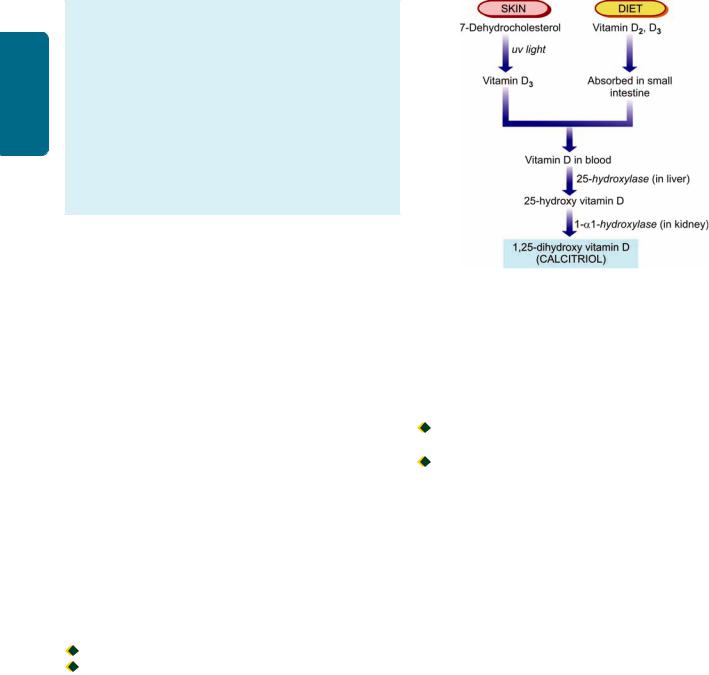
248
Techniques Basic and Pathology General I SECTION
ii)Squamous metaplasia of pancreatic ductal epithelium may lead to obstruction and cystic dilatation.
iii)Squamous metaplasia of urothelium of the pelvis of kidney may predispose to pyelonephritis and perhaps to renal calculi.
iv)Long-standing metaplasia may cause progression to anaplasia under certain circumstances.
v)Bone growth in vitamin A deficient animals is retarded.
vi)Immune dysfunction may occur due to damaged barrier epithelium and compromised immune defenses.
vii)Pregnant women may have increased risk of maternal infection, mortality and impaired embryonic development.
HYPERVITAMINOSIS A. Very large doses of vitamin A can produce toxic manifestations in children as well as in adults. These may be acute or chronic.
Acute toxicity. This results from a single large dose of vitamin A. The effects include neurological manifestations resembling brain tumour e.g. headache, vomiting, stupor, papilloedema.
Chronic toxicity. The clinical manifestations of chronic vitamin A excess are as under:
i)Neurological such as severe headache and disordered vision due to increased intracranial pressure.
ii)Skeletal pains due to loss of cortical bone by increased osteoclastic activity as well as due to exostosis.
iii)Cutaneous involvement may be in the form of pruritus, fissuring, sores at the corners of mouth and coarseness of hair.
iv)Hepatomegaly with parenchymal damage and fibrosis.
v)Hypercarotenaemia is yellowness of palms and skin due to excessive intake of β-carotene containing foods like carrots or due to inborn error of metabolism.
The effects of toxicity usually disappear on stopping excess of vitamin A intake.
Vitamin D (Calcitriol)
PHYSIOLOGY. This fat-soluble vitamin exists in 2 activated
sterol forms:

 Vitamin D2 or calciferol; and
 Vitamin D3 or cholecalciferol.
The material originally described as vitamin D1 was subsequently found to be impure mixture of sterols. Since vitamin D2 and D3 have similar metabolism and functions, they are therefore referred to as vitamin D.
There are 2 main sources of vitamin D:
i)Endogenous synthesis. 80% of body’s need of vitamin D is met by endogenous synthesis from the action of ultraviolet light on 7-dehydrocholesterol widely distributed in oily secretions of the skin. The vitamin so formed by irradiation enters the body directly through the skin. Pigmentation of the skin reduces the beneficial effects of ultraviolet light.
ii)Exogenous sources. The other source of vitamin D is diet such as deep sea fish, fish oil, eggs, butter, milk, some plants and grains.
Irrespective of the source of vitamin D, it must be converted to its active metabolites (25-hydroxy vitamin D and 1,25-
Figure 9.9 
 Normal metabolism of vitamin D.
dihydroxy vitamin D or calcitriol) after its metabolism in the liver and kidney for being functionally active (Fig. 9.9).
1, 25-dihydroxy vitamin D (calcitriol) is 5-10 times more potent biologically than 25-hydroxy vitamin D. The production of calcitriol by the kidney is regulated by:

 plasma levels of calcitriol (hormonal feedback);
 plasma calcium levels (hypocalcaemia stimulates synthesis); and

 plasma phosphorus levels (hypophosphataemia stimulates synthesis).
The main storage site of vitamin D is the adipose tissue rather than the liver which is the case with vitamin A.
The main physiologic functions of the most active metabolite of vitamin D, calcitriol, are mediated by its binding to nuclear receptor superfamily, vitamin D receptor, expressed on a wide variety of cells. These actions are as under:
1. Maintenance of normal plasma levels of calcium and phosphorus. The major essential function of vitamin D is to promote mineralisation of bone. This is achieved by the following actions of vitamin D:
i)Intestinal absorption of calcium and phosphorus is stimulated by vitamin D.
ii)On bones. Vitamin D is normally required for mineralisation of epiphyseal cartilage and osteoid matrix. However, in hypocalcaemia, vitamin D collaborates with parathyroid hormone and causes osteoclastic resorption of calcium and phosphorus from bone so as to maintain the normal blood levels of calcium and phosphorus.
iii)On kidneys. Vitamin D stimulates reabsorption of calcium at distal renal tubular level, though this function is also parathyroid hormone-dependent.
2. Antiproliferative effects. Vitamin D receptor is expressed on the parathyroid gland cells by which active form of vitamin D causes antiproliferative action on parathyroid cells and suppresses the parathormone gene. Besides, vitamin D receptor is also expressed on cells of organs which do not
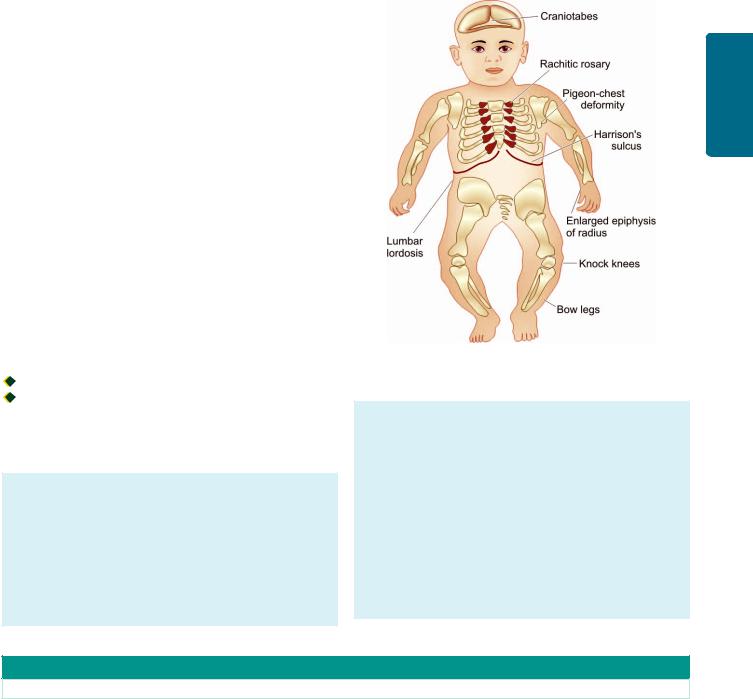
have any role in mineral ion homeostasis and has antiproliferative effects on them e.g. in skin, breast cancer cells, prostate cancer cells.
LESIONS IN VITAMIN D DEFICIENCY. Deficiency of vitamin D may result from:
i)reduced endogenous synthesis due to inadequate exposure to sunlight;
ii)dietary deficiency of vitamin D;
iii)malabsorption of lipids due to lack of bile salts such as in intrahepatic biliary obstruction, pancreatic insufficiency and malabsorption syndrome;
iv)derangements of vitamin D metabolism as occur in kidney disorders (chronic renal failure, nephrotic syndrome, uraemia), liver disorders (diffuse liver disease) and genetic disorders; and
v)resistance of end-organ to respond to vitamin D. Deficiency of vitamin D from any of the above
mechanisms results in 3 types of lesions:
1.rickets in growing children;
2.osteomalacia in adults; and
3.hypocalcaemic tetany due to neuromuscular dysfunction.
RICKETS. The primary defects in rickets are:

 interference with mineralisation of bone; and
 deranged endochondral and intramembranous bone growth.
The pathogenesis of lesions in rickets is better understood by contrasting them with sequence of changes in normal bone growth as outlined in Table 9.5.
MORPHOLOGIC FEATURES. Rickets occurs in growing children from 6 months to 2 years of age. The disease has the following lesions and clinical characteristics (Fig. 9.10):
Skeletal changes. These are as under:
i) Craniotabes is the earliest bony lesion occurring due to small round unossified areas in the membranous bones of the skull, disappearing within 12 months of birth. The skull looks square and box-like.
Figure 9.10 
 Lesions in rickets.
ii)Harrison’s sulcus appears due to indrawing of soft ribs on inspiration.
iii)Rachitic rosary is a deformity of chest due to cartilaginous overgrowth at costochondral junction.
iv)Pigeon-chest deformity is the anterior protrusion of sternum due to action of respiratory muscles.
v)Bow legs occur in ambulatory children due to weak bones of lower legs.
vi)Knock knees may occur due to enlarged ends of the femur, tibia and fibula.
vii)Lower epiphyses of radius may be enlarged.
viii)Lumbar lordosis is due to involvement of the spine and pelvis.
TABLE 9.5: Contrasting Features of Rickets with Normal Bone Growth.
Normal Bone Growth |
Rickets |
|
|
I.ENDOCHONDRAL OSSIFICATION (OCCURRING IN LONG TUBULAR BONES)
i. |
Proliferation of cartilage cells at the epiphyses |
i. |
Proliferation of cartilage cells at the epiphyses |
|
followed by provisional mineralisation |
|
followed by inadequate provisional mineralisation |
ii. |
Cartilage resorption and replacement by osteoid |
ii. Persistence and overgrowth of epiphyseal cartilage; |
|
|
matrix |
|
deposition of osteoid matrix on inadequately |
|
|
|
mineralised cartilage resulting in enlarged and expanded |
|
|
|
costochondral junctions |
iii. Mineralisation to form bone |
iii. Deformed bones due to lack of structural rigidity |
||
iv. |
Normal vascularisation of bone |
iv. |
Irregular overgrowth of small blood vessels in |
|
|
|
disorganised and weak bone |
II. INTRAMEMBRANOUS OSSIFICATION |
|
|
|
(OCCURRING IN FLAT BONES) |
|
|
|
Mesenchymal cells differentiate into osteoblasts which |
Mesenchymal cells differentiate into osteoblasts with |
||
develop osteoid matrix and subsequent mineralisation |
laying down of osteoid matrix which fails to get mineralised |
||
|
|
resulting in soft and weak flat bones |
|
|
|
|
|
249
Diseases Nutritional and Environmental 9 CHAPTER

250 |
Biochemical changes. These are as follows: |
||
|
i) Lowered levels of active metabolites of vitamin D (25- |
||
SECTION |
hydroxy vitamin D and 1, 25-dihydroxy vitamin D). |
||
ii) Plasma calcium levels are normal or slightly low. |
|||
iii) Plasma phosphate levels are lowered. |
|||
iv) Plasma alkaline phosphatase is usually raised due to |
|||
osteoblastic activity. |
|||
|
Vitamin D-dependent rickets is an autosomal dominant |
||
I |
disorder of vitamin D. The disease responds rapidly to |
||
|
administration of 1,25-dihydroxy vitamin D. |
||
General |
OSTEOMALACIA. Osteomalacia is the adult counterpart |
||
poor endogenous synthesis of vitamin D, or as a result of |
|||
|
of rickets in which there is failure of mineralisation of the |
||
|
osteoid matrix. It may occur following dietary deficiency, |
||
Pathology |
conditioned deficiency. |
||
|
|
||
MORPHOLOGIC FEATURES. Due to deficiency of |
|||
|
|||
|
vitamin D, osteoid matrix laid down fails to get minera- |
||
|
lised. In H and E stained microscopic sections, this is |
||
and |
identified by widened and thickened osteoid seams |
||
(stained pink) and decreased mineralisation at the borders |
|||
|
|||
Basic |
between osteoid and bone (stained basophilic). von Kossa’s |
||
stain for calcium may be employed to mark out the wide |
|||
seams of unstained osteoid while the calcified bone is |
|||
Techniques |
stained black. In addition, there may be increased |
||
ii) |
vague bony pains; |
||
|
osteoclastic activity and fibrosis of marrow. |
||
|
Clinical features. Osteomalacia is characterised by: |
||
|
i) |
muscular weakness; |
|
iii) fractures following trivial trauma;
iv) incomplete or greenstick fractures; and
v) looser’s zones or pseudofractures at weak places in bones.
Biochemical changes. These are:
i)normal or low serum calcium levels;
ii)plasma phosphate levels lowered; and
iii)raised serum alkaline phosphatase due to increased osteoblastic activity.
It may be worthwhile to note here that another chronic disorder of skeleton seen in elderly, osteoporosis, is clinically similar but biochemically different disease (Chapter 28).
HYPERVITAMINOSIS D. Very large excess of vitamin D may cause increased intestinal absorption of calcium and phosphorus, leading to hypercalcaemia, hyperphosphataemia and increased bone resorption. These changes may result in the following effects:
i)increased urinary excretion of calcium and phosphate;
ii)predisposition to renal calculi;
iii)osteoporosis; and
iv)widespread metastatic calcification, more marked in the renal tubules, arteries, myocardium, lungs and stomach.
Vitamin E (α-Tocopherol)
PHYSIOLOGY. Out of many naturally-occurring tocoferols and tocotrienols, α-tocopherol is biologically the most active fat soluble compound for humans. Vitamin E is found in most of the ordinary foods such as vegetables, grains, nuts and oils. It is absorbed from the intestine and transported in blood
in the form of chylomicrons. It is stored in fat depots, liver and muscle.
The main physiologic functions of vitamin E are as under:
1.Anti-oxidant activity. Active form of Vitamin E acts as an antioxidant and prevents the oxidative degradation of cell membranes containing phospholipids.
2.Scavenger of free radicals. Vitamin E scavenges free radicals formed by redox reaction in the body (Chapter 3) and thus maintains the integrity of the cell.
3.Inhibits prostaglandin synthesis.
4.Activates protein kinase C and phospholipase A2.
LESIONS IN VITAMIN E DEFICIENCY. The deficiency of vitamin E is mainly by conditioning disorders affecting its absorption and transport such as abetalipoproteinaemia, intraand extrahepatic biliary cholestasis, cystic fibrosis of the pancreas and malabsorption syndrome. Low birth weight neonates, due to physiologic immaturity of the liver and bowel, may also develop vitamin E deficiency. Lesions of vitamin E deficiency are as follows:
1.Neurons with long axons develop degeneration in the posterior columns of spinal cord.
2.Peripheral nerves may also develop myelin degeneration in the axons.
3.Skeletal muscles may develop denervation.
4.Retinal pigmentary degeneration may occur.
5.Red blood cells deficient in vitamin E such as in premature infants have reduced lifespan.
6.In experimental animals, vitamin E deficiency can produce sterility in both male and female animals.
Vitamin K
PHYSIOLOGY. Vitamin K (K for Koagulations in Danish) exists in nature in 2 forms:
 Vitamin K1 or phylloquinone, obtained from exogenous dietary sources such as most green leafy vegetables; and
 Vitamin K2 or menaquinone, produced endogenously by normal intestinal flora. Phylloquinone can be converted into menaquinone in some organs.
Like other fat-soluble vitamins, vitamin K is absorbed from the small intestine and requires adequate bile flow and intact pancreatic function.
The main physiologic function of vitamin K is in hepatic microsomal carboxylation reaction for vitamin K- dependent coagulation factors (most importantly factor II or prothrombin; others are factors VII, IX and X).
LESIONS IN VITAMIN K DEFICIENCY. Since vitamin K is necessary for the manufacture of prothrombin, its deficiency leads of hypoprothrombinaemia (Chapter 13). Estimation of plasma prothrombin, thus, affords a simple in vitro test for determining whether there is deficiency of vitamin K. Subjects with levels below 70% of normal should receive therapy with vitamin K.
Because most of the green vegetables contain vitamin K and that it can be synthesised endogenously, vitamin K deficiency is frequently a conditioned deficiency. The conditions which may bring about vitamin K deficiency are as follows:
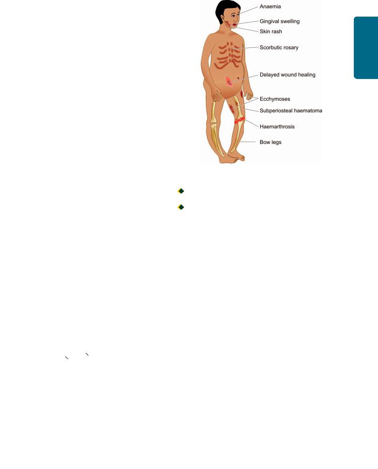
1.Haemorrhagic disease of newborn. The newborn infants are deficient in vitamin K because of minimal stores of vitamin K at birth, lack of established intestinal flora for endogenous synthesis and limited dietary intake since breast milk is a poor source of vitamin K. Hence the clinical practice is to routinely administer vitamin K at birth.
2.Biliary obstruction. Bile is prevented from entering the bowel due to biliary obstruction which prevents the absorption of this fat-soluble vitamin. Surgery in patients of obstructive jaundice, therefore, leads to marked tendency to bleeding.
3.Due to malabsorption syndrome. Patients suffering from malabsorption of fat develop vitamin K deficiency e.g. coeliac disease, sprue, pancreatic disease, hypermotility of bowel etc.
4.Due to anticoagulant therapy. Patients on warfarin group of anticoagulants have impaired biosynthesis of vitamin K- dependent coagulation factors.
5.Due to antibiotic therapy. The use of broad-spectrum antibiotics and sulfa drugs reduces the normal intestinal flora.
6.Diffuse liver disease. Patients with diffuse liver disease (e.g. cirrhosis, amyloidosis of liver, hepatocellular carcinoma, hepatoblastoma) have hypoprothrombinaemia due to impaired synthesis of prothrombin. Administration of vitamin K to such patients is of no avail since liver, where prothrombin synthesis utilising vitamin K takes place, is diseased.
WATER-SOLUBLE VITAMINS
Vitamin C (Ascorbic Acid)
PHYSIOLOGY. Vitamin C exists in natural sources as L- ascorbic acid closely related to glucose. The major sources of vitamin C are citrus fruits such as orange, lemon, grape fruit and some fresh vegetables like tomatoes and potatoes. It is present in small amounts in meat and milk. The vitamin is easily destroyed by heating so that boiled or pasteurised milk may lack vitamin C. It is readily absorbed from the small intestine and is stored in many tissues, most abundantly in adrenal cortex.
The physiologic functions of vitamin C are due to its ability to carry out oxidation-reduction reactions:
L-Ascorbic Acid |
|
|
Dehydro L-Ascorbic |
|
|||
|
|
||
|
|
|
acid + 2H+ + 2e |
1.Vitamin C has been fond to have antioxidant properties and can scavenge free radicals.
2.Ascorbic acid is required for hydroxylation of proline to form hydroxyproline which is an essential component of
collagen.
3.Besides collagen, it is necessary for the ground substance of other mesenchymal structures such as osteoid, chondroitin sulfate, dentin and cement substance of vascular endothelium.
4.Vitamin C being a reducing substance has other functions such as:

 hydroxylation of dopamine to norepinephrine;
Figure 9.11 
 Lesions in scurvy.

 maintenance of folic acid levels by preventing oxidation of tetrahydrofolate; and
 role in iron metabolism in its absorption, storage and keeping it in reduced state.
LESIONS IN VITAMIN C DEFICIENCY. Vitamin C deficiency in the food or as a conditioned deficiency results in scurvy. The lesions and clinical manifestations of scurvy are seen more commonly at two peak ages: in early childhood and in the very aged. These are as under (Fig. 9.11):
1.Haemorrhagic diathesis. A marked tendency to bleeding is characteristic of scurvy. This may be due to deficiency of intercellular cement which holds together the cells of capillary endothelium. There may be haemorrhages in the skin, mucous membranes, gums, muscles, joints and underneath the periosteum.
2.Skeletal lesions. These changes are more pronounced in growing children. The most prominent change is the deranged
formation of osteoid matrix and not deranged mineralisation (c.f. the pathological changes underlying rickets already described). Growing tubular bones as well as flat bones are affected. The epiphyseal ends of growing long bones have cartilage cells in rows which normally undergo provisional mineralisation. However, due to vitamin C deficiency, the next step of laying down of osteoid matrix by osteoblasts is poor and results in failure of resorption of cartilage. Consequently, mineralised cartilage under the widened and irregular epiphyseal plates project as scorbutic rosary. The skeletal changes are further worsened due to haemorrhages and haematomas under the periosteum and bleeding into the joint spaces.
3. Delayed wound healing. There is delayed healing of wounds in scurvy due to following:

 deranged collagen synthesis;
251
Diseases Nutritional and Environmental 9 CHAPTER
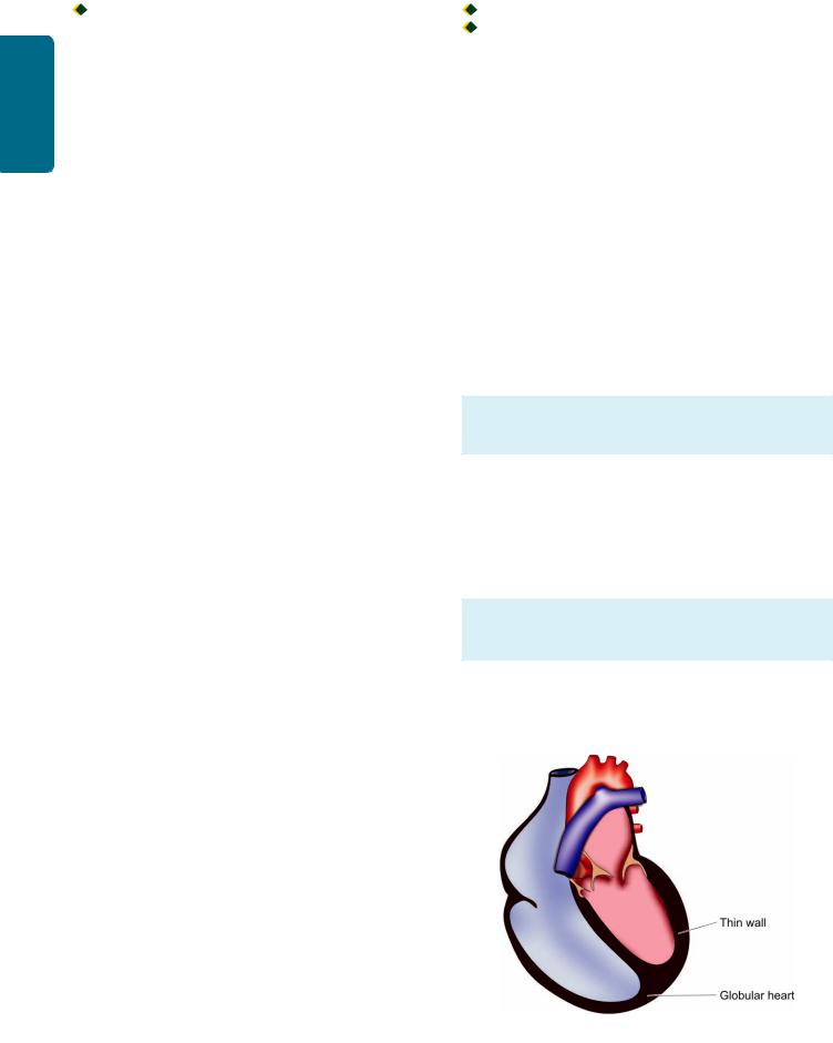
252
Techniques Basic and Pathology General I SECTION
 poor preservation and maturation of fibroblasts; and
 localisation of infections in the wounds.
4.Anaemia. Anaemia is common in scurvy. It may be the result of haemorrhage, interference with formation of folic acid or deranged iron metabolism. Accordingly, anaemia is most often normocytic normochromic type; occasionally it may be megaloblastic or even iron deficiency type.
5.Lesions in teeth and gums. Scurvy may interfere with development of dentin. The gums are soft and swollen, may bleed readily and get infected commonly.
6.Skin rash. Hyperkeratotic and follicular rash may occur in scurvy.
VITAMIN B COMPLEX
The term vitamin B was originally coined for a substance capable of curing beriberi (B from beriberi). Now, vitamin B complex is commonly used for a group of essential compounds
which are biochemically unrelated but occur together in certain foods such as green leafy vegetables, cereals, yeast, liver and milk. Most of the vitamins in this group are involved in metabolism of proteins, carbohydrates and fats.
The principal members of vitamin B complex are thiamine (vitamin B1), riboflavin (vitamin B2), niacin/nicotinic acid (vitamin B3), pantothenic acid (vitamin B5), pyridoxine (vitamin B6), folate (folic acid), cyanocobalamin (vitamin B12) and biotin. There is no definite evidence that any clinical disorder results from deficiency of pantothenic acid (vitamin B5).
Thiamine (Vitamin B1)
PHYSIOLOGY. Thiamine was the first in the family of vitamin B complex group and hence named B1. Thiamine hydrochloride is available in a variety of items of diet such as peas, beans, pulses, yeast, green vegetable roots, fruits, meat, pork, rice and wheat bran. The vitamin is lost in refined foods such as polished rice, white flour and white sugar. A few substances in the diet (strong tea, coffee) act as antithiamines. Since the vitamin is soluble in water, considerable amount of the vitamin is lost during cooking of vegetables. The vitamin is absorbed from the intestine either by passive diffusion or by energy-dependent transport. Reserves of vitamin B1 are stored in the skeletal muscles, heart, liver, kidneys and bones.
The main physiologic function of thiamine is in carbohydrate metabolism. Thiamine after absorption is phosphorylated to form thiamine pyrophosphate which is the functionally active compound. This compound acts as coenzyme for carboxylase so as to decarboxylate pyruvic acid, synthesises ATP and also participates in the synthesis of fat from carbohydrate. In addition, thiamin plays a role in peripheral nerve conduction by an unknown mechanism.
LESIONS IN THIAMINE DEFICIENCY. Thiamine deficiency can occur from primary or conditioned causes, chronic alcoholism being an important cause. The deficiency state leads to failure of complete combustion of carbohydrate and accumulation of pyruvic acid. This results in beriberi which produces lesions at 3 target tissues (peripheral nerves, heart and brain). Accordingly, beriberi is of 3 types:

 dry beriberi (peripheral neuritis);
 wet beriberi (cardiac manifestations), and cerebral beriberi (Wernicke-Korsakoff’s syndrome).
It is worth-noting that lesions in beriberi are mainly located in the nervous system and heart. This is because the energy requirement of the brain and nerves is solely derived from oxidation of carbohydrates which is deranged in beriberi, while lesions in the heart appear to arise due to reduced ATP synthesis in beriberi which is required for cardiac functions.
The features of 3 forms of beriberi are as under:
1.Dry beriberi (peripheral neuritis). This is marked by neuromuscular symptoms such as weakness, paraesthesia and sensory loss. The nerves show polyneuritis, myelin degeneration and fragmentation of axons.
2.Wet beriberi (cardiac manifestations). This is characterised by cardiovascular involvement, generalised oedema, serous effusions and chronic passive congestion of viscera. The heart in beriberi is flabby (due to thin and weak myocardium), enlarged and globular in appearance due to 4-chamber dilatation (Fig. 9.12).
Microscopic examination of the heart shows hydropic degeneration of myocardial fibres, loss of striations, interstitial oedema and lymphocytic infiltration.
3. Cerebral beriberi (Wernicke-Korsakoff’s syndrome).
It consists of the following features:
i) Wernicke’s encephalopathy occurs more often due to conditioned deficiencies such as in chronic alcoholism. It is characterised by degeneration of ganglia cells, focal demyelination and haemorrhage in the nuclei surrounding the region of ventricles and aqueduct.
Microscopic examination shows degeneration and necrosis of neurons, hypertrophy-hyperplasia of small blood vessels and haemorrhages.
ii) Korsakoff’s psychosis results from persistence of psychotic features following brain haemorrhage in Wernicke’s encephalopathy.
Figure 9.12 
 Wet (Cardiac) beriberi. Flabby, thin-walled, enlarged and globular appearance of the heart due to four-chamber dilatation.

Riboflavin (Vitamin B2)
PHYSIOLOGY. Riboflavin used to be called ‘yellow respiratory enzyme’ (flavus = yellow), now known as ‘cytochrome oxidase enzyme’ which is important in view of its role as cellular respiratory coenzyme. The vitamin is usually distributed in plant and animal foods such as the liver, beaf, mutton, pork, eggs, milk and green vegetables. Like other water-soluble vitamins, it is rapidly absorbed from the bowel and stored in tissues like liver.
LESIONS IN RIBOFLAVIN DEFICIENCY. Lesions due to primary or conditioned deficiency of riboflavin (ariboflavinosis) are as follows:
1.Ocular lesions consist of vascularisation of normally avascular cornea due to proliferation of capillaries from limbus. Subsequently, conjunctivitis, interstitial keratitis and corneal ulcers may develop.
2.Cheilosis and angular stomatitis are characterised by occurrence of fissures and cracks at the angles of mouth.
3.Glossitis is development of red, cyanosed and shiny tongue due to atrophy of mucosa of tongue (‘bald tongue’).
4Skin changes appear in the form of scaly dermatitis resembling seborrheic dermatitis on nasolabial folds on the face, scrotum and vulva.
5.Anaemia may develop in some cases.
Niacin/Nicotinic Acid (Vitamin B3)
PHYSIOLOGY. As with thiamine and riboflavin, niacin or nicotinic acid or vitamin B3 is also widely distributed in plant and animal foods such as the liver, kidney, meat, green vegetables and whole grain cereals. Niacin includes biologically active derivative nicotinamide which is essential for the formation of 2 oxidative coenzymes (dehydrogenases):  NAD (nicotinamide adenine dinucleotide) which is required for dehydrogenation in the metabolism of fat, carbohydrates and proteins.

 NADP (nicotinamide adenine dinucleotide phosphate) which is essential for dehydrogenation in the hexose monophosphate shunt of glucose metabolism.
LESIONS IN NIACIN DEFICIENCY. Deficiency of niacin causes pellagra, so named because of the rough skin of such patients (Italian pelle agra = rough skin). Pellagra may result from dietary deficiency in those who largely subsist on maize since niacin in maize is present in bound form and hence not absorbable. Since niacin can be endogenously synthesised from tryptophan, a diet deficient in this amino acid or disorders of tryptophan metabolism such as in carcinoid syndrome or Hartnup syndrome results in niacin deficiency.
Lesions in pellagra are characterised by 3Ds:
1 Dermatitis: The sun-exposed areas of skin develop erythema resembling sunburn. This may progress to chronic type of dermatitis with blister formation.
2. Diarrhoea: Lesions similar to those seen in skin may develop in mucous membrane of the alimentary tract resulting in glossitis, lesions in the mouth, oesophagus, stomach and colon and cause diarrhoea, nausea, vomiting and burning sensation.
3. Dementia: Degeneration of neurons of the brain and of spinal tract results in neurological symptoms such as dementia, peripheral neuritis, ataxia and visual and auditory disturbances.
TOXICITY OF NIACIN. Toxicity due to administration of high doses of niacin as therapy for dyslipidaemia has been observed but not due dietary excess. It is characterised by flushing of skin and liver derangement.
Pyridoxine (Vitamin B6)
PHYSIOLOGY. Pyridoxine or vitamin B6 is widely distributed in all animal and plant foods such as meat, liver, eggs, green vegetables and whole grain cereals. Pyridoxine exists in 3 closely related naturally-occurring substances—
pyridoxine, pyridoxal and pyridoxamine. All of these can be converted into biologically active coenzyme, pyridoxal 5- phosphate.
The major physiologic functions of pyridoxine are related to:

 fat metabolism;
 protein metabolism;
 amino acid metabolism such as decarboxylation of amino acids, transmethylation of methionine, conversion of tryptophan to niacin;

 steroid metabolism;
 neurotransmitter synthesis; and

 haem synthesis.
LESIONS IN PYRIDOXINE DEFICIENCY. Vitamin B6 deficiency may result from inadequate dietary intake or may result from secondary deficiency such as increased demand in pregnancy and lactation, chronic alcoholism and intake of certain drugs (e.g. isoniazid in the treatment of tuberculosis, penicillamine, oestrogen in oral contraceptives etc).
The lesions of pyridoxine deficiency include the following:
1.Convulsions in infants born to mothers who had been
administered large doses of vitamin B6 for hyperemesis gravidarum (pyridoxine dependence)
2.Dermatitis and seborrhoea
3.Cheilosis and angular stomatitis
4.Glossitis (bald tongue)
5.Neuropathy
6.Depression, confusion
7.Sideroblastic anaemia.
Folate (Folic Acid) and Cyanocobalamin (Vitamin B12)
Both these vitamins included in the B complex group are required for red cell formation. Their deficiency leads to megaloblastic anaemia which is discussed in Chapter 12.
Biotin
PHYSIOLOGY. Biotin is a water-soluble vitamin and a member of vitamin B complex group. It is available in food sources such as organ meat, soya beans, egg yolk; however
253
Diseases Nutritional and Environmental 9 CHAPTER

254
Techniques Basic and Pathology General I SECTION
egg-white has a protein avidin which binds to biotin and blocks its bioavailability.
The major physiologic functions of biotin are as under:
1.In gene expression
2.In gluconeogenesis
3.In fatty acid synthesis
4.In catabolism of certain amino acids such as leucine
4.As carrier of CO2 in carboxylase enzymes.
LESIONS IN BIOTIN DEFICIENCY. Biotin deficiency is rare and develops due to inborn errors of metabolism and in patients on parenteral nutrients devoid of biotin. The lesions of biotin deficiency are as under:
1.Mental and neurologic symptoms such as hallucination, depression, paraesthesia
2.Anorexia
3.Nausea
4.Scaly, seborrhoeic dermatitis
5.In infants, hypotonia, alopecia and rash near ears.
In concluding the discussion of vitamin B complex, it must be mentioned that many of the animal and plant foods contain vitamin B complex group of vitamins. Their deficiency, whether primary from poverty, ignorance etc, or secondary from conditioning factors like chronic alcoholism, is more frequently multiple vitamin deficiency. Hence, the clinical practice is to administer combination of these members of vitamin B complex.
CHOLINE
PHYSIOLOGY. Choline is precursor form of acetylcoline and betaine. Choline is widely distributed as lecithin in foods such as egg yolk, milk, wheat and organ meat. Choline is also synthesised in the liver.
The major physiologic functions of choline are as under:
1.In maintenance structural integrity of cell membranes
2.In transmembrane signaling pathways
3.In cholinergic neurotransmission
4.In metabolism of lipids and cholesterol.
LESIONS IN CHOLINE DEFICIENCY. Choline deficiency develops in patients on choline-free parenteral nutrients. The lesions of choline deficiency are as under:
1.Fatty liver with deranged liver enzymes
2.Skeletal muscle damage with elevated CPK levels.
FLAVONOIDS
PHYSIOLOGY. Flavoonoids are a form of polyphenols present in several fruits and vegetables and are the constituents which imparts colour, flavour and taste to these edible products. Particular food and vegetables rich in flavonoids are berries, grapes, apples, broccoli, onions, legumes etc.
The major physiologic functions of flavonoids are as under:
1.As antioxidants
2.In cell signaling pathways
LESIONS IN BIOTIN DEFICIENCY. Flavonoids have been a recent addition to the family of vitamins. Present data on animal experiments and human clinical studies indicates that they play a role in prevention of neurodegenerative diseases, osteoporosis and diabetes.
METALS AND TRACE ELEMENTS
Several minerals in trace amounts are essential for health since they form components of enzymes and cofactors for metabolic functions. Besides calcium and phosphorus required for vitamin D manufacture, others include: iron, copper, iodine,
zinc, selenium, manganese, nickel, chromium, molybdenum, fluorine. However, out of these, the dietary deficiency of first five trace elements is associated with deficiency states which are discussed in detail in respective chapters later. These are as under:
i)Iron: Microcytic hypochromic anaemia.
ii)Calcium: reduced bone mass, osteoporosis.
iii)Phosphorous: Rickets, osteomalacia.
iv)Copper: Muscle weakness, neurologic defect, anaemia, growth retardation.
v)Iodine: Goitre and hyperthyroidism, cretinism.
vi)Zinc: Growth retardation, infertility, alopecia.
vii)Selenium: Cardiomyopathy, muscle degeneration.
viii)Fluoride: Dental caries.
ix)Manganese: Impaired growth and skeletal development.
x)Molybdenum: Severe neurological abnormalities.
DIET AND CANCER
Before closing the discussion of nutritional pathology, it is worthwhile to sum up relationship of these factors to carcinogenesis discussed in previoius chapter. There are three possible mechanisms on which the story of this relationship can be built up:
1. Dietary content of exogenous carcinogens:
i) The most important example in this mechanism comes from naturally-occurring carcinogen aflatoxin which is strongly associated with high incidence of hepatocellular carcinoma in those consuming grain contaminated with
mould, Aspergillus flavus.
ii) Artificial sweeteners (e.g. saccharine cyclomates), food additives and pesticide contamination of food are implicated as carcinogens derived from diet.
2. Endogenous synthesis of carcinogens or promoters:
i)In the context of etiology of gastric carcinoma, nitrites, nitrates and amines from the digested food are transformed in the body to carcinogens—nitrosamines and nitrosamides.
ii)In the etiology of colon cancer, low fibre intake and high animal-derived fats are implicated. High fat diet results in rise in the level of bile acids and their intermediate metabolites produced by intestinal bacteria which act as
carcinogens. The low fibre diet, on the other hand, does not provide adequate protection to the mucosa and reduces the stool bulk and thus increases the time the stools remain in the colon.
iii) In the etiology of breast cancer, epidemiologic studies have implicated the role of animal proteins, fats and obesity with as yet unsubstantiated evidence.
3. Inadequate protective factors:
As already mentioned, some components of diet such as vitamin C, A, E, selenium, and β-carotenes have protective role against cancer. These substances in normal amounts in the body act as antioxidants and protect the cells against free radical injury but their role of supplementation in diet as prevention against cancer is unproven.
q
255
Diseases Nutritional and Environmental 9 CHAPTER

256
Techniques Basic and Pathology General I SECTION
Chapter 10
Genetic and
Paediatric Diseases
This chapter deals with the group of disorders affecting the foetus during intrauterine life (genetic as well as developmental) and paediatric age group. In the western countries, developmental and genetic birth defects constitute about 50% of total mortality in infancy and childhood, while in the developing and underdeveloped countries 95% of infant mortality is attributed to environmental factors such as poor sanitation and undernutrition.
For the purpose of convenience of discussion, genetic and paediatric diseases are covered under the following headings:
1Developmental defects: Errors in morphogenesis
2.Cytogenetic (Karyotypic) defects: chromosomal abnormalities
3.Single-gene defects: Mendelian disorders
4.Multifactorial inheritance disorders
5.Other paediatric diseases
Though many of diseases included in the groups above have been discussed along with relevant chapters later, broad overview of these disorders is presented below.
DEVELOPMENTAL DEFECTS
Developmental defects are a group of abnormalities during foetal life due to errors in morphogenesis. The branch of science dealing with the study of developmental anomalies is called teratology. Certain chemicals, drugs, physical and biologic agents are known to induce such birth defects and are called teratogens. The morphologic abnormality or defect in an organ or anatomic region of the body so produced is
called malformation.
Pathogenesis
The teratogens may result in one of the following outcomes:
i)Intrauterine death
ii)Intrauterine growth retardation (IUGR)
iii)Functional defects
iv)Malformation
The effects of teratogens in inducing developmental
defects are related to the following factors:

 Variable individual susceptibility to teratogen: All patients exposed to the same teratogen do not develop birth defect.

 Intrauterine stage at which patient is exposed to teratogen:
Most teratogens induce birth defects during the first trimester of pregnancy.
 Dose of teratogen: Higher the exposure dose of teratogen, greater the chances of inducing birth defects.

 Specificity of developmental defect for specific teratogen: A particular teratogen acts in a particular way and induces the same specific developmental defect.
Classification
Various developmental anomalies resulting from teratogenic effects are categorised as under:
Agenesis means the complete absence of an organ e.g. unilateral or bilateral agenesis of kidney.
Aplasia is the absence of development of an organ with presence of rudiment or anlage e.g. aplasia of lung with rudimentary bronchus.
Hypoplasia is incomplete development of an organ not reaching the normal adult size e.g. microglossia.
Atresia refers to incomplete formation of lumen in hollow viscus e.g. oesophageal atresia.
Developmental dysplasia is defective development of cells and tissues resulting in abnormal or primitive histogenetic structures e.g. renal dysplasia (Developmental dysplasia is quite different from cellular dysplasia in relation to precancerous lesions discussed earlier on page 58).
Dystraphic anomalies are the defects resulting from failure of fusion e.g. spina bifida.
Ectopia or heterotopia refers to abnormal location of tissue at ectopic site e.g. pancreatic heterotopia in the wall of stomach.
Examples of Developmental Defects
A few common clinically important examples are given below:
1.Anencephaly-spina bifida complex. This is the group of anomalies resulting from failure to fuse (dystraphy). While anencephaly results from failure of neural tube closure, spina bifida occurs from incomplete closure of the spinal cord and vertebral column, often in the lumbar region. The latter results in meningocele or meningomyelocele.
2.Thalidomide malformations. Thalidomide is the best known example of teratogenic drug which was used as a sedative by pregnant women in 1960s in England and Germany and resulted in high incidence of limb-reduction anomalies (phocomelia) in the newborns.
3.Foetal hydantoin syndrome. Babies born to mothers on anti-epileptic treatment with hydantoin have characteristic facial features and congenital heart defects.
4.Foetal alcohol syndrome. Ethanol is another potent teratogen. Consumption of alcohol by pregnant mother in first trimester increases the risk of miscarriages, stillbirths, growth retardation and mental retardation in the newborn.
5.TORCH complex. Infection with TORCH group of organisms (Toxoplasma, Others, Rubella, Cytomegalovirus,
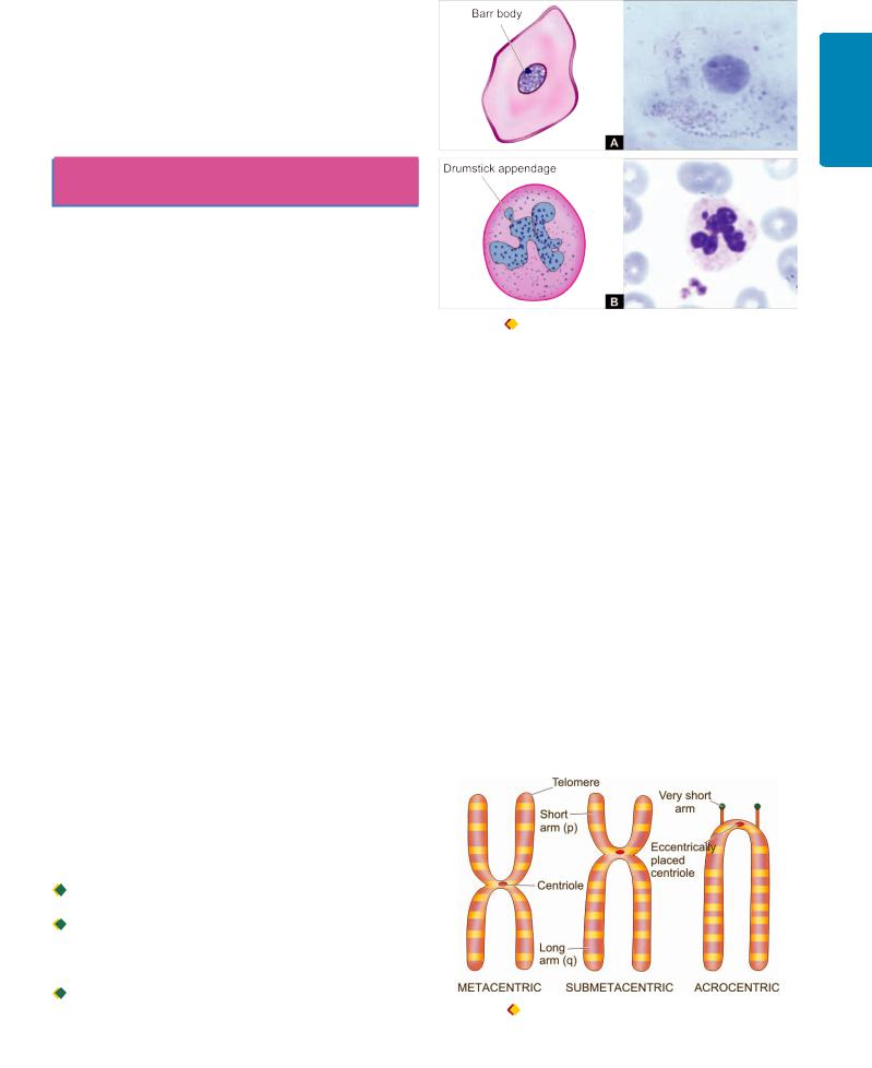
and Herpes simplex) during pregnancy is associated with multisystem anomalies and TORCH syndrome in the newborn (page 191).
6. Congenital syphilis. As discussed in Chapter 6, vertical transmission of syphilis from mother to foetus is characterised by Hutchinson’s triad: interstitial keratitis, sensorineural deafness and deformed Hutchinson’s teeth, along with saddle-nose deformity.
CYTOGENETIC (KARYOTYPIC)
ABNORMALITIES
Human germ cells (ova and sperms) contain 23 chromosomes (haploid or 1N) while all the nucleated somatic cells of the human body contain 23 pairs of chromosomes (diploid or 2N)—44 autosomes and 2 sex chromosomes, being XX in females (46, XX) and XY in males (46, XY). The branch of science dealing with the study of human chromosomal abnormalities is called cytogenetics (discussed in Chapter 2).
In a female, one of the two X chromosomes (paternal or maternal derived) is inactivated during embryogenesis as stated in Lyon hypothesis. This inactivation is passed to all the somatic cells while the germ cells in the female remain unaffected i.e. ovary will always have active X chromosome. Such an inactive X chromosome in the somatic cells in females lies condensed in the nucleus and is called as sex chromatin seen specifically in the somatic cells in females. Nuclear sexing can be done for genetic female testing by preparing and staining the smears of squamous cells scraped from oral cavity, or by identifying the Barr body in the circulating neutrophils as drumstick appendage attached to one of the nuclear lobes (Fig. 10.1). A minimum of 30% cells positive for sex chromatin is indicative of genetically female composition.
Though chromosomes can be studied in any human nucleated cells, circulating lymphocytes are more often used for this purpose. The study is done by arresting the dividing cells in metaphase by colchicine and then spreading them on glass slide and staining them with Giemsa stain.
Karyotype is the photographic representation of the stained preparation of chromosomes.
Each chromosome is composed of a pair of identical double helix of chromosomal DNA called chromatids. The chromosomes are classified based on their length and location of the centromere; centromere is the point where the two chromatids cross each other (Fig. 10.2). The distal end of each chromosome is called telomere.
Based on centromeric location, they are classified into 3 groups:

 Metacentric chromosomes (numbers 1, 3, 16, 19, 20) are those in which the centromere is exactly in the middle.
 Submetacentric chromosomes (numbers 1, 3) in which the centromere divides the chromosomes into short arm (p arm; petit means short in French) and long arm (q arm; for alphabet next to p).

 Acrocentric chromosomes (numbers 13, 14, 15, 21, 22, and Y) have very short arm and the centromere is eccentrically located.
257
|
|
10 CHAPTER |
|
|
|
and Genetic |
|
Figure 10.1 |
Nuclear sexing. A, sex chromatin as seen in scraped |
Paediatric |
|
|
|||
squamous cells from oral cavity. B, Barr body seen as drumstick |
|
||
appendage attached to a lobe of a circulating neutrophil. |
Diseases |
||
Based on length of chromosomes, they are divided into 7 |
|||
|
|||
groups—A to G, called Denver classification adopted at a meeting in Denver, Colorado in US.
Chromosomal banding techniques are employed for study of classes of chromosomes. Chromosomal bands are unique alternate dark and light staining patterns. Banding techniques include:
i)G-banding (Giemsa stain);
ii)Q-banding (quinacrine fluorescence stain);
iii)R-banding (reverse Giemsa staining); and
iv)C-banding (constitutive heterochromatin demonstration).
With these brief introductory comments, we can now turn to abnormalities of chromosomes which can be divided into 2 types:
1.Numerical abnormalities; and
2.Structural abnormalities.
Numerical Abnormalities
As mentioned above, normal karyotype of a human nucleated somatic cell is diploid or 2N (46 chromosomes)
Figure 10.2 |
Classification of chromosomes based on size and |
location of centromere.
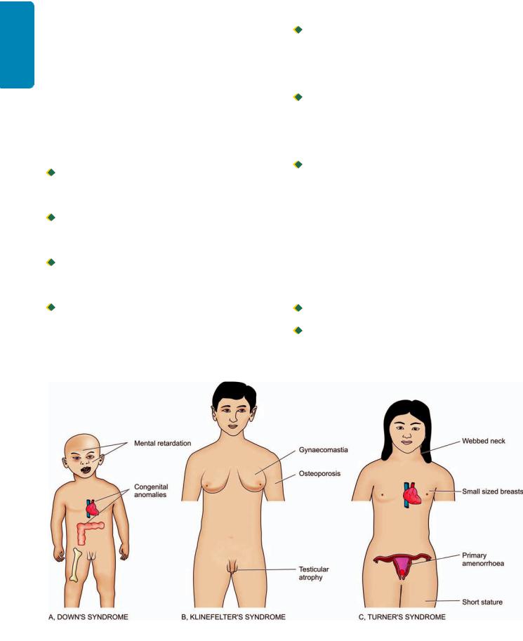
258 while the germ cells have haploid or 1N (23 chromosomes).
SECTION |
1. Polyploidy is the term used for the number of chromo- |
|
somes which is a multiple of haploid number e.g. triploid or |
||
|
||
|
3N (69 chromosomes), tetraploid or 4N (92 chromosomes). |
|
|
Polyploidy occurs normally in megakaryocytes and dividing |
|
|
liver cells. Polyploidy in somatic cells of conceptus results |
|
I |
in spontaneous abortions. |
|
2. Aneuploidy is the number of chromosomes which is not |
||
|
||
General |
an exact multiple of haploid number e.g. hypodiploid or |
|
2N-1 (45 chromosomes) monosomy, hyperdiploid or 2 N+1 |
||
(47 chromosomes) trisomy. |
||
The most common mechanism of aneuploidy is |
||
nondisjunction. Nondisjunction is the failure of chromo- |
||
Pathology |
somes to separate normally during cell division during first |
|
failure to separate while the other two gametes will have no |
||
|
or second stage of meiosis, or in mitosis. |
|
|
Nondisjunction during first meiotic division stage will result |
|
|
in two gametes from both the parental chromosomes due to |
|
and |
chromosomes (nullisomic). |
|
Nondisjunction during second meiotic division stage results |
||
Basic |
in one gamete with two identical copies of the same |
|
chromosome, one nullisomic gamete, and two gametes with |
||
|
||
Techniques |
normal chromosome number. |
|
Anaphase lag is a form of nondisjunction involving single |
||
|
Nondisjunction during mitosis results in mosaicism, |
|
|
meaning thereby that the individual has two or more types |
|
|
of cell lines derived from the same zygote. Mosaicism of |
|
|
mitotic nondisjunction of chromosomes occurs in cancers. |
|
|
pair of chromosomes in which one chromosome in meiosis |
|
|
or a chromatid in mitosis fails to reach the pole of dividing |
|
|
cell at the same time (i.e. it lags behind) and is left out of the |
|
|
nucleus of daughter cell. This results in one normal daughter |
|
|
cell and the other monosomic for the missing chromosome. |
Three clinically important syndromes resulting from numerical aberrations of chromosomes due to nondisjunction are as under and their main clinical features are illustrated in Fig. 10.3:

 Down’s syndrome. There is trisomy 21 in about 95% cases of Down’s syndrome due to nondisjunction during meiosis in one of the parents. Down’s syndrome is the most common chromosomal disorder and is the commonest cause of mental retardation. The incidence of producing offspring with Down’s syndrome rises in mothers over 35 years of age.
 Klinefelter’s syndrome. Klinefelter’s syndrome is the most important example of sex chromosome trisomy. About 80% cases have 47, XXY karyotype while others are mosaics. Typically, these patients have testicular dysgenesis. In general, sex chromosome trisomies are more common than trisomies of autosomes.

 Turner’s syndrome. Turner’s syndrome is an example of monosomy (45, X0) most often due to loss of X chromosome in paternal meiosis.
Structural Abnormalities
During cell division (meiosis as well as mitosis), certain structural abnormalities of chromosomes may appear. These may occur during gametogenesis and then transmitted to all somatic cells and cause hereditary transmissible disorders, or may produce somatic cell mutations and result in changes varying from no effect to some forms of cancers. Structural abnormalities may be balanced or unbalanced.

 Balanced structural alteration means no change in total number of genes or genetic material.
 Unbalanced structural alteration refers to gene rearrangement resulting in loss or gain of genetic material.
Some common forms of structural abnormalities are as under (Fig. 10.4):
Figure 10.3 
 Clinical features of important forms of numerical chromosomal abnormalities.
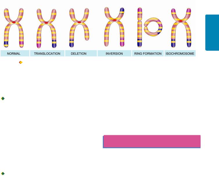
Figure 10.4 Common structural abnormalities of human chromosomes.
TRANSLOCATIONS. Translocation means crossing over or exchange of fragment of chromosome which may occur between non-homologous or homologous chromosomes. There are two main types of translocations: reciprocal in about two-third and Robertsonian in one-third cases:

 Reciprocal translocation is the exchange of genetic material between two non-homologous (heterologous) chromosomes without involving centromere (acentric). Such translocations occur due to single breaks in both the chromosomes and the exchange is detected by banding techniques. Reciprocal translocation may be balanced (without any loss of genetic material during the exchange) or unbalanced (with some loss of genetic material).
i)Balanced reciprocal translocation is more common and the individual is phenotypically normal e.g. translocation between long arm (q) of chromosomes 22 and long arm (q) of chromosome 9 written as 46, XX, t (9;22). This translocation is termed Philadelphia chromosome seen in most cases of chronic myeloid leukaemia (page 355).
ii)Unbalanced reciprocal translocations are less common and account for repeated abortions and malformed children.

 Robertsonian translocation is less common than reciprocal translocation. In this, there is fusion of two acrocentric chromosomes (having very short arms) at the centromere (centric fusion) with loss of short arms. The result of this fusion is one very large chromosome and the other very small one. Individuals born with Robertsonian translocation may be phenotypically normal but suffer from infertility and are at higher risk of producing malformed children in the next progeny.
DELETIONS. Loss of genetic material from the chromosome is called deletion. Deletion may be from the terminal or middle portion of the chromosome. The examples of deletion are: cri du chat (named after cry of infant like that of a cat) syndrome (deletion of short arm of chromosome 5) and several cancers with hereditary basis (e.g. retinoblastoma with deletion of long arm of chromosome 13, Wilms’ tumour with deletion of short arm of chromosome 11).
INVERSION. Inversion is a form of rearrangement involving breaks of a single chromosome at two points. Inversion may be pericentric or paracentric, depending upon whether the rotation occurs at the centromere or at the acentric portion of the arm of chromosome. Inversions are not associated with any abnormality.
RING CHROMOSOME. A ring of chromosome is formed by a break at both the telomeric (terminal) ends of a chromosome followed by deletion of the broken fragment and then end-to-end fusion. The consequences of ring chromosome depend upon the amount of genetic material lost due to break.
ISOCHROMOSOME. When centromere, rather than dividing parallel to the long axis, instead divides transverse to the long axis of chromosome, it results in either two short arms only or two long arms only called isochromosomes. The example involving isochromosome of X-chromosome is seen in some cases (15%) of Turner’s syndrome.
SINGLE-GENE DEFECTS
(MENDELIAN DISORDERS)
In order to unravel causes of disease at genetic level, spectacular advances have been made in human genetics. With mapping of human genome consisting of about 30,000 genes, it is possible to perform molecular profiling of diseases at genetic level.
The classic laws of inheritance of characteristics or traits were outlined by Austrian monk Gregor Mendel in 1866 based on his observations of cross-breeding of red and white garden peas. Single-gene defects follow the classic mendelian patterns of inheritance and are also called mendelian disorders. These disorders are the result of mutation of a single gene of large effect.
MUTATIONS. The term mutation is applied to permanent change in the DNA of the cell. Mutations affecting germ cells are transmitted to the next progeny producing inherited diseases, while the mutations affecting somatic cells give rise to various cancers and congenital malformations. Presently, following types of mutations have been described:
i)Point mutation is the result of substitution of a single nucleotide base by a different base i.e. replacement of an amino acid by another e.g. in sickle cell anaemia there is point mutation by substitution of glutamic acid by valine in the polypeptide chain.
ii)Stop codon or nonsense mutation refers to a type of point mutation in which the protein chain is prematurely terminated or truncated.
259
Diseases Paediatric and Genetic 10 CHAPTER

260 iii) Frameshift mutation occurs when there is insertion or deletion of one or two base pairs in the DNA sequence e.g. in cystic fibrosis of pancreas.
SECTION |
iv) Trinucleotide repeat mutation is characterised by |
||
amplification of a sequence of three nucleotides. |
|||
Thus it can be summed up from above that single-gene |
|||
defects are synonymous with various types of heritable |
|||
mutations. Currently, approximately 5000 single-gene defects |
|||
I |
have been described—some major and others of minor |
||
|
consequence. While most of these disorders are discussed in |
||
General |
relevant chapters later, the group of storage diseases (inborn |
||
errors of metabolism) is considered below. |
|||
INHERITANCE PATTERN. The inheritance pattern of |
|||
genetic abnormalities may be dominant or recessive, autosomal |
|||
Pathology |
alleles of a gene pair are expressed in heterozygous state, it |
||
|
or sex-linked: |
||
|
A dominant gene* produces its effects, whether combined |
||
|
with similar dominant or recessive gene. Recessive genes are |
||
|
effective only if both genes are similar. However, when both |
||
and |
is called codominant inheritance. A single gene may express |
||
in multiple allelic forms known as polymorphism. |
|||
Basic |
Autosomal dominant inheritance pattern is characterised by |
||
one faulty copy of gene (i.e. mutant allele) in any autosome |
|||
|
|||
Techniques |
and one copy of normal allele; disease phenotype is seen in |
||
mutated. Usually, it occurs when both parents are carriers |
|||
|
all such individuals. Patients having autosomal dominant |
||
|
inheritance disease have 50% chance of passing on the disease |
||
|
to the next generation. |
||
|
In autosomal recessive inheritance, both copies of genes are |
||
|
of the defective gene, i.e. having one normal allele and one |
||
|
defective allele in each parent, and each parent passes on |
||
|
their defective gene to the next progeny causing disease. |
||
|
There is 25% chance of transmission of autosomal recessive |
||
|
disease when both parents are carriers. |
||
|
X-linked disorders are caused by mutations in genes on X- |
||
|
chromosome, derived from either one of the two X- |
||
|
chromosomes in females, or from the single X-chromosome |
||
|
of the male. There are much fewer genes on Y-chromosome |
||
|
and are determinant for testis; they are not known to cause |
||
|
any sex-linked disorder. Therefore, all sex-linked disorders |
||
|
are, in fact, X-linked disorders. |
||
|
Table 10.1 lists important examples of groups of genetic |
||
|
disorders: autosomal recessive (the largest group), codominant |
||
|
(intermediate), and dominant, and sex-(X-) linked recessive |
||
|
and dominant disorders. |
||
|
|
|
|
|
STORAGE DISEASES |
||
|
|||
|
(INBORN ERRORS OF METABOLISM) |
||
|
Storage diseases or inborn errors of metabolism are bio- |
||
|
chemically distinct groups of disorders occurring due to |
||
|
|
|
|
|
*A particular characteristic of an individual is determined by a pair of |
||
|
single genes, located at the same specific site termed locus, on a pair of |
||
|
homologous chromosomes. These paired genes are called alleles which |
||
|
may be homozygous when alike, and heterozygous if dissimilar. Genotype |
||
|
is the genetic composition of an individual while phenotype is the effect |
||
|
of genes produced. |
||
TABLE 10.1: Important Examples of Mendelian Disorders (Single Gene Defects).
I.AUTOSOMAL RECESSIVE INHERITANCE
1.β-thalassaemia
2.Sickle cell anaemia
3.Haemochromatosis
4.Cystic fibrosis of pancreas
5.Albinism
6.Wilson’s disease
7.Xeroderma pigmentosum
8.Inborn errors of metabolism (Lysosomal storage diseases, glycogenosis, alkaptonuria, phenylketonuria)
II.AUTOSOMAL CODOMINANT INHERITANCE
1.ABO blood group antigens
2.α 1-antitrypsin deficiency
3.HLA antigens
III.AUTOSOMAL DOMINANT INHERITANCE
1.Familial polyposis coli
2.Adult polycystic kidney
3.Hereditary spherocytosis
4.Neurofibromatosis (von Recklinghausen’s disease)
5.Marfan’s syndrome
6.von Willebrand’s disease
7.Hereditary haemorrhagic telangiectasia
8.Acute intermittent porphyria
9.Familial hypercholesterolaemia
10.Osteogenesis imperfecta
IV. SEX-(X-) LINKED RECESSIVE INHERITANCE
1.Haemophilia A
2.G6PD deficiency
3.Diabetes insipidus
4.Chronic granulomatous disease
5.Colour blindness
6.Bruton’s agammaglobulinaemia
7.Muscular dystrophies
V. SEX-(X-) LINKED DOMINANT INHERITANCE
1.Hypophosphataemic rickets
2.Incontinentia pigmenti
genetic defect in the metabolism of carbohydrates, lipids, and proteins resulting in intracellular accumulation of metabolites. These substances may collect within the cells throughout the body but most commonly affected organ or site is the one where the stored material is normally found and degraded. Since lysosomes comprise the chief site of intracellular digestion (autophagy as well as heterophagy), the material is naturally stored in the lysosomes, and hence the generic name ‘lysosomal storage diseases’. Cells of mono- nuclear-phagocyte system are particularly rich in lysosomes; therefore, reticuloendothelial organs containing numerous phagocytic cells like the liver and spleen are most commonly involved in storage disease.
Based on the biochemical composition of the accumulated material within the cells, storage diseases are classified into distinct groups, each group containing a number of diseases depending upon the specific enzyme deficiency. A summary

TABLE 10.2: Storage Diseases (Inborn Errors of Metabolism).
Disease |
Enzyme Deficiency |
Accumulating Metabolite |
Organs Involved |
GLYCOGEN STORAGE DISEASE |
|
|
|
Type I (von Gierke’s disease) |
Glucose-6-phosphatase |
Glycogen |
Liver, kidney |
Type II (Pompe’s disease) |
Acid-α-glucosidase (acid maltase) |
Glycogen |
Heart, skeletal muscle |
Type III (Forbes’/Cori’s disease) |
Amyloglucosidase (debrancher) |
Limit dextrin |
Heart, skeletal muscle |
Type IV (Anderson’s disease) |
Amylotransglucosidase (brancher) |
Amylopectin |
Liver |
Type V (McArdle’s disease) |
Muscle phosphorylase |
Glycogen |
Skeletal muscle |
Type VI (Hers’ disease) |
Liver phosphorylase |
Glycogen |
Liver |
Type VII |
Phosphofructokinase |
Glycogen |
Muscle |
Type VIII |
Phosphorylase kinase |
Glycogen |
Liver |
MUCOPOLYSACCHARIDOSES (MPS) |
|
|
|
Type I to type VI MPS |
Different lysosomal |
Chondroitin sulphate, |
Connective tissue, liver, |
syndromes |
enzymes |
dermatan sulphate, |
spleen, bone marrow, |
|
|
heparan sulphate, |
lymph nodes, kidneys, |
|
|
keratan sulphate |
heart, brain |
SPHINGOLIPIDOSES (GANGLIOSIDOSES) |
|
|
|
GM1-gangliosidosis |
GM1 ganglioside-galactose |
GM1-ganglioside |
Liver, kidney, |
(infantile and juvenile types) |
|
|
spleen, heart, brain |
GM2-gangliosidosis |
Hexosaminidase |
GM2-ganglioside |
Liver, kidney, spleen, |
(Tay-Sachs, Sandhoff’s disease) |
|
|
heart, brain |
SULFATIDOSES |
|
|
|
Metachromatic |
Aryl sulfatase A |
Sulfatide |
Brain, liver, spleen, |
leucodystrophy |
|
|
heart, kidney |
Krabbe’s disease |
Galactocerebrosidase |
Galactocerebroside |
Nervous system, kidney |
Fabry’s disease |
α-Galactosidase |
Ceramide |
Skin, kidney, heart, spleen |
Gaucher’s disease |
Glucocerebrosidase |
Glucocerebroside |
Spleen, liver, bone marrow |
Niemann-Pick disease |
Sphingomyelinase |
Sphingomyelin |
Spleen, liver, bone marrow, |
|
|
|
lymph nodes, lung |
|
|
|
|
261
Diseases Paediatric and Genetic 10 CHAPTER
of major groups of storage diseases along with their respective enzyme deficiencies, major accumulating metabolites and the organs involved is presented in Table 10.2. A few general comments can be made about all storage diseases:

 All the storage diseases occur either as a result of autosomal recessive, or sex-(X-) linked recessive genetic transmission.
 Most, but not all, of the storage diseases are lysosomal storage diseases. Out of the glycogen storage diseases, only type II (Pompe’s disease) is due to lysosomal enzyme deficiency.
A few important forms of storage diseases are described below:
Glycogen Storage Diseases (Glycogenoses)
These are a group of inherited disorders in which there is defective glucose metabolism resulting in excessive intracellular accumulation of glycogen in various tissues. Based on specific enzyme deficiencies, glycogen storage diseases are divided into 8 main types designated by Roman numerals I to VIII. However, based on pathophysiology, glycogen storage diseases can be divided into 3 main subgroups:
1. Hepatic forms are characterised by inherited deficiency of hepatic enzymes required for synthesis of glycogen for
storage (e.g. von Gierke’s disease or type I glycogenosis) or due to lack of hepatic enzymes necessary for breakdown of glycogen into glucose (e.g. type VI glycogenosis).
2.Myopathic forms on the other hand, are those disorders in which there is genetic deficiency of glycolysis to form lactate in the striated muscle resulting in accumulation of glycogen in the muscles (e.g. McArdle’s disease or type V glycogenosis, type VII disease).
3.Other forms are those in which glycogen storage does not occur by either hepatic or myopathic mechanisms. In Pompe’s disease or type II glycogenosis, there is lysosomal storage of glycogen, while in type IV there is deposition of abnormal metabolites of glycogen in the brain, heart, liver and muscles.
The prototypes of these three forms are briefly considered below.
VON GIERKE’S DISEASE (TYPE I GLYCOGENOSIS).
This condition is inherited as an autosomal recessive disorder due to deficiency of enzyme, glucose-6-phos- phatase. In the absence of glucose-6-phosphatase, excess of normal type of glycogen accumulates in the liver and also results in hypoglycaemia due to reduced formation of free glucose from glycogen. As a result, fat is metabolised for energy requirement leading to hyperlipoproteinaemia and ketosis. Other changes due to

262
Techniques Basic and Pathology General I SECTION
deranged glucose metabolism are hyperuricaemia and accumulation of pyruvate and lactate.
The disease manifests clinically in infancy with failure to thrive and stunted growth. Most prominent feature is enormous hepatomegaly with intracytoplasmic and intranuclear glycogen. The kidneys are also enlarged and show intracytoplasmic glycogen in tubular epithelial cells. Other features include gout, skin xanthomas and bleeding tendencies due to platelet dysfunction.
POMPE’S DISEASE (TYPE II GLYCOGENOSIS). This is also an autosomal recessive disorder due to deficiency of a lysosomal enzyme, acid maltase, and is the only example of lysosomal storage disease amongst the various types of glycogenoses. Acid maltase is normally present in most cell types and is responsible for the degradation of glycogen. Its deficiency, therefore, results in accumulation of glycogen in many tissues, most often in the heart and skeletal muscle, leading to cardiomegaly and hypotonia.
McARDLE’S DISEASE (TYPE V GLYCOGENOSIS). The condition occurs due to deficiency of muscle phosphorylase resulting in accumulation of glycogen in the muscle (deficiency of liver phosphorylase results in type VI glycogenosis). The disease is common in 2nd to 4th decades of life and is characterised by painful muscle cramps, especially after exercise, and detection of myoglobinuria in half the cases.
Mucopolysaccharidoses (MPS)
Mucopolysaccharidoses are a group of six inherited syndromes numbered from MPS I to MPS VI. Each of these results from deficiency of specific lysosomal enzyme involved in the degradation of mucopolysaccharides or glycosaminoglycans, and are, therefore, a form of lysosomal storage diseases. Mucopolysaccharides which accumulate in the MPS are: chondroitin sulphate, dermatan sulphate, heparan sulphate and keratan sulphate. All these syndromes are autosomal recessive disorders except MPS II (Hunter’s syndrome) which has X-linked recessive transmission.
Syndrome of MPS manifests in infancy or early childhood and involves multiple organs and tissues, chiefly connective tissues, liver, spleen, bone marrow, lymph nodes, kidneys, heart and brain. The mucopolysaccharides accumulate in mononuclear phagocytic cells, endothelial cells, intimal smooth muscle cells and fibroblasts. The material is finely granular and PAS-positive by light microscopy. By electron microscopy, it appears in the swollen lysosomes and can be identified biochemically as mucopolysaccharide.
Gaucher’s Disease
This is an autosomal recessive disorder in which there is mutation in lysosomal enzyme, acid β-glucosidase (earlier called glucocerebrosidase), which normally cleaves glucose from ceramide. This results in lysosomal accumulation of glucocerebroside (ceramide-glucose) in phagocytic cells of the body and sometimes in the neurons. The main sources of glucocerebroside in phagocytic cells are the membrane glycolipids of old leucocytes and erythrocytes, while the deposits in the neurons consist of gangliosides.
Clinically, 3 subtypes of Gaucher’s disease are identified based on neuronopathic involvement:
 Type I or classic form is the adult form of disease in which there is storage of glucocerebrosides in the phagocytic cells of the body, principally involving the spleen, liver, bone marrow, and lymph nodes. This is the most common type comprising 80% of all cases of Gaucher’s disease.

 Type II is the infantile form in which there is progressive involvement of the central nervous system.
 Type III is the juvenile form of the disease having features in between type I and type II i.e. they have systemic involvement like in type I and progressive involvement of the CNS as in type II.
The clinical features depend upon the clinical subtype of Gaucher’s disease. In addition to involvement of different organs and systems (splenomegaly, hepatomegaly, lymphadenopathy, bone marrow and cerebral involvement), a few other features include pancytopenia, or thrombocytopenia secondary to hypersplenism, bone pains and pathologic fractures.
Microscopy shows large number of characteristically distended and enlarged macrophages called Gaucher cells which are found in the spleen, liver, bone marrow and lymph nodes, and in the case of neuronal involvement, in the Virchow-Robin space. The cytoplasm of these cells is abundant, granular and fibrillar resembling crumpled tissue paper. They have mostly a single nucleus but occasionally may have two or three nuclei (Fig. 10.5, A, C). Gaucher cells are positive with PAS, oil red O, and Prussian-blue reaction indicating the nature of accumulated material as glycolipids admixed with haemosiderin. These cells often show erythrophagocytosis and are rich in acid phosphatase.
Niemann-Pick Disease
This is also an autosomal recessive disorder characterised by accumulation of sphingomyelin and cholesterol due to defect in acid sphingomyelinase.
Two types have been described: type A and B.
 Type A is more common and typically presents in infancy and is characterised by hepatosplenomegaly, lymphadenopathy, rapidly progressive deterioration of CNS and physical underdevelopment. About a quarter of patients present with familial amaurotic idiocy with characteristic cherry-red spots in the macula of the retina (amaurosis = loss of vision without apparent lesion of the eye).

 Type B develops later and has a progressive hepatosplenomegaly with development of cirrhosis due to replacement of the liver by foam cells, and impaired lung function due to infiltration in lung alveoli.
Microscopy shows storage of sphingomyelin and cholesterol within the lysosomes, particularly in the cells of mononuclear phagocyte system. The cells of NiemannPick disease are somewhat smaller than Gaucher cells and their cytoplasm is not wrinkled but is instead foamy
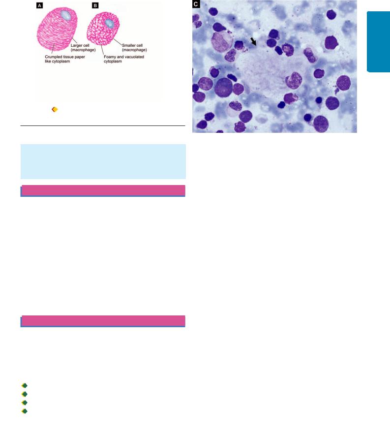
Figure 10.5 |
Diagrammatic view of comparative features of typical |
Gaucher cell (A). B, typical macrophage in Niemann-Pick disease. C, A Gaucher cell (arrow) in bone marrow aspirate smear.
and vacuolated which stains positively with fat stains (Fig. 10.5, B). These cells are widely distributed in the spleen, liver, lymph nodes, bone marrow, lungs, bowel and brain.
MULTIFACTORIAL INHERITANCE
Some normal phenotypic characteristics have also multifactorial inheritance e.g. colour of hair, eye, skin, height and intelligence. Multifactorial disorders are those disorders which result from the combined effect of genetic composition and environmental influences. Some common examples of such disorders in which environmental influences mask the mutant genes are as under:
1.Cleft lip and cleft palate
2.Pyloric stenosis
3.Diabetes mellitus
4.Hypertension
5.Congenital heart disease
6.Coronary heart disease.
OTHER PAEDIATRIC DISEASES
As mentioned in the foregoing discussion, many diseases affecting infancy and childhood are genetic or developmental in origin. Here, other diseases affecting the period from birth to puberty are discussed under the heading of paediatric diseases. This period is conventionally subdivided into 4 stages:

 Neonatal period: birth to first 4 weeks

 Infancy: first year of life

 Early childhood: 1-4 years

 Late childhood: 5-14 years
Each of these four stages has distinct anatomic, physiologic and immunologic development compared to adults and, therefore, has different groups of diseases unique to particular age groups. Before discussing these diseases affecting different age groups, a few general comments about these stages can be made:
263
|
10 CHAPTER |
|
and Genetic |
1. Neonatal period is the period of continuation of |
Paediatric |
|
|
dependent intrauterine foetal life to independent postnatal |
|
period. Therefore, this is the period of maximum risk to life |
Diseases |
adequate postnatal medical care is not provided, neonatal |
|
due to perinatal causes (e.g. prematurity, low birth weight, |
|
perinatal infections, respiratory distress syndrome, birth |
|
asphyxia, birth trauma etc) and congenital anomalies. If |
|
mortality is high. Neonatal mortality in first week after birth |
|
is about 10-times higher compared to second week, and |
|
shows improvement with every passing week at this stage. |
|
2. In infancy, the major health problems are related to |
|
congenital anomalies, infections of lungs and bowel, and |
|
sudden infant death syndrome (often during sleep). |
|
3. Young children from 1-4 years are exposed to higher risk |
|
of sustaining injuries, and manifest certain congenital |
|
anomalies. Some malignant tumours are peculiar to this age |
|
group. |
|
4. Older children from 5-14 years too have higher risk of |
|
injuries from accidents and have other problems related to |
|
congenital anomalies and certain malignant tumours at this |
|
age. |
|
Thus, hazardous effects of congenital anomalies are a |
|
common denominator for all age groups from birth to |
|
adolescence. Specific tumours peculiar to infants and |
|
children are discussed along with discussion in related |
|
chapters of Systemic Pathology. However, a short note on |
|
general aspects of this subject is given below. |
|
TUMOURS OF INFANCY AND CHILDHOOD |
|
Tumours of infancy and childhood comprise 2% of all |
|
malignant tumours but they are the leading cause of death |
|
in this age group exceeded only by accidents. Benign tumours |
|
are more common than malignant neoplasms but they are |
|
generally of little immediate consequence. Another aspect |
|
requiring consideration here is the difficulty in differentiating |
|
benign tumours from tumour-like lesions. |
|
HISTOGENESIS. Histogenetic evolution of tumours at |
|
different age groups takes place as under: |
|

264 
 Some tumours have probably evolved in utero and are apparent at birth or in immediate postnatal period. Such
tumours are termed developmental tumours.
SECTION |
Many other tumours originate in abnormally developed |
|
organs and organ rests; they become apparent subsequently |
||
|
||
|
and are termed embryonic tumours. |
|
|
In embryonic tumours, proliferation of embryonic cells |
|
I |
occurs which have not reached the differentiation stage essential |
|
for specialised functions i.e. the cells proliferate as |
||
|
||
|
undifferentiated or as partially differentiated stem cells and an |
|
General |
embryonal tumour is formed. |
|
growth promoting influence of oncogenes and suffer from |
||
|
Tumours of infancy and childhood have some features of |
|
|
normal embryonic or foetal cells in them which proliferate under |
|
Pathology |
mutations which make them appear morphologically |
|
proliferating mature differentiated cells e.g. a neonatal |
||
|
malignant. |
|
|
Under appropriate conditions, these malignant embryo- |
|
|
nal cells may cease to proliferate and transform into non- |
|
and |
neuroblastoma may mature and differentiate into benign |
|
ganglioneuroma; tissues in foetal sacrococcygeal teratoma |
||
Basic |
may mature with age to adult tissues and is assigned better |
|
prognosis. |
||
|
||
Techniques |
Thus, normal somatic cell maturation and neoplastic |
|
Benign Tumours and Tumour-like Conditions |
||
|
development in embryonal tumours represent two opposite |
|
|
ends of ontogenesis, with capability of some such tumours to |
|
|
mature and differentiate to turn benign from malignant. |
|
|
Many of the benign tumours seen in infancy and childhood |
|
|
are actually growth of displaced cells and masses of tissues |
|
|
and their proliferation takes place along with the growth of |
|
|
the child. Some of these tumours undergo a phase of |
|
|
spontaneous regression subsequently—a feature usually not |
|
|
seen in true benign tumours. While some consider such |
|
|
lesions as mere ‘tumour-like lesions or malformations’, others |
|
|
call them benign tumours. A few such examples are as under: |
1.Hamartomas. Hamartomas are focal accumulations of cells normally present in that tissue but are arranged in an abnormal manner i.e. though present at normal site they do not reproduce normal architecture identical to adjacent tissues (page 193).
2.Choristoma (heterotopia). Choristoma or heterotopia is collection of normal cells and tissues at aberrant locations e.g. heterotopic pancreatic tissue in the wall of small bowel or stomach.
A list of common benign tumours and tumour-like lesions is presented in Table 10.3.
Malignant Tumours
Cancers of infancy and childhood differ from those in adults in the following respects:
1. Sites. Cancers of this age group more commonly pertain to haematopoietic system, neural tissue and soft tissues compared to malignant tumours in adults at sites such as the lung, breast, prostate, colon and skin.
2.Genetic basis. Many of paediatric malignant tumours have underlying genetic abnormalities.
3.Regression. Foetal and neonatal malignancies have a tendency to regress spontaneously or to mature.
4.Histologic features. These tumours have unique histologic features in having primitive or embryonal appearance rather than pleomorphic-anaplastic histologic appearance.
5.Management. Many of paediatric malignant tumours are curable by chemotherapy and/or radiotherapy but may develop second malignancy.
A few generalisations can be drawn about paediatric cancers:

 In infants and children under 4 years of age: the most common malignant tumours are various types of blastomas.
  Children between 5 to 9 years of age: haematopoietic malignancies are more common.

 In the age range of 10-14 years (prepubertal age): soft tissue and bony sarcomas are the prominent tumours.
TABLE 10.3: Common Paediatric Benign Tumours and Tumour-like Lesions.
Nomenclature |
Main Features |
|
|
|
|
BENIGN TUMOURS |
|
|
i. Haemangioma |
• Most common in infancy |
|
|
|
• Commonly on skin (e.g. port-wine stain) |
|
|
• May regress spontaneously |
ii. Lymphangioma |
• Cystic and cavernous type common |
|
|
|
• Located in skin or deeper tissues |
|
|
• Tends to increase in size after birth |
iii. |
Sacrococcygeal teratoma |
• Often accompanied with other congenital malformations |
|
|
• Majority (75%) are benign; rest are immature or malignant |
iv. |
Fibromatosis |
• Solitary (which generally behaves as benign) to multifocal (aggressive lesions) |
TUMOUR-LIKE LESIONS/BENIGN TUMOURS |
|
|
i. |
Naevocellular naevi |
• Very common lesion on the skin |
ii. |
Liver cell adenoma |
• Most common benign tumour of liver |
iii. |
Rhabdomyoma |
• Rare foetal and cardiac tumour |
|
|
|
TABLE 10.4: Common Paediatric Malignant Tumours.
|
System |
Age < 4 Yrs |
Age 5-9 Yrs |
Age 10-14 Yrs |
|
|
|
|
|
|
|
1. |
Haematopoietic |
Acute leukaemia |
Acute leukaemia |
Hodgkin’s |
|
|
|
|
Lymphoma |
|
|
2. |
Blastomas |
Neuroblastoma |
Neuroblastoma |
Hepatocellular |
|
|
|
Hepatoblastoma |
Hepatocellular carcinoma |
carcinoma |
|
|
|
Retinoblastoma |
|
|
|
|
|
Nephroblastoma (Wilms’ tumour) |
|
|
|
3. |
Soft tissues |
Rhabdomyosarcoma |
Soft tissue sarcoma |
Soft tissue sarcoma |
|
4. |
Bony |
— |
Ewing’s sarcoma |
Osteogenic sarcoma |
|
5. |
Neural |
CNS tumours |
CNS tumours |
— |
|
6. |
Others |
Teratoma |
— |
Thyroid cancer |
|
|
|
|
|
|
|
Based on these broad guidelines, classification of common |
presented in Table 10.4. These have been discussed in related |
paediatric malignant tumours at different age groups is |
chapters later. |
|
|
265
Diseases Paediatric and Genetic 10 CHAPTER

266
Techniques Basic and Pathology General I SECTION
Chapter 11 Basic Diagnostic Cytology*
INTRODUCTION
The last chapter in General Pathology deals with the techniques and applications of basic cytologic methods in a diagnostic pathology laboratory.
As mentioned in the beginning of this book, surgical pathology developed as a prospective diagnostic branch around the turn of 19th Century. Its basis was application of knowledge of morphological details of cells for diagnosis of disease in biopsy material. This generated interest of workers towards obtaining cellular material by non-biopsy techniques to arrive at the diagnosis. As a result, cytopathologic diagnosis was initially introduced purely as Exfoliative Cytology in the 1920s by Dr. George N. Papanicolaou. Subsequently, success of bone marrow aspiration for diagnosis of haematopoietic diseases generated interest in aspiration biopsy for diagnosis of solid lesions in the 1930s, which evolved over the next three decades mainly in Scandinavian countries in Europe and later spread to the rest of the world to become the mainstay branch of diagnostic cytology known
as Interventional Cytology.
In general, diagnostic cytology pertains to the interpretation of cells from the human body that either exfoliate (desquamate) spontaneously from epithelial surfaces, or the cells are obtained from various organs/ tissues by different clinical procedures. While histopathologic diagnosis is based on interpretation of changes in tissue architecture, the cytopathologic diagnosis rests upon alterations in morphology observed in single cells or small groups of cells.
Role of Diagnostic Cytology
Among the numerous applications of cytodiagnostic techniques, the following are more important:
1. Diagnosis and management of cancer. In the field of oncology, establishing a ‘tissue diagnosis’ (i.e. pathologic diagnosis based on microscopic evidence of malignancy) is an essential pre-requisite for proper management of a cancer patient.
i)Cytodiagnosis in its traditional role is a valuable adjunct to histopathology for establishing the vital tissue diagnosis e.g. diagnosis of lymphomas where imprint smears prove valuable, and in some respects superior to histopathology in typing the lymphoma.
ii)Cytologic techniques also provide a preliminary diagnosis of cancer for later confirmation by histopathology e.g. detection of ovarian cancer cells in ascitic fluid.
iii)In some fields, cytodiagnosis has replaced histopathology as the primary method of establishing a tissue diagnosis e.g. in breast cancer, where positive cytologic report is considered sufficient for planning the management, obviating the need for diagnostic surgical biopsy.
iv)Cytodiagnosis has a major role in the detection and diagnosis of clinically silent early cancer e.g. carcinoma in situ of the uterine cervix.
v)In the management of cancer, cytodiagnosis may help in assessing response to therapy e.g. cervical smears for response to radiotherapy in carcinoma cervix, and urinary cytology for response to chemotherapy in carcinoma of the urinary bladder.
vi)In the follow-up of previously diagnosed cases of cancer, it is of particular value in detecting dissemination (metastasis) or recurrence of tumour.
2.Identification of benign neoplasms. This application is derived from its ability to distinguish between benign and malignant neoplasms e.g. fibroadenoma of the breast versus carcinoma.
3.Intraoperative pathologic diagnosis. In this role, cytodiagnosis complements histopathologic diagnosis e.g. imprint smears alongwith frozen section for breast lumps.
4.Diagnosis of non-neoplastic/inflammatory conditions.
Cytodiagnosis allows recognition of specific conditions which
TABLE 11.1: Nuclear Criteria of Malignancy.
1. Nuclear size |
: Usually larger than benign nuclei; |
|
variation in size (anisonucleosis) more |
|
significant. |
2.Nucleus-cytoplasmic : Increased. (N:C) ratio
3. |
Nuclear shape |
: Moderate to marked variation. |
||
4. |
Nuclear membrane |
: Irregular thickening, angulation and |
||
|
|
|
indentations. |
|
5. |
Nuclear chromatin |
: |
Hyperchromatic (less significant), |
|
|
|
|
uneven distribution, coarse irregular |
|
|
|
|
angulated |
chromatin clumping, |
|
|
|
parachromatin clearing (more signi- |
|
|
|
|
ficant). |
|
6. |
Nucleoli |
: Increased |
size and number less |
|
|
|
|
significant; irregular angular outlines |
|
|
|
|
more significant. |
|
7. |
Number of nuclei |
: |
Multinucleation unreliable; nuclear |
|
|
|
|
character more important. |
|
8. |
Mitoses |
|
Increased mitoses unreliable; abnormal |
|
|
|
|
mitoses significant. |
|
|
|
|
|
|
*The contribution of this chapter by Dr Rohit Sharma, MD, to the second edition of this book (1994) is gratefully acknowledged.

TABLE 11.2: Differences between Exfoliative and Interventional Cytology.
|
Feature |
Exfoliative Cytology |
Interventional Cytology |
|
|
|
|
1. |
Cell samples |
Exfoliated from epithelial surfaces |
Obtained by intervention/aspiration |
2. |
Smears |
Require screening to locate |
Abundance of cells for study in most |
|
|
suitable cells for study |
smears |
3. |
Diagnostic basis |
Individual cell morphology |
Cell patterns and morphology of groups of cells |
4. |
Morphologic criteria of |
Nuclear characteristics most |
Nuclear characteristics important; cytoplasmic |
|
diagnostic significance |
important |
character and background equally significant |
|
|
|
|
267
11 CHAPTER
do not routinely require surgical intervention e.g. Hashimoto’s thyroiditis.
5.Diagnosis of specific infections. A variety of bacterial, viral, protozoal and fungal infections can be identified by cytologic methods e.g. tubercle bacilli in lymph node aspirates, herpetic inclusions and Trichomonas in cervicovaginal smears, fungal hyphae in touch preparations from draining sinuses.
6.Cytogenetics. Cytodiagnostic techniques can be employed for chromosomal studies including leucocyte and tissue cultures and for demonstrating sex-chromatin e.g. buccal smear for Barr body.
7.Assessment of hormonal status in women. Vaginal smears accurately reflect changes in female sex hormonal levels e.g. to confirm the onset of menopause.
8.Identification of cell of origin. For example, identification of spermatogenic elements in aspirates from the testes in cases of male infertility.
For cytomorphological recognition of cancer, nuclear characteristics are used to determine the presence or absence of malignancy (Table 11.1). Cytoplasmic characteristics help in typing the malignancy e.g.keratinisation in squamous cell carcinoma, mucin droplets in adenocarcinoma, melanin pigment in melanomas.
Branches of Diagnostic Cytology
Two branches of diagnostic cytology are currently recognised: exfoliative and interventional.
EXFOLIATIVE CYTOLOGY. This is the older branch that essentially involves the study of cells spontaneously shed off (as a result of continuous growth of epithelial linings) from epithelial surfaces into body cavities or body fluids. Exfoliative cytology is facilitated by the fact that the rate of exfoliation is enhanced in disease-states thereby yielding a larger number of cells for study. In addition, cells for study may also be obtained by scraping, brushing, or washing various mucosal surfaces (abrasive cytology).
INTERVENTIONAL CYTOLOGY. This is the branch in which samples are obtained by clinical procedures or surgical intervention. It is dominated by, and is virtually synonymous with, Fine Needle Aspiration Cytology (FNAC) which is also known as Aspiration Biopsy Cytology (ABC). In interventional cytology, the cytopathologist often performs the clinical procedure himself and interacts
with the patient in contrast to exfoliative cytology where the samples are prepared and forwarded by the clinician to the pathologist and the cytopathologist interprets them in isolation.
A brief account of both main branches of cytology is given below while their contrasting features are presented in Table 11.2.
EXFOLIATIVE CYTOLOGY
Type of samples that can be obtained from different organ systems for exfoliative cytodiagnosis are listed in Table 11.3. Apparently, a comprehensive review of each type of sample is beyond the scope of this text. Cytology of female genital tract is discussed in detail below while brief mention is made about other samples.
I. FEMALE GENITAL TRACT
Smears from the female genital tract have traditionally been known as ‘Pap smears’. These smears may be prepared by
TABLE 11.3: Types of Samples for Exfoliative Cytology
Obtainable from Different Organ Systems/Body Sites.
1.Female genital tract — Lateral vaginal smears (LVS)
—Vaginal ‘pool’ smears
—Cervical smears
—Combined (fast) smears
—Triple smears (CVE)
—Endocervical/Endometrial aspiration
2. Respiratory tract |
— Sputum |
—Bronchial washings/brushing/ bronchioalveolar lavage (BAL)
3.Gastrointestinal tract — Endoscopic lavage/brushing
4. |
Urinary tract |
— |
Urinary sediment |
|
|
— |
Bladder washings |
|
|
— |
Retrograde catheterisation |
|
|
— |
Prostatic massage (secretions) |
5. |
Body fluids |
— |
Effusions |
—Fluids of small volume
i.Cerebrospinal fluid (CSF)
ii.Synovial fluid
iii.Amniotic fluid
iv.Hydrocele fluid
v.Seminal fluid (semen)
vi.Nipple discharge
6.Other samples — Buccal smears (for sex chromatin)
Cytology Diagnostic Basic
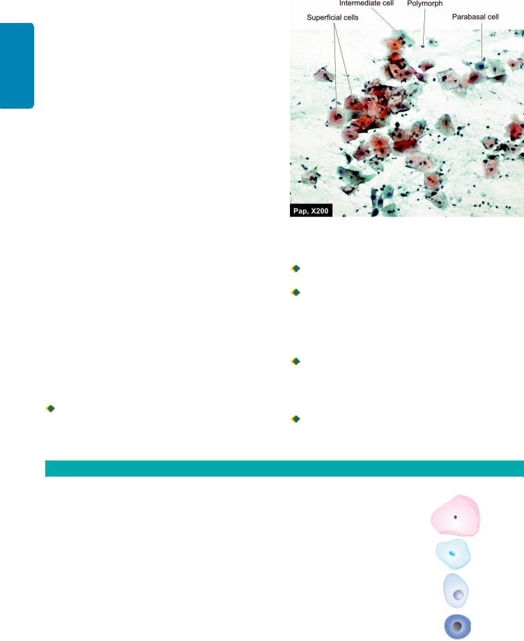
268 different methods depending upon the purpose for which they are intended:
ISECTION |
i) Lateral vaginal smears (LVS) obtained by scraping the |
|
and are recommended for detection of endometrial and |
||
|
upper third of the lateral walls of the vagina are ideal for |
|
|
cytohormonal assessment. |
|
|
ii) Vaginal ‘pool’ or ‘vault’ smears are obtained by scraping or |
|
|
aspirating material from the posterior fornix of the vagina |
|
|
ovarian cancer. |
|
General |
iii) Cervical smears obtained by cotton swabs or Ayre’s spatula |
|
from the portio of the cervix are ideal for detection of cervical |
||
|
||
|
carcinoma. |
|
|
iv) Combined (Fast) smears are a combination of vaginal pool |
|
Pathology |
and cervical scrapings. They offer the advantages of both |
|
v) Triple (cervical-vaginal-endocervical or CVE) smears contain |
||
|
and are recommended for routine population screening as |
|
|
they allow detection of up to 97% of cervical cancers and |
|
|
about 90% of endometrial cancers when properly prepared. |
|
and |
three distinct samples representing the ectocervix, vagina and |
|
endocervix on three separate areas of the same slide. These |
||
Basic |
smears are also recommended for routine screening as they |
|
allow localisation of lesions but are difficult to prepare. |
||
|
||
Techniques |
vi) Endocervical and endometrial smears may also be prepared |
|
by aspirating the contents of the endocervical canal and |
||
|
||
|
endometrial cavity respectively. |
|
|
CELLS IN NORMAL COMBINED SMEARS. Normally, |
|
|
combined smear contains two types of cells: epithelial and |
|
|
others (Fig. 11.1): |
|
|
Epithelial cells. There are 4 types of squamous epithelial cells |
|
|
in the cervical smear: superficial, intermediate, parabasal and |
|
|
basal cells. Morphological features of these cells are summed |
|
|
up in Table 11.4. A few variants of morphological forms and |
|
|
other epithelial cells are as under: |
|
|
Navicular cells are boat-shaped intermediate cells with |
|
|
folded cell borders. These cells appear in latter half of the |
|
|
menstrual cycle, during pregnancy and menopause. |
Figure 11.1 
 Various types of epithelial cells seen in normal Pap smear.
 Lactation cells are parabasal cells with strongly acidophilic cytoplasm. These cells are seen so long as lactation persists.

 Endocervical cells appear either as single dispersed nuclei due to degeneration, or as clusters of columnar cells giving it honey-combed appearance. Nuclei of endocervical cells are vesicular, with fine granular chromatin and contain 1-2 nucleoli, while the cytoplasm is slightly basophilic or vacuolated.
 Endometrial cells are seen up to 12th day of menstrual cycle. They are slightly smaller than endocervical cells, appear as tight rounded clusters of overlapping cells with moderately dark oval nuclei and scanty basophilic, vacuolated cytoplasm.

 Trophoblastic cells are seen following abortion or after delivery.
TABLE 11.4: Squamous Epithelial Cells Found in Normal Combined (Fast) Smears.
Cell Type |
Size |
Nuclei |
Cytoplasm |
Morphology |
|
|
|
|
|
Superficial |
30-60 μm |
< 6 μm |
Polyhedral, thin, broad, |
|
|
|
dark, pyknotic |
acidophilic or cyanophilic |
|
|
|
|
with keratohyaline granules. |
|
Intermediate |
20-40 μm |
6-9 μm |
Polyhedral or elongated, |
|
|
|
vesicular |
thin, cyanophilic with |
|
|
|
|
folded edges. |
|
Parabasal |
15-25 μm |
6-11 μm |
Round to oval, thick, |
|
|
|
vesicular |
well-defined, basophilic |
|
|
|
|
with occasional small |
|
|
|
|
vacuoles. |
|
Basal |
13-20 μm |
Large, (> one-half of cell |
Round to oval, |
|
|
|
volume), hyperchromatic, |
deeply basophilic. |
|
|
|
may have small nucleoli |
|
|
|
|
|
|
|

Other cells. Besides epithelial cells, other cells in cervical smears are leucocytes and Döderlein bacilli:
 Leucocytes in cervical smear include polymorphonuclear neutrophils (in large numbers normally), lymphocytes (isolated and entrapped in mucus normally), plasma cells (in chronic cervicitis), macrophages (normally in first 10 days of the menstrual cycle) and multinucleate cells (in specific inflammation).

 Döderlein bacilli (Bacillus vaginalis/Lactobacillus acidophilis), which belong to the group of lactobacilli, are the predominant organisms of the normal vaginal flora. It is a slender, grampositive, rod-like organism staining pale blue with the Papanicolaou technique. These organisms utilise the glycogen contained in the cytoplasm of intermediate and parabasal cells resulting in their disintegration (cytolysis). They are most numerous in the luteal phase and during pregnancy.
APPLICATIONS OF PAP SMEAR
Cytohormonal Evaluation
Assessment of hormonal status is best carried out from lateral vaginal smears although vaginal ‘pool’ or fast smears may also be used. Ideally, at least 3 smears obtained on alternate days should be scrutinised and cytologic indices determined for each smear for accurate assessment.
Several indices are available for description of cytohormonal patterns. The most commonly used are as under:
i)Acidophilic index (AI): The relative proportion of cells containing acidophilic (pink) and basophilic (blue) cytoplasm are determined by AI.
ii)Pyknotic index (PI): The percentage of cells having small, dark, shrunken nuclei (less than 6 μm in size) is determined by PI. The PI is more reliable than AI as it is not influenced by many of the cytoplasmic artefacts e.g. acidophilia caused by drying of smears prior to fixation that affects the latter.
iii)Maturation index (MI): MI is the most widely used method. One hundred squamous cells are counted and grouped according to their type—parabasal, intermediate, or superficial (basal cells are virtually absent from normal vaginal smears). Their proportions are expressed as a percentage; for example 10/80/10 represents parabasal, intermediate and superficial cells respectively. Some representative MIs at different stages of life are listed in
Table 11.5.
Abnormal Combined Smears
In order to evolve a system acceptable to clinicians and cytopathologists, National Cancer Institute Workshop in 1988 developed the Bethesda System (TBS) for uniformity in evaluation as well as limitations of reporting in cervicovaginal cytopathology; this was subsequently modified in 1991 and further updated in 2001. Criteria followed in the Bethesda system are given in Chapter 26. Briefly, it has three basic components in recommnedations:
1. Specimen adequacy: It is an important component of quality assurance and provides feedback regarding sampling technique. It implies properly labelled, adequately fixed
TABLE 11.5: Representative Maturation Indices at Different Stages of Life.
Stage |
Maturation Index* (%) |
Comment |
|
|
|
Neonatal |
0/90/10 |
As in pregnancy |
Infancy |
90/10/0 |
With infections |
|
|
shows midzone |
|
|
shift |
Preovulatory |
0/40/60 |
Shift-to-right |
Post-ovulatory |
0/70/30 |
Midzone shift |
Pregnancy |
0/95/5 |
Midzone shift |
Postpartum |
90/10/0 |
Shift-to-left |
Menopausal |
0/80/10 |
Estatrophy |
(early) |
|
|
Menopausal |
95/5/0 |
Teleatrophy |
(late) |
|
|
*MI = Parabasal/intermediate/superficial
smears having sufficient number of evenly spread, well preserved cells as evaluated microscopically.
2.General categorisation: It includes categorising the smear in one of the three broad categories: within normal limits, benign cellular changes, and epithelial cell abnormalities.
3.Descriptive diagnosis: Final aspect of the Bethesda system includes detailed description of the benign cellular changes or epithelial cell abnormalities in the smear.
Based on it, the cellular changes in cervical smears are described under 2 headings: non-neoplastic (or benign) and neoplastic epithelial cell abnormalities.
NON-NEOPLASTIC (BENIGN) CELLULAR CHANGES:
i) NON-SPECIFIC INFLAMMATORY CHANGES.
Inflammatory changes not associated with any specific infection or identifiable infectious agent (i.e. non-specific inflammation) are commonly seen in smears of the female genital tract as under:
 Acute inflammatory changes are characterised by an increase in the number of parabasal cells (due to disruption of superficial layers of the epithelium resulting in exposure of deeper layers), cytoplasmic acidophilia and vacuolisation, leucocytic migration into cytoplasm, and perinuclear halos with nuclear pyknosis or enlargement.

 Chronic inflammatory changes (Reactive changes) manifest in squamous cells as nuclear enlargement, hyperchromatism, and nucleolar prominence, with multinucleation in some instances. Endocervical cells may show reparative hyperplasia and/or squamous metaplasia.
ii) SPECIFIC INFLAMMATORY CHANGES. Specific inflammatory changes may be associated with a variety of infectious agents, the common among which are listed in
Table 11.6.
a) Bacterial agents:

 N. gonorrhoeae (the gonococcus) is a gram-negative diplococcus which is an intracellular micro-organism. It may be observed under oil-immersion within intermediate or parabasal squamous cells and polymorphonuclear leucocytes
269
Cytology Diagnostic Basic 11 CHAPTER
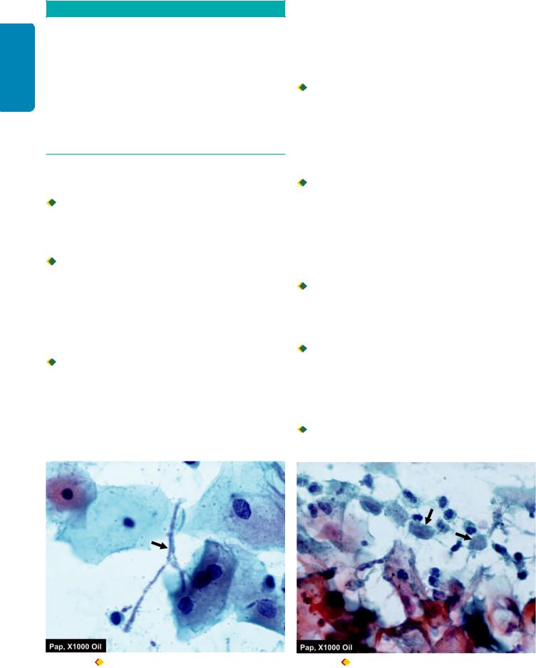
270
Techniques Basic and Pathology General I SECTION
TABLE 11.6: Common Infections Detectable on Pap Smears.
BACTERIAL AGENTS
Neisseria gonorrhoeae
Gardnerella vaginalis
Mycobacterium tuberculosis
VIRAL AGENTS
Herpes simplex virus (HSV)
Human papillomavirus (HPV)
FUNGAL AGENTS
Candida albicans
Torulopsis glabrata
PARASITIC AGENTS
Trichomonas vaginalis
Entamoeba histolytica
as tiny paired coffee-bean organisms. However, a definitive diagnosis requires bacterial culture.
 Gardnerella vaginalis, a gram-negative rod, is a frequent cause of bacterial vaginitis. It manifests in smears as blue stained cocco-bacillary organism covering and imparting a grainy appearance to affected squamous cells (so called ‘clue cells’) against a clean background devoid of Döderlein bacilli.

 Tuberculosis of the female genital tract usually manifests as tuberculous salpingitis or endometritis, but may involve the uterine cervix. While Mycobacterium tuberculosis cannot be identified in Papanicolaou stained smears, tuberculous granulomas composed of epithelioid macrophages with accompanying Langhans’ giant cells may occasionally be identified.
b) Viral agents:

 Human papilloma virus (HPV) is one of the commonest sexually-transmitted infections. There are 2 types of HPVs: low-risk and high-risk types. Low-risk HPV, most commonly types 6 and 11, are involved in causation of anogenital wart or condyloma acuminatum. High-risk HPV, most often types 16 and 18, are strongly implicated in precancerous lesions of ectocervical epithelium leading to cervical cancer. Morphologically, smears from HPV-infected subjects show
aggregates of rounded to oval squamous cells with blunted cell margins, peripheral cytoplasmic condensation and crisply outlined perinuclear halos, with dysplastic or degenerative changes of nuclei; these changes are collectively termed ‘koilocytotic changes.’ (see Fig. 11.4). HPV can also be identified by Southern blot, PCR, in situ hybridization and by hybrid capture.
 Genital infection by herpes simplex virus (HSV) commonly affects the squamous epithelium of the vulva, vagina and cervix, but may ascend to involve endocervical and endometrial epithelium. Infected cells show enlarged nuclei with watery, ‘ground-glass’ chromatin, multinucleation with internuclear moulding, and intranuclear acidophilic inclusions.
c) Fungal agents:
 Moniliasis (infection by Candida albicans) is the commonest fungal infection of the female genital tract and is particularly associated with diabetes, pregnancy and use of antibacterial agents (which alter the vaginal flora). Candida appears in smears in two forms—the yeast form (unicellular) appears as round to oval budding organisms with inconspicuous capsules, and the fungal form (pseudohyphae) as thin, elongated, pseudoseptate, bamboo-like filaments
(Fig. 11.2).
 Torulopsis glabrata causes less than 10% of mycotic vaginal infections. It is related to Candida, appears in smears as round budding organisms with thick capsule-like halos, and does not form pseudohyphae in vivo.
d) Parasitic agents:
 Up to 25% of adult women are estimated to harbour Trichomonas vaginalis in their lower genital tract. Smears from infected women may not show any cytomorphological changes or may show non-specific acute inflammatory changes. The protozoan appears in the background as a fuzzy, grey-green, round or elliptical structure 8 to 20 μm in size, containing a small vesicular nucleus (Fig. 11.3).

 Entamoeba histolytica in trophozoite form appears in Pap smears as basophilic, round to oval structures, 15 to 20 μm
Figure 11.2 |
Inflammatory smear with Candida (candidiasis, Figure 11.3 |
Trichomonas in inflammatory smear (arrow). |
moniliasis) (arrow). |
|
|
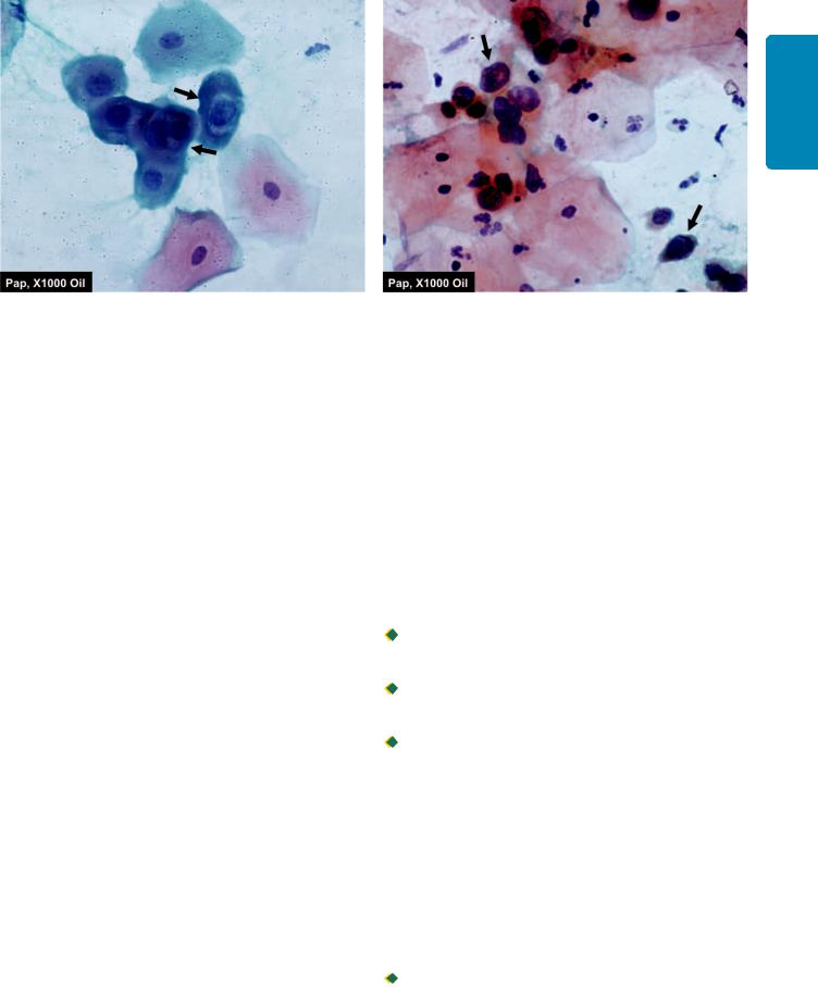
Figure 11.4 
 L-SIL. The smear shows koilocytes having abundant vacuolated cytoplasm and nuclear enlargement (arrow).
in size, with ingested erythrocytes and a round eccentric nucleus with a central karyosome.
NEOPLASTIC EPITHELIAL CELL ABNORMALITIES:
Carcinoma of the uterine cervix still ranks high in the list as the most frequent cancer in developing countries of the world and is the leading cause of cancer morbidity and mortality. Vast majority of cervical cancers are of the squamous cell type, and the diagnosis of squamous cell carcinoma of the cervix and its precursor lesions is considered as the most important application of exfoliative cytology.
Neoplastic epithelial changes are described below under 2 headings: squamous cell abnormalities and glandular cell abnormalities.
1. SQUAMOUS CELL ABNORMALITIES: The fullydeveloped invasive squamous cell carcinoma of the uterine cervix is preceded by a pre-invasive intraepithelial neoplastic process that is recognisable on histologic and cytologic examination.
Morphogenesis and nomenclature. The earliest recognisable change is hyperplasia of basal or reserve cells which normally constitute a single layer at the deepest part of the epithelium. The proliferating reserve cells next develop certain atypical features i.e. hyperchromasia and increased nuclear size. The continued proliferation of these atypical cells with loss of polarity, a concomitant increase in mitotic activity, and progressive involvement of more and more layers of the epithelium is known as dysplasia (disordered growth). When dysplasia involves the full thickness of the epithelium and the lesion morphologically resembles squamous cell carcinoma without invasion of underlying stroma, it is termed carcinoma in situ (CIS). CIS further evolves through the stage of microinvasive carcinoma (with depth of stromal invasion not exceeding 3 mm) into full-blown invasive
squamous cell carcinoma.
Previously depending on the degree of epithelial involvement, three grades of dysplasia were recognised: mild, moderate and severe. As the stages of dysplasia and CIS represented a continuous spectrum of lesions seen in
Figure 11.5 
 H-SIL. The squamous cells have scanty cytoplasm and markedly hyperchromatic nuclei having irregular nuclear outlines. The background shows numerous PMNs.
the precancerous state, they were collectively termed ‘cervical intraepithelial neoplasia’ (CIN) and categorised as under (also see Fig. 24.5):
CIN I |
Mild |
Primitive (atypical) cells, |
|
dysplasia |
proliferating in lower |
|
|
third of epithelium. |
CIN II |
Moderate |
Involvement up to |
|
dysplasia |
middle-third of epithelium. |
CIN III |
Severe |
Involvement of upper-third |
|
dysplasia |
of epithelium. |
|
Carcinoma |
Involvement of full |
|
in situ (CIS) |
thickness of epithelium. |
Presently, the Bethesda system divides squamous cell abnormalities into four categories:

 Atypical squamous cells of undetermined significance (ASCUS) which represents cellular changes falling short of intraepithelial lesion.
 Low-grade squamous intraepithelial lesion (L-SIL) that includes CIN-I and cellular changes associated with HPV infection.

 High-grade squamous intraepithelial lesion (H-SIL) includes CIN grade II, III and CIS.

 Squamous cell carcinoma.
Cytomorphology. Precancerous states can be distinguished from invasive carcinoma on the basis of cytomorphological features observed in smears. In dysplastic epithelium, stratification and maturation of cytoplasm occurs above the layers of proliferating primitive cells while the nuclear abnormalities persist. Cells exfoliating from the surface, thus, display cytoplasmic maturation and differentiation with nuclear atypia and are known as dyskaryotic cells.
The character and type of dyskaryotic cells observed in smears reflect the severity of dysplasia:

 In mild dysplasia (CIN-I or L-SIL), maturation occurs in the upper two thirds of the epithelium and exfoliated dyskaryotic cells are of the superficial type. These cells show
271
Cytology Diagnostic Basic 11 CHAPTER
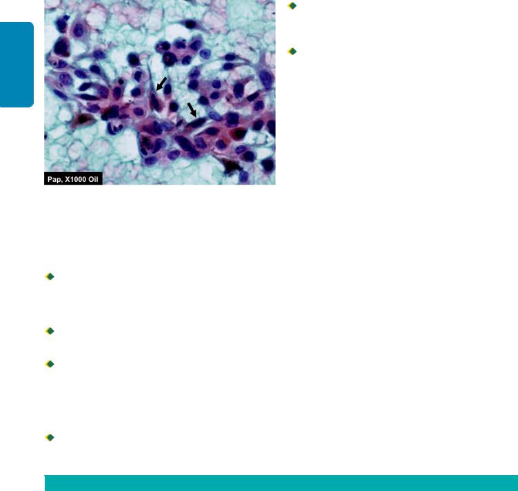
272
Techniques Basic and Pathology General I SECTION
Figure 11.6 
 Invasive squamous cell carcinoma. The malignant epithelial cells have anisonucleosis with irregular nuclear chromatin with prominent nucleoli. A few fibre cells and caudate cells are also seen
(arrow). The background shows abundant haemorrhage and some necrotic debris.
cytoplasmic vacuolation (koilocytosis) and nuclear enlargement (Fig. 11.4).

 With increasing dysplasia (CIN-II and CIN-III, or H-SIL), the proliferating primitive cells reach closer to the epithelial surface, less cytoplasmic maturation/differentiation occurs, and dyskaryotic intermediate and parabasal cells are observed in smears (Fig. 11.5).
 Progression to CIS (CIN-III or H-SIL) manifests as subtle alterations in cell arrangement and morphology (with predominantly basal and parabasal cells in smears).

 Onset of invasive carcinoma is heralded by the appearance of macronucleoli, cytoplasmic orangeophilia and presence of tumour diathesis (dirty, necrotic background)
(Fig. 11.6).
2. GLANDULAR CELL ABNORMALITIES. The Bethesda system categorises glandular abnormalities as under:

 Atypical glandular cells of undetermined significance (AGUS) which represent nuclear atypia of endocervical and endometrial cells exceeding reparative changes.
 Endocervical and endometrial adenocarcinoma, both of which can be detected from Pap smears; cytomorphological features allowing distinction between these two types of malignancies are summed up in Table 11.7.

 Cells from extrauterine cancers may also be present in Pap smears, majority originating from the ovaries. Clues to the extrauterine origin of cancer cells include: the absence of a tumour diathesis, arrangement of cancer cells in glandular and papillary configuration (accompanied by psammoma bodies in some instances), and absence of dysplastic changes in coexistent cervico-endometrial cells.
Automation in Cervical Cytology
Introduction of automated devices like PapNet for primary screening is a major technologic achievement in recent times. Automation offers routine pre-screening of hundreds of Pap smears, decreasing the workload of cytopathologists and at the same time providing quality assurance. It can be applied to conventional Pap smears or to Thin-Preps (discussed later).
II. RESPIRATORY TRACT
Cellular material from the respiratory tract may be obtained as a result of spontaneous expectoration (sputum), aspiration or brushing during bronchoscopic procedures.
1.SPUTUM EXAMINATION. Sputum is produced by a spontaneous deep cough bringing up material from small airways and alveoli. The cough reflex may also be triggered artificially by aerosol-inhalation of cough-stimulating substances (e.g. propylene glycol with hypertonic saline). Sputum examination is advantageous as samples are easily obtained and the cellular content is representative of the entire respiratory tract (satisfactory specimens allow diagnosis of over 80% of lung cancers). However, a large number of cells have to be scrutinised and the specimen does not allow localisation of the lesion within the respiratory tract.
2.BRONCHIAL ASPIRATION (WASHINGS) AND BRONCHIAL BRUSHING. Cellular material obtained by aspiration brushing or bronchial washing, or by bronchio-
TABLE 11.7: Cytomorphological Features of Endocervical and Endometrial Adenocarcinoma.
|
Feature |
Endocervical Carcinoma |
Endometrial Carcinoma |
|
|
|
|
1. |
Background |
Clean |
Dirty, bloody |
2. |
Cell yield |
High (found mainly on cervical smears) |
Low (found mainly on vaginal smears) |
3. |
Cell arrangement |
Clusters or sheets with ‘side-by-side’ grouping |
Three-dimensional ‘cell ball’ or grape-like clusters |
4.Cytoplasm
i) |
Amount |
Moderate (often in columnar configuration) |
Small |
ii) |
Character |
Granular, well-stained, often producing mucin |
Finely vacuolated, translucent, with ingested |
|
|
|
leucocytes |
5.Nuclei
i) |
Size |
Large |
Fairly small |
ii) |
Character |
Finely granular chromatin (mild hyperchromasia) |
Coarse chromatin (marked hyperchromasia) |
iii) |
Nucleoli |
Macronucleoli |
Micronucleoli |
|
|
|
|

alveolar lavage (BAL) during bronchoscopy allows localisation of lesions to specific areas of the respiratory tract. Moreover, specimens are easier to study as the cellularity is less than that of sputum. However, bronchoscopic procedures are unpleasant for patients, time-consuming and require considerable expertise.
III. GASTROINTESTINAL TRACT
Lesions in the oral cavity may be sampled by scraping the surface with wooden and metal tongue-depressors. For the oesophagus and stomach, samples are obtained under direct vision by brushing or lavage through fibreoptic endoscopes. For cytologic samples from the stomach, the older method of blind lavage through a Ryle’s tube using isotonic saline or Ringer’s solution have largely been replaced nowadays by direct mucosal visualisation by the endoscopist and collection of cytologic sample while doing a biopsy. Cytology samples from the colon may also obtained by brushing during colonoscopy or lavage following enema to clean the colon.
IV. URINARY TRACT
1.URINARY SEDIMENT CYTOLOGY. Cytological evaluation of the urinary tract is most often carried out by examining the sediment of voided or catheterised specimens of urine. It is convenient for the patient and a useful method for study of both the upper and lower urinary tracts, provided the samples are collected and processed in the correct manner (page 276). While voided specimens are satisfactory in men, catheterisation is often preferred in women to avoid contamination by vaginal cells and menstrual blood.
2.BLADDER IRRIGATION (WASHINGS). Washings of the urinary bladder obtained at cystoscopy are preferred in symptomatic patients when bladder tumours are suspected. The procedure provides excellent cytological preparations.
3.RETROGRADE CATHETERISATION. For suspected lesions of the upper urinary tract, voided urine is usually satisfactory. While renal parenchymal cells are infrequent in urine, material obtained from the renal pelvis and ureter contains adequate quantity of these cells. In some instances, retrograde catheterisation and brushing of the ureter and renal pelvis are utilised for localisation of lesions.
4.PROSTATIC MASSAGE. Prostatic secretions are obtained by prostatic massage and the sample is collected directly onto a glass slide and smeared. The procedure is rarely used nowadays with the advent of direct sampling of the prostate by FNAC.
Diagnostic Utility of Urinary Cytology
While evaluating the utility of urinary cytology in the diagnosis of urothelial tumours, following aspects need to be kept in mind:
1. Papillary tumours of low-grade are lined by urothelium showing no morphological abnormalities or only slight cellular and nuclear abnormalities. Such tumours cannot be diagnosed on cytologic material with any degree of certainty.
2.High-grade papillary tumours, sessile tumours and carcinoma in situ where urothelial cells exhibit cytomorphological abnormalities, are readily diagnosed by urinary cytology.
3.Urothelial tumours are often synchronous or metachronous and may involve different regions of the urinary tract.
Urinary cytology is, thus, of immense value in the follow-up of patients with previously diagnosed urothelial tumours.
V. BODY FLUIDS
For the sake of convenience, the cytology of body fluids can be discussed under following two headings:
A.Effusions in body cavities
B.Fluids of small volume.
A. EFFUSIONS
Effusion refers to the accumulation of fluid in any of the three body cavities (pleural, pericardial and peritoneal). An effusion in the peritoneal cavity is also known as ascites. Effusions have traditionally been classed as transudates or exudates. This distinction is important in diagnostic cytology as malignant effusions are invariably exudates with a protein content greater than 3 gm/dl (see Table 5.1, page 96).
Diagnostic cytology of effusions on samples obtained by paracentesis is mainly related to the identification of malignancy, and wherever possible, its classification. In benign effusions, cytological findings are mostly non-specific.
Cellular Components in Effusions
Two main primary component cells of effusions are mesothelial cells and macrophages or histiocytes. Effusion causes disruption of the mesothelial lining and these cells collect in the fluid individually or as small groups. Macrophages appear as mononuclear cells of the size of mesothelial cells distributed singly or as loose clusters. However, macrophages have ill-defined cell border compared to mesothelial and may have cytoplasmic vacuoles. Moreover, the nuclei in the macrophages are eccentric and kidney-shaped while those of mesothelial cells are central and round.
In addition, the effusion may have the following cellular components:
CELLS IN BENIGN EFFUSIONS. These include reactive proliferations of mesothelial cells in inflammation, polymorphonulcear neutrophils in acute suppurative inflammation, and lymphocytes in chronic fluid collections. When lymphocytes are dominant cells in the effusion fluid in fibrin-rich background, the possibility of tuberculosis is considered. In tuberculous effusions, granulomas, epithelioid cells or Langhans’ giant cells may not be seen; stain for tubercle bacilli may sometimes be of value.
CELLS IN MALIGNANT EFFUSIONS. Malignant cells in effusion may of origin from primary tumour (e.g. mesothelioma) or from secondary/metastatic tumour; the latter being more common the case.
Mesothelioma. It is uncommon and is more often epithelial type since fibrous mesothelioma does not exfoliate cells in
273
Cytology Diagnostic Basic 11 CHAPTER
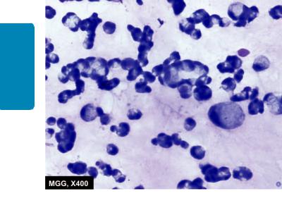
274
Techniques Basic and Pathology General I SECTION
Figure 11.7 
 Pleural fluid positive for signet ring cells (adenocarcinoma stomach). There are large number of malignant cells scattered singly or in small clusters having characteristic cytoplasmic vacuoles, nuclear hyperchromasia and prominent nucleoli.
the effusion. Epithelial cells of mesothelioma require to be distinguished from adenocarcinoma cells.
Adenocarcinomas. These are the most common malignant cellular component in the effusions. They mostly represent metastasis from primary adenocarcinomas such as from the stomach, lung, breast, colon, and ovary. Less frequent sources are adenocarcinoma of the biliary system, pancreas and liver. Most adenocarcinomas show similar morphologic appearance and thus it may not be possible to comment on the possible primary site of origin of the adenocarcinoma on morphology alone (Fig. 11.7).
Squamous cell carcinoma. Effusion may rarely have malignant squamous cells in it and represent metastasis from carcinoma lung, oesophagus or uterine cervix.
Small cell carcinoma. Cytologic features of small cell carcinoma of the lung are quite characteristic—hyper- chromatic moulded nuclei and scanty or no cytoplasm.
Lymphomas-leukaemias. Effusions may sometimes have malignant cells of leukameia and lymphoma in line with primary disease in the body.
B. FLUIDS OF SMALL VOLUME
Of the miscellaneous fluids included in this category, the most common samples submitted for cytological evaluation are cerebrospinal fluid (CSF) and seminal fluid (semen).
Cerebrospinal Fluid (CSF)
CSF examination is an important part of complete neurologic evaluation, both in non-neoplastic and neoplastic diseases of the central nervous system. Samples are usually obtained by lumbar puncture. Factors that are critical to the successful cytological evaluation of CSF are as under:
i) Speed of delivery to the laboratory and immediate processing, as diagnostic material may disintegrate within an hour resulting in false-negative diagnosis.
ii) Correct cytological processing and technique, since the cell yield of CSF is generally low and faulty technique may result in loss of cellular material or poor morphology.
Normal CSF. CSF is an ultrafiltrate of plasma with a total volume of about 150 ml (in the adult). Cell content is low (0-4/μl) comprising mainly lymphocytes or monocytes.
CSF in non-neoplastic diseases. Changes in CSF in various non-neoplastic diseases are discussed in Chapter 30 (see Table 30.1).
CSF in neoplastic disease. Neoplasms that are in direct contact with the CSF are most likely to shed cells which are recoverable for scrutiny. Metastatic cancers (leukaemia, lymphoma and adenocarcinoma), medulloblastomas, and ependymomas shed the greatest amount of material. Though meningioma occurs in the subarachnoid space, it rarely exfoliates sufficient material for diagnosis. Low-grade gliomas do not generally shed sufficient cells although highgrade gliomas may yield adequate cellular material for diagnosis.
Seminal Fluid (Semen)
Examination of seminal fluid (semen analysis) is one of the tests for investigating infertile couples, and is also used to check the adequacy of vasectomy. Samples are obtained by masturbation or coitus interruptus after observing at least 4 days of sexual abstinence and assessed on the following lines:
1.Volume. Normal volume is between 2.5 and 5 ml.
2.Viscosity and pH. When ejaculated, semen is fairly viscid but liquefies in about 10 to 30 minutes. It is usually alkaline (pH about 8).
3.Motility. Normally, within 2 hours of ejaculation, at least 60% of the spermatozoa are vigorously motile; in 6 to 8 hours
25to 40% are still motile.
4.Count. Counting is done in a Neubauer chamber after suitable dilution. Normally, 60 million or more spermatozoa are present per ml. Currently, automated counters for semen are also available.
5.Morphology. Stained smears are used to assess morphology. Normally, not more than 20% of spermatozoa are morphologically abnormal (e.g. double-head, pointedhead, or swollen).
6.Fructose. Seminal fructose estimation (normal levels 150-
600mg/dl) complements cytological analysis. Low levels of seminal fructose indicate obstruction at the level of ejaculatory ducts.
Results of semen analysis need to be interpreted with caution as infertility is caused by several factors in the couple. As discussed in Chapter 23, other causes of infertility need to be evaluated before it is ascribed to abnormalities in semen. Even specimens with several abnormal features need not necessarily be the cause of infertility.
VI. BUCCAL SMEARS FOR SEX CHROMATIN BODIES
Sex-specific chromatin bodies are observed in interphase nuclei and comprise the Barr body (X chromatin) and the
fluorescent or F body (Y chromatin). The number of Barr bodies observed in interphase nuclei is one less than the number of X chromosome (n–1), whereas the number of F bodies observed is equal to the number of Y chromosomes present (n). In effect, the Barr body is specific for females and the F body for males (page 257).
1.DEMONSTRATION OF BARR BODY (X CHROMATIN). In buccal smears, the Barr body appears as a planoconvex mass about 1 μm in diameter applied to the inner surface of the nuclear membrane. The interphase nuclei of one hundred intermediate squamous epithelial cells are scrutinised. In normal females, Barr bodies are present in at least 20% of nuclei (Fig. 11.8, A). Males have a Barr body count of less than 2%. Vaginal smears may also be used for Barr body counts (page 257).
2.DEMONSTRATION OF THE F BODY (Y CHROMATIN). Demonstration of the F body requires fluorescent staining in contrast to the Barr body which may be observed on routine Papanicolaou-stained smears. On staining buccal smears with quinacrine mustard, the intensely fluorescent F body is observed in about 60% of interphase nuclei in males and in less than 8% of nuclei in females. The F body is also demonstrable in the nuclei of lymphocytes in a peripheral blood film stained with quinacrine mustard.
3.COUNTING OF DRUMSTICK APPENDAGE. The presence and frequency of drumstick appendages attached to nuclei of polymorphonuclear leucocytes on a peripheral blood film may also be used for determining sex. At least
500neutrophilic leucocytes are scrutinised in a Romanowskystained blood film. Genetic females show drumsticks in 3- 6% of neutrophils (Fig. 11.8, B). In males, the frequency of drumsticks is less than 0.3%.
TECHNIQUES IN EXFOLIATIVE CYTOLOGY
A brief resume of methods for collection of specimens and processing of samples for exfoliative cytodiagnosis is outlined below under 4 headings: collection, fixation and fixatives, processing in the laboratory, and staining.
A. Collection of Samples
1. PREPARATION OF COMBINED (FAST) SMEARS.
Smears should not be taken during menstrual bleeding. The patient should not douche for at least 24 hours before the smear is obtained.
i)Smears are obtained under direct vision after introducing a Cusco’s speculum with the patient in lithotomy position.
ii)Ideally, lubricants and medical jellies should not be used for introducing the speculum since use of lubricants results in contamination of the smear. If required, the speculum may be moistened with a few drops of normal saline.
iii)The posterior fornix of the vagina is aspirated with a blunt-ended glass pipette fitted with a rubber bulb. A drop of the aspirate is placed at the unlabelled end of a glass slide.
Figure 11.8 
 Sex chromatin. A, Buccal smear showing condensation of chromatin under the nuclear membrane. B, Blood film showing presence of drumstick appendage attached to a nuclear lobe of the neutrophil.
iv)The ectocervix is sampled with the Ayre’s spatula (Fig. 11.9). The longer limb of the spatula is fitted into the external os and the spatula rotated through 360° to sample the entire cervix. The scraped material on the spatula is then placed on the drop aspirated from the vaginal pool and the smear prepared with the spatula itself or with the tip of the gloved finger (cotton swabs may be used instead of the Ayre’s spatula, although the quality of smears is not as good).
v)Thin uniform smears should be prepared and the slide immediately immersed in fixative to avoid artefacts in cells caused by drying.
vi)Smears should be transported to the laboratory in the fixative in a Coplin’s jar (discussed later).
Slides should ideally be labelled by using a diamond pencil. If labels of sticking plaster are used, the labels must not come into contact with the fixative.
2.LIQUID-BASED CYTOLOGY PREPARATIONS (THIN PREPS). This is a special technique for preparation of gynaecologic and non-gynaecologic samples which provides uniform monolayered dispersion of cells on smears, without overlapping or clump formation. It is a pre-requisite for quantitative analysis and automated devices.
3.PREPARATION OF LATERAL VAGINAL SMEARS (LVS). The LVS is obtained by scraping the lateral walls of the upper third of the vagina (at the level of the cervical external os) with the flat surface of a wooden tongue depressor and smearing the material directly onto labelled slides. The patient is prepared and positioned as described for the combined (fast) smear.
4.COLLECTION OF SPUTUM. Fresh, unfixed, early morning specimens resulting from overnight accumulation of secretions are best for diagnostic purposes. A minimum of at least three specimens collected on three successive days should be examined. The patient is instructed to cough deeply on waking up and to expectorate all sputum into a
275
Cytology Diagnostic Basic 11 CHAPTER
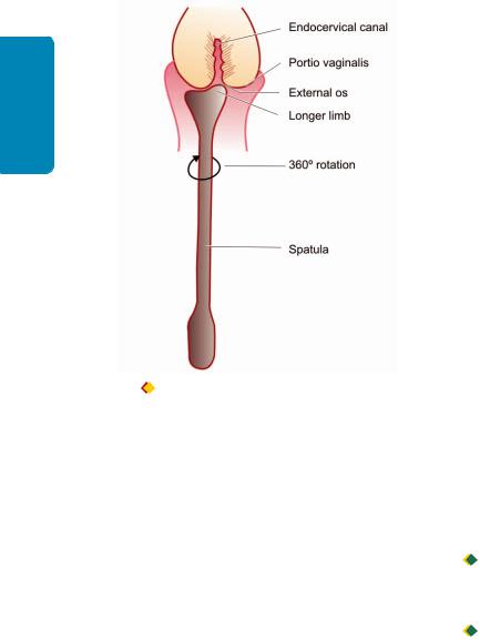
276
Techniques Basic and Pathology General I SECTION
Figure 11.9 
 Method of obtaining cervical smear with Ayre spatula (fast smear).
clean, dry, wide-mouthed glass container or a petridish. Care should be taken to avoid spitting into the container. The container is capped or covered, labelled and transported to the laboratory where smears are prepared.
5.BRONCHIAL MATERIALS. All aspirated bronchial secretions, lavage, washings and brushings must be despatched to the laboratory without delay. If immediate despatch is not possible, the sample should be collected in fixative (50% ethanol in volumes equal to that of the sample).
6.BUCCAL SMEARS. The mouth is rinsed with water or normal saline and the buccal mucosa scraped vigorously with a wooden or metal tongue depressor. The material is smeared directly onto labelled glass slides which are placed in fixative.
7.G.I. CYTOLOGY. In a case of suspected malignancy or specific infections of GI tract, or for screening for Barrett’s oesophagus, cytologic examination is indicated. The cytology specimen is collected during fibreoptic endoscopy of the part being visualised.
8.URINE. Fresh catheterised specimens are preferred in female patients while voided urine is satisfactory in males.
After initial morning voiding (which is discarded), samples of about 50 to 100 ml are collected on three consecutive days. Hydration by forced intake of fluids (1 glass of water every 30 minutes over 3 hour period) is recommended by some workers for production of high volume specimens. If delay is anticipated in despatch to the laboratory, the sample should be collected in an equal volume of 50% ethanol.
The initial morning specimen is discarded as cells deteriorate extremely quickly in acidic urine, and the morphology of cells accumulating overnight in bladder urine is distorted to an extreme degree. For the same reason,
24-hour collections of urine are useless for cytodiagnostic purposes.
9.EFFUSIONS. Pericardial, pleural and peritoneal fluids are obtained by paracentesis. As these fluids are often exudative in character, they may clot after removal from respective cavities. Anticoagulants may be used to prevent coagulation—heparin (3-5 units per ml), 3.8% sodium citrate (1 ml per 10 ml), or EDTA (1 mg per ml) may be used for this purpose.
If the fluids cannot be processed within 12 hours of collection, an equal volume of 50% ethanol or 10% formalin should be added.
10.CEREBROSPINAL FLUID (CSF). CSF samples should be despatched without delay to the laboratory for immediate processing as the cells contained are extremely fragile. A gap of even 1 hour between removal and processing may result in loss of diagnostic cellular material.
11.SEMEN. Samples of seminal fluid obtained by masturbation are best collected at the laboratory. Samples obtained by coitus interruptus are collected in clean, dry test tubes (or vials) and transported to the laboratory within 30 minutes. The patient is instructed to note the time of ejaculation.
B. Fixation and Fixatives
All material for cytological examination must be properly fixed to ensure preservation of cytomorphological details. Methods of fixation vary depending upon the type of staining employed:
 Material for exfoliative cytodiagnosis is usually wet-fixed i.e. smears are immersed in fixative without allowing them to dry. These smears are then stained with Papanicolaou (Pap) or haematoxylin and eosin (H & E) stains.

 Sometimes, exfoliative cytology smears are air-dried for use with the Romanowsky stains as are used in haematologic studies. In Romanowsky staining, fixation is effected during the staining procedure.
1.ROUTINE FIXATIVES. The ideal fixative for routine use is Papanicolaou’s fixative comprising a solution of equal parts of ether and 95% ethanol. However, the flammability of ether makes it hazardous. Most laboratories use 95% ethanol alone with excellent results. Where ethanol is not available, 100% methanol, 95% denatured alcohol, or 85% isopropyl alcohol (isopropanolol) may be used.
Smears prepared at the bedside as well as those prepared in the laboratory from fluid samples are immediately placed in 95% ethanol without allowing them to dry prior to fixation.
Drying causes distortion of cells and induces cytoplasmic staining artefacts. Fixation time of 10 to 15 minutes at room temperature is adequate. Smears may also be left in the fixative for 24 hours or more without any detrimental effect as regards cytomorphological detail. Smears should be transported to the laboratory in the fixative; screw-capped Coplin jars are best for this purpose.
2.COATING FIXATIVES. Coating fixatives are applied as aerosol-sprays or with a dropper to the surface of freshly

prepared smears. While commercial preparations (e.g. cytospray) are available, a standard hairspray with a high alcohol content is also a suitable alternative. Smears while still wet, are placed face-up on a table and sprayed with the nozzle held at a distance of 10 to 12 inches. A period of 10 minutes is sufficient for drying of coated smears, which may then be wrapped or put in a box for transport to the laboratory. Coating fixatives are ideal when unstained smears are to be transported over long distances.
3.SPECIAL PURPOSE FIXATIVES. Buffered neutral formalin, Bouin’s fluid and picric acid are used for specific purposes when required. Formalin vapour fixation is also employed for some staining procedures.
4.PRESERVATION OF FLUID SAMPLES. Samples of fluids are best submitted to the laboratory in a fresh state for immediate processing. Where a delay is anticipated in despatch to the laboratory or in processing, the sample is collected in a suitable preservative for ‘prefixation’ so that cellular morphology is preserved. When the samples have been processed and smears prepared, ‘fixation’ is effected as described earlier.
Fluid samples with a high protein content (e.g. pericardial, pleural and peritoneal fluids) may be preserved under refrigeration for up to 12 hours without prefixation. However, fluids with a low protein content (e.g. urine and cerebrospinal fluid) deteriorate in 1 to 2 hours even if refrigerated. Fluids with low pH (e.g. gastric lavage samples) must be collected in pre-cooled preservative since cells deteriorate extremely rapidly in such specimens.
The best preservative for general use is 50% ethanol in volumes equal to that of the fluid sample. Ninety-five per cent ethanol precipitates proteins and hardens the sediment making smear preparation difficult; it is used only for gastric aspirates. Pericardial, pleural and peritoneal fluids may be pre-fixed with an equal volume of 10% formalin if ethanol is not available. Solutions containing ether and acetone are not used as preservatives as they cause extreme hardening of the sediment making smear preparation virtually impossible.
C. Processing of Samples in the Laboratory
1.PREPARED SMEARS. Smears prepared at the bedside and wet-fixed in ethanol need no further processing in the laboratory prior to staining.
2.SPUTUM. The sample is prepared as under:
i)The sample is placed in a petridish and inspected against a dark background.
ii)Bloody, discoloured or solid particles are removed with wooden applicator sticks and placed on glass slides. Strands of ropy mucus are also selected (exfoliated cells adhere to mucus strands).
iii)Clean glass slides are used to crush the particles/mucus and spread the material evenly.
iv)Four such smears are prepared and immediately placed in 95% ethanol for fixation.
3.FLUID SPECIMENS. Large volumes of fluid received are allowed to stand in the refrigerator for half to one hour.
After that, all except the bottom 200 ml is discarded. The |
277 |
|
retained fluid is then processed. In specimens containing |
|
|
excessive amounts of blood, erythrocytes may be lysed by |
CHAPTER |
|
the addition of 1 ml of glacial acetic acid for every 50 ml of |
||
|
||
specimen. |
|
|
Commonly employed techniques for processing of fluids |
|
|
are as under: |
|
|
Sediment smears. The sample is poured into 50 ml centrifuge |
11 |
|
tubes and centrifuged at 600 g for 10 minutes. (To achieve a |
||
relative centrifugal force of 600 g, the speed of the centrifuge |
|
|
in revolutions per minute varies with the rotating radius of |
Basic |
|
speed is 1500 rpm). |
||
the centrifuge. With a rotating radius of 25 cm the required |
|
|
Following centrifugation, the supernatant is decanted and |
Diagnostic |
|
preservative require albuminised slides as cell adhesiveness |
||
smears prepared from the sediment or cell button by |
|
|
recovering the material with a glass pipette or a platinum |
|
|
wire loop. Smear preparation from samples collected in a |
|
|
is reduced by prefixation. Smears are wet-fixed in 95% |
Cytology |
|
methods are most useful for small volume fluids of low cell |
||
ethanol (or air-dried in some instances). |
|
|
Cytocentrifuge and membrane filter preparations. These |
|
|
content. The interested reader is referred to specialised texts |
|
|
for descriptions of these methods. |
|
|
D. Staining of Smears |
|
|
Three staining procedures are commonly employed: |
|
|
Papanicolaou and H&E stains are used for wet-fixed smears |
|
|
while Romanowsky stains are used for air-dried smears. |
|
1.PAPANICOLAOU STAIN. Papanicolaou staining is the best method for routine cytodiagnostic studies. Three solutions are used comprising a nuclear stain and two cytoplasmic counter-stains. Harris’ haematoxylin is the nuclear stain. Orange G (OG-6) and eosin-alcohol (EA-65 or EA-50) are the two cytoplasmic counterstains which impart the orange and cyanophilic tints to cytoplasm respectively.
2.H & E STAIN. The stain is essentially the same as that used for histological sections. Harris’ haematoxylin is the nuclear stain, and eosin is the cytoplasmic counterstain.
3.ROMANOWSKY STAINS. Romanowsky stains used in haematological preparations may also be used for cytological smear preparations. Leishman’s, May-Grünwald-Giemsa (MGG) and Wright’s stains are most commonly used.
INTERVENTIONAL CYTOLOGY
In interventional cytology, samples are obtained by aspiration or surgical biopsy. This branch includes fine needle aspiration cytology, imprint cytology, crush smear cytology and biopsy sediment cytology.
I. FINE NEEDLE ASPIRATION CYTOLOGY
Interventional cytology is virtually synonymous with Fine Needle Aspiration Cytology (FNAC). The technique has gained wide acceptance in the last four decades and is increasingly being used to sample a wide variety of body
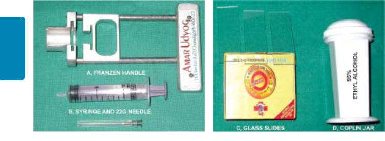
278
Techniques Basic and Pathology General I SECTION
Figure 11.10 
 Equipments required for transcutaneous FNAC.
tissues. Almost all organ systems are accessible to the fine needle and versatility of the technique has enormously increased the scope of diagnostic cytology. The applications and advantages of FNAC, its procedure, complications and limitations are detailed below.
A. APPLICATIONS OF FNAC
In routine practice, FNAC is most often used for diagnosis of palpable mass lesions. Palpable lesions commonly sampled are: breast masses, enlarged lymph nodes, enlarged thyroid and superficial soft tissue masses. The salivary glands, palpable abdominal lesions and the testicles are also frequently sampled for FNAC. Other sites and lesions accessible to FNAC are the prostate, pelvic organs, bone and joint spaces, lungs, retroperitoneum and orbit.
B. ADVANTAGES OF FNAC
i)FNAC is an OPD procedure and requires no hospitalisation while surgical biopsies are obtained in the operation theatre and hospitalisation is often required.
ii)No anaesthesia is required (except in specific circumstances), while surgical biopsy is performed under local or general anaesthesia.
iii)The procedure is quick, safe and painless.
iv)Multiple attempts (or repeating the procedure) are possible without inconvenience, whereas repeating a surgical biospy is uncomfortable and inconvenient for the patient.
v)Results are obtained rapidly with reports being available in a matter of hours (turn-around time of 2 to 24 hours depending on the urgency), while histopathological reports are available after a longer duration (turn-around time of 2 to 4 days on account of time required for processing and sectioning of tissues).
vi)It is a low-cost procedure which is cost-effective.
vii)As the cytopathologist performs the procedure himself, he gains first-hand knowledge of the clinical findings which facilitates interpretation of slides and enhances diagnostic accuracy.
C. GENERAL PROCEDURE FOR FNAC
Materials
For performing FNAC, a syringe with a well-fitting needle, syringe holder, some microscopic glass slides and a suitable fixative are the only material required in most instances
(Fig. 11.10):
NEEDLES. Fine needles range from 25 to 20 gauge (0.6 mm to 0.9 mm outer diameter). The standard 21 gauge disposable needle of 38 mm length is suitable for routine transcutaneous FNAC of palpable masses; 25 or 24 gauge disposable needles of 25 mm length are used for lymph nodes and in children. Larger needles, 80 to 160 mm in length, are required for sampling the lung and abdominal viscera; 22 to 20 gauge Chiba spinal puncture needles may be profitably employed for this purpose. Needles of up to 200 mm length are used for transrectal and transvaginal FNAC of the prostate and ovary respectively. Aspiration of bony lesions may require 18 gauge (1 mm outer diameter) needles although superficial lytic lesions are adequately sampled with a 21 gauge needle.
SYRINGES. Syringes of 10 to 20 ml capacity are suitable. Syringe holders such as the Franzen handle permit a singlehand grip during aspiration, employing disposable syringes. For needles with metal hubs (e.g. spinal puncture needles), disposable syringes should be used to ensure a proper fit between the needle hub and syringe nozzle.
GLASS SLIDES AND FIXATIVE. Four or six standard microscopic glass slides and a Coplin jar containing 95% ethyl alcohol (as a fixative) are the only other material required for routine FNAC.
Method of Aspiration
Transcutaneous FNAC of palpable masses is routinely performed without anaesthesia as per the following procedure; aspiration of sites/lesions requiring anaesthesia or special technique are discussed separately (Fig. 11.11):
i) The patient is asked to lie down in a position that best exposes the target area.
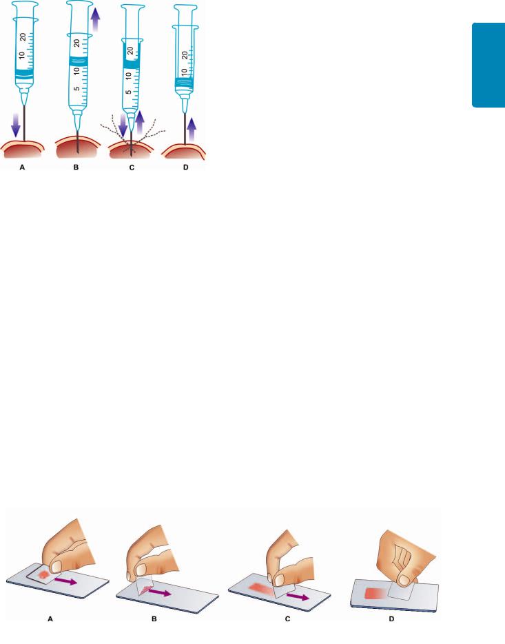
Figure 11.11 
 Procedure for FNAC of palpable masses. Needle is introduced into the mass (A). Plunger is retracted after needle enters the mass (B). Suction is maintained while needle is moved back and forth within the mass (C). Suction is released and plunger returned to original position before needle is withdrawn (D).
ii)The target area is thoroughly palpated and the firmest portion of the lesion or mass delineated.
iii)The skin is cleaned with an alcohol pad.
iv)The mass is fixed by the palpating hand of the operator or by an assistant; gloves may be used for protection of the operator and the assistant.
v)The needle is inserted into the target area. On reaching the lesion, the plunger of the syringe is retracted and at least 10 ml of suction applied while moving the needle back and forth within the lesion; the direction or angle of the needle may be changed to access different areas of the lesion.
vi)Aspiration is terminated when aspirated material or blood becomes visible at the base or hub of the needle. For diagnostic purposes, cellular material contained within the needle is more than adequate; material drawn into the barrel of the syringe is not recovered since it is of no use for cytologic diagnosis.
vii)On completion of aspiration, suction is released and pressure within the syringe allowed to equalise before withdrawing the needle. Withdrawing the needle with negative pressure or suction results in blood being aspirated and cellular material being sucked into the barrel of the syringe, thus lost to interpretation.
viii)Following withdrawal of the needle from the lesion, pressure is applied to the site of puncture by the assistant or patient himself for 2 to 3 minutes in order to arrest bleeding and prevent haematoma formation.
ix)Aspirated material is recovered by detaching the needle from the syringe and filling the syringe with air; the syringe and needle are then reconnected and the aspirate expressed onto one end of a glass slide.
Preparation of Smears
Preparation of smears is crucial to the success of FNAC. Poorly-prepared smears with distorted cellular morphology will frustrate the best efforts of the most competent cytopathologist, and often result in errors of interpretation or in failure to arrive at any specific diagnosis. The procedure consists of the following steps (Fig. 11.12):
i)Aspirates deposited on the slide are inspected with the naked eye.
ii)Semisolid particulate aspirates are crush-smeared by flat pressure with a glass slide or a thick coverslip. Even and gentle pressure is required to avoid traumatising cells. Droplets of fluid or bloody material are gathered under the edge of the angled smearing slide or coverslip and pulled like a blood smear. Particulate material, which collects along the edges and at the end of the smear, is then crush-smeared by flattening the smearing slide.
iii)Prepared smears are either wet-fixed or air-dried. Half the number of smears are immediately immersed in 95% ethanol, transported to the laboratory in the fixative, and used for Papanicolaou or H&E staining. The remaining smears are air-dried, wrapped in tissue paper for transport to the laboratory, and stained by Romanowsky stains (e.g. Leishman’s, May-Grünwald-Giemsa). Most cytopathologists use both wet-fixed and air-dried smears—the wet-fixed smears provide excellent nuclear detail while the air-dried smears yields information about the cytoplasm and the background. The general properties of wet-fixed and air-dried smears outlined in Table 11.8.
Special and Ancillary Studies
Aspirates may also be studied by special stains and techniques for specific purposes as under:
279
Cytology Diagnostic Basic 11 CHAPTER
Figure 11.12 
 Preparation of smears. Semisolid aspirates are crush-smeared by flat pressure with cover slip or glass slide (A). Fluid or blood droplet is collected along edge of spreader (B), and pulled as for peripheral blood films (C). Particles at the end of the smear are crush-smeared (D).

280
Techniques Basic and Pathology General I SECTION
TABLE 11.8: General Properties of Wet-Fixed and Air-Dried Smears.
|
Feature |
Wet-fixed |
Air-dried |
|
|
|
|
1. |
Staining |
Papanicolaou, H&E |
Leishman, May-Grünwald-Giemsa |
2. |
Cell size |
Comparable to tissue sections |
Exaggerated |
3. |
Nuclear detail |
Excellent |
Fair |
4. |
Nucleoli |
Well demonstrated |
Not always discernible |
5. |
Cytoplasmic details |
Poorly demonstrated |
Well demonstrated |
6. |
Stromal components |
Poorly demonstrated |
Well demonstrated |
7. |
Partially necrotic material |
Single intact cells, well defined |
Cell details poorly defined |
|
|
|
|
i)SPECIAL STAINS. Wet-fixed smears are used for a variety of special stains such as Alcian blue, mucicarmine and PAS (for mucin and carbohydrates); methyl violet or congo red (for amyloid); and bacterial and fungal stains (for microbial agents).
ii)MICROBIOLOGICAL STUDIES. Aspirates may also be submitted for viral, fungal, mycobacterial and bacterial culture. When infection is suspected, an additional aspirate is obtained and expressed into a sterile culture tube. If additional aspiration is not feasible, the needle is flushed with sterile isotonic saline or foetal calf serum and the rinsed fluid submitted for microbial culture.
iii)CELL BLOCK. Aspirated material may be processed as surgical pathology material by preparing paraffin blocks from cell button of the centrifuged deposit. Sections provide recognition of histologic patterns and can also be used for ancillary techniques.
iv)IMMUNOCYTOCHEMICAL STUDIES. The smears or cell block sections can be further evaluated by immunocytochemical stains by employing panel of antibodies selected on the basis of differential diagnosis made after routine morphologic examination of smears.
v)IMAGE ANALYSIS AND MORPHOMETRY. These techniques when applied to cytological smears bring quantitation and objectivity to cytodiagnosis. They determine the cellular parameters like N/C ratio, nuclear area, shape and size of nuclei and nucleoli etc.
vi)FLOW CYTOMETRY. Determination of ploidy status and S phase fraction of tumour cells using flow cytometry enhances the diagnostic and prognostic information available on routine cytology.
vii)ULTRASTRUCTURAL STUDIES. Aspirates obtained by FNAC are also suitable for electron microscopy (both TEM and SEM).
viii)MOLECULAR BIOLOGIC TECHNIQUES. These techniques are now being widely applied to cytopathology also. Detection of oncogenes like ERBB-2 in breast cancer, BCL-2 in lymphomas, DNA sequence analysis, gene expression profiling and fluorescence in situ hybridisation (FISH) can all be used for aspiration samples.
Aspiration of Specific Lesions/Body Sites
In addition to the general procedure for FNAC described above, aspiration from certain specific sites/lesions
require greater expertise and additional material as outlined below.
i)CYSTS. Cysts of the neck, thyroid, breast and other sites are often encountered during FNAC. During aspiration, the entire fluid content is evacuated by drawing into the syringe and collected in test tube for centrifugation and smear preparation. If a residual mass is palpable after removal of fluid, a fresh syringe is used for additional aspiration of the mass in the usual manner.
ii)THYROID. Lesions of the thyroid are aspirated with the patient either sitting up or lying supine with the neck extended. Solitary nodules are fixed between two palpating fingers, while a diffusely enlarged lobe is fixed by asking the patient to swallow and applying two fingers to the base of the lobe to hold it against the trachea. The patient is asked to avoid swallowing during aspiration. Subsequently, firm pressure is applied to the puncture site for about 3 minutes to prevent haematoma formation.
iii)LUNG AND RETROPERITONEUM. FNAC of these two sites is usually carried out under the guidance of radiological imaging techniques. Local anaesthesia (1% xylocaine) is advisable with infiltration of skin and deeper tissues. Spinal puncture needles (22 or 20 gauge) are used. Since these long needles are flexible they are rotated during insertion to enable an accurate approach to the target lesion.
iv)PROSTATE. The prostate is sampled transrectally during per-rectal examination with the patient in lithotomy position. A custom-made or commercially available needle guide (such as the Franzen needle guide) may be used. Alternatively, a 16 gauge blunt-tipped venous cannula may serve as a needle guide. The cannula is taped to the index finger of the gloved hand with its tip just proximal to the pulp of the index-finger, and a fingercot drawn over it. The prostatic lesion is palpated with the pulp of the finger and sampled by puncturing through the fingercot with a long 22 gauge needle introduced through the cannula.
v)TESTIS. Local anaesthesia is advisable when FNAC of the testis is carried out for infertility as pain may be considerable when suction is applied. Spermatic cord block is employed for the purpose. Infiltration of scrotal skin is not required. For suspected testicular tumours, local anaesthesia is not required; however, thinner needles (25 or 24 gauge) are advocated by some workers to avoid seeding of tumour along the needle track.

vi) ABDOMINAL FAT ASPIRATION. FNA of the paraumbilical abdominal adipose tissue is currently accepted method for diagnosis of secondary systemic amyloidosis. Amyloid is demonstrated as rings around fat cells by the conventional Congo red staining (congophilia) and applegreen birefringence when viewed under polarising microscopy.
Radiological Imaging Aids for FNAC
Non-palpable lesions require some form of localisation by radiological aids for FNAC to be carried out. Plain X-ray films are usually adequate for lesions within bones and for some lesions within the chest. FNAC of the chest may also be attempted under image amplified fluoroscopy which allows visualisation of needle placement on the television monitor.
Computerised tomographic-(CT) guidance is also used for lesions within the chest and abdomen. The most versatile radiological aid is ultrasonographic (US)-guidance which allows direct visualisation of needle placement in real time and is free from radiation hazards. It is an extremely valuable aid for FNAC of thyroid nodules, soft tissue masses, intra-abdo- minal lesions and for intrathoracic lesions which abut the chest wall, but is of no help in deep intrathoracic lesions or in bony lesions.
D. COMPLICATIONS AND HAZARDS OF FNAC
FNAC is associated with relatively few complications. Possible hazards and more commonly encountered complications are as follows:
1.Haematomas. Bleeding from the puncture site and haematoma formation are the commonest complications of the procedure, particularly in the breast and the thyroid. Firm finger pressure for 2 to 3 minutes immediately after the procedure greatly reduces the frequency of these complications.
2.Infection. Introduction of infection is not a significant hazard; even transabdominal aspiration does not result in peritoneal contamination despite puncture of bowel walls. Transrectal aspiration in cases of acute prostatitis may, however, result in bacteraemia and septicaemia.
3.Pneumothorax. Transcutaneous aspiration of the lung causes pneumothorax in about 20% of cases; most resolve spontaneously although intercostal intubation may be required in some instances. Transient haemoptysis may also be associated with lung aspiration.
4.Dissemination of tumour. Generalised dissemination of malignant cells via lymphatics and blood vessels following FNAC is a theoretical possibility but no definite instances have been recorded. Local dissemination by seeding of malignant cells along the needle tract is a rare complication and has been reported in cancers of the lung, prostate and pancreas. Aspiration of malignant ovarian cysts may result in release of cyst contents into the peritoneal cavity with peritoneal implants.
E. PRECAUTIONS AND CONTRAINDICATIONS OF FNAC
While FNAC is generally a safe procedure, precautions have to be taken when aspiration is contemplated of some sites under certain circumstances:
1.Bleeding disorders. Thrombocytopenia per se is not a contraindication to FNAC. In patients with coagulopathies such as haemophilia, aspiration of joint spaces, chest and abdominal viscera is contraindicated; superficial lesions may be aspirated and pressure applied to the puncture site for at least 5 minutes following the procedure.
2.Liver. Estimation of prothrombin time is an essential prerequisite for aspiration of the liver. FNAC is not advisable if prothrombin index (PTI) is less than 80%. Obstructive jaundice is a relative contraindication for FNAC on account of the risk of bile peritonitis.
3.Lung. FNAC of the lung should not be undertaken in elderly patients with emphysema or pulmonary hypertension because of the enhanced risk of pneumothorax and haemoptysis respectively.
4.Pancreas. FNAC is contraindicated in acute pancreatitis as it aggravates the inflammatory process.
5.Prostate. Transrectal aspiration in acute prostatitis may cause bacteraemia/septicaemia and is contraindicated.
6.Testis. Aspiration is extremely painful in acute epididymo-orchitis and should be deferred till such time the acute inflammatory process subsides. The patient is treated with anti-inflammatory agents and antibiotics and FNAC undertaken at a later date.
7.Adrenal. FNAC of a suspected pheochromocytoma is inadvisable as it may sometimes provoke extreme fluctuations in blood pressure.
F. CYTOLOGIC DIAGNOSIS
The cytopathologist can render a preliminary diagnosis within one hour after the FNAC procedure when urgently required. Basic cytologic features in FNAC are similar to those in histopathology but smear cytology depends upon the technique for smear and stains employed. Emphasis in FNA cytology is on pattern recognition or arrangement of cells, nuclear and cytoplasmic features of individual cells or groups of cells, and comment on the background morphology.
Understandably, it is beyond the scope of this book to delve into morphological details of various lesions which can be sampled by FNAC; interested readers in this subject are referred to the specialised texts listed at the end of the book. However, a few common and classical examples of FNAC applications in lymph node (tuberculous lymphadenitis, Fig. 11.13), thyroid (follicular neoplasm, Fig. 11.14), breast (fibroadenoma and infiltrating carcinoma, Figs. 11.15 and 11.16), abdominal fat (for amyloid, Fig. 11.17) and cell-block preparation along with immunocytochemistry (Fig. 11.18) are illustrated.
281
Cytology Diagnostic Basic 11 CHAPTER
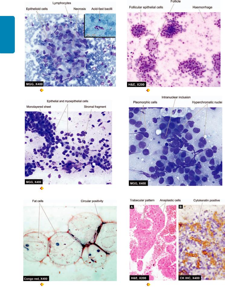
282
Techniques Basic and Pathology General I SECTION
Figure 11.13 |
FNA lymph node showing tuberculous lymphadenitis. Figure 11.14 |
FNA thyroid showing follicular neoplasm. |
Inbox shows Ziehl-Neelsen staining having many tubercle bacilli. |
|
|
Figure 11.15  FNA breast lump showing features of fibroadenoma. Figure 11.16
 FNA breast showing features of infiltrating ductal carcinoma.
Figure 11.17 |
Abdominal fat aspirate stained with Congo red stain Figure 11.18 |
A, Section of cellblock preparation showing hepato- |
for amyloid. |
cellular carcinoma. B, Same cellblock, stained with cytokeratin for |
|
|
epithelial cells. |
|

G. LIMITATIONS OF FNAC
The main limitation of FNAC lies in the fact that only a small population of cells is sampled by the procedure. The reliability of the test, thus, depends upon the adequacy of the sample and its representative character. An inadequate sample which is not representative of the true lesion results in a ‘false-negative’ diagnosis. If the FNAC report is ‘negative’ despite a strong clinical suspicion of malignancy, the patient should be investigated further. FNAC may be repeated or a surgical biopsy performed to obtain a tissue diagnosis in such instances.
Lack of requisite clinical information (e.g. size, site and character of mass) or relevant investigative results (e.g. X- ray findings) further limit the utility of FNAC. Knowledge of the exact site from where the aspirate has been obtained is crucial to the accurate interpretation of FNAC smears and lack of this information severely compromises the ability of the cytopathologist to provide a diagnosis.
II. IMPRINT CYTOLOGY
In imprint cytology, touch preparations from cut surfaces of fresh unfixed surgically excised mass lesions are examined. Imprints may also be obtained from draining sinuses or ulcerated areas.
For surgically resected specimens (e.g. lymph nodes) smears are prepared by bisecting or slicing the specimen and lightly touching or pressing a glass slide onto the freshly exposed surface without smearing it. Smears cannot be prepared from fixed specimens. Smears are wet-fixed or airdried and stained as per routine.
The main advantage of the imprint smear is that the cell distribution reflects, and to some extent, recapitulates tissue architecture thus aiding in interpretation. The technique is used in the intraoperative diagnosis of malignancy as a
complement to frozen-section, and is also valuable as an 283 adjunct to histopathology in the typing of lymphomas.
III. CRUSH SMEAR CYTOLOGY |
CHAPTER |
|
|
||
Crush smear preparations of tissue particles obtained by |
|
|
craniotomy have been used in the diagnosis of brain tumours. |
|
|
These smears are preferred by many workers as they allow |
|
|
recognition of tissue architecture to some degree, in addition |
11 |
|
to better cytological details. |
||
|
||
IV. BIOPSY SEDIMENT CYTOLOGY |
Basic |
|
Biopsy sediment cytology entails the examination of |
||
|
||
sediment obtained by centrifugation of fixatives/fluids in |
Diagnostic |
|
which surgical biopsy specimens are despatched to the |
||
|
||
laboratory. The method may be useful in the rapid diagnosis |
|
|
of bone tumours as histological sections are usually obtained |
|
|
after many days on account of the delay necessitated by |
|
|
decalcification. For soft tissue specimens, the technique offers |
Cytology |
|
It is imperative for the student in pathology as well as the |
||
no particular advantage. |
|
|
In conclusion, both exfoliative cytology and FNA |
|
|
cytology have now become a part of diagnostic pathology. |
|
|
clinician to be familiar with the advantages and limitations |
|
|
of cytologic diagnosis. It is acknowledged that a marked |
|
|
decline in incidence of cervical cancer in developed countries |
|
|
is attributable to highly successful preventive Pap smear |
|
|
screening programme. Similarly, large number of surgical |
|
|
diagnostic procedures are now avoided by rational use of |
|
|
FNAC. However, in view of unique responsibility of |
|
|
pathologist in patient management, reporting cyto- |
|
|
pathologist should be adequately trained in the skill and |
|
|
should not hesitate to ask for ancillary diagnostic techniques, |
|
|
or advise the use of core biopsy or open biopsy, wherever |
|
|
appropriate. |
|

284
Tissues Lymphoreticular and Haematology II SECTION
Section II |
HAEMATOLOGY AND |
|
LYMPHORETICULAR TISSUES |
|
Introduction to Haematopoietic |
Chapter 12 |
System and Disorders of |
|
Erythroid Series |
The section on disorders of the haematopoietic system is concerned with diseases of the blood and the bone marrow. Conventionally, it includes study of constituents of circulating blood and thus comprises of discussion on diseases of red blood cells, white blood cells, platelets and bleeding disorders, and an account of blood groups and blood transfusion. However, with wide acceptance of the newer WHO classification of malignancies of haematopoietic system and lymphoreticular tissues over previous FAB (French-American-British) Cooperative Group classification, this section needed rational redistribution. While the FAB group had taken the criteria of location of leucocytes (in blood and in tissues) for classification, the WHO classification has taken into consideration both ‘circulating’ leucocytes and ‘fixed’ leucocytes present anywhere in the tissues together since these diseases tend to disseminate from one pool to the other.
Thus, in this edition, the present chapter encompasses basic introduction to the haematopoietic system and the bone marrow, followed by discussion on erythroid series of disorders. The disorders of leucocytes (myeloid and lymphoid series) and diseases of lymphoreticular tissues have been put together in Chapter 14. A separate chapter on disorders of platelets and bleeding along with transfusion medicine (Chapter 13) has been placed interposed between these two chapters.
Since study of haematopoietic system involves two phases—laboratory haematology and clinical haematology, understanding of broad principles of management of common haematological diseases has been considered desirable part of the discussion on these topics.
BONE MARROW
The pluripotent stem cells in the bone marrow give rise to two types of multipotent stem cells: non-lymphoid stem cells which differentiate in the bone marrow, and lymphoid stem cells which differentiate in the bone marrow and then migrate to the lymphoid tissues. The non-lymphoid stem cells form the circulating erythrocytes, granulocytes, monocytes and platelets. Monocytes on entering the tissues form a variety
of phagocytic macrophages, both of which together constitute mononuclear-phagocyte system (Chapter 4). Lymphopoietic cells in the marrow undergo differentiation to form B, T and natural killer (NK) cells of the immune system.
Circulating blood normally contains 3 main types of mature cells—the red cells (erythrocytes), the white cells (leucocytes) and the platelets (thrombocytes). These blood cells perform their respective physiologic functions: erythrocytes are largely concerned with oxygen transport, leucocytes play various roles in body defense against infection and tissue injury, while thrombocytes are primarily involved in maintaining integrity of blood vessels and in preventing blood loss. The lifespan of these cells in circulating blood is variable—neutrophils have a lifespan of 6-8 hours, followed by platelets with a lifepan of 10 days, while the RBCs have the longest lifespan of 90-120 days. The rates of production of these blood cells are normally regulated in healthy individuals in such a way so as to match the rate at which they are lost from circulation. Their concentration is normally maintained within well-defined limits unless the balance is disturbed due to some pathologic processes.
HAEMATOPOIESIS
In the human embryo, the yolk sac is the main site of haematopoiesis in the first few weeks of gestation. By about 3rd month, however, the liver and spleen are the main sites of blood cell formation and continue to do so until about 2 weeks after birth. Haematopoiesis commences in the bone marrow by 4th and 5th month and becomes fully active by 7th and 8th month so that at birth practically all the bones contain active marrow. During normal childhood and adult life, therefore, the marrow is the only source of new blood cells. However, during childhood, there is progressive fatty replacement throughout the long bones so that by adult life the haematopoietic marrow is confined to the central skeleton (vertebrae, sternum, ribs, skull, sacrum and pelvis) and proximal ends of femur, tibia and humerus (Fig. 12.1). Even in these haematopoietic areas, about 50% of the marrow consists of fat (Fig. 12.2). Non-haematopoietic marrow in the adult is, however, capable of reverting to active haemato-
Figure 12.1 |
Sites of haematopoiesis in the bone marrow in the |
adult. |
|
poiesis in certain pathologic conditions. The spleen and liver can also resume their foetal haematopoietic role in certain pathologic conditions and is called extramedullary
haematopoiesis.
In the bone marrow, developing blood cells are situated outside the marrow sinuses, from where after maturation they enter the marrow sinuses, the marrow microcirculation and thence released into circulation.
HAEMATOPOIETIC STEM CELLS
Haematopoieisis involves two stages: mitotic division or proliferation, and differentiation or maturation.
It is widely accepted that blood cells develop from a small population of common multipotent haematopoietic stem cells. The stem cells express a variety of cell surface proteins such as CD34 and adhesion proteins which help the stem cells to “home” to the bone marrow when infused. The stem cells have the appearance of small or intermediate-sized lymphocytes and their presence in the marrow can be demonstrated by cell culture techniques by the growth of colony-forming units (CFU) pertaining to different cell lines. The stem cells have the capability of maintaining their progeny by self-replication. The bone marrow provides a suitable environment for growth and development of stem cells. For instance, if haematopoietic stem cells are infused intravenously into a suitably-prepared recipient, they seed the marrow successfully but do not thrive at other sites. This principle forms the basis of bone marrow (or stem cell) transplantation performed for various haematologic diseases.
Figure 12.2 
 A normal bone marrow in an adult as seen in a section after trephine biopsy. Bony trabeculae support the marrow-containing tissue. Approximately 50% of the soft tissue of the bone consists of haematopoietic tissue and 50% is fatty marrow.
The stem cells, after a series of divisions, differentiate into two types of progenitors—lymphoid (immune system) stem cells, and non-lymphoid or myeloid (trilineage) stem cells. The former develop into T, B and NK cells while the latter differentiate into 3 types of cell lines—granulocyte-monocyte progenitors (producing neutrophils, eosinophils, basophils and monocytes), erythroid progenitors (producing red cells), and megakaryocytes (as the source of platelets). The development of mature cells (i.e. poiesis)—red cells (erythropoiesis), granulocytes (granulopoiesis), monocytes, lymphocytes (lymphopoiesis) and platelets (thrombopoiesis) are considered in detail later in relevant sections.
Myeloid haematopoiesis or myelopoiesis includes differentiation and maturation of granulocytes, monocytes, erythroid cells and megakaryocytes (Fig. 12.3). The differentiation and maturation of each series of these cells from stem cells are regulated by endogenous glycoproteins called as growth factors, cytokines and hormones. These are as under:

 Erythropoietin

 Granulocyte colony-stimulating factor (G-CSF)

 Granulocyte-macrophage colony-stimulating factor (GMCSF)

 Thrombopoietin
Each of these growth factors acts on specific receptors for growth factor to initiate further cell events as shown schematically in Fig. 12.3.
BONE MARROW EXAMINATION
Examination of the bone marrow provides an invaluable diagnostic help in some cases, while in others it is of value in confirming a diagnosis suspected on clinical examination or on the blood film. A peripheral blood smear examination, however, must always precede bone marrow examination.
285
Series Erythroid of Disorders and System Haematopoietic to Introduction 12 CHAPTER
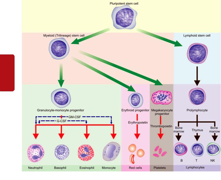
286
Tissues Lymphoreticular and Haematology II SECTION
Figure 12.3 
 Schematic representation of differentiation of multipotent stem cells into blood cells.
Bone marrow examination may be performed by two methods—aspiration and trephine biopsy. A comparison of the two methods is summarised in Table 12.1.
BONE MARROW ASPIRATION. The method involves suction of marrow via a strong, wide bore, short-bevelled needle fitted with a stylet and an adjustable guard in order to prevent excessive penetration; for instance Salah bone marrow aspiration needle (Fig. 12.4,A). Smears are prepared immediately from the bone marrow aspirate and are fixed in 95% methanol after air-drying. The usual Romanowsky technique is employed for staining and a stain for iron is performed routinely so as to assess the reticuloendothelial stores of iron.
The marrow film provides assessment of cellularity, details of developing blood cells (i.e. normoblastic or megaloblastic, myeloid, lymphoid, macrophages and megakaryocytic), ratio between erythroid and myeloid cells, storage diseases, and for the presence of cells foreign to the marrow such as secondary carcinoma, granulomatous
conditions, fungi (e.g. histoplasmosis) and parasites (e.g. malaria, leishmaniasis, trypanosomiasis). Estimation of the proportion of cellular components in the marrow, however, can be provided by doing a differential count of at least 500 cells (myelogram, Table 12.2). In some conditions, the marrow cells can be used for more detailed special tests such as cytogenetics, microbiological culture, biochemical analysis, and immunological and cytological markers.
TREPHINE BIOPSY. Trephine biopsy is performed by a simple Jamshidi trephine needle by which a core of tissue from periosteum to bone marrow cavity is obtained (Fig. 12.4,B). The tissue is then fixed, soft decalcified and processed for histological sections and stained with haematoxylin and eosin and for reticulin. Trephine biospy is useful over aspiration since it provides an excellent view of the overall marrow architecture, cellularity, and presence or absence of infiltrates, but is less valuable than aspiration as far as individual cell morphology is concerned.
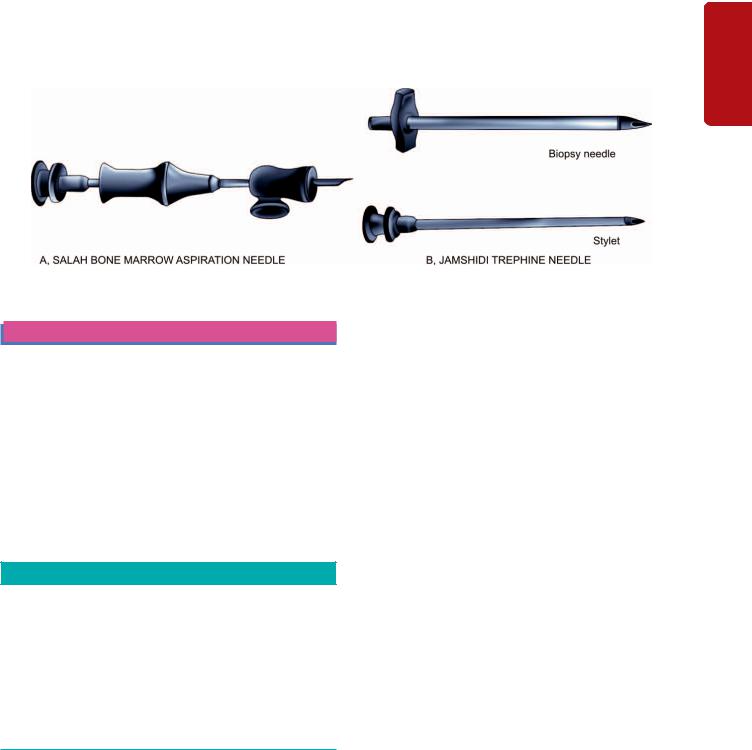
TABLE 12.1: Comparison of Bone Marrow Aspiration and Trephine Biopsy. |
|
287 |
|||
|
Feature |
Aspiration |
Trephine |
|
|
|
|
|
|
|
|
1. |
Site |
Sternum, posterior iliac crest; tibial head in infants |
Posterior iliac crest |
||
2. |
Instrument |
Salah BM aspiration needle |
Jamshidi trephine needle |
||
3. |
Stains |
Romanowsky, Perls’ reaction for iron on smears |
Haematoxylin and eosin, reticulin on tissue sections |
||
4. |
Time |
Within 1-2 hours |
Within 1-7 days |
||
5. |
Morphology |
Better cellular morphology of aspiration smears |
Better marrow architectural pattern but cell morphology |
||
|
|
but marrow architecture is indistinct |
is not as distinct since tissue sections are examined |
||
|
|
|
and not |
smears |
|
6. Indications |
Anaemias, suspected leukaemias, neutropenia |
Additional indications are: |
|
thrombocytopenia, polycythaemia, myeloma, |
myelosclerosis, aplastic anaemia and in |
|
lymphomas, carcinomatosis, lipid storage |
cases with ‘dry tap’ on aspiration. |
|
diseases, granulomatous conditions, parasites, |
|
|
fungi, and unexplained enlargements of liver, |
|
|
spleen or lymph nodes. |
|
|
|
|
Figure 12.4 
 The Salah bone marrow aspiration needle (A), Jamshidi trephine needle (B).
RED BLOOD CELLS
ERYTHROPOIESIS
Although the stem cells which eventually form the mature erythrocytes of the peripheral blood cannot be recognised morphologically, there is a well-defined and readily recognisable lineage of nucleated red cells (i.e. the erythroid series) in the marrow.
Erythroid Series
The cells in this series are as under (Fig. 12.5):
1. PROERYTHROBLAST. The earliest recognisable cell in the marrow is a proerythroblast or pronormoblast. It is a large
TABLE 12.2: Normal Adult Bone Marrow Counts (Myelogram).
Fat/cell ratio : 50:50
Myeloid/erythroid (M/E) ratio : 2-4:1 (mean 3:1)
Myeloid series: 30-45% (37.5%)
•Myeloblasts : 0.1-3.5%
•Promyelocytes: 0.5-5%
Erythroid series: 10-15% (mean 12.5%)
Megakaryocytes: 0.5%
Lymphocytes: 5-20%
Plasma cells: < 3%
Reticulum cells: 0.1-2%
cell, 15-20 μm in diameter having deeply basophilic cytoplasm and a large central nucleus containing nucleoli. The deep blue colour of the cytoplasm is due to high content of RNA which is associated with active protein synthesis. As the cells mature, the nuclei lose their nucleoli and become smaller and denser, while the cytoplasm on maturation leads to replacement of dense blue colour progressively by pinkstaining haemoglobin. Each proerythroblast undergoes 4-5 replications and forms 16-32 mature RBCs.
2.BASOPHILIC (EARLY) ERYTHROBLAST. It is a round cell having a diameter of 12-16 μm with a large nucleus which is slightly more condensed than the proerythroblast and contains basophilic cytoplasm. Basophilic erythroblast undergoes rapid proliferation.
3.POLYCHROMATIC (INTERMEDIATE) ERYTHROBLAST. Next maturation stage has a diameter of 12-14 μm. The nucleus at this stage is coarse and deeply basophilic. The cytoplasm is characteristically polychromatic i.e. contains admixture of basophilic RNA and acidophilic haemoglobin. The cell at this stage ceases to undergo proliferative activity.
4.ORTHOCHROMATIC (LATE) ERYTHROBLAST. The final stage in the maturation of nucleated red cells is the orthochromatic or late erythroblast. The cell at this stage is smaller, 8-12 μm in diameter, containing a small and pyknotic
Series Erythroid of Disorders and System Haematopoietic to Introduction 12 CHAPTER
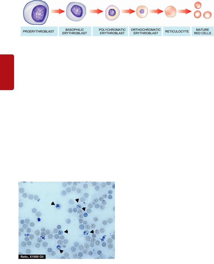
288
Tissues Lymphoreticular and Haematology II SECTION
Figure 12.5 
 The erythroid series. There is progressive condensation of the nuclear chromatin which is eventually extruded from the cell at the late erythroblast stage. The cytoplasm contains progressively less RNA and more haemoglobin.
nucleus with dark nuclear chromatin. The cytoplasm is characteristically acidophilic with diffuse basophilic hue due to the presence of large amounts of haemoglobin.
5. RETICULOCYTE. The nucleus is finally extruded from the late erythroblast within the marrow and a reticulocyte results. The reticulocytes are juvenile red cells devoid of nuclei but contain ribosomal RNA so that they are still able to synthesise haemoglobin. A reticulocyte spends 1-2 days in the marrow and circulates for 1-2 days in the peripheral blood before maturing in the spleen, to become a biconcave red cell. The reticulocytes in the peripheral blood are distinguished from mature red cells by slightly basophilic hue in the cytoplasm similar to that of an orthochromatic erythroblast. Reticulocytes can be counted in the laboratory by vital staining with dyes such as new methylene blue or brilliant cresyl blue. The reticulocytes by either of these staining methods contain deep blue reticulofilamentous material (Fig. 12.6). While erythroblasts are not normally present in human peripheral blood, reticulocytes are found normally in the peripheral blood. Normal range of reticulocyte count in health is 0.5-2.5% in adults and 2-6% in infants. Their percentage in the peripheral blood is a fairly accurate reflection of erythropoietic activity. Their proportion is increased in conditions of rapid red cell regeneration e.g. after haemorrhage, haemolysis and haematopoietic response of anaemia to treatment.
Figure 12.6 
 Reticulocytes in blood as seen in blood stained by supravital dye, new methylene blue.
Erythropoietin
Erythropoietic activity in the body is regulated by the hormone, erythropoietin, which is produced in response to anoxia. The principal site of erythropoietin production is the kidney though there is evidence of its extra-renal production in certain unusual circumstances. Its levels are, therefore, lowered in chronic renal diseases, while a case of renal cell carcinoma may be associated with its increased production and erythrocytosis. Erythropoietin acts on the marrow at the various stages of morphologically unidentifiable as well as identifiable erythroid precursors.
There is an increased production of erythropoietin in various types of anaemias but in anaemia of chronic diseases (e.g. in infections and neoplastic conditions) there is no such enhancement of erythropoietin. In polycythaemia rubra vera, there is erythrocytosis but depressed production of erythropoietin. This is because of an abnormality of the stem cell class which is not under erythropoietin control.
Immunoassay of erythropoietin in plasma or serum can be done by sensitive techniques (ELISA and radioimmunoassay) due to its quite low values; normal values are 10-25 U/L.
The Red Cell
The mature erythrocytes of the human peripheral blood are non-nucleated cells and lack the usual cell organelles. The normal human erythrocyte is a biconcave disc, 7.2 μm in diameter, and has a thickness of 2.4 μm at the periphery and 1 μm in the centre. The biconcave shape renders the red cells quite flexible so that they can pass through capillaries whose minimum diameter is 3.5 μm. More than 90% of the weight of erythrocyte consist of haemoglobin. The lifespan of red cells is 120 + 30 days.
NORMAL VALUES AND RED CELL INDICES. Range of normal red cell count in health is 5.5 ± 1.0 × 10¹²/L in men and 4.8 ± 1.0 × 10¹²/L in women. The packed cell volume (PCV) or haematocrit is the volume of erythrocytes per litre of whole blood indicating the proportion of plasma and red cells and ranges 0.47 ± 0.07 L/L (40-54%) in men and 0.42 ± 0.05 L/L (37-47%) in women. The haemoglobin content in health is 15.5
± 2.5 g/dl (13-18 g/dl) in men and 14.0 ± 2.5 g/dl (11.5-16.5 g/dl) in women. Based on these normal values, a series of absolute values or red cell indices can be derived which have diagnostic importance. These are as under:

1. Mean corpuscular volume (MCV) =
PCV in L/L
_________________
RBC count/L
The normal value is 85 ± 8 fl (77-93 fl)*.
2. Mean corpuscular haemoglobin (MCH) =
Hb/L
________________
RBC count/L
The normal range is 29.5 ± 2.5 pg (27-32 pg)*.
3. Mean corpuscular haemoglobin concentration (MCHC) =
Hb/dl
_________________
PCV in L/L
The normal value is 32.5 ± 2.5 g/dl (30-35 g/dl).
Since MCHC is independent of red cell count and size, it is considered to be of greater clinical significance as compared to other absolute values. It is low in iron deficiency anaemia but is usually normal in acrostic anaemia.
RED CELL MEMBRANE. The red cell membrane is a trilaminar structure having a bimolecular lipid layer interposed between two layers of proteins. The important proteins in red cell membrane are band 3 protein (named on the basis of the order in which it migrates during electrophoresis), glycophorin and spectrin; important lipids are glycolipids, phospholipids and cholesterol; and carbohydrates form skeleton of erythrocytes having a latticelike network which is attached to the internal surface of the membrane and is responsible for biconcave form of the erythrocytes.
A number of inherited disorders of the red cell membrane and cytoskeletal components produce abnormalities of the shape such as: spherocytosis (spherical shape from loss of part of the membrane), ovalocytosis (oval shape from loss of elasticity of cytoskeleton), echinocytosis (spiny processes from external surface due to metabolic abnormalities of red cells), and stomatocytosis (bowl-shaped red cells from expansion of inner membrane on one side).
NUTRITIONAL REQUIREMENTS FOR ERYTHROPOIESIS. New red cells are being produced each day for which the marrow requires certain essential substances. These substances are as under:
1.Metals. Iron is essential for red cell production because it forms part of the haem molecule in haemoglobin. Its deficiency leads to iron deficiency anaemia. Cobalt and manganese are certain other metals required for red cell production.
2.Vitamins. Vitamin B12 and folate are essential for biosynthesis of nucleic acids. Deficiency of B12 or folate causes megaloblastic anaemia. Vitamin C (ascorbic acid) plays an indirect role by facilitating the iron turnover in the body.
*For conversions, the multiples used are as follows: ‘deci (d) = 10–1, milli
(m) = 10–3, micro (μ) = 10–6, nano (n) = 10–9, pico (p) = 10–12, femto (f) =
10–15
Vitamin B6 (pyridoxine), vitamin E (tocopherol) and riboflavin are the other essential vitamins required in the synthesis of red cells.
3.Amino acids. Amino acids comprise the globin component of haemoglobin. Severe amino acid deficiency due to protein deprivation causes depressed red cell production.
4.Hormones. As discussed above, erythropoietin plays a significant regulatory role in the erythropoietic activity. Besides erythropoietin, androgens and thyroxine also appear to be involved in the red cell production.
HAEMOGLOBIN. Haemoglobin consists of a basic protein, globin, and the iron-porphyrin complex, haem. The molecular weight of haemoglobin is 68,000. Normal adult haemoglobin (HbA) constitutes 96-98% of the total haemoglobin content and consists of four polypeptide chains, α2β2. Small quantities of 2 other haemoglobins present in adults are: HbF containing α2γ2 globin chains comprising 0.5-0.8% of total haemoglobin, and HbA2 having α2δ2 chains and constituting 1.5-3.2% of total haemoglobin. Most of the haemoglobin (65%) is synthesised by the nucleated red cell precursors in the marrow, while the remainder (35%) is synthesised at the reticulocyte stage.
Synthesis of haem occurs largely in the mitochondria by a series of biochemical reactions summarised in Fig. 12.7. Coenzyme, pyridoxal-6-phosphate, derived from pyridoxine (vitamin B6) is essential for the synthesis of amino levulinic acid (ALA) which is the first step in the biosynthesis of protoporphyrin. The reaction is stimulated by erythropoietin and inhibited by haem. Ultimately, protoporphyrin combines with iron supplied from circulating transferrin to form haem. Each molecule of haem combines with a globin chain synthesised by polyribosomes. A tetramer of 4 globin chains, each having its own haem group, constitutes the haemoglobin molecule (Fig. 12.8, A).
RED CELL FUNCTIONS. The essential function of the red cells is to carry oxygen from the lungs to the tissue and to transport carbon dioxide to the lungs. In order to perform these functions, the red cells have the ability to generate energy as ATP by anaerobic glycolytic pathway (EmbdenMeyerhof pathway). This pathway also generates reducing power as NADH and NADPH by the hexose monophosphate (HMP) shunt.
1. Oxygen carrying. The normal adult haemoglobin, HbA, is an extremely efficient oxygen-carrier. The four units of tetramer of haemoglobin molecule take up oxygen in succession, which, in turn, results in stepwise rise in affinity of haemoglobin for oxygen. This is responsible for the sigmoid shape of the oxygen dissociation curve.
The oxygen affinity of haemoglobin is expressed in term of P50 value which is the oxygen tension (pO2) at which 50% of the haemoglobin is saturated with oxygen. Pulmonary capillaries have high pO2 and, thus, there is virtual saturation of available oxygen-combining sites of haemoglobin. The tissue capillaries, however, have relatively low pO2 and, thus, part of haemoglobin is in deoxy state. The extent to which oxygen is released from haemoglobin at pO2, in tissue capillaries depends upon 3 factors—the nature of globin
289
Series Erythroid of Disorders and System Haematopoietic to Introduction 12 CHAPTER

290
II SECTION
Figure 12.7 
 Schematic diagram of haemoglobin synthesis in the developing red cell.
Tissues Lymphoreticular and Haematology
chains, the pH, and the concentration of 2,3- biphosphoglycerate (2,3-BPG) as follows (Fig 12.8, B):

 Normal adult haemoglobin (HbA) has lower affinity for oxygen than foetal haemoglobin and, therefore, releases greater amount of bound oxygen at pO2 of tissue capillaries.
 A fall in the pH (acidic pH) lowers affinity of oxyhaemoglobin for oxygen, so called the Bohr effect, thereby causing enhanced release of oxygen from erythrocytes at the lower pH in tissue capillaries.

 A rise in red cell concentration of 2,3-BPG, an intermediate product of Embden-Meyerhof pathway, as occurs in anaemia and hypoxia, causes decreased affinity of HbA for oxygen. This, in turn, results in enhanced supply of oxygen to the tissue.
2. CO2 transport. Another important function of the red cells is the CO2 transport. In the tissue capillaries, the pCO2 is high so that CO2 enters the erythrocytes where much of it is converted into bicarbonate ions which diffuse back into the plasma. In the pulmonary capillaries, the process is reversed and bicarbonate ions are converted back into CO2. Some of the CO2 produced by tissues is bound to deoxyhaemoglobin forming carbamino-haemoglobin. This compound dissociates in the pulmonary capillaries to release CO2.
RED CELL DESTRUCTION. Red cells have a mean lifespan of 120 days, after which red cell metabolism gradually deteriorates as the enzymes are not replaced. The destroyed red cells are removed mainly by the macrophages of the reticuloendothelial (RE) system of the marrow, and to some
Figure 12.8 
 A, Normal adult haemoglobin molecule (HbA) consisting of α2 β2 globin chains, each with its own haem group in oxy and deoxy state. The haemoglobin tetramer can bind up to four molecules of oxygen in the iron containing sites of the haem molecules. As oxygen is bound, salt bridges are broken, and 2,3-BPG and CO2 are expelled. B, Hb-dissociation curve. On dissociation of oxygen from Hb molecule i.e. on release of oxygen to the tissues, salt bridges are formed again, and 2,3-BPG and CO2, are bound. The shift of the curve to higher oxygen delivery is affected by acidic pH, increased 2,3-BPG and HbA molecule while oxygen delivery is less with high pH, low 2,3-BPG and HbF.
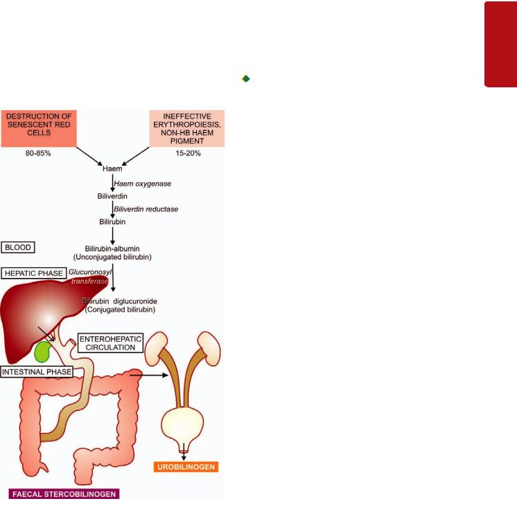
extent by the macrophages in the liver and spleen (Fig.12.9). The breakdown of red cells liberates iron for recirculation via plasma transferrin to marrow erythroblasts, and protoporphyrin which is broken down to bilirubin. Bilirubin circulates to the liver where it is conjugated to its diglucuronide which is excreted in the gut via bile and converted to stercobilinogen and stercobilin excreted in the faeces. Part of stercobilinogen and stercobilin is reabsorbed and excreted in the urine as urobilinogen and urobilin. A small fragment of protoporphyrin is converted to carbon monoxide and excreted in expired air from the lungs. Globin chains are broken down to amino acids and reused for protein synthesis in the body.
ANAEMIA—GENERAL CONSIDERATIONS
Anaemia is defined as a haemoglobin concentration in blood below the lower limit of the normal range for the age and sex of the individual. In adults, the lower extreme of the
Figure 12.9 
 Normal red cell destruction in the RE system.
normal haemoglobin is taken as 13.0 g/dl for males and 11.5 g/dl for females. Newborn infants have higher haemoglobin level and, therefore, 15 g/dl is taken as the lower limit at birth, whereas at 3 months the normal lower level is 9.5 g/dl. Although haemoglobin value is employed as the major parameter for determining whether or not anaemia is present, the red cell counts, haematocrit (PCV) and absolute values (MCV, MCH and MCHC) provide alternate means of assessing anaemia.
Pathophysiology of Anaemia
Subnormal level of haemoglobin causes lowered oxygencarrying capacity of the blood. This, in turn, initiates compensatory physiologic adaptations such as follows:

 increased release of oxygen from haemoglobin;

 increased blood flow to the tissues;

 maintenance of the blood volume; and
 redistribution of blood flow to maintain the cerebral blood supply.
Eventually, however, tissue hypoxia develops causing impaired functions of the affected tissues. The degree of functional impairment of individual tissues is variable depending upon their oxygen requirements. Tissues with high oxygen requirement such as the heart, CNS and the skeletal muscle during exercise, bear the brunt of clinical effects of anaemia.
Clinical Features of Anaemia
The haemoglobin level at which symptoms and signs of anaemia develop depends upon 4 main factors:
1.The speed of onset of anaemia: Rapidly progressive anaemia causes more symptoms than anaemia of slow-onset as there is less time for physiologic adaptation.
2.The severity of anaemia: Mild anaemia produces no symptoms or signs but a rapidly developing severe anaemia (haemoglobin below 6.0 g/dl) may produce significant clinical features.
3.The age of the patient: The young patients due to good cardiovascular compensation tolerate anaemia quite well as compared to the elderly. The elderly patients develop cardiac and cerebral symptoms more prominently due to associated cardiovascular disease.
4.The haemoglobin dissociation curve: In anaemia, the affinity of haemoglobin for oxygen is depressed as 2,3-BPG in the red cells increases. As a result, oxyhaemoglobin is dissociated more readily to release free oxygen for cellular use, causing a shift of the oxyhaemoglobin dissociation curve to the right.
SYMPTOMS. In symptomatic cases of anaemia, the presenting features are: tiredness, easy fatiguability, generalised muscular weakness, lethargy and headache. In older patients, there may be symptoms of cardiac failure, angina pectoris, intermittent claudication, confusion and visual disturbances.
SIGNS. A few general signs common to all types of anaemias are as under:
291
Series Erythroid of Disorders and System Haematopoietic to Introduction 12 CHAPTER

292 1. Pallor. Pallor is the most common and characteristic sign which may be seen in the mucous membranes, conjunctivae and skin.
2. Cardiovascular system. A hyperdynamic circulation may be present with tachycardia, collapsing pulse, cardiomegaly, midsystolic flow murmur, dyspnoea on exertion, and in the case of elderly, congestive heart failure.
3. Central nervous system. The older patients may develop symptoms referable to the CNS such as attacks of faintness, giddiness, headache, tinnitus, drowsiness, numbness and tingling sensations of the hands and feet.
SECTION |
4. Ocular manifestations. Retinal haemorrhages may occur |
|
if there is associated vascular disease or bleeding diathesis. |
||
5. Reproductivesystem. Menstrual disturbances such as |
||
|
||
|
amenorrhoea and menorrhagia and loss of libido are some |
|
|
of the manifestations involving the reproductive system in |
|
II |
anaemic subjects. |
|
6. Renal system. Mild proteinuria and impaired concen- |
||
|
||
|
trating capacity of the kidney may occur in severe anaemia. |
|
Haematology |
7. Gastrointestinalsystem. Anorexia, flatulence, nausea, |
|
constipation and weight loss may occur. |
||
|
||
|
In addition to the general features, specific signs may be |
|
|
associated with particular types of anaemia which are |
|
|
described later together with discussion of specific types of |
|
and |
anaemias. |
|
Investigations of the Anaemic Subject |
||
|
||
Lymphoreticular |
After obtaining the full medical history pertaining to different |
|
tenderness are looked for. |
||
|
general and specific signs and symptoms, the patient is |
|
|
examined for evidence of anaemia. Special emphasis is placed |
|
|
on colour of the skin, conjunctivae, sclerae and nails. Changes |
|
|
in the retina, atrophy of the papillae of the tongue, rectal |
|
|
examination for evidence of bleeding, and presence of |
|
|
hepatomegaly, splenomegaly, lymphadenopathy and bony |
|
Tissues |
In order to confirm or deny the presence of anaemia, its |
|
type and its cause, the following plan of investigations is |
||
|
||
|
generally followed, of which complete blood counts (CBC) |
|
|
with reticulocyte count is the basic test. |
|
|
A. HAEMOGLOBIN ESTIMATION. The first and foremost |
|
|
investigation in any suspected case of anaemia is to carry |
|
|
out a haemoglobin estimation. Several methods are available |
|
|
but most reliable and accurate is the cyanmethaemoglobin |
|
|
(HiCN) method employing Drabkin’s solution and a |
|
|
spectrophotometer. If the haemoglobin value is below the |
|
|
lower limit of the normal range for particular age and sex, |
|
|
the patient is said to be anaemic. In pregnancy, there is |
|
|
haemodilution and, therefore, the lower limit in normal |
|
|
pregnant women is less (10.5 g/dl) than in the non-pregnant |
|
|
state. |
|
|
B. PERIPHERAL BLOOD FILM EXAMINATION. The |
|
|
haemoglobin estimation is invariably followed by |
|
|
examination of a peripheral blood film for morphologic |
|
|
features after staining it with the Romanowsky dyes (e.g. |
|
|
Leishman’s stain, May-Grünwald-Giemsa’s stain, Jenner- |
|
|
Giemsa’s stain, Wright’s stain etc). The blood smear is |
evaluated in an area where there is neither Rouleaux formation nor so thin as to cause red cell distortion. Such an area can usually be found at junction of the body with the tail of the film, but not actually at the tail. The following abnormalities in erythroid series of cells are particularly looked for in a blood smear:
1.Variation in size (Anisocytosis). Normally, there is slight variation in diameter of the red cells from 6.7-7.7 μm (mean value 7.2 μm). Increased variation in size of the red cell is termed anisocytosis. Anisocytosis may be due to the presence of cells larger than normal (macrocytosis) or cells smaller than normal (microcytosis). Sometimes both microcytosis and macrocytosis are present (dimorphic).

 Macrocytes are classically found in megaloblastic anaemia; other causes are aplastic anaemia, other dyserythropoietic anaemias, chronic liver disease and in conditions with increased erythropoiesis.

 Microcytes are present in iron deficiency anaemia, thalassaemia and spherocytosis. They may also result from fragmentation of erythrocytes such as in haemolytic anaemia.
2.Variation in shape (Poikilocytosis). Increased variation in shape of the red cells is termed poikilocytosis. The nature of the abnormal shape determines the cause of anaemia. Poikilocytes are produced in various types of abnormal erythropoiesis e.g. in megaloblastic anaemia, iron deficiency anaemia, thalassaemia, myelosclerosis and microangiopathic haemolytic anaemia.
3.Inadequate haemoglobin formation (Hypochromasia).
Normally, the intensity of pink staining of haemoglobin in a Romanowsky-stained blood smear gradually decreases from the periphery to the centre of the cell. Increased central pallor is referred to as hypochromasia. It may develop either from lowered haemoglobin content (e.g. in iron deficiency anaemia, chronic infections), or due to thinness of the red cells (e.g. in thalassaemia, sideroblastic anaemia). Unusually deep pink staining of the red cells due to increased haemoglobin concentration is termed hyperchromasia and may be found in megaloblastic anaemia, spherocytosis and in neonatal blood.
4.Compensatory erythropoiesis. A number of changes are associated with compensatory increase in erythropoietic activity. These are as under:
i)Polychromasia is defined as the red cells having more than one type of colour. Polychromatic red cells are slightly larger, generally stained bluish-grey and represent reticulocytes and, thus, correlate well with reticulocyte count.
ii)Erythroblastaemia is the presence of nucleated red cells in the peripheral blood film. A small number of erythroblasts (or normoblasts) may be normally found in cord blood at birth. They are found in large numbers in haemolytic disease of the newborn, other haemolytic disorders and in extramedullary erythropoiesis. They may also appear in the blood in various types of severe anaemias except in aplastic anaemia. Erythroblastaemia may also occur after splenectomy.
iii)Punctate basophilia or basophilic stippling is diffuse and uniform basophilic granularity in the cell which does not stain positively with Perls’ reaction (in contrast to
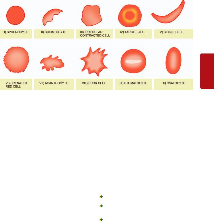
293
12 CHAPTER
Figure 12.10 
 Some of the common morphologic abnormalities of red cells (The serial numbers in the illustrations correspond to the order in which they are described in the text).
Pappenheimer bodies which stain positively). Classical punctate basophilia is seen in aplastic anaemia, thalassaemia, myelodysplasia, infections and lead poisoning.
iv) Howell-Jolly bodies are purple nuclear remnants, usually found singly, and are larger than basophilic stippling. They are present in megaloblastic anaemia and after splenectomy.
5. Miscellaneous changes. In addition to the morphologic changes of red cells described above, several other abnormal red cells may be found in different haematological disorders. Some of these are as follows (Fig. 12.10):
i)Spherocytosis is characterised by presence of spheroidal rather than biconcave disc-shaped red cells. Spherocytes are seen in hereditary spherocytosis, autoimmune haemolytic anaemia and in ABO haemolytic disease of the newborn.
ii)Schistocytosis is identified by fragmentation of erythrocytes. Schistocytes are found in thalassaemia, hereditary elliptocytosis, megaloblastic anaemia, iron deficiency anaemia, microangiopathic haemolytic anaemia and in severe burns.
iii)Irregularly contracted red cells are found in drug and chemical induced haemolytic anaemia and in unstable haemoglobinopathies.
iv)Leptocytosis is the presence of unusually thin red cells. Leptocytes are seen in severe iron deficiency and thalassaemia. Target cell is a form of leptocyte in which there is central round stained area and a peripheral rim of haemoglobin. Target cells are found in iron deficiency, thalassaemia, chronic liver disease, and after splenectomy.
v)Sickle cells or drepanocytes are sickle-shaped red cells found in sickle cell disease.
vi)Crenated red cells are the erythrocytes which develop numerous projections from the surface. They are present in blood films due to alkaline pH, presence of traces of fatty substances on the slides and in cases where the film is made from blood that has been allowed to stand overnight.
vii)Acanthocytosis is the presence of coarsely crenated red cells. Acanthocytes are found in large number in blood film made from splenectomised subjects, and in chronic liver disease.
viii)Burr cells are cell fragments having one or more spines. They are particularly found in uraemia.
ix)Stomatocytosis is the presence of stomatocytes which have central area having slit-like or mouth-like appearance. They are found in hereditary stomatocytosis, or may be seen in chronic alcoholism.
x)Ovalocytosis or elliptocytosis is the oval or elliptical shape of red cells. Their highest proportion (79%) is seen in hereditary ovalocytosis and elliptocytosis; other conditions showing such abnormal shapes of red cells are megaloblastic anaemia and hypochromic anaemia.
C.RED CELL INDICES. An alternative method to diagnose and detect the severity of anaemia is by measuring the red cell indices:

 In iron deficiency and thalassaemia, MCV, MCH and MCHC are reduced.

 In anaemia due to acute blood loss and haemolytic anaemias, MCV, MCH and MCHC are all within normal limits.

 In megaloblastic anaemias, MCV is raised above the normal range.
D.LEUCOCYTE AND PLATELET COUNT. Measurement of leucocyte and platelet count helps to distinguish pure anaemia from pancytopenia in which red cells, granulocytes and platelets are all reduced. In anaemias due to haemolysis or haemorrhage, the neutrophil count and platelet counts are often elevated. In infections and leukaemias, the leucocyte counts are high and immature leucocytes appear in the blood.
E.RETICULOCYTE COUNT. Reticulocyte count (normal 0.5-2.5%) is done in each case of anaemia to assess the marrow
Series Erythroid of Disorders and System Haematopoietic to Introduction

294 erythropoietic activity. In acute haemorrhage and in haemolysis, the reticulocyte response is indicative of impaired marrow function.
F. ERYTHROCYTE SEDIMENTATION RATE. The ESR is a non-specific test used as a screening test for anaemia. It usually gives a clue to the underlying organic disease but anaemia itself may also cause rise in the ESR.
|
G. BONE MARROW EXAMINATION. Bone marrow |
|
|
aspiration is done in cases where the cause for anaemia is |
|
|
not obvious. The procedures involved for marrow aspiration |
|
|
and trephine biopsy and their relative advantages and |
|
SECTION |
disadvantages have already been discussed (page 285). |
|
In addition to these general tests, certain specific tests |
||
|
||
|
are done in different types of anaemias which are described |
|
|
later under the discussion of specific anaemias. |
II |
Classification of Anaemias |
|||
Several types of classifications of anaemias have been |
||||
|
||||
|
proposed. Two of the widely accepted classifications are |
|||
Haematology |
based on the pathophysiology and morphology (Table 12.3). |
|||
PATHOPHYSIOLOGIC CLASSIFICATION. Depending |
||||
upon the pathophysiologic mechanism, anaemias are |
||||
classified into 3 groups: |
||||
I. Anaemia due to blood loss. This is further of 2 types: |
||||
A. Acute post-haemorrhagic anaemia |
||||
and |
B. Anaemia of chronic blood loss |
|||
II. Anaemia due to impaired red cell formation. A |
||||
Lymphoreticular |
||||
disturbance due to impaired red cell production from various |
||||
causes may produce anaemia. These are as under: |
||||
|
||||
TABLE 12.3: Classification of Anaemias. |
||||
A. PATHOPHYSIOLOGIC |
||||
I. Anaemia due to increased blood loss |
||||
a) |
Acute post-haemorrhagic anaemia |
|||
b) |
Chronic blood loss |
|||
Tissues |
||||
II. Anaemias due to impaired red cell production |
||||
a) |
Cytoplasmic maturation defects |
|||
|
1. |
Deficient haem synthesis: |
||
|
|
Iron deficiency anaemia |
||
|
|
|
||
|
|
2. |
Deficient globin synthesis: |
|
|
|
|
Thalassaemic syndromes |
|
|
b) |
Nuclear maturation defects |
||
|
|
Vitamin B12 and/or folic acid deficiency: |
||
|
|
Megaloblastic anaemia |
||
|
c) |
Defect in stem cell proliferation and differentiation |
||
|
|
1. |
Aplastic anaemia |
|
|
|
2. Pure red cell aplasia |
||
|
d) |
Anaemia of chronic disorders |
||
|
e) |
Bone marrow infiltration |
||
|
f) |
Congenital anaemia |
||
III. Anaemias due to increased red cell destruction (Haemolytic anaemias) (Details in Table 12.9)
A.Extrinsic (extracorpuscular) red cell abnormalities
B.Intrinsic (intracorpuscular) red cell abnormalities
B. MORPHOLOGIC
I. Microcytic, hypochromic
II.Normocytic, normochromic III. Macrocytic, normochromic
A.Cytoplasmic maturation defects
1.Deficient haem synthesis: iron deficiency anaemia
2.Deficient globin synthesis: thalassaemic syndromes
B.Nuclear maturation defects
Vitamin B12 and/or folic acid deficiency: megaloblastic anaemia
C. Haematopoietic stem cell proliferation and differentiation abnormality e.g.
1.Aplastic anaemia
2.Pure red cell aplasia
D.Bone marrow failure due to systemic diseases (anaemia of chronic disorders) e.g.
1.Anaemia of inflammation/infections, disseminated malignancy
2.Anaemia in renal disease
3.Anaemia due to endocrine and nutritional deficiencies (hypometabolic states)
4.Anaemia in liver disease
E.Bone marrow infiltration e.g.
1.Leukaemias
2.Lymphomas
3.Myelosclerosis
4.Multiple myeloma
F.Congenital anaemia e.g.
1.Sideroblastic anaemia
2.Congenital dyserythropoietic anaemia.
The term hypoproliferative anaemias is also used to denote impaired marrow proliferative activity and includes 2 main groups: hypoproliferation due to iron deficiency and that due to other hypoproliferative disorders; the latter category includes anaemia of chronic inflammation/infection, renal disease, hypometabolic states, and causes of bone marrow failure.
III. Anaemia due to increased red cell destruction (haemolytic anaemias). This is further divided into 2 groups:
A.Intracorpuscular defect (hereditary and acquired).
B.Extracorpuscular defect (acquired haemolytic anaemias).
MORPHOLOGIC CLASSIFICATION. Based on the red cell size, haemoglobin content and red cell indices, anaemias are classified into 3 types:
1.Microcytic, hypochromic: MCV, MCH, MCHC are all reduced e.g. in iron deficiency anaemia and in certain noniron deficient anaemias (sideroblastic anaemia, thalassaemia, anaemia of chronic disorders).
2.Normocytic, normochromic: MCV, MCH, MCHC are all normal e.g. after acute blood loss, haemolytic anaemias, bone marrow failure, anaemia of chronic disorders.
3.Macrocytic: MCV is raised e.g. in megaloblastic anaemia
due to deficiency of vitamin B12 or folic acid.
With these general comments on anaemias, a discussion of the specific types of anaemias is given in the following pages.
ANAEMIA OF BLOOD LOSS
Depending upon the rate of blood loss due to haemorrhage, the effects of post-haemorrhagic anaemia appear.
ACUTE BLOOD LOSS. When the loss of blood occurs suddenly, the following events take place:
i)Immediate threat to life due to hypovolaemia which may result in shock and death.
ii)If the patient survives, shifting of interstitial fluid to intravascular compartment with consequent haemodilution with low haematocrit.
iii)Hypoxia stimulates production of erythropoietin resulting in increased marrow erythropoiesis.
LABORATORY FINDINGS
i)Normocytic and normochromic anaemia
ii)Low haematocrit
iii)Increased reticulocyte count in peripheral blood (10-15% after one week) reflecting accelerated marrow erythropoiesis.
CHRONIC BLOOD LOSS. When the loss of blood is slow and insidious, the effects of anaemia will become apparent only when the rate of loss is more than rate of production and the iron stores are depleted. This results in iron deficiency anaemia as seen in other clinical conditions discussed below.
HYPOCHROMIC ANAEMIA
Hypochromic anaemia due to iron deficiency is the commonest cause of anaemia the world over. It is estimated that about 20% of women in child-bearing age group are iron deficient, while the overall prevalence in adult males is about 2%. It is the most important, though not the sole, cause of microcytic hypochromic anaemia in which all the three red cell indices (MCV, MCH and MCHC) are reduced and occurs due to defective haemoglobin synthesis. Hypochromic anaemias, therefore, are classified into 2 groups:
I. Hypochromic anaemia due to iron deficiency.
II.Hypochromic anaemias other than iron deficiency. The latter category includes 3 groups of disorders—
sideroblastic anaemia, thalassaemia and anaemia of chronic disorders.
IRON DEFICIENCY ANAEMIA
The commonest nutritional deficiency disorder present throughout the world is iron deficiency but its prevalence is higher in the developing countries. The factors responsible for iron deficiency in different populations are variable and are best understood in the context of normal iron metabolism.
Iron Metabolism
The amount of iron obtained from the diet should replace the losses from the skin, bowel and genitourinary tract. These losses together are about 1 mg daily in an adult male or in a non-menstruating female, while in a menstruating woman there is an additional iron loss of 0.5-1 mg daily. The iron required for haemoglobin synthesis is derived from 2 primary sources—ingestion of foods containing iron (e.g. leafy vegetables, beans, meats, liver etc) and recycling of iron from senescent red cells (Fig. 12.11).
ABSORPTION. The average Western diet contains 10-15 mg of iron, out of which only 5-10% is normally absorbed. In pregnancy and in iron deficiency, the proportion of absorption is raised to 20-30%. Iron is absorbed mainly in the duodenum and proximal jejunum. Iron from diet containing haem is better absorbed than non-haem iron. Absorption of nonhaem iron is enhanced by factors such as ascorbic acid (vitamin C), citric acid, amino acids, sugars, gastric secretions and hydrochloric acid. Iron absorption is impaired by factors like medicinal antacids, milk, pancreatic secretions, phytates, phosphates, ethylene diamine tetra-acetic acid (EDTA) and tannates contained in tea.
Non-haem iron is released as ferrous or ferric form but is absorbed almost exclusively as ferrous form; reduction of ferric to ferrous form when required takes place at the intestinal brush border by ferric reductase. Transport across the membrane is accomplished by divalent metal transporter 1 (DMT 1). Once inside the gut cells, ferric iron may be either stored as ferritin or further transported to transferrin by two vehicle proteins—ferroportin and hephaestin. The mechanism of dietary haem iron absorption is not clearly understood yet but it is through a different transport than DMT 1. After absorption of both non-haem and haem forms of iron, it comes into mucosal pool.
Major mechanism of maintaining iron balance in the body is by intestinal absorption of dietary iron. When the demand for iron is increased (e.g. during pregnancy, menstruation, periods of growth and various diseases), there is increased iron absorption, while excessive body stores of iron cause reduced intestinal iron absorption (Fig. 12.12,A page 297).
TRANSPORT. Iron is transported in plasma bound to a β-globulin, transferrin, synthesised in the liver. Transferrinbound iron is made available to the marrow where the developing erythroid cells having transferring receptors utilise iron for haemoglobin synthesis. It may be mentioned here that tranferrin receptors are present on cells of many tissues of the body but their number is greatest in the developing erythroblasts. Transferrin is reutilised after iron is released from it. A small amount of transferrin iron is delivered to other sites such as parenchymal cells of the liver. Normally, transferrin is about one-third saturated. But in conditions where transferrin-iron saturation is increased, parenchymal iron uptake is increased. Virtually, no iron is deposited in the mononuclear-phagocyte cells (RE cells) from the plasma transferrin-iron but instead these cells derive most of their iron from phagocytosis of senescent red cells. Storage form of iron (ferritin and haemosiderin) in RE cells is normally not functional but can be readily mobilised in response to increased demands for erythropoiesis. However, conditions such as malignancy, infection and inflammation interfere with the release of iron from iron stores causing ineffective erythropoiesis.
EXCRETION. The body is unable to regulate its iron content by excretion alone. The amount of iron lost per day is 0.5-1 mg which is independent of iron intake. This loss is nearly twice more (i.e. 1-2 mg/day) in menstruating women. Iron is lost from the body in both sexes as a result of desquamation of epithelial cells from the gastrointestinal
295
Series Erythroid of Disorders and System Haematopoietic to Introduction 12 CHAPTER

296
Tissues Lymphoreticular and Haematology II SECTION
Figure 12.11 
 Daily iron cycle. Iron on absorption from upper small intestine circulates in plasma bound to transferrin and is transported to the bone marrow for utilisation in haemoglobin synthesis. The mature red cells are released into circulation, which on completion of their lifespan of 120 days, die. They are then phagocytosed by RE cells and iron stored as ferritin and haemosiderin. Stored iron is mobilised in response to increased demand and used for haemoglobin synthesis, thus completing the cycle (M = males; F = females).
tract, from excretion in the urine and sweat, and loss via hair and nails. Iron excreted in the faeces mainly consists of unabsorbed iron and desquamated mucosal cells.
DISTRIBUTION. In an adult, iron is distributed in the body as under:
1.Haemoglobin—present in the red cells, contains most of the body iron (65%).
2.Myoglobin—comprises a small amount of iron in the muscles (3.5%).
3.Haem and non-haem enzymes—e.g. cytochrome, catalase, peroxidases, succinic dehydrogenase and flavoproteins constitute a fraction of total body iron (0.5%).
4.Transferrin-bound iron—circulates in the plasma and constitutes another fraction of total body iron (0.5%).
All these forms of iron are in functional form.
5. Ferritin and haemosiderin—are the storage forms of excess iron (30%). They are stored in the mononuclear-phagocyte
cells of the spleen, liver and bone marrow and in the parenchymal cells of the liver.
Pathogenesis
Iron deficiency anaemia develops when the supply of iron is inadequate for the requirement of haemoglobin synthesis. Initially, negative iron balance is covered by mobilisation from the tissue stores so as to maintain haemoglobin synthesis. It is only after the tissue stores of iron are exhausted that the supply of iron to the marrow becomes insufficient for haemoglobin formation and thus a state of iron deficiency anaemia develops. The development of iron deficiency depends upon one or more of the following factors:
1.Increased blood loss
2.Increased requirements
3.Inadequate dietary intake
4.Decreased intestinal absorption.
The relative significance of these factors varies with the
age and sex of the patient (Table 12.4). Accordingly, certain
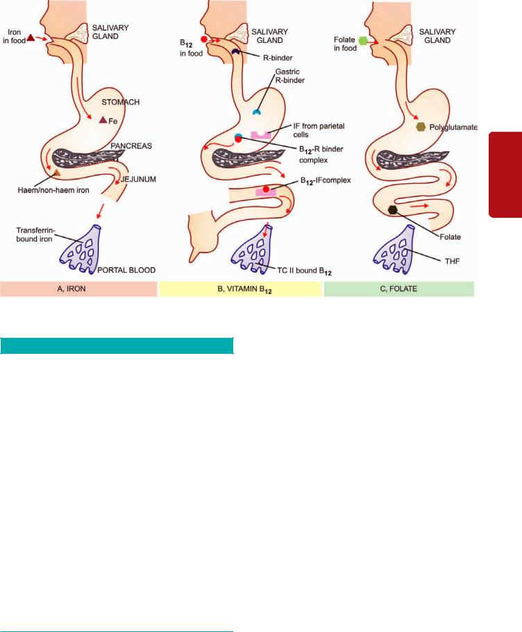
Figure 12.12 
 Contrasting pathways of absorption and transport of iron, vitamin B12 and folic acid.
TABLE 12.4: Etiology of Iron Deficiency Anaemia.
I. INCREASED BLOOD LOSS
1.Uterine e.g. excessive menstruation in reproductive years, repeated miscarriages, at onset of menarche, post-menopausal uterine bleeding.
2.Gastrointestinal e.g. peptic ulcer, haemorrhoids, hookworm infestation, cancer of stomach and large bowel, oesophageal varices, hiatus hernia, chronic aspirin ingestion, ulcerative colitis, diverticulosis.
3.Renal tract e.g. haematuria, haemoglobinuria.
4.Nose e.g. repeated epistaxis.
5.Lungs e.g. haemoptysis.
II.INCREASED REQUIREMENTS
1.Spurts of growth in infancy, childhood and adolescence.
2.Prematurity.
3.Pregnancy and lactation.
III.INADEQUATE DIETARY INTAKE
1.Poor economic status.
2.Anorexia e.g. in pregnancy.
3.Elderly individuals due to poor dentition, apathy and financial constraints.
IV. DECREASED ABSORPTION
1.Partial or total gastrectomy
2.Achlorhydria
3.Intestinal malabsorption such as in coeliac disease.
groups of individuals at increased risk of developing iron deficiency can be identified (see below). In general, in developed countries the mechanism of iron deficiency is usually due to chronic occult blood loss, while in the developing countries poor intake of iron or defective absorption are responsible for iron deficiency anaemia.
Etiology
Iron deficiency anaemia is always secondary to an underlying disorder. Correction of the underlying cause, therefore, is essential part of its treatment. Based on the above-mentioned pathogenetic mechanisms, following etiologic factors are involved in development of iron deficiency anaemia at different age and sex (Table 12.4):
1. FEMALES IN REPRODUCTIVE PERIOD OF LIFE. The highest incidence of iron deficiency anaemia is in women during their reproductive years of life. It may be from one or more of the following causes:
i) Blood loss. This is the most important cause of anaemia in women during child-bearing age group. Commonly, it is due to persistent and heavy menstrual blood loss such as occurs in various pathological states and due to insertion of IUCDs. Young girls at the onset of menstruation may develop mild anaemia due to blood loss. Significant blood loss may occur as a result of repeated miscarriages.
297
Series Erythroid of Disorders and System Haematopoietic to Introduction 12 CHAPTER
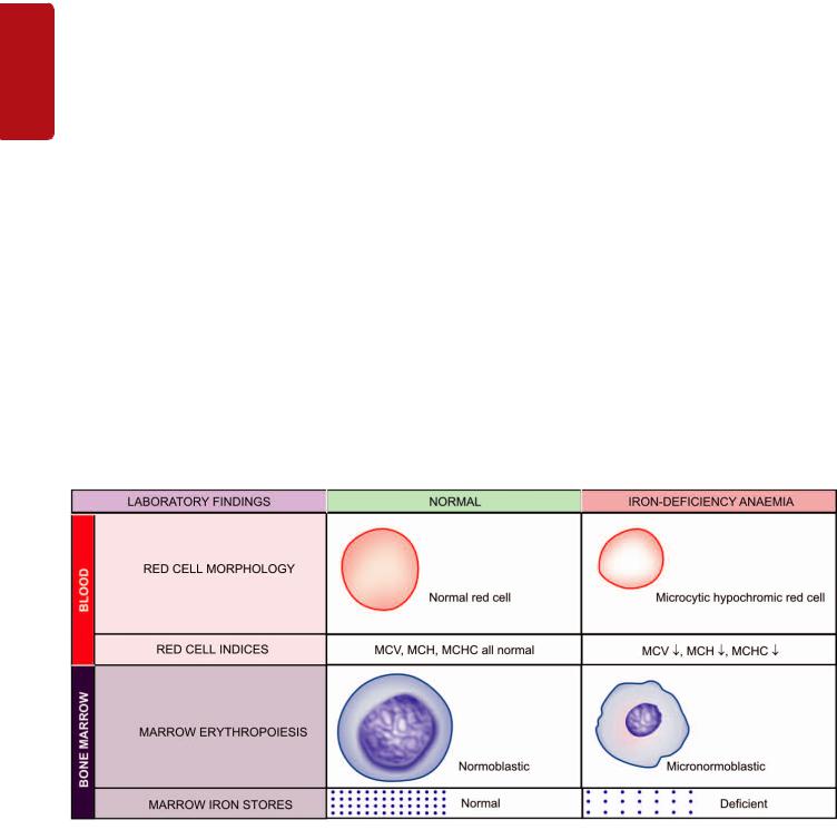
298ii) Inadequate intake. Inadequate intake of iron is prevalent in women of lower economic status. Besides diet deficient in iron, other factors such as anorexia, impaired absorption and diminished bioavailability may act as contributory factors.
iii)Increased requirements. During pregnancy and adolescence, the demand of body for iron is increased. During a normal pregnancy, about 750 mg of iron may be siphoned off from the mother—about 400 mg to the foetus, 150 mg to the placenta, and 200 mg is lost at parturition and lactation. If several pregnancies occur at short intervals, iron deficiency anaemia certainly follows.
SECTION |
2. |
POST-MENOPAUSAL FEMALES. Though the |
|
physiological demand for iron decreases after cessation of |
|||
|
|||
|
menstruation, iron deficiency anaemia may develop in post- |
||
|
menopausal women due to chronic blood loss. Following are |
||
|
among the important causes during these years: |
||
II |
i) Post-menopausal uterine bleeding due to carcinoma of the |
||
uterus. |
|||
|
|||
|
ii) Bleeding from the alimentary tract such as due to carcinoma |
||
Haematology |
of stomach and large bowel and hiatus hernia. |
||
3. ADULT MALES. It is uncommon for adult males to |
|||
|
|||
|
develop iron deficiency anaemia in the presence of normal |
||
|
dietary iron content and iron absorption. The vast majority |
||
|
of cases of iron deficiency anaemia in adult males are due to |
||
|
chronic blood loss. The cause for chronic haemorrhage may |
||
and |
lie at one of the following sites: |
||
i) |
Gastrointestinal tract is the usual source of bleeding which |
||
Lymphoreticular |
may be due to peptic ulcer, haemorrhoids, hookworm |
||
infestation, carcinoma of stomach and large bowel, |
|||
|
|||
|
oesophageal varices, hiatus hernia, chronic aspirin ingestion |
||
|
and ulcerative colitis. Other causes in the GIT are |
||
|
malabsorption and following gastrointestinal surgery. |
||
|
ii) Urinary tract e.g. due to haematuria and haemoglobinuria. |
||
|
iii) Nose e.g. in repeated epistaxis. |
||
Tissues |
iv) Lungs e.g. in haemoptysis from various causes. |
||
|
|
||
4. INFANTS AND CHILDREN. Iron deficiency anaemia is fairly common during infancy and childhood with a peak incidence at 1-2 years of age. The principal cause for anaemia at this age is increased demand of iron which is not met by the inadequate intake of iron in the diet. Normal full-term infant has sufficient iron stores for the first 4-6 months of life, while premature infants have inadequate reserves because iron stores from the mother are mainly laid down during the last trimester of pregnancy. Therefore, unless the infant is given supplemental feeding of iron or ironcontaining foods, iron deficiency anaemia develops.
Clinical Features
As already mentioned, iron deficiency anaemia is much more common in women between the age of 20 and 45 years than in men; at periods of active growth in infancy, childhood and adolescence; and is also more frequent in premature infants. Initially, there are usually no clinical abnormalities. But subsequently, in addition to features of the underlying disorder causing the anaemia, the clinical consequences of iron deficiency manifest in 2 ways—anaemia itself and epithelial tissue changes.
1.ANAEMIA. The onset of iron deficiency anaemia is generally slow. The usual symptoms are weakness, fatigue, dyspnoea on exertion, palpitations and pallor of the skin, mucous membranes and sclerae. Older patients may develop angina and congestive cardiac failure. Patients may have unusual dietary cravings such as pica. Menorrhagia is a common symptom in iron deficient women.
2.EPITHELIAL TISSUE CHANGES. Long-standing chronic iron deficiency anaemia causes epithelial tissue changes in some patients. The changes occur in the nails (koilonychia or spoon-shaped nails), tongue (atrophic glossitis), mouth (angular stomatitis), and oesophagus causing dysphagia from development of thin, membranous webs at the postcricoid area (Plummer-Vinson syndrome).
Figure 12.13 
 Laboratory findings in iron deficiency anaemia.
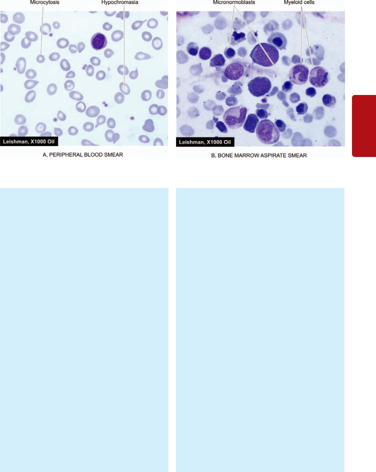
Figure 12.14 
 Iron deficiency anaemia. A, PBF showing microcytic hypochromic anaemia. There is moderate microcytosis and hypochromia. B, Examination of bone marrow aspirate showing micronormoblastic erythropoiesis.
Laboratory Findings
The development of anaemia progresses in 3 stages:
 Firstly, storage iron depletion occurs during which iron reserves are lost without compromise of the iron supply for erythropoiesis.
 The next stage is iron deficient erythropoiesis during which the erythroid iron supply is reduced without the development of anaemia.
 The final stage is the development of frank iron deficiency anaemia when the red cells become microcytic and hypochromic.
The following laboratory tests can be used to assess the varying degree of iron deficiency (Fig. 12.13):
1.BLOOD PICTURE AND RED CELL INDICES. The degree of anaemia varies. It is usually mild to moderate but occasionally it may be marked (haemoglobin less than 6 g/dl) due to persistent and severe blood loss. The salient haematological findings in these cases are as under.
i)Haemoglobin. The essential feature is a fall in haemoglobin concentration up to a variable degree.
ii)Red cells. The red cells in the blood film are hypochromic and microcytic, and there is anisocytosis and poikilocytosis (Fig. 12.14,A). Hypochromia generally precedes microcytosis. Hypochromia is due to poor filling of the red cells with haemoglobin so that there is increased central pallor. In severe cases, there may be only a thin rim of pink staining at the periphery. Target cells, elliptical forms and polychromatic cells are often present. Normoblasts are uncommon. RBC count is below normal but is generally not proportionate to the fall in haemoglobin value. When iron deficiency is associated
with severe folate or vitamin B12 deficiency, a dimorphic blood picture occurs with dual population of red cells— macrocytic as well as microcytic hypochromic.
iii)Reticulocyte count. The reticulocyte count is normal or reduced but may be slightly raised (2-5%) in cases after haemorrhage.
iv)Absolute values. The red cell indices reveal a diminished MCV (below 50 fl), diminished MCH (below 15 pg), and diminished MCHC (below 20 g/dl).
v)Leucocytes. The total and differential white cell counts are usually normal.
vi)Platelets. Platelet count is usually normal but may be slightly to moderately raised in patients who have had recent bleeding.
2.BONE MARROW FINDINGS. Bone marrow examination is not essential in such cases routinely but is done in complicated cases so as to distinguish from other hypochromic anaemias. The usual findings are as follows
(Fig.12.14,B):
i)Marrow cellularity. The marrow cellularity is increased due to erythroid hyperplasia (myeloid-erythroid ratio decreased).
ii)Erythropoiesis. There is normoblastic erythropoiesis with predominance of small polychromatic normoblasts (micronormoblasts). These normoblasts have a thin rim of cytoplasm around the nucleus and a ragged and irregular cell border. The cytoplasmic maturation lags behind so that the late normoblasts have pyknotic nucleus but persisting polychromatic cytoplasm (compared from megaloblastic anaemia in which the nuclear maturation lags behind, page 307).
iii)Other cells. Myeloid, lymphoid and megakaryocytic cells are normal in number and morphology.
iv)Marrow iron. Iron staining (Prussian blue reaction) carried out on bone marrow aspirate smear shows
299
Series Erythroid of Disorders and System Haematopoietic to Introduction 12 CHAPTER

300 deficient reticuloendothelial iron stores and absence of siderotic iron granules from developing normoblasts.
|
3.BIOCHEMICAL FINDINGS. In addition to blood and |
|
|
bone marrow examination, the following biochemical tests |
|
|
are of value: |
|
|
i) The serum iron level is low (normal 40-140 μg/dl); it is |
|
|
often under 50 μg/dl. When serum iron falls below |
|
|
15 μg/dl, marrow iron stores are absent. |
|
|
ii) Total iron binding capacity (TIBC) is high (normal 250- |
|
|
450 μg/dl) and rises to give less than 10% saturation |
|
|
(normal 33%). In anaemia of chronic disorders, however, |
|
SECTION |
serum iron as well as TIBC are reduced. |
|
iii) Serum ferritin is very low (normal 30-250 ng/ml) |
||
|
||
|
indicating poor tissue iron stores. The serum ferritin is |
|
|
raised in iron overload and is normal in anaemia of chronic |
|
|
disorders. |
|
II |
iv) Red cell protoporphyrin is very low (normal 20-40 |
|
μg/dl) as a result of insufficient iron supply to form haem. |
||
|
||
|
v) Serum transferrin receptor protein which is normally |
|
Haematology |
present on developing erythroid cells and reflects total |
|
red cell mass, is raised in iron deficiency due to its release |
||
|
||
|
in circulation (normal level 4-9 μg/L as determined by |
|
|
immunoassay). |
|
|
Treatment |
|
and |
The management of iron deficiency anaemia consists of |
|
2 essential principles: correction of disorder causing the |
||
|
||
Lymphoreticular |
anaemia, and correction of iron deficiency. |
|
1. CORRECTION OF THE DISORDER. The underlying |
||
|
||
|
cause of iron deficiency is established after thorough check- |
|
|
up and investigations. Appropriate surgical, medical or |
|
|
preventive measures are instituted to correct the cause of |
|
|
blood loss. |
|
|
2. CORRECTIONOF IRON DEFICIENCY. The lack of |
|
Tissues |
iron is corrected with iron therapy as under: |
|
i) Oral therapy. Iron deficiency responds very effectively |
||
|
||
|
to the administration of oral iron salts such as ferrous sulfate, |
|
|
ferrous fumarate, ferrous gluconate and polysaccharide iron. |
|
|
These preparations have varying amount of elemental iron |
|
|
in each tablet ranging from 39 mg to 105 mg. Optimal |
|
|
absorption is obtained by giving iron fasting, but if side- |
|
|
effects occur (e.g. nausea, abdominal discomfort, diarrhoea) |
|
|
iron can be given with food or by using a preparation of lower |
|
|
iron content (e.g. ferrous gluconate containing 39 mg |
|
|
elemental iron). Oral iron therapy is continued long enough, |
|
|
both to correct the anaemia and to replenish the body iron |
|
|
stores. The response to oral iron therapy is observed by |
|
|
reticulocytosis which begins to appear in 3-4 days with a |
|
|
peak in about 10 days. Poor response to iron replacement |
|
|
may occur from various causes such as: incorrect diagnosis, |
|
|
non-compliance, continuing blood loss, bone marrow |
|
|
suppression by tumour or chronic inflammation, and |
|
|
malabsorption. |
|
|
ii) Parenteral therapy. Parenteral iron therapy is indicated |
|
|
in cases who are intolerant to oral iron therapy, in GIT |
|
|
disorders such as malabsorption, or a rapid replenishment |
of iron stores is desired such as in women with severe anaemia a few weeks before expected date of delivery. Parenteral iron therapy is hazardous and expensive when compared with oral administration. The haematological response to parenteral iron therapy is no faster than the administration of adequate dose of oral iron but the stores are replenished much faster. Before giving the parenteral iron, total dose is calculated by a simple formula by multiplying the grams of haemoglobin below normal with 250 (250 mg of elemental iron is required for each gram of deficit haemoglobin), plus an additional 500 mg is added for building up iron stores. A common preparation is iron dextran which may be given as a single intramuscular injection, or as intravenous infusion after dilution with dextrose or saline. The adverse effects with iron dextran include hypersensitivity or anaphylactoid reactions, haemolysis, hypotension, circulatory collapse, vomiting and muscle pain. Newer iron complexes such as sodium ferric gluconate and iron sucrose can be administered as repeated intravenous injections with much lower side effects.
SIDEROBLASTIC ANAEMIA
The sideroblastic anaemias comprise a group of disorders of diverse etiology in which the nucleated erythroid precursors in the bone marrow, show characteristic ‘ringed sideroblasts.’
Siderocytes and Sideroblasts
Siderocytes and sideroblasts are erythrocytes and normoblasts respectively which contain cytoplasmic granules of iron (Fig.12.15).
SIDEROCYTES. These are red cells containing granules of non-haem iron. These granules stain positively with Prussian blue reaction as well as stain with Romanowsky dyes when they are referred to as Pappenheimer bodies. Siderocytes are normally not present in the human peripheral blood but a small number may appear following splenectomy. This is because the reticulocytes on release from the marrow are finally sequestered in the spleen to become mature red cells. In the absence of spleen, the final maturation step takes place in the peripheral blood and hence siderocytes make their appearance in the blood after splenectomy.
SIDEROBLASTS. These are nucleated red cells (normoblasts) containing siderotic granules which stain positively
Figure 12.15 
 A siderocyte containing Pappenheimer bodies, a normal sideroblast and a ring sideroblast.
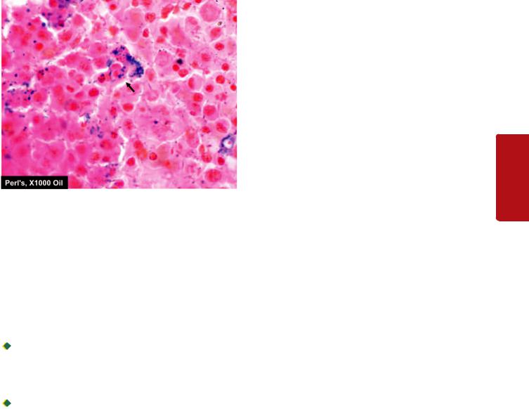
Figure 12.16 
 Sideroblastic anaemia bone marrow aspirate smear in Perls’ stain shows marked excess of reticular iron and a ringed sideroblast (arrow) showing Prusian blue granules in the cytoplasm.
with Prussian blue reaction. Depending upon the number, size and distribution of siderotic granules, sideroblasts may be normal or abnormal (Fig. 12.16).
Normal sideroblasts contain a few fine, scattered cytoplasmic granules representing iron which has not been utilised for haemoglobin synthesis. These cells comprise 30-50% of normoblasts in the normal marrow but are reduced or absent in iron deficiency.
Abnormal sideroblasts are further of 2 types:
 One type is a sideroblast containing numerous, diffusely scattered, coarse cytoplasmic granules and are seen in conditions such as dyserythropoiesis and haemolysis. In this type, there is no defect of haem or globin synthesis but the percentage saturation of transferrin is increased.

 The other type is ringed sideroblast in which haem synthesis is disturbed as occurs in sideroblastic anaemias. Ringed sideroblasts contain numerous large granules, often forming a complete or partial ring around the nucleus. The ringed arrangement of these granules is due to the presence of iron-laden mitochondria around the nucleus.
Types of Sideroblastic Anaemias
Based on etiology, sideroblastic anaemias are classified into hereditary and acquired types. The acquired type is further divided into primary and secondary forms:
I. HEREDITARY SIDEROBLASTIC ANAEMIA. This is a rare X-linked disorder associated with defective enzyme activity of aminolevulinic acid (ALA) synthetase required for haem synthesis. The affected males have moderate to marked anaemia while the females are carriers of the disorder and do not develop anaemia. The condition manifests in childhood or in early adult life.
II. ACQUIRED SIDEROBLASTIC ANAEMIA. The acquired sideroblastic anaemias are classified into primary and secondary types.
A. Primary acquired sideroblastic anaemia. Primary, idiopathic, or refractory acquired sideroblastic anaemia occurs
spontaneously in middle-aged and older individuals of both |
301 |
||
sexes. The disorder has its pathogenesis in disturbed growth |
|
||
and maturation of erythroid precursors at the level of |
|
||
haematopoietic stem cell, possibly due to reduced activity |
|
||
of the enzyme, ALA synthetase. The anaemia is of moderate |
|
||
to severe degree and appears insidiously. The bone marrow |
|
||
cells commonly show chromosomal abnormalities, neutro- |
|
||
penia and thrombocytopenia with associated bleeding |
|
||
diathesis. The spleen and liver may be either normal or mildly |
|
||
enlarged, while the lymph nodes are not enlarged. Unlike |
|
||
other types of sideroblastic anaemia, this type is regarded as |
|
||
a myelodysplastic disorder in the FAB (French-American- |
|
||
British) classification and thus, can be a preleukaemic |
CHAPTER |
||
disorder (page 361). About 10% of individuals with refractory |
|||
|
|||
acquired sideroblastic anaemia develop acute myelogenous |
|
||
leukaemia. |
|
||
B. Secondary acquired sideroblastic anaemia. Acquired |
|
||
sideroblastic anaemia may develop secondary to a variety |
12 |
||
of drugs, chemicals, toxins, haematological and various other |
|||
diseases. |
|
||
1. Drugs, chemicals and toxins: Isoniazid, an anti-tuberculous |
Introduction |
||
are: cycloserine, chloramphenicol and alkylating agents (e.g. |
|||
drug and a pyridoxine antagonist, is most commonly |
|
||
associated with development of sideroblastic anaemia by |
|
||
producing abnormalities in pyridoxine metabolism. Other |
|
||
drugs occasionally causing acquired sideroblastic anaemia |
|
||
cyclophosphamide). Alcohol and lead also cause |
to |
||
sideroblastic anaemia. All these agents cause reversible |
|||
Haematopoietic |
|||
sideroblastic anaemia which usually resolves following |
|||
|
|||
removal of the offending agent. |
|
||
2. Haematological disorders: These include myelofibrosis, |
|
||
polycythaemia vera, acute leukaemia, myeloma, lymphoma |
|
||
and haemolytic anaemia. |
|
||
3. Miscellaneous: Occasionally, secondary sideroblastic |
|
||
anaemia may occur in association with a variety of inflam- |
System |
||
matory, neoplastic and autoimmune diseases such as |
|||
|
|||
carcinoma, myxoedema, rheumatoid arthritis and SLE. |
|
||
|
|
||
Laboratory Findings |
|
and |
|
Sideroblastic anaemias usually show the following |
|
||
haematological features: |
|
Disorders |
|
1. There is generally moderate to severe degree of anaemia. |
|
||
|
|
||
2.The blood picture shows hypochromic anaemia which |
|
|
|
may be microcytic, or there may be some normocytic red |
|
|
|
cells as well (dimorphic). |
|
of |
|
3. Absolute values (MCV, MCH and MCHC) are reduced |
|
||
|
Erythroid |
||
in hereditary type but MCV is often raised in acquired |
|
||
|
|
||
type. |
|
|
|
4. Bone marrow examination shows erythroid hyperplasia |
|
|
|
with usually macronormoblastic erythropoiesis. Marrow |
|
Series |
|
iron stores are raised and pathognomonic ring sideroblasts |
|
||
|
|
||
are present. |
|
|
|
5. Serum ferritin levels are raised. |
|
|
|
6. Serum iron is usually raised with almost complete |
|
|
|
saturation of TIBC. |
|
|
|
7. There is increased iron deposition in the tissue. |
|
|
|
|
|
|
|

302
Tissues Lymphoreticular and Haematology II SECTION
TABLE 12.5: Laboratory Diagnosis of Hypochromic Anaemias.
|
Test |
Iron Deficiency |
Chronic Disorders |
Thalassaemia |
Sideroblastic Anaemia |
|
|
|
|
|
|
1. |
MCV, MCH, MCHC |
Reduced |
Low normal-to-reduced |
Very low |
Very low |
|
|
|
|
|
(except MCV raised in aquired type) |
2. |
Serum iron |
Reduced |
Reduced |
Normal |
Raised |
3. |
TIBC |
Raised |
Low-to-normal |
Normal |
Normal |
4. |
Serum ferritin |
Reduced |
Raised |
Normal |
Raised |
|
|
|
|
|
(complete saturation) |
5. |
Marrow-iron stores |
Absent |
Present |
High |
High |
6. |
Iron in normoblasts |
Absent |
Absent |
Present |
Ring sideroblasts |
7. |
Hb electrophoresis |
Normal |
Normal |
Abnormal |
Normal |
|
|
|
|
|
|
Treatment
The treatment of secondary sideroblastic anaemia is primarily focussed on removal of the offending agent. No definite treatment is available for hereditary and idiopathic types of sideroblastic anaemias. However, pyridoxine is administered routinely to all cases of sideroblastic anaemia (200 mg per day for 2-3 months). Blood transfusions and other supportive therapy are indicated in all patients.
Differential diagnosis of various types of hypochromic anaemias by laboratory tests is summarised in Table 12.5.
ANAEMIA OF CHRONIC DISORDERS
One of the commonly encountered anaemia is in patients of a variety of chronic systemic diseases in which anaemia develops secondary to a disease process but there is no actual invasion of the bone marrow. A list of such chronic systemic diseases is given in Table 12.6. In general, anaemia in chronic disorders is usually normocytic normochromic but can have mild degree of microcytosis and hypochromia unrelated to iron deficiency. The severity of anaemia is usually directly related to the primary disease process. The anaemia is corrected only if the primary disease is alleviated.
Pathogenesis
A number of factors may contribute to the development of anaemia in chronic systemic disorders, and in many condi-
TABLE 12.6: Anaemias Secondary to Chronic Systemic Disorders.
1.ANAEMIA IN CHRONIC INFECTIONS/INFLAMMATION
a.Infections e.g. tuberculosis, lung abscess, pneumonia, osteomyelitis, subacute bacterial endocarditis, pyelonephritis.
b.Non-infectious inflammations e.g. rheumatoid arthritis, SLE, vasculitis, dermatomyositis, scleroderma, sarcoidosis, Crohn’s disease.
c.Disseminated malignancies e.g. Hodgkin’s disease, disseminated carcinomas and sarcomas.
2.ANAEMIA OF RENAL DISEASE e.g. uraemia, renal failure
3.ANAEMIA OF HYPOMETABOLIC STATE e.g.
endocrinopathies (myxoedema, Addison's disease, hyperthyroidism, hypopituitarism, Addison’s disease), protein malnutrition, scurvy and pregnancy, liver disease.
tions, the anaemia is complicated by other causes such as iron, B12 and folate deficiency, hypersplenism, renal failure with consequent reduced erythropoietic activity, endocrine abnormalities etc. However, in general, 2 factors appear to play significant role in the pathogenesis of anaemia in chronic disorders. These are: defective red cell production and reduced
red cell lifespan.
1.Defective red cell production. Though there is abundance of storage iron in these conditions but the amount of iron available to developing erythroid cells in the marrow is subnormal. The mononuclear phagocyte system is hyperplastic which traps all the available free iron due to the activity of iron binding protein, lactoferrin. A defect in the transfer of iron from macrophages to the developing erythroid cells in the marrow leads to reduced availability of iron for haem synthesis despite adequate iron stores, elevating serum ferritin levels. The defect lies in suppression of erythropoieitn by inflammatory cytokines at some stage in erythropoiesis, and hepcidin which is the key iron regulatory hormone. These inflammatory cytokines include TNF and IFN-β released in bacterial infections and tumours, and IL-1 and IFN-γ released in patients of rheumatoid arthritis and autoimmune vasculitis (Fig. 12.17).
2.Reduced red cell lifespan. Decreased survival of circulating red cells in chronic renal disease is attributed to hyperplastic mononuclear phagocyte system.
Laboratory Findings
The characteristic features of anaemia in these patients uncomplicated by other deficiencies are as under:
i)Haemoglobin. Anaemia is generally mild to moderate. A haemoglobin value of less than 8 g/dl suggests the presence of additional contributory factors.
ii)Blood picture. The type of anaemia in these cases is generally normocytic normochromic but may have slight microcytosis and hypochromia.
iii)Absolute values. Red cell indices indicate that in spite of normocytic normochromic anaemia, MCHC is slightly low.
iv)Reticulocyte count. The reticulocyte count is generally low.
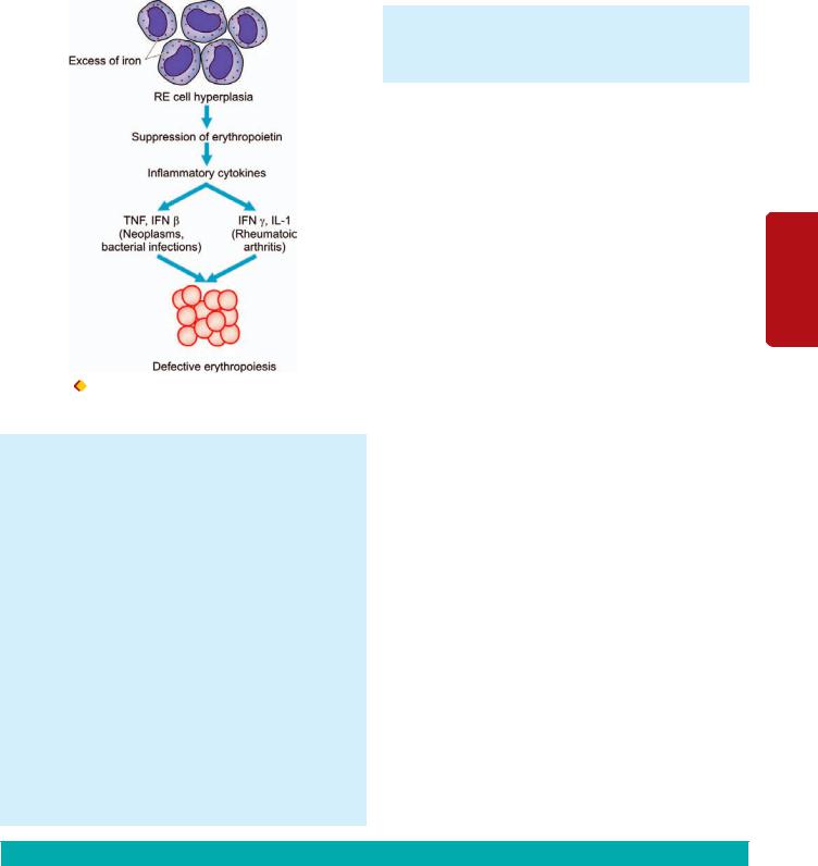
Figure 12.17 |
Pathogenesis of anaemia of chronic disorders through |
suppression of erythropoiesis by cytokines.
v)Red cell survival. Measurement of erythrocyte survival generally reveals mild to moderate shortening of their lifespan.
vi)Bone marrow. Examination of the marrow generally reveals normal erythroid maturation. However, the red cell precursors have reduced stainable iron than normal, while macrophages in the marrow usually contain increased amount of iron. Cases of chronic infection often have myeloid hyperplasia and increase in plasma cells.
vii)Serum iron and TIBC. Serum iron is characteristically reduced in this group of anaemias while TIBC is low-to- normal (in contrast to iron deficiency where there is reduction in serum iron but high TIBC, see Table12.5).
viii)Serum ferritin. Serum ferritin levels are increased in these patients and is the most distinguishing feature between true iron-deficiency anaemia and iron-deficient erythropoieisis in anemia of chronic diseases.
ix)Other plasma proteins. In addition, certain other plasma proteins called ‘phase reactants’ are raised in patients with chronic inflammation, probably under the stimulus of interleukin-1 released by activated
macrophages. These proteins include γ-globulin, C3, haptoglobin, α1-antitrypsin and fibrinogen. Elevation of these proteins is responsible for raised ESR commonly present in these patients.
MEGALOBLASTIC ANAEMIA
The megaloblastic anaemias are disorders caused by impaired DNA synthesis and are characterised by a distinctive abnormality in the haematopoietic precursors in the bone marrow in which the maturation of the nucleus is delayed relative to that of the cytoplasm. Since cell division is slow but cytoplasmic development progresses normally, the nucleated red cell precursors tend to be larger which Ehrlich in 1880 termed megaloblasts. Megaloblasts are both morphologically and functionally abnormal with the result that the mature red cells formed from them and released into the peripheral blood are also abnormal in shape and size, the most prominent abnormality being macrocytosis.
The underlying defect for the asynchronous maturation of the nucleus is defective DNA synthesis due to deficiency of vitamin B12 (cobalamin) and/or folic acid (folate). Less common causes are interference with DNA synthesis by congenital or acquired abnormalities of vitamin B12 or folic acid metabolism. Before considering the megaloblastic anaemia, an outline of vitamin B12 and folic acid metabolism is given for a better understanding of the subject.
The salient nutritional aspects and metabolic functions of vitamin B12 and folic acid are summarised in Table12.7.
Vitamin B12 Metabolism
BIOCHEMISTRY. Vitamin B12 or cobalamin is a complex organometallic compound having a cobalt atom situated within a corrin ring, similar to the structure of porphyrin from which haem is formed. In humans, there are 2 metabolically active forms of cobalamin—methyl- cobalamin and adenosyl-cobalamin, which act as coenzymes. The therapeutic vitamin B12 preparation is called cyanocobalamin.
SOURCES. The only dietary sources of vitamin B12 are foods of animal protein origin such as kidney, liver, heart, muscle meats, fish, eggs, cheese and milk. In contrast to folate, fruits and vegetables contain practically no vitamin B12 unless contaminated with bacteria. Cooking has little effect on its activity. Vitamin B12 is synthesised in the human large bowel by microorganisms but is not absorbed from this site and,
TABLE 12.7: Salient Features of Vitamin B12 and Folate Metabolism.
|
Feature |
Vitamin B12 |
Folate |
1. |
Main foods |
Animal proteins only |
Green vegetables, meats |
2. |
Cooking |
Little effect |
Easily destroyed |
3. |
Daily requirements |
2-4 μg |
100-200 μg |
4. |
Daily intake |
5-30 μg |
100-500 μg |
5. |
Site of absorption |
Ileum |
Duodenum and jejunum |
6. |
Mechanism of absorption |
Intrinsic factor |
Conversion to methyl-THF |
7. |
Body stores |
2-3 mg (enough for 2-4 yrs) |
10-12 mg (enough for 4 months) |
|
|
|
|
303
Series Erythroid of Disorders and System Haematopoietic to Introduction 12 CHAPTER
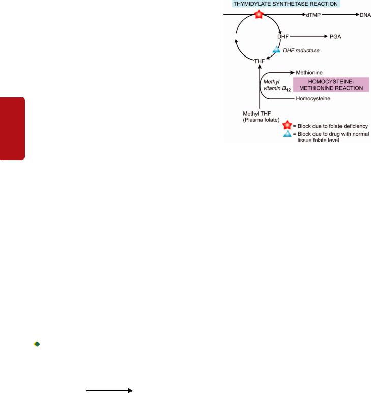
304 thus, the humans are entirely dependent upon dietary sources. The average daily requirement for vitamin B12 is 2-4 μg.
ABSORPTION. After ingestion, vitamin B12 in food is released and forms a stable complex with gastric R-binder. R-binder is a form of glycoprotein found in various secretions
|
(e.g. saliva, milk, gastric juice, bile), phagocytes and plasma. |
|
|
On entering the duodenum, the vitamin B12-R-binder |
|
|
complex is digested releasing vitamin B12 which then binds |
|
|
to intrinsic factor (IF). The IF is a glycoprotein of molecular |
|
|
weight 50,000 produced by the parietal cells of the stomach |
|
|
and its secretion roughly parallels that of hydrochloric acid. |
|
SECTION |
The vitamin B12-IF complex, on reaching the distal ileum, |
|
binds to the specific receptors on the mucosal brush border, |
||
thereby enabling the vitamin to be absorbed. The IF, |
||
therefore, acts as cell-directed carrier protein similar to |
||
transferrin. The receptor-bound vitamin B12-IF complex is |
||
II |
taken into the ileal mucosal cells where after several hours |
|
the IF is destroyed, vitamin B12 released and is transferred |
||
|
||
|
to another transport protein, transcobalamin (TC) II. The |
|
Haematology |
vitamin B12-TC II complex is finally secreted into the portal |
|
circulation from where it is taken by the liver, bone marrow |
||
|
||
|
and other cells. There are 2 major vitamin B12 binding |
|
|
proteins—TC I and TC II, and a minor protein TC III. TC I is |
|
|
not essential for vitamin B12 transport but functions primarily |
|
|
as a storage protein while TC III is similar to TC II and binds |
|
and |
a small amount of vitamin B12 (see Fig. 12.12,B). |
|
TISSUE STORES. Normally, the liver is the principal storage |
||
Lymphoreticular |
site of vitamin B12 and stores about 2 mg of the vitamin, while |
|
other tissues like kidney, heart and brain together store about |
||
|
||
|
2 mg. The body stores of vitamin B12 are adequate for 2-4 |
|
|
years. Major source of loss is via bile and shedding of |
|
|
intestinal epithelial cells. A major part of the excreted vitamin |
|
|
B12 is reabsorbed in the ileum by the IF resulting in |
|
|
enterohepatic circulation. |
|
Tissues |
FUNCTIONS. Vitamin B12 plays an important role in general |
|
nervous system. Vitamin B12 acts as a co-enzyme for 2 main |
||
|
cell metabolism, particulary essential for normal |
|
|
haematopoiesis and for maintenance of integrity of the |
|
|
biochemical reactions in the body: |
|
|
Firstly, as methyl cobalamin (methyl B12) in the methylation |
|
|
of homocysteine to methionine by methyl tetrahydrofolate |
|
|
(THF). The homocysteine-methionine reaction is closely |
|
|
linked to folate metabolism (Fig. 12.18): |
Methyl B
Homocysteine |
12 |
Methionine |
|
When this reaction is impaired, folate metabolism is deranged and results in defective DNA synthesis responsible for megaloblastic maturation.
Secondly, as adenosyl cobalamin (adenosyl B12) in propionate metabolism for the conversion of methyl malonyl co-enzyme A to succinyl co-enzyme A:
Adenosyl B12
Propionyl CoA → Methyl malonyl CoA → Succinyl CoA
Figure 12.18 
 Biochemical basis of megaloblastic anaemia (THF = tetrahydrofolate; DHF = dihydrofolate; PGA = pteroyl glutamic acid; dUMP = deoxy uridylate monophosphate; dTMP = deoxy thymidylate monophosphate).
Lack of adenosyl B12 leads to large increase in the level of methyl malonyl CoA and its precursor, propionyl CoA. This results in synthesis of certain fatty acids which are incorporated into the neuronal lipids. This biochemical abnormality may contribute to the neurologic complications of vitamin B12 deficiency.
Folate Metabolism
BIOCHEMISTRY. Folate or folic acid, a yellow compound, is a member of water-soluble B complex vitamins with the chemical name of pteroyl glutamic acid (PGA). Folic acid does not exist as such in nature but exists as folates in polyglutamate form (conjugated folates). For its metabolic action as co-enzyme, polyglutamates must be reduced to dihydroand tetrahydrofolate forms.
SOURCES. Folate exists in different plants, bacteria and animal tissues. Its main dietary sources are fresh green leafy vegetables, fruits, liver, kidney, and to a lesser extent, muscle meats, cereals and milk. Folate is labile and is largely destroyed by cooking and canning. Some amount of folate synthesised by bacteria in the human large bowel is not available to the body because its absorption takes place in the small intestine. Thus, humans are mainly dependent upon diet for its supply. The average daily requirement is 100-200 μg.
ABSORPTION AND TRANSPORT. Folate is normally absorbed from the duodenum and upper jejunum and to a lesser extent, from the lower jejunum and ileum. However, absorption depends upon the form of folate in the diet. Polyglutamate form in the foodstuffs is first cleaved by the enzyme, folate conjugase, in the mucosal cells to monoand diglutamates which are readily assimilated. Synthetic folic acid preparations in polyglutamate form are also absorbed as rapidly as monoand diglutamate form because of the
absence of natural inhibitors. Monoand diglutamates undergo further reduction in the mucosal cells to form tetrahydrofolate (THF), a monoglutamate. THF circulates in the plasma as methylated compound, methyl THF, bound to a protein. Once methyl THF is transported into the cell by a carrier protein, it is reconverted to polyglutamate (see
Fig. 12.12,C).
TISSUE STORES. The liver and red cells are the main storage sites of folate, largely as methyl THF polyglutamate form. The total body stores of folate are about 10-12 mg enough for about 4 months. Normally, folate is lost from the sweat, saliva, urine and faeces.
FUNCTIONS. Folate plays an essential role in cellular metabolism. It acts as a co-enzyme for 2 important biochemical reactions involving transfer of 1-carbon units (viz. methyl and formyl groups) to various other compounds. These reactions are as under:

 Thymidylate synthetase reaction. Formation of deoxy thymidylate monophosphate (dTMP) from its precursor form, deoxy uridylate monophosphate (dUMP).
 Methylation of homocysteine to methionine. This reaction is linked to vitamin B12 metabolism (Fig. 12.18).
These biochemical reactions are considered in detail below together with biochemical basis of the megaloblastic anaemia.
Biochemical Basis of Megaloblastic Anaemia
The basic biochemical abnormality common to both vitamin B12 and folate deficiency is a block in the pathway of DNA synthesis and that there is an inter-relationship between vitamin B12 and folate metabolism in the methylation reaction of homocysteine to methionine (Fig. 12.18).
As stated above, folate as co-enzyme methylene THF, is required for transfer of 1-carbon moieties (e.g. methyl and formyl) to form building blocks in DNA synthesis. These 1-carbon moieties are derived from serine or formiminoglutamic acid (FIGLU). Two of the important folatedependent (1-carbon transfer) reactions for formation of building blocks in DNA synthesis are as under:
1.Thymidylate synthetase reaction. This reaction involves synthesis of deoxy thymidylate monophosphate (dTMP) from deoxy uridylate monophosphate (dUMP). The methyl group of dUMP → dTMP reaction is supplied by the coenzyme, methylene-THF. After the transfer of 1-carbon from methylene-THF, dihydrofolate (DHF) is produced which must be reduced to active THF by the enzyme DHF-reductase before it can participate in further 1-carbon transfer reaction. Drugs like methotrexate (anti-cancer) and pyrimethamine (antimalarial) are inhibitory to the enzyme, DHF-reductase, thereby inhibiting the DNA synthesis.
2.Homocysteine-methionine reaction. Homocysteine is converted into methionine by transfer of a methyl group from methylene-THF. After transfer of 1-carbon from methyleneTHF, THF is produced. This reaction requires the presence
of vitamin B12 (methyl-B12).
Deficiency of folate from any cause results in reduced supply of the coenzyme, methylene-THF, and thus interferes
with the synthesis of DNA. Deficiency of vitamin B12 traps folate as its transport form, methyl-THF, thereby resulting in reduced formation of the active form, methylene-THF, needed for DNA synthesis. This is referred to as methyl-folate trap hypothesis. An alternative hypothesis of inter-relationship of B12 and folate is the formate-saturation hypothesis. According to this hypothesis, the active substrate is formyl-THF. Vitamin B12 deficiency results in reduced supply of formate to THF causing reduced generation of the active compound, formyl THF.
Etiology and Classification of Megaloblastic Anaemia
The etiology of megaloblastic anaemia varies in different parts of the world. As outlined in Table 12.8,megaloblastic anaemia is classified into 3 broad groups: vitamin B12 deficiency, folate deficiency, and deficiency from other causes.
1.VITAMIN B12 DEFICIENCY. In Western countries, deficiency of vitamin B12 is more commonly due to pernicious (Addisonian) anaemia. True vegetarians like traditional Indian Hindus and breast-fed infants have dietary lack of
vitamin B12. Gastrectomy by lack of intrinsic factor, and small intestinal lesions involving distal ileum where absorption of
vitamin B12 occurs, may cause deficiency of the vitamin. Deficiency of vitamin B12 takes at least 2 years to develop when the body stores are totally depleted.
2.FOLATE DEFICIENCY. Folate deficiency is more often due to poor dietary intake. Other causes include
TABLE 12.8: Etiologic Classification of Megaloblastic Anaemia.
I.VITAMIN B12 DEFICIENCY
A.Inadequate dietary intake e.g. strict vegetarians, breast-fed infants.
B.Malabsorption
1.Gastric causes: pernicious anaemia, gastrectomy, congenital lack of intrinsic factor.
2.Intestinal causes: tropical sprue, ileal resection, Crohn’s disease, intestinal blind loop syndrome, fish-tapeworm infestation.
II. FOLATE DEFICIENCY
A.Inadequate dietary intake e.g. in alcoholics, teenagers, infants, old age, poverty.
B.Malabsorption e.g. in tropical sprue, coeliac disease, partial gastrectomy, jejunal resection, Crohn’s disease.
C.Excess demand
1.Physiological: pregnancy, lactation, infancy.
2.Pathological : malignancy, increased haematopoiesis, chronic exfoliative skin disorders, tuberculosis, rheumatoid arthritis.
D.Excess urinary folate loss e.g. in active liver disease, congestive heart failure.
III. OTHER CAUSES
A.Impaired metabolism e.g. inhibitors of dihydrofolate (DHF) reductase such as methotrexate and pyrimethamine; alcohol, congenital enzyme deficiencies.
B.Unknown etiology e.g. in Di Guglielmo’s syndrome, congenital dyserythropoietic anaemia, refractory megaloblastic anaemia.
305
Series Erythroid of Disorders and System Haematopoietic to Introduction 12 CHAPTER
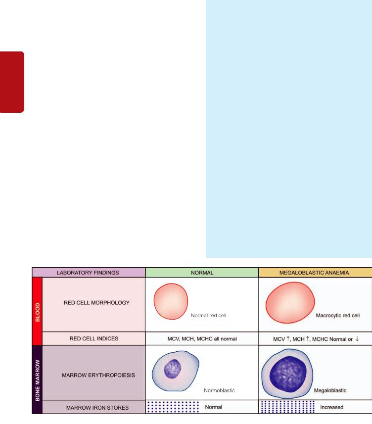
306 malabsorption, excess folate utilisation such as in pregnancy and in various disease states, chronic alcoholism, and excess urinary folate loss. Folate deficiency arises more rapidly than vitamin B12 deficiency since the body’s stores of folate are relatively low which can last for up to 4 months only.
Patients with tropical sprue are often deficient in both vitamin B12 and folate. Combined deficiency of vitamin B12 and folate may occur from severe deficiency of vitamin B12 because of the biochemical interrelationship with folate metabolism.
|
3. OTHER CAUSES. In addition to deficiency of vitamin |
|
|
B12 and folate, megaloblastic anaemias may occasionally be |
|
SECTION |
induced by other factors unrelated to vitamin deficiency. |
|
These include many drugs which interfere with DNA |
||
|
||
|
synthesis, acquired defects of haematopoietic stem cells, and |
|
|
rarely, congenital enzyme deficiencies. |
II |
Clinical Features |
|
Deficiency of vitamin B12 and folate may cause following |
||
|
||
Haematology |
clinical manifestations which may be present singly or in |
|
combination and in varying severity: |
||
1. Anaemia. Macrocytic megaloblastic anaemia is the |
||
|
||
|
cardinal feature of deficiency of vitamin B12 and/or folate. |
|
|
The onset of anaemia is usually insidious and gradually |
|
|
progressive. |
|
|
2. Glossitis. Typically, the patient has a smooth, beefy, red |
|
and |
tongue. |
|
3. Neurologic manifestations. Vitamin B12 deficiency, parti- |
||
Lymphoreticular |
cularly in patients of pernicious anaemia, is associated with |
|
symptoms include numbness, paraesthesia, weakness, |
||
|
significant neurological manifestations in the form of |
|
|
subacute combined, degeneration of the spinal cord and |
|
|
peripheral neuropathy (Chapter 30), while folate deficiency |
|
|
may occasionally develop neuropathy only. The underlying |
|
|
pathologic process consists of demyelination of the peripheral |
|
|
nerves, the spinal cord and the cerebrum. Signs and |
|
Tissues |
ataxia, poor finger coordination and diminished reflexes. |
|
|
4. Others. In addition to the cardinal features mentioned above, patients may have various other symptoms. These include: mild jaundice, angular stomatitis, purpura, melanin pigmentation, symptoms of malabsorption, weight loss and anorexia.
Laboratory Findings
The investigations of a suspected case of megaloblastic anaemia are aimed at 2 aspects:
A.General laboratory investigations of anaemia which include blood picture, red cell indices, bone marrow findings, and biochemical tests.
B.Special tests to establish the cause of megaloblastic anaemia
as to know whether it is due to deficiency of vitamin B12 or folate.
Based on these principles, the following scheme of investigations is followed:
A. General Laboratory Findings
1. BLOOD PICTURE AND RED CELL INDICES. Estimation of haemoglobin, examination of a blood film and evaluation of absolute values are essential preliminary investigations (Fig. 12.19):
i)Haemoglobin. Haemoglobin estimation reveals values below the normal range. The fall in haemoglobin concentration may be of a variable degree.
ii)Red cells. Red blood cell morphology in a blood film shows the characteristic macrocytosis. However, macrocytosis can also be seen in several other disorders such as: haemolysis, liver disease, chronic alcoholism, hypothyroidism, aplastic anaemia, myeloproliferative disorders and reticulocytosis. In addition, the blood smear demonstrates marked anisocytosis, poikilocytosis and presence of macroovalocytes. Basophilic stippling and occasional normoblast may also be seen (Fig. 12.20, A).
iii)Reticulocyte count. The reticulocyte count is generally low to normal in untreated cases.
Figure 12.19 
 General laboratory findings in megaloblastic anaemia.
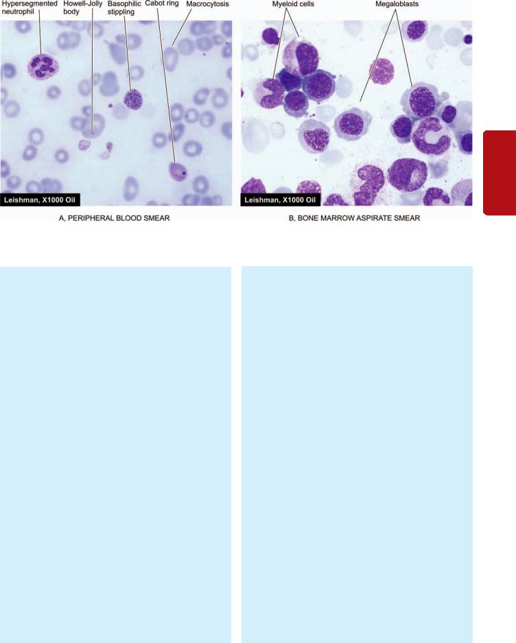
307
12 CHAPTER
Figure 12.20 
 Megaloblastic anaemia. A, PBF showing prominent macrocytosis of red cells and hypersegmented neutrophils. B, Examination of bone marrow aspirate showing megaloblastic erythropoiesis.
iv) Absolute values. The red cell indices reveal an elevated MCV (above 120 fl) proportionate to the severity of macrocytosis, elevated MCH (above 50 pg) and normal or reduced MCHC.
v)Leucocytes. The total white blood cell count may be reduced. Presence of characteristic hypersegmented neutrophils (having more than 5 nuclear lobes) in the blood film should raise the suspicion of megaloblastic anaemia. An occasional myelocyte may also be seen.
vi)Platelets. Platelet count may be moderately reduced in severely anaemic patients. Bizarre forms of platelets may be seen.
2. BONE MARROW FINDINGS. The bone marrow examination is very helpful in the diagnosis of megaloblastic anaemia. Significant findings of marrow examination are as under (Fig. 12.20,B):
i)Marrow cellularity. The marrow is hypercellular with a decreased myeloid-erythroid ratio.
ii)Erythropoiesis. Erythroid hyperplasia is due to characteristic megaloblastic erythropoiesis. Megaloblasts are abnormal, large, nucleated erythroid precursors, having nuclear-cytoplasmic asynchrony i.e. the nuclei are less mature than the development of cytoplasm. The nuclei are large, having fine, reticular and open chromatin that stains lightly, while the haemoglobinisation of the cytoplasm proceeds normally or at a faster rate i.e. nuclear maturation lags behind that of cytoplasm (compared from iron deficiency anaemia in which cytoplasmic maturation lags behind, page 299). Megaloblasts with abnormal mitoses may be seen. Features of ineffective erythropoiesis such as presence of degenerated erythroid precursors may be present.
iii) Other cells. Granulocyte precursors are also affected to some extent. Giant forms of metamyelocytes and band cells may be present in the marrow. Megakaryocytes are usually present in normal number but may occasionally be decreased and show abnormal morphology such as hypersegmented nuclei and agranular cytoplasm.
iv)Marrow iron. Prussian blue staining for iron in the marrow shows an increase in the number and size of iron granules in the erythroid precursors. Ring sideroblasts are, however, rare. Iron in the reticulum cells is increased.
v) Chromosomes. Marrow cells may show variety of random chromosomal abnormalities such as chromosome breaks, centromere spreading etc.
3.BIOCHEMICAL FINDINGS. In addition to the general blood and marrow investigations and specific tests to determine the cause of deficiency (described below), the following biochemical abnormalities are observed in cases of megaloblastic anaemia:
i)There is rise in serum unconjugated bilirubin and LDH as a result of ineffective erythropoiesis causing marrow cell breakdown.
ii)The serum iron and ferritin may be normal or elevated.
B. Special Tests for Cause of Specific Deficiency
In evaluating a patient of megaloblastic anaemia, it is important to determine the specific vitamin deficiency by assay of vitamin B12 and folate. In sophisticated clinical laboratories, currently automated multiparametric, random access analysers are employed based on separation techniques by chemiluminescence and enzyme-linked fluorescence detection systems which have largely
Series Erythroid of Disorders and System Haematopoietic to Introduction

308replaced the traditional microbiologic assays for vitamin B12 and folate. Traditional tests are briefly described below.
TESTS FOR VITAMIN B12 DEFICIENCY. The normal range of vitamin B12 in serum is 280-1000 pg/ml. Values less than 100 pg/ml indicate clinically deficient stage.
Traditional tests employed to establish vitamin B12 deficiency are serum vitamin B12 assay, Schilling (24-hour urinary excretion) test and serum enzyme levels.
1. SERUM VITAMIN B12 ASSAY. Assay of vitamin B12 in blood can be done by 2 methods—microbiological assay and radioassay.
SECTION |
i) Microbiological assay. In this test, the serum sample to |
||
be assayed is added to a medium containing all other |
|||
essential growth factors required for |
a vitamin |
||
B12-dependent microorganism. The medium along with |
|||
microorganism is incubated and the amount of vitamin |
|||
II |
B12 is determined turbimetrically which is then compared |
||
|
with the growth produced by a known amount of vitamin |
||
Haematology |
B12. Several organisms have been used for this test such |
||
as Euglena gracilis, Lactobacillus leichmannii, Escherichia coli |
|||
|
|||
|
and Ochromonas malhamensis. E. gracilis is, however, consi- |
||
|
dered more sensitive and accurate. The addition of anti- |
||
|
biotics to the test interferes with the growth and yields |
||
|
false low result. |
|
|
and |
ii) Radioassay. Assays of serum B12 by radioisotope |
||
dilution (RID) and radioimmunoassay (RIA) have been |
|||
developed. These tests are more sensitive and have the |
|||
Lymphoreticular |
advantage over microbiologic assays in that they are |
||
simpler and more rapid, and the results are unaffected by |
|||
|
|||
|
antibiotics and other drugs which may affect the living |
||
|
organisms. |
|
|
|
2. SCHILLING TEST (24 HOUR URINARY EXCRETION |
||
|
TEST). Schilling test is done to detect vitamin B12 |
||
|
deficiency as well as to distinguish and detect lack of IF |
||
Tissues |
and malabsorption syndrome. The results of test also |
||
or 57Co. The test is performed in 3 stages as under: |
|||
|
depend upon good renal function and proper urinary |
||
|
collection. Radioisotope used for labeling B |
is either 58Co |
|
|
12 |
|
|
Stage I: Without IF. The patient after an overnight fasting is administered oral dose of 1 μg of radioactively labelled vitamin B12 (‘hot’ B12) in 200 ml of water. At the same time, 1 mg of unlabelled vitamin B12 (‘cold’ B12) is given by intramuscular route; this ‘cold’ B12 will saturate the serum as well as the tissue binding sites. The patient is kept fasting for a further period of 2 hours, following which urinary excretion of B12 is estimated:
 In normal individuals, 24-hour urinary excretion is >10% of the oral dose of ‘hot’ B12.
 Patients with IF deficiency excrete lower quantity of ‘hot’ B12 which is further confirmed by repeating the test as in stage II given below.
Stage II: With IF. If the 24-hour urinary excretion of ‘hot’ B12 is low, the test is repeated using the same procedure as in stage I but in addition high oral dose of IF is administered along with ‘hot’ B12.
 If the 24-hour urinary excretion of ‘hot’ B12 is now normal, the low value in first stage of the test was due to IF deficiency (i.e. pernicious anaemia).
 Patients with pernicious anaemia have abnormal test even after treatment with vitamin B12 due to IF deficiency. However, abnormal 24-hour urinary excretion of ‘hot’ B12 is further investigated in stage III for a cause in intestinal malabsorption of ‘hot’ B’12.
Stage III: Test for malabsorption of vitamin B12. Some patients absorb vitamin B12 in water as was stipulated in the original Schilling test. Modified Schilling test employs the use of protein-bound vitamin B12. In conditions causing malabsorption, the test is repeated after a course of treatment with antibiotics or anti-inflammatory drugs.
3. SERUM ENZYME LEVELS. Besides Schilling test, another way of distinguishing whether megaloblastic anaemia is due to cobalamine or folate is by serum determination of methylmalonic acid and homocysteine by sophisticated enzymatic assays. Both are elevated in cobalamine deficiency, while in folate deficiency there is only elevation of homocysteine and not of methylmalonic acid.
TESTS FOR FOLATE DEFICIENCY. The normal range of serum folate is 6-18 ng/ml. Values of 4 ng/ml or less are generally considered to be diagnostic of folate deficiency. Measurement of formiminoglutamic acid (FIGLU) urinary excretion after histidine load was used formerly for assessing folate status but it is less specific and less sensitive than the serum assays. Currently, there are 3 tests used to detect folate deficiency—urinary excretion of FIGLU, serum and red cell folate assay.
1.URINARY EXCRETION OF FIGLU. Folic acid is required for conversion of formiminoglutamic acid (FIGLU) to glutamic acid in the catabolism of histidine. Thus, on oral administration of histidine, urinary excretion of FIGLU is increased if folate deficiency is present.
2.SERUM FOLATE ASSAY. The folate in serum can be estimated by 2 methods—microbiological assay and radioassay.
i)Microbiological assay. This test is based on the principle that the serum folate acid activity is mainly due to the presence of a folic acid co-enzyme, 5-methyl THF, and that this compound is required for growth of the microorganism, Lactobacillus casei. The growth of L. casei is inhibited by addition of antibiotics.
ii)Radioassay. The principle and method of radioassay by radioisotope dilution (RID) test are similar to that for
serum B12 assay. The test employs labelled pteroylglutamic acid or methyl-THF. Commercial kits are available which permit simultaneous assay of both vitamin B12 and folate.
3.RED CELL FOLATE ASSAY. Red cells contain 20-50 times more folate than the serum; thus red cell folate assay is more reliable indicator of tissue stores of folate than serum folate assay. Microbiological radioassay and protein-binding assay methods can be used for estimation

of red cell folate. Red cell folate values are decreased in patients with megaloblastic anaemia as well as in patients with pernicious anaemia.
Treatment
Most cases of megaloblastic anaemia need therapy with appropriate vitamin. This includes: hydroxycobalamin as intramuscular injection 1000 μg for 3 weeks and oral folic acid 5 mg tablets daily for 4 months. Severely-anaemic patients in whom a definite deficiency of either vitamin cannot be established with certainty are treated with both vitamins concurrently. Blood transfusion should be avoided since it may cause circulatory overload. Packed cells may, however, be infused slowly.
Treatment of megaloblastic anaemia is quite gratifying. The marrow begins to revert back to normal morphology within a few hours of initiating treatment and becomes normoblastic within 48 hours of start of treatment. Reticulocytosis appears within 4-5 days after therapy is started and peaks at day 7. Haemoglobin should rise by 2-3 g/dl each fortnight. The peripheral neuropathy may show some improvement but subacute combined degeneration of the spinal cord is irreversible.
PERNICIOUS ANAEMIA
Pernicious anaemia (PA) was first described by Addison in 1855 as a chronic disorder of middle-aged and elderly individual of either sex in which intrinsic factor (IF) secretion ceases owing to atrophy of the gastric mucosa. The condition is, therefore, also termed Addisonian megaloblastic anaemia. The average age at presentation is 60 years but rarely it can be seen in children under 10 years of age (juvenile pernicious anaemia). PA is seen most frequently in individuals of northern European descent and African Americans and is uncommon in South Europeans and Orientals.
Pathogenesis
There is evidence to suggest that the atrophy of gastric mucosa in PA resulting in absence or low level of IF is caused by an autoimmune reaction against gastric parietal cells. The evidences in support of immunological abnormalities in pernicious anaemia are as under:
1.The incidence of PA is high in patients with other autoimmune diseases such as Graves’ disease, myxoedema, thyroiditis, vitiligo, diabetes and idiopathic adrenocortical insufficiency.
2.Patients with PA have abnormal circulating autoantibodies such as anti-parietal cell antibody (90% cases) and antiintrinsic factor antibody (50% cases).
3.Relatives of patients with PA have an increased incidence of the disease or increased presence of autoantibodies.
4.Corticosteroids have been reported to be beneficial in curing the disease both pathologically and clinically.
5.PA is more common in patients with agammaglobulinaemia supporting the role of cellular immune system in destruction of parietal cells.
MORPHOLOGIC FEATURES. The most characteristic pathologic finding in PA is gastric atrophy affecting the acidand pepsin-secreting portion of the stomach and sparing the antrum (Chapter 20). Gastric epithelium may show cellular atypia. About 2-3% cases of PA develop carcinoma of the stomach. Other pathologic changes are secondary to vitamin B12 deficiency and include megaloblastoid alterations in the gastric and intestinal epithelium and neurologic abnormalities such as peripheral neuropathy and spinal cord damage.
Clinical Features
The disease has insidious onset and progresses slowly. The clinical manifestations are mainly due to vitamin B12 deficiency. These include: anaemia, glossitis, neurological abnormalities (neuropathy, subacute combined degeneration of the spinal cord, retrobulbar neuritis), gastrointestinal manifestations (diarrhoea, anorexia, weight loss, dyspepsia), hepatosplenomegaly, congestive heart failure and haemorrhagic manifestations. Other autoimmune diseases such as autoimmune thyroiditis may be associated.
Diagnostic Criteria
Since diagnosis of PA requires the patient to receive lifelong parenteral B12 therapy, the diagnosis of PA is made by combined clinical and laboratory evaluation as per following diagnostic criteria:
I. Major criteria:
i)Low serum B12 level in presence of normal renal function
ii)Megaloblastic anaemia in bone marrow examination, which should not be due to folate deficiency
iii)Positive test for IF antibody
II. Minor laboratory criteria:
i)Macrocytosis in PBF
ii)Anaemia of variable degree
iii)Hypergastrinaemia
iv)Positive gastric parietal cell antibody
v)Raised plasma homocysteine level
vi)Gastric pH above 6
III. Minor clinical criteria:
i)Neurologic features of parasthaesia, numbness or ataxia
ii)Hypothyroidism
iii)Family history of PA or hypothyroidism
iv)Vitiligo
IV. Reference standard criteria:
i) Schilling test showing malabsorption of oral cyanocobalamin corrected by simultaneous administration of IF.
Treatment
Patients of PA are treated with vitamin B12 in the following way:
1.Parenteral vitamin B12 replacement therapy.
2.Symptomatic and supportive therapy such as physiotherapy for neurologic deficits and occasionally blood transfusion.
3.Follow-up for early detection of cancer of the stomach.
309
Series Erythroid of Disorders and System Haematopoietic to Introduction 12 CHAPTER
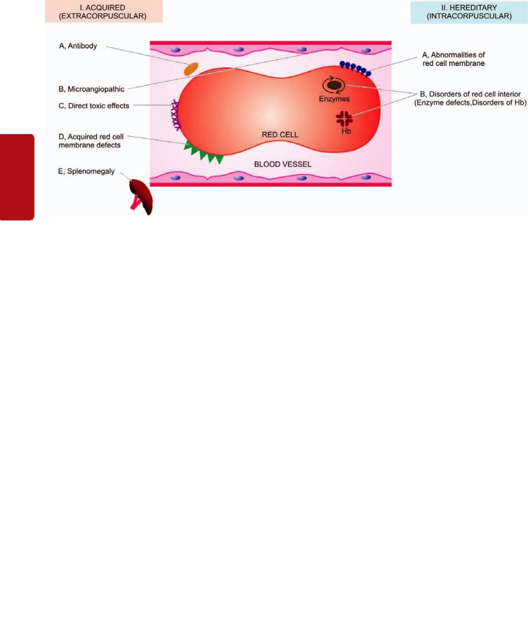
310
II SECTION
Tissues Lymphoreticular and Haematology
Figure 12.21 
 Diagrammatic representation of classification of haemolytic anaemias based on principal mechanisms of haemolysis.
Most of the abnormalities due to vitamin B12 deficiency can be corrected except the irreversible damage to the spinal cord. Corticosteroid therapy can improve the gastric lesion with a return of acid secretion but the higher incidence of gastric polyps and cancer of the stomach in these patients can only be detected by frequent follow-up.
HAEMOLYTIC ANAEMIAS
GENERAL ASPECTS
Definition and Classification
Haemolytic anaemias are defined as anaemias resulting from an increase in the rate of red cell destruction. Normally, effete red cells undergo lysis at the end of their lifespan of 120+30 days within the cells of reticuloendothelial (RE) system in the spleen and elsewhere (extravascular haemolysis), and haemoglobin is not liberated into the plasma in appreciable amounts. The red cell lifespan is shortened in haemolytic anaemia i.e. there is accelerated haemolysis. However, shortening of red cell lifespan does not necessarily result in anaemia. In fact, compensatory bone marrow hyperplasia may cause 6 to 8-fold increase in red cell production without causing anaemia to the patient, so-called compensated
haemolytic disease.
The premature destruction of red cells in haemolytic anaemia may occur by 2 mechanisms:

 Firstly, the red cells undergo lysis in the circulation and release their contents into plasma (intravascular haemolysis). In these cases the plasma haemoglobin rises substantially and part of it may be excreted in the urine (haemoglobinuria).

 Secondly, the red cells are taken up by cells of the RE system where they are destroyed and digested (extravascular haemolysis). In extravascular haemolysis, plasma haemoglobin level is, therefore, barely raised.
Extravascular haemolysis is more common than the former. One or more factors may be involved in the pathogenesis of various haemolytic anaemias.
Haemolytic anaemias are broadly classified into 2 main categories:
I. Acquired haemolytic anaemias caused by a variety of extrinsic environmental factors (extracorpuscular).
II. Hereditary haemolytic anaemias are usually the result of intrinsic red cell defects (intracorpuscular).
A simplified classification based on these mechanisms is given in Table 12.9 and diagrammatically represented in
Fig. 12.21.
Features of Haemolysis
A number of clinical and laboratory features are shared by various types of haemolytic anaemias. These are briefly described below:
GENERAL CLINICAL FEATURES. Some of the general clinical features common to most congenital and acquired haemolytic anaemias are as under:
1.Presence of pallor of mucous membranes.
2.Positive family history with life-long anaemia in patients with congenital haemolytic anaemia.
3.Mild fluctuating jaundice due to unconjugated hyperbilirubinaemia.
4.Urine turns dark on standing due to excess of urobilinogen in urine.
5.Splenomegaly is found in most chronic haemolytic anaemias, both congenital and acquired.
6.Pigment gallstones are found in some cases.
LABORATORY EVALUATION OF HAEMOLYSIS.
Pathways by which haemoglobin derived from effete red cells is metabolised is already discussed on page 290. The

TABLE 12.9: Classification of Haemolytic Anaemias.
I. ACQUIRED (EXTRACORPUSCULAR)
A.Antibody: Immunohaemolytic anaemias
1.Autoimmune haemolytic anaemia (AIHA)
i)Warm antibody AIHA
ii)Cold antibody AIHA
2.Drug-induced immunohaemolytic anaemia
3.Isoimmune haemolytic anaemia (page 340)
B.Mechanical trauma: Microangiopathic haemolytic anaemia
C.Direct toxic effects: Malaria, bacterial, infection and other agents
D.Acquired red cell membrane abnormalities: paroxysmal nocturnal haemoglobinuria (PNH)
E.Splenomegaly
II. HEREDITARY (INTRACORPUSCULAR)
A.Abnormalities of red cell membrane
1.Hereditary spherocytosis
2.Hereditary elliptocytosis (hereditary ovalocytosis)
3.Hereditary stomatocytosis
B.Disorders of red cell interior
1.Red cell enzyme defects (Enzymopathies)
i)Defects in the hexose monophosphate shunt: G6PD deficiency
ii)Defects in the Embden-Meyerhof (or glycolytic) pathway: pyruvate kinase deficiency
2.Disorders of haemoglobin (Haemoglobinopathies)
i)Structurally abnormal haemoglobins: sickle syndromes, other haemoglobinopathies
ii)Reduced globin chain synthesis: thalassaemias
investigations of a patient suspected to have haemolytic anaemia should provide answers to 3 vital questions:
1.Is there evidence of haemolysis?
2.What is the type of haemolytic mechanism?
3.What is the precise diagnosis?
The laboratory findings are conveniently divided into the
following 4 groups:
I. Tests of increased red cell breakdown:
1. Serum bilirubin—unconjugated (indirect) bilirubin is raised.
4.Serum haptoglobin (α-globulin binding protein) is reduced or absent.
5.Plasma lactic dehydrogenase is raised.
6.Evidences of intravascular haemolysis in the form of haemoglobinaemia, haemoglobinuria, methaemoglobinaemia and haemosiderinuria.
II. Tests of increased red cell production:
1.Reticulocyte count reveals reticulocytosis which is generally early and is hence most useful initial test of marrow erythroid hyperplasia.
2.Routine blood film shows macrocytosis, polychromasia and presence of normoblasts.
3.Bone marrow shows erythroid hyperplasia with usually raised iron stores.
4.X-ray of bones shows evidence of expansion of marrow space, especially in tubular bones and skull.
III. Tests of damage to red cells:
1. Routine blood film shows a variety of abnormal morphological appearances of red cells described on page 366 and illustrated in Fig. 12.10 already. A summary of contributory features of morphology of RBCs in arriving at the diagnosis of haemolytic anaemia and its cause is given in Table 12.10.
2.Osmotic fragility is increased or decreased.
3.Autohaemolysis test with or without addition of glucose.
4.Coombs’ antiglobulin test.
5.Electrophoresis for abnormal haemoglobins.
6.Estimation of HbA2.
7.Estimation of HbF.
8.Tests for sickling.
9.Screening test for G6PD deficiency and other enzymes (e.g. Heinz bodies test).
IV.Tests for shortened red cell lifespan. A shortened red cell survival is best tested by 51Cr labelling method. Normal RBC lifespan of 120 days is shortened to 20-40 days in moderate haemolysis and to 5-20 days in severe haemolysis.
I.ACQUIRED (EXTRACORPUSCULAR) HAEMOLYTIC ANAEMIAS
2. |
Urine urobilinogen is raised but there is no bilirubinuria. Acquired haemolytic anaemias are caused by a variety of |
||
3. |
Faecal stercobilinogen is raised. |
extrinsic factors, namely: antibody (immunohaemolytic |
|
|
|||
TABLE 12.10: Red Cell Morphologic Features in Various Types of Haemolytic Anaemias. |
|||
|
Feature |
Etiology |
Type of Haemolytic Anaemia |
|
|
|
|
1. |
Spherocytes |
Loss of spectrin from membrane |
Hereditary spherocytosis |
|
|
|
AIHA |
2. |
Target cells (Leptocytes) |
Increased ratio of surface area: volume |
Thalassaemias |
|
|
|
Liver disease |
|
|
|
HbS disease |
|
|
|
HbC disease |
3. |
Schistocytes |
Traumatic damage to red cell membrane |
Microangiopathy |
4. |
Sickle cells |
Polymerisation of HbS |
Sickle syndromes |
5. |
Acanthocytes (Spur cells) |
Abnormality in membrane lipids |
Severe liver disease |
6. |
Heinz bodies |
Precipitated Hb |
Unstable Hb |
|
|
|
|
311
Series Erythroid of Disorders and System Haematopoietic to Introduction 12 CHAPTER

312anaemia), mechanical factors (microangiopathic haemolytic anaemia), direct toxic effect (in malaria, clostridial infection etc), splenomegaly, and certain acquired membrane abnormalities (paroxysmal nocturnal haemoglobinuria). These are discussed below:
|
A. IMMUNOHAEMOLYTIC ANAEMIAS |
|
|
Immunohaemolytic anaemias are a group of anaemias |
|
|
occurring due to antibody production by the body against |
|
|
its own red cells. Immune haemolysis in these cases may be |
|
|
induced by one of the following three types of antibodies: |
|
SECTION |
1. Autoimmune haemolytic anaemia (AIHA) characterised by |
|
formation of autoantibodies against patient’s own red cells. |
||
Depending upon the reactivity of autoantibody, AIHA is |
||
|
||
|
further divided into 2 types: |
|
|
i) ‘Warm’ antibody AIHA in which the autoantibodies are |
|
II |
reactive at body temperature (37°C). |
|
ii) ‘Cold’ antibody AIHA in which the autoantibodies react |
||
|
||
|
better with patient’s own red cells at 4°C. |
|
Haematology |
2. Drug-induced immunohaemolytic anaemia. |
|
3. Isoimmune haemolytic anaemia in which the antibodies are |
||
|
||
|
acquired by blood transfusions, pregnancies and haemolytic |
|
|
disease of the newborn. |
|
|
An important diagnostic tool in all cases of immuno- |
|
|
haemolytic anaemias is Coombs’ antiglobulin test for |
|
and |
detection of incomplete Rh-antibodies in saline directly |
|
(direct Coombs’) or after addition of albumin (indirect |
||
|
||
Lymphoreticular |
Coombs’). |
|
Autoimmune Haemolytic Anaemia (AIHA) |
||
|
||
|
‘WARM’ ANTIBODY AIHA |
|
|
PATHOGENESIS. Warm antibodies reactive at body |
|
|
temperature and coating the red cells are generally IgG class |
|
Tissues |
antibodies and occasionally they are IgA. Little is known |
|
about the origin of these acquired red blood cell antibodies |
||
|
||
|
in AIHA but the mechanism of destruction of red cells coated |
|
|
with IgG is better understood. Human red cells coated with |
|
|
IgG antibodies are bound to the surface of RE cells, especially |
|
|
splenic macrophages. A part of the coated cell membrane is |
|
|
lost resulting in spherical transformation of the red cells |
|
|
(acquired spherocytosis). Red cells coated with IgG along |
|
|
with C3 on the surface further promote this red cell-leucocyte |
|
|
interaction, accounting for more severe haemolysis. The |
|
|
spleen is particularly efficient in trapping red cells coated |
|
|
with IgG antibodies. It is, thus, the major site of red cell |
|
|
destruction in warm antibody AIHA. |
|
|
CLINICAL FEATURES. Warm antibody AIHA may occur |
|
|
at any age and in either sex. The disease may occur without |
|
|
any apparent cause (idiopathic) but about a quarter of |
|
|
patients develop this disorder as a complication of an |
|
|
underlying disease affecting the immune system such as SLE, |
|
|
chronic lymphocytic leukaemia, lymphomas and certain |
|
|
drugs such as methyl DOPA, penicillin etc (Table 12.11). |
|
|
The disease tends to have remissions and relapses. The |
|
|
usual clinical features are as follows: |
TABLE 12.11: Conditions Predisposing to Autoimmune Haemolytic Anaemia (AIHA).
A.WARM ANTIBODY AIHA
1.Idiopathic (primary)
2.Lymphomas-leukaemias e.g. non-Hodgkin’s lymphoma, CLL, Hodgkin's disease.
3.Collagen vascular diseases e.g SLE
4.Drugs e.g. methyldopa, penicillin, quinidine group
5.Post-viral
B. COLD ANTIBODY AIHA
1.Cold agglutinin disease
a)Acute: Mycoplasma infection, infectious mononucleosis
b)Chronic: Idiopathic, lymphomas
2.PCH (Mycoplasma infection, viral flu, measles, mumps, syphilis)
1.Chronic anaemia of varying severity with remissions and relapses.
2.Splenomegaly.
3.Occasionally hyperbilirubinaemia.
Treatment of these cases consists of removal of the cause
whenever present, corticosteroid therapy, and in severe cases blood transfusions. Splenectomy is the second line of therapy in this disorder.
LABORATORY FINDINGS. The haematological and biochemical findings in such cases are as under:
1.Mild to moderate chronic anaemia.
2.Reticulocytosis.
3.Prominent spherocytosis in the peripheral blood film.
4.Positive direct Coombs’ (antiglobulin) test for presence of warm antibodies on the red cell, best detected at 37°C.
5.A positive indirect Coombs’ (antiglobulin) test at 37°C may indicate presence of large quantities of warm antibodies in the serum.
6.Unconjugated (indirect) hyperbilirubinaemia.
7.Co-existent immune thrombocytopenia alongwith occasional venous thrombosis may be present (termed Evans’ syndrome).
8.In more severe cases, haemoglobinaemia and haemoglobinuria may be present.
‘COLD’ ANTIBODY AIHA
PATHOGENESIS. Antibodies which are reactive in the cold (4°C) may induce haemolysis under 2 conditions: cold agglutinin disease and paroxysmal cold haemoglobinuria.
1. Cold agglutinin disease. In cold agglutinin disease, the antibodies are IgM type which bind to the red cells best at 4°C. These cold antibodies are usually directed against the I antigen on the red cell surface. Agglutination of red blood cells by IgM cold agglutinins is most profound at very low temperature but upon warming to 37°C or above, disagglutination occurs quickly. Haemolytic effect is mediated through fixation of C3 to the red blood cell surface and not by agglutination alone. Most cold agglutinins affect juvenile red blood cells.

The etiology of cold antibody remains unknown. It is seen in the course of certain infections (e.g. Mycoplasma pneumonia, infectious mononucleosis) and in lymphomas.
2. Paroxysmal cold haemoglobinuria (PCH). In PCH, cold antibody is an IgG antibody (Donath-Landsteiner antibody) which is directed against P blood group antigen and brings about complement-mediated haemolysis. Attacks of PCH are precipitated by exposure to cold.
PCH is uncommon and may be seen in association with tertiary syphilis or as a complication of certain infections such as Mycoplasma pneumonia, flu, measles and mumps.
CLINICAL FEATURES. The clinical manifestations are due to haemolysis and not due to agglutination. These include the following:
1.Chronic anaemia which is worsened by exposure to cold.
2.Raynaud’s phenomenon.
3.Cyanosis affecting the cold exposed regions such as tips of nose, ears, fingers and toes.
4.Haemoglobinaemia and haemoglobinuria occur on exposure to cold.
Treatment consists of keeping the patient warm and treating the underlying cause.
LABORATORY FINDINGS. The haematologic and biochemical findings are somewhat similar to those found in warm antibody AIHA except the thermal amplitude. These findings are as follows:
1.Chronic anaemia.
2.Low reticulocyte count since young red cells are affected
more.
3.Spherocytosis is less marked.
4.Positive direct Coombs’ test for detection of C3 on the red cell surface but IgM responsible for C3 coating on red cells is not found.
5.The cold antibody titre is very high at 4°C and very low at 37°C (Donath-Landsteiner test). IgM class cold antibody has specificity for I antigen, while the rare IgG class antibody of PCH has P blood group antigen specificity.
Drug-induced Immunohaemolytic Anaemia
Drugs may cause immunohaemolytic anaemia by 3 different mechanisms:
1.α -METHYL DOPA TYPE ANTIBODIES. A small proportion of patients receiving α-methyl dopa develop immunohaemolytic anaemia which is identical in every respect to warm antibody AIHA described above.
2.PENICILLIN-INDUCED IMMUNOHAEMOLYSIS.
Patients receiving large doses of penicillin or penicillin-type antibiotics develop antibodies against the red blood cell-drug complex which induces haemolysis.
3.INNOCENT BYSTANDER IMMUNOHAEMOLYSIS.
Drugs such as quinidine form a complex with plasma proteins to which an antibody forms. This drug-plasma protein-antibody complex may induce lysis of bystanding red blood cells or platelets.
In each type of drug-induced immunohaemolytic 313 anaemia, discontinuation of the drug results in gradual disappearance of haemolysis.
Isoimmune Haemolytic Anaemia
Isoimmune haemolytic anaemias are caused by acquiring isoantibodies or alloantibodies by blood transfusions, pregnancies and in haemolytic disease of the newborn. These antibodies produced by one individual are directed against red blood cells of the other. These conditions are considered on page 340.
B. MICROANGIOPATHIC HAEMOLYTIC ANAEMIA |
CHAPTER |
|
|
||
Microangiopathic haemolytic anaemia is caused by |
|
|
abnormalities in the microvasculature. It is generally due to |
|
|
mechanical trauma to the red cells in circulation and is |
|
|
characterised by red cell fragmentation (schistocytosis). There |
12 |
|
are 3 different ways by which microangiopathic haemolytic |
||
|
||
anaemia results: |
|
|
1. EXTERNAL IMPACT. Direct external trauma to red |
Introduction |
|
blood cells when they pass through microcirculation, espe- |
||
|
||
cially over the bony prominences, may cause haemolysis |
|
|
during various activities e.g. in prolonged marchers, joggers, |
|
|
karate players etc. These patients develop haemoglobi- |
|
|
naemia, haemoglobinuria (march haemoglobinuria), and |
|
|
sometimes myoglobinuria as a result of damage to muscles. |
to |
|
|
||
2. CARDIAC HAEMOLYSIS. A small proportion of |
Haematopoietic |
|
patients who received prosthetic cardiac valves or artificial |
||
|
||
grafts develop haemolysis. This has been attributed to direct |
|
|
mechanical trauma to the red cells or shear stress from |
|
|
turbulent blood flow. |
|
|
3. FIBRIN DEPOSIT IN MICROVASCULATURE. |
|
|
Deposition of fibrin in the microvasculature exposes the red |
|
|
cells to physical obstruction and eventual fragmentation of |
System |
|
red cells and trapping of the platelets. Fibrin deposits in the |
||
small vessels may occur in the following conditions: |
||
i) Abnormalities of the vessel wall e.g. in hypertension, |
||
eclampsia, disseminated cancers, transplant rejection, |
and |
|
haemangioma etc. |
||
ii) Thrombotic thrombocytopenic purpura. |
Disorders |
|
|
||
iii) Haemolytic-uraemic syndrome. |
|
|
iv) Disseminated intravascular coagulation (DIC) |
|
|
v) Vasculitis in collagen diseases. |
|
|
All these conditions are described in relevant sections |
of |
|
separately. |
||
Haemolysis may result from direct toxic effects of certain |
Erythroid |
|
C. HAEMOLYTIC ANAEMIA FROM |
|
|
DIRECT TOXIC EFFECTS |
|
|
agents. These include the following examples: |
Series |
|
fever) (Fig. 12.22). |
||
1. Malaria by direct parasitisation of red cells (black-water |
|
|
2. Bartonellosis by direct infection of red cells by the |
|
|
microorganisms. |
|
|
3. Septicaemia with Clostridium welchii by damaging the red |
|
|
cells. |
|
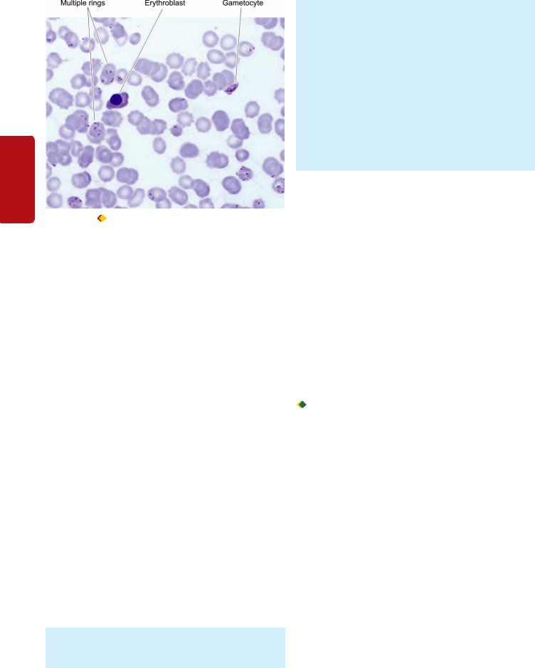
314
Tissues Lymphoreticular and Haematology II SECTION
Figure 12.22 |
Malarial parasite, Plasmodium falciparum, in the |
peripheral blood showing numerous ring stages and a crescent of gametocyte. The background shows a normoblast.
4. Other microorganisms such as pneumococci, staphylococci and Escherichia coli.
5.Copper by direct haemolytic effect on red cells in Wilson’s disease and patients on haemodialysis.
6.Lead poisoning shows basophilic stippling of red blood cells.
7.Snake and spider bites cause haemolysis by their venoms.
8.Extensive burns.
D.PAROXYSMAL NOCTURNAL HAEMOGLOBINURIA (PNH)
PNH is a rare acquired disorder of red cell membrane in which there is chronic intravascular haemolysis due to undue sensitivity of red blood cells to complement due to defective synthesis of a red cell membrane protein. The defect affects all the cells of myeloid progenitor lineage (RBCs, WBCs, platelets) suggesting a deficient haematopoiesis. The disorder generally presents in adult life.
PATHOGENESIS. PNH is considered as an acquired clonal disease of the cell membrane while normal clone also continues to proliferate. The defect is a mutation in the stem cells affecting myeloid progenitor cells that is normally required for the biosynthesis of glycosyl phosphatidyl inositol (GPI) essential for anchoring of the cell; the mutant form of the gene is an X-linked gene called PIG-A (phosphatidyl inositol glycan). Thus, as a result of mutation, there is partial or complete deficiency of anchor protein. Out of about 20 such proteins described so far, the lack of two of the proteins—decay accelerating factor (DAF, CD55) and a
membrane inhibitor of reactive lysis (MIRL, CD59), makes the RBCs unduly sensitive to the lytic effect of complement.
CLINICAL AND LABORATORY FINDINGS. Clinical and laboratory findings are as under:
i) Haemolytic anaemia.
ii)Pancytopenia (mild granulocytopenia and thrombocytopenia frequent).
iii)Intermittent clinical haemoglobinuria; acute haemolytic episodes occur at night identified by passage of brown urine in the morning.
iv)Haemosiderinuria very common.
v)Venous thrombosis as a common complication.
The presence of inordinate sensitivity of red blood cells, leucocytes and platelets to complement in PNH can be demonstrated in vitro by Ham’s test using red cell lysis at acidic pH or by sucrose haemolysis test.
About 20% cases of PNH may develop myeloproliferative or myelodysplastic disorder and some even develop acute myeloid leukaemia.
E. HAEMOLYTIC ANAEMIA IN SPLENOMEGALY
Haemolytic anaemia is common in splenic enlargement from any cause (Chapter 14). Normally, the spleen acts as a filter and traps the damaged red blood cells, destroys them and the splenic macrophages phagocytose the damaged red cells. A normal spleen poses no risk to normal red blood cells. But splenomegaly exaggerates the damaging effect to which the red cells are exposed. Besides haemolytic anaemia, splenomegaly is usually associated with pancytopenia. Splenectomy or reduction in size of spleen by appropriate therapy relieves the anaemia as well as improves the leucocyte and platelet counts.
II. HEREDITARY (INTRACORPUSCULAR)
HAEMOLYTIC ANAEMIA
Hereditary haemolytic anaemias are usually the result of intracorpuscular defects. Accordingly, they are broadly classified into 2 groups (see Table 12.9):

 Hereditary abnormalities of red cell membrane.
 Hereditary disorders of the interior of the red cells.
A.HEREDITARY ABNORMALITIES OF RED CELL MEMBRANE
The abnormalities of red cell membrane are readily identified on blood film examination. There are 3 important types of inherited red cell membrane defects: hereditary spherocytosis, hereditary elliptocytosis (hereditary ovalocytosis) and hereditary stomatocytosis.
Hereditary Spherocytosis
Hereditary spherocytosis is a common type of hereditary haemolytic anaemia of autosomal dominant inheritance in which the red cell membrane is abnormal.
PATHOGENESIS. The molecular abnormality in hereditary spherocytosis is a defect in proteins which anchor the lipid bilayer to the underlying cytoskeleton. These protein abnormalities are as under and are schematically illustrated in Fig.12.23:
1. Spectrin deficiency. Almost all cases have deficiency in the structural protein of the red cell membrane, spectrin.
Figure 12.23 
 Diagrammatic representation of pathogenesis of hereditary spherocytosis. A, Normal red cell with biconcave surface and normal size. B, Red cell membrane as seen in cross section in hereditary spherocytosis. Mutations in membrane proteins—α-spectrin, β-spectrin and ankyrin, result in defect in anchoring of lipid bilayer of the membrane to the underlying cytoskeleton. C, This results in spherical contour and small size so as to contain the given volume of haemoglobin in the deformed red cell. D, During passage through the spleen, these rigid spherical cells lose their cell membrane further. This produces a circulating subpopulation of hyperspheroidal spherocytes while splenic macrophages in large numbers phagocytose defective red cells causing splenomegaly.
Spectrin deficiency correlates with the severity of anaemia. Mutation in spectrin by recessive inheritance called α−spectrin causes more severe form of anaemia, while mutation by dominant inheritance forming β-spectrin results in mild form of the disease.
2. Ankyrin abnormality.About half the cases of hereditary spherocytosis have defect in ankyrin, protein that binds protein 3 and spectrin. Homozygous state with recessive inheritance pattern has severe anaemia while heterozygotes with more common dominant inheritance pattern have milder anaemia.
Inherited mutation in spectrin or ankyrin causes defect in anchoring of lipid bilayer cell membrane. Red cells with such unstable membrane but with normal volume, when released in circulation, lose their membrane further, till they can accommodate the given volume. This results in formation of spheroidal contour and smaller size of red blood cells, termed microspherocytes. These deformed red cells are not flexible, unlike normal biconcave red cells. These rigid cells are unable to pass through the spleen, and in the process they lose their surface membrane further. This produces a subpopulation of hyperspheroidal red cells in the peripheral blood which are subsequently destroyed in the spleen.
CLINICAL FEATURES. The disorder may be clinically apparent at any age from infancy to old age and has equal sex incidence. The family history may be present. The major clinical features are as under:
1.Anaemia is usually mild to moderate.
2.Splenomegaly is a constant feature.
3.Jaundice occurs due to increased concentration of unconjugated (indirect) bilirubin in the plasma (also termed congenital haemolytic jaundice).
4.Pigment gallstones are frequent due to increased bile pigment production. Splenectomy offers the only reliable mode of treatment.
LABORATORY FINDINGS. The usual haematological and biochemical findings are as under:
1.Anaemia of mild to moderate degree.
2.Reticulocytosis, usually 5-20%.
3.Blood film shows the characteristic abnormality of erythrocytes in the form of microspherocytes (Fig.12.24).
4.MCV is usually normal or slightly decreased but MCHC is increased.
5.Osmotic fragility test is helpful in testing the spheroidal nature of red cells which lyse more readily in solutions of low salt concentration i.e. osmotic fragility is increased
(Fig.12.25).
6.Autohaemolysis test is similar to osmotic fragility test after incubation and shows increased spontaneous autohaemolysis (10-15% red cells) as compared to normal red cells (less than 4%). Autohaemolysis is correctable by addition of glucose.
7.Direct Coombs’ (antiglobulin) test is negative so as to distinguish this condition from acquired spherocytosis of AIHA in which case it is positive.
Spherocytes may also be seen in blood film in acquired immune haemolytic anaemia and following red cell transfusion.
Hereditary Elliptocytosis (Hereditary Ovalocytosis)
Hereditary elliptocytosis or hereditary ovalocytosis is another autosomal dominant disorder involving red cell membrane protein spectrin. Some patients have an inherited defect in erythrocyte membrane protein 4.1 that interconnects spectrin with actin in the cytoskeleton. The disorder is similar in all respects to hereditary spherocytosis except that the blood film shows oval or elliptical red cells and is clinically a milder disorder than hereditary spherocytosis.
Acquired causes of elliptocytosis include iron deficiency and myeloproliferative disorders.
315
Series Erythroid of Disorders and System Haematopoietic to Introduction 12 CHAPTER
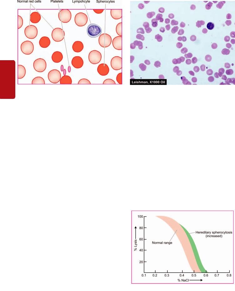
316
Tissues Lymphoreticular and Haematology II SECTION
Figure 12.24 
 Peripheral blood film findings in hereditary spherocytosis.
Hereditary Stomatocytosis
Stomatocytes are cup-shaped RBCs having one surface concave and the other side as convex. This causes a central slit-like or mouth-like appearance of red cells. The underlying defect is in membrane protein, stomatin, having autosomal dominant pattern of inheritance. The stomatocytes are swollen red cells (overhydrated red cells) due to increased permeability to sodium and potassium. The affected patients have mild anaemia and splenomegaly.
B. HEREDITARY DISORDERS OF RED CELL INTERIOR
Inherited disorders involving the interior of the red blood cells are classified into 2 groups:
1.Red cell enzyme defects (Enzymopathies): These cause defective red cell metabolism involving 2 pathways
(Fig. 12.26):
i)Defects in the hexose monophosphate shunt: Common example is glucose-6-phosphate dehydrogenase (G6PD) deficiency.
ii)Defects in the Embden-Meyerhof (glycolytic) pathway:
Example is pyruvate kinase (PK) deficiency.
2.Disorders of haemoglobin (haemoglobinopathies):
These are divided into 2 subgroups:
i)Structurally abnormal haemoglobin: Examples are sickle syndromes and other haemoglobinopathies.
ii)Reduced globin chain synthesis: Common examples are various types of thalassaemias.
These disorders are discussed below.
Red Cell Enzyme Defects (Enzymopathies)
G6PD DEFICIENCY
Among the defects in hexose monophosphate shunt, the most common is G6PD deficiency. It affects millions of people throughout the world. G6PD gene is located on the X chromosome and its deficiency is, therefore, a sex (X)-linked trait affecting males, while the females are carriers and are
asymptomatic. Several variants of G6PD have been described. The normal G6PD variant is designated as type B but blacks have normally A+ (positive) type G6PD variant. The most common and significant clinical variant is A– (negative) type found in black males. Like the HbS gene, the A–type G6PD variant confers protection against malaria. Individuals with A– G6PD variant have shortened red cell lifespan but without anaemia. However, these individuals develop haemolytic episodes on exposure to oxidant stress such as viral and bacterial infections, certain drugs (antimalarials, sulfonamides, nitrofurantoin, aspirin, vitamin K), metabolic acidosis and on ingestion of fava beans (favism).
PATHOGENESIS. Normally, red blood cells are well protected against oxidant stress because of adequate generation of reduced glutathione via the hexose monophosphate shunt. Individuals with inherited deficiency of G6PD, an enzyme required for hexose monophosphate shunt for glucose metabolism, fail to develop adequate levels of reduced glutathione in their red cells. This results
Figure 12.25 
 Osmotic fragility test in hereditary spherocytosis showing increased fragility.
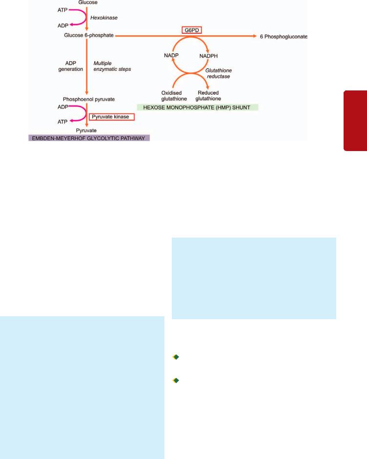
Figure 12.26 
 Abbreviated pathways of anaerobic glycolysis (Embden-Meyerhof) and hexose monophosphate (HMP) shunt in the metabolism of erythrocyte. The two red cell enzyme defects, glucose-6 phosphate dehydrogenase (G6PD) and pyruvate kinase, are shown bold.
in oxidation and precipitation of haemoglobin within the red cells forming Heinz bodies. Besides G6PD deficiency, deficiency of various other enzymes involved in the hexose monophosphate shunt may also infrequently cause clinical problems.
CLINICAL FEATURES. The clinical manifestations are those of an acute haemolytic anaemia within hours of exposure to oxidant stress. The haemolysis is, however, self-limiting even if the exposure to the oxidant is continued since it affects the older red cells only. Haemoglobin level may return to normal when the older population of red cells has been destroyed and only younger cells remain. Some patients may have only darkening of the urine from haemoglobinuria but more severely affected ones develop constitutional symptoms including jaundice. Treatment is directed towards the prevention of haemolytic episodes such as stoppage of offending drug. Blood transfusions are rarely indicated.
LABORATORY FINDINGS. These are as under:
1.During the period of acute haemolysis, there is rapid fall in haematocrit by 25-30%, features of intravascular haemolysis such as rise in plasma haemoglobin, haemoglobinuria, rise in unconjugated bilirubin and fall in plasma haptoglobin. Formation of Heinz bodies is visualised by means of supravital stains such as crystal violet, also called Heinz body haemolytic anaemia. However, Heinz bodies are not seen after the first one or two days since they are removed by the spleen, leading to formation of ‘bite cells’ and fragmented red cells.
2. Between the crises, the affected patient generally has no anaemia. The red cell survival is, however, shortened.
The diagnosis of G6PD enzyme deficiency is made by one of the screening tests (e.g. methaemoglobin reduction test, fluorescent screening test, ascorbate cyanide screening test), or by direct enzyme assay on red cells.
PK DEFICIENCY
Pyruvate kinase (PK) deficiency is the only significant enzymopathy of the Embden-Meyerhof glycolytic pathway.The disorder is inherited as an autosomal recessive pattern. Heterozygote state is entirely asymptomatic, while the homozygous individual presents during early childhood with anaemia, jaundice and splenomegaly.
LABORATORY FINDINGS: These are as under:
1.Normocytic and normochromic anaemia.
2.Reticulocytosis.
3.Blood film shows bizarre red cells.
4.Osmotic fragility is usually normal but after incubation it is increased.
5.Autohaemolysis is increased, but unlike hereditary spherocytosis, is not corrected by addition of glucose.
6.Direct specific enzyme assay on red cells is the only method of establishing the diagnosis.
Haemoglobinopathies
Haemoglobin in RBCs may be abnormally synthesised due to inherited defects. These disorders may be of two types: 
 Qualitative disorders in which there is structural abnormality in synthesis of haemoglobin e.g. sickle cell syndrome, other haemoglobinopathies.
 Quantitative disorders in which there quantitatively decreased globin chain synthesis of haemoglobin e.g. thalassaemias.
Both these groups of disorders may occur as homozygous state in which both genes coding for that character are abnormal, or heterozygous when one gene is abnormal and the other gene is normal. However, there are examples of combined disorders too in which there are two different mutations in loci of two corresponding genes (i.e. double heterozygous) e.g. HbS gene from one parent and β-thal from the other parent resulting in βSβthal. These disorders may vary
317
Series Erythroid of Disorders and System Haematopoietic to Introduction 12 CHAPTER
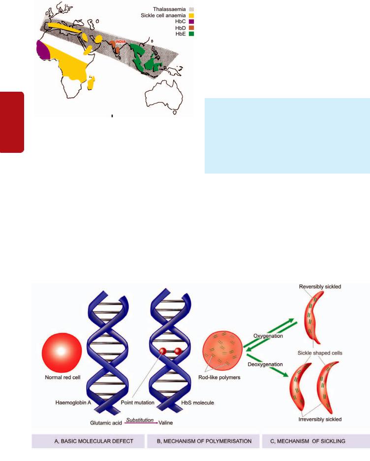
318
Tissues Lymphoreticular and Haematology II SECTION
Figure 12.27 
 The geographic distribution of major haemoglobinopathies and thalassaemias. Thalassaemia and HbD are the haemoglobin disorders common in India.
in presentation from asymptomatic laboratory abnormalities to intrauterine death.
There are geographic variations in the distribution of various haemoglobinopathies world over as shown in Fig. 12.27. Major examples of these disorders are described below.
STRUCTURALLY ABNORMAL HAEMOGLOBINS
SICKLE SYNDROMES
The most important and widely prevalent type of haemoglobinopathy is due to the presence of sickle haemoglobin (HbS) in the red blood cells. The red cells with HbS develop ‘sickling’ when they are exposed to low oxygen tension. Sickle syndromes have the highest frequency in black race and in Central Africa where falciparum malaria is endemic.
Patients with HbS are relatively protected against falciparum malaria. Sickle syndromes occur in 3 different forms:
1.As heterozygous state for HbS: sickle cell trait (AS).
2.As homozygous state for HbS: sickle cell anaemia (SS).
3.As double heterozygous states e.g. sickle β-thalassaemia, sickle-C disease (SC), sickle-D disease (SD).
Heterozygous State:Sickle Cell Trait
Sickle cell trait (AS) is a benign heterozygous state of HbS in which only one abnormal gene is inherited. Patients with AS develop no significant clinical problems except when they become severely hypoxic and may develop sickle cell crises.
LABORATORY FINDINGS. These patients have no anaemia and have normal appearance of red cells. But in hypoxic crisis, sickle cell crises develop. The diagnosis is made by 2 tests:
1.Demonstration of sickling done under condition of reduced oxygen tension by an oxygen consuming reagent, sodium metabisulfite.
2.Haemoglobin electrophoresis reveals 35-40% of the total haemoglobin as HbS.
Homozygous State:Sickle Cell Anaemia
Sickle cell anaemia (SS) is a homozygous state of HbS in the red cells in which an abnormal gene is inherited from each parent. SS is a severe disorder associated with protean clinical manifestations and decreased life expectancy.
PATHOGENESIS. Following abnormalities are observed
(Fig. 12.28):
1.Basic molecular lesion: In HbS, basic genetic defect is the single point mutation in one amino acid out of 146 in haemoglobin molecule— there is substitution of valine for glutamic acid at 6-residue position of the β-globin, producing Hb α2β2s.
Figure 12.28 
 Pathogenesis of sickle cell anaemia. A, Basic molecular defect. B, Mechanism of polymerisation and consequent sickling of red cells containing HbS. C, Mechanism of sickling on oxygenation-deoxygenation.
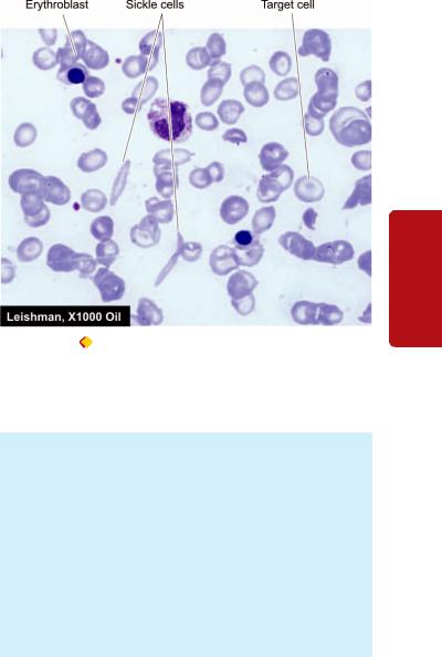
2.Mechanism of sickling: During deoxygenation, the red cells containing HbS change from biconcave disc shape to an elongated crescent-shaped or sickle-shaped cell. This process termed sickling occurs both within the intact red cells and in vitro in free solution. The mechanism responsible for sickling upon deoxygenation of HbS-containing red cells is the polymerisation of deoxygenated HbS which aggregates to form elongated rod-like polymers. These elongated fibres align and distort the red cell into classic sickle shape.
3.Reversible-irreversible sickling: The oxygen-dependent sickling process is usually reversible. However, damage to red cell membrane leads to formation of irreversibly sickled red cells even after they are exposed to normal oxygen tension.
4.Factors determining rate of sickling: Following factors determine the rate at which the polymerisation of HbS and consequent sickling take place:
i)Presence of non-HbS haemoglobins: The red cells in patients of SS have predominance of HbS and a small part consists of non-HbS haemoglobins, chiefly HbF (2-20% of the total haemoglobin). HbF-containing red cells are protected from sickling while HbA-containing red cells participate readily in co-polymerisation with HbS.
ii)Intracellular concentration of HbS.
iii)Total haemoglobin concentration.
iv)Extent of deoxygenation.
v)Acidosis and dehydration.
vi)Increased concentration of 2, 3-BPG in the red cells.
CLINICAL FEATURES. The clinical manifestations of homozygous sickle cell disease are widespread. The symptoms begin to appear after 6th month of life when most of the HbF is replaced by HbS. Infection and folic acid deficiency result in more severe clinical manifestations. These features are as under:
1.Anaemia. There is usually severe chronic haemolytic anaemia (primarily extravascular) with onset of aplastic crisis in between. The symptoms of anaemia are generally mild since HbS gives up oxygen more readily than HbA to the tissues.
2.Vaso-occlusive phenomena. Patients of SS develop recurrent vaso-occlusive episodes throughout their lives due to obstruction to capillary blood flow by sickled red cells upon deoxygenation or dehydration. Vaso-obstruction affecting different organs and tissues results in infarcts which may be of 2 types:
i)Microinfarcts affecting particularly the abdomen, chest, back and joints and are the cause of recurrent painful crises in SS.
ii)Macroinfarcts involving most commonly the spleen (splenic sequestration, autosplenectomy), bone marrow (pains), bones (aseptic necrosis, osteomyelitis), lungs (pulmonary infections), kidneys (renal cortical necrosis), CNS (stroke), retina (damage) and skin (ulcers), and result in anatomic and functional damage to these organs.
3. Constitutional symptoms. In addition to the features of anaemia and infarction, patients with SS have impaired
Figure 12.29 |
Sickle cell anaemia. PBF shows crescent shaped |
elongated red blood cells, a few target cells and a few erythroblasts.
growth and development and increased susceptibility to infection due to markedly impaired splenic function.
LABORATORY FINDINGS. The diagnosis of SS is considered high in blacks with haemolytic anaemia. The laboratory findings in these cases are as under (Fig. 12.29):
1.Moderate to severe anaemia (haemoglobin concentration 6-9 g/dl).
2.The blood film shows sickle cells and target cells and features of splenic atrophy such as presence of HowellJolly bodies.
3.A positive sickling test with a reducing substance such as sodium metabisulfite (described above).
4.Haemoglobin electrophoresis shows no normal HbA but shows predominance of HbS and 2-20% HbF.
Double Heterozygous States
Double heterozygous conditions involving combination of HbS with other haemoglobinopathies may occur. Most common among these are sickle-β-thalassaemia (βSβthal), sickle C disease (SC), and sickle D disease (SD). All these disorders behave like mild form of sickle cell disease. Their diagnosis is made by haemoglobin electrophoresis and separating the different haemoglobins.
OTHER STRUCTURAL HAEMOGLOBINOPATHIES
Besides sickle haemoglobin, about 400 structurally different abnormal human haemoglobins have been discovered in different parts of the world. Some of them are associated with clinical manifestations, while others are of no consequence. A few important and common variants are briefly described below:
HbC Haemoglobinopathy
HbC haemoglobinopathy is prevalent in West Africa and in American blacks. The molecular lesion in HbC is substitution of lysine for glutamic acid at β-6 globin chain position. The
319
Series Erythroid of Disorders and System Haematopoietic to Introduction 12 CHAPTER

320 disorder of HbC may occur as benign homozygous HbC disease, or as asymptomatic heterozygous HbC trait, or as double heterozygous combinations such as sickle-HbC disease and HbC-β thalassaemia.
HbD Haemoglobinopathy
HbD occurs in North-West India, Pakistan and Iran. About 3% of Sikhs living in Punjab are affected with HbD haemoglobinopathy (called HbD Punjab, also known as HbLos Angeles). HbD Punjab arises from the substitution of glutamine for glutamic acid at β-121 globin chain position.
SECTION |
HbE Haemoglobinopathy |
|
HbE is predominantly found in South-East Asia, India, |
||
|
||
|
Burma and Sri Lanka. HbE arises from the substitution of |
|
|
lysine for glutamic acid at β-26 globin chain position. Like |
|
|
other abnormal haemoglobins, HbE haemoglobinopathy may |
|
II |
also occur as asymptomatic heterozygous HbE trait, |
|
compensated haemolytic homozygous HbE disease, or as |
||
|
||
|
double heterozygous states in combination with other |
|
Haematology |
haemoglobinopathies such as HbE-β thalassaemia and HbE- |
|
homozygous form of the disease appears as mild haemolytic |
||
|
α thalassaemia. |
Haemoglobin O-Arab Disease
Hb O-Arab disease was first identified in an Arab family but has now been detected in American blacks too. The
and |
anaemia with splenomegaly. |
|
Unstable-Hb Haemoglobinopathy |
||
Lymphoreticular |
||
The unstable haemoglobins are those haemoglobin variants |
||
which undergo denaturation and precipitation within the red |
||
cells as Heinz bodies. These give rise to what is known as |
||
congenital non-spherocytic haemolytic anaemia or congenital |
||
Heinz body haemolytic anaemia. These disorders have either |
||
autosomal dominant inheritance or develop from sponta- |
||
neous mutations. The unstable haemoglobins arise from |
||
Tissues |
either a single amino acid substitution in the globin chain or |
|
due to deletion of one or more amino acids within the β- |
||
globin chain so that the firm bonding of the haem group |
||
within the molecule is disturbed leading to formation of |
||
|
||
|
methaemoglobin and precipitation of globin chains as Heinz |
|
|
bodies. |
|
|
Over 100 unstable haemoglobins have been described. |
|
|
They are named according to the place where they are |
|
|
encountered. For instance: Hb-Koln, Hb-Hammersmith, Hb- |
|
|
Zurich, Hb-Sydney, and so on. The diagnosis of unstable Hb |
|
|
disease is made by test for Heinz bodies and by haemoglobin |
|
|
electrophoresis. |
|
|
REDUCED GLOBIN CHAIN SYNTHESIS: |
|
|
THALASSAEMIAS |
|
|
DEFINITION |
|
|
The thalassaemias are a diverse group of hereditary disorders |
|
|
in which there is reduced synthesis of one or more of the |
|
|
globin polypeptide chains. Thus, thalassaemias, unlike |
|
|
haemoglobinopathies which are qualitative disorders of |
|
|
haemoglobin, are quantitative abnormalities of polypeptide globin |
chain synthesis. Thalassaemias were first described in people of Mediterranean countries (North Africa, Southern Europe) from where it derives its name ‘Mediterranean anaemia.’ The Word ‘thalassa’ in Greek means ‘the sea’ since the condition was found commonly in regions surrounding the Mediterranean sea. The condition also occurs in the Middle East, Indian subcontinent, South-East Asia and, in general in blacks (see Fig. 12.27).
GENETICS AND CLASSIFICATION
Thalassaemias are genetically transmitted disorders. Normally, an individual inherits two β-globin genes located one each on two chromosomes 11, and two α-globin genes one each on two chromosomes 16, from each parent i.e. normal adult haemoglobin (HbA) is α2 β2*. Depending upon whether the genetic defect or deletion lies in transmission of α- or β-globin chain genes, thalassaemias are classified into α- and β- thalassaemias. Thus, patients with α-thalassaemia have structurally normal α-globin chains but their production is impaired. Similarly, in β-thalassaemia, β-globin chains are structurally normal but their production is decreased. Each of the two main types of thalassaemias may occur as heterozygous (called α- and β-thalassaemia minor or trait), or as homogygous state (termed α- and β-thalassaemia major). The former is generally asymptomatic, while the latter is a severe congenital haemolytic anaemia.
A classification of various types of thalassaemias alongwith the clinical syndromes produced and salient laboratory findings are given in Table 12.12.
PATHOPHYSIOLOGY OF ANAEMIA IN THALASSAEMIA
A constant feature of all forms of thalassaemia is the presence of anaemia which occurs from following mechanisms:
α-Thalassaemia: In α-thalassaemia major, the obvious cause of anaemia is the inability to synthesise adult haemoglobin, while in α-thalassaemia trait there is reduced production of normal adult haemoglobin.
β-Thalassaemia: In β-thalassaemia major, the most important cause of anaemia is premature red cell destruction brought about by erythrocyte membrane damage caused by the precipitated α-globin chains. Other contributory factors are: shortened red cell lifespan, ineffective erythropoiesis, and haemodilution due to increased plasma volume. A deficiency of β-globin chains in β-thalassaemia leads to large excess of α-chains within the developing red cells. Part of these excessive α-chains are removed by pairing with γ-globin chains as HbF, while the remainder unaccompanied α-chains precipitate rapidly within the red cell as Heinz bodies. The precipitated α-chains cause red cell membrane damage. During their passage through the splenic sinusoids, these red cells are further damaged and develop pitting due to removal of the precipitated aggregates. Thus, such red cells
*In a normal adult, distribution of haemoglobin is as under: HbA (α2β2 ) = 95-98%, HbA2 (α2δ2) (a minor variant of HbA) = 1.5-3.5%, HbF (α2 γ2) = less than 1%. But the level of HbF in children under 6 months is slightly higher.

TABLE 12.12: Classification of Thalassaemias.
|
Type |
Hb |
Hb-electrophoresis |
Genotype |
Clinical Syndrome |
|
|
|
|
|
|
α-THALASSAEMIAS |
|
|
|
|
|
1. |
Hydrops foetalis |
3-10 gm/dl |
Hb Barts (g4) (100%) |
Deletion of four α-genes |
Fatal in utero or in early infancy |
2. |
Hb-H disease |
2-12 gm/dl |
HbF (10%), HbH (2-4%) |
Deletion of three α-genes |
Haemolytic anaemia |
3. |
α-Thalassaemia trait |
10-14 gm/dl |
Almost normal |
Deletion of two α-genes |
Microcytic hypochromic blood |
|
|
|
|
|
picture but no anaemia |
β-THALASSAEMIAS |
|
|
|
|
|
1. |
β-Thalassaemia major |
< 5 gm/dl |
HbA (0-50%), |
βthal/βthal |
Severe congenital haemolytic |
|
|
|
HbF(50-98%) |
|
anaemia, requires blood transfusions |
2. |
β-Thalassaemia intermedia 5-10 gm/dl |
Variable |
Multiple mechanisms |
Severe anaemia, but regular blood |
|
|
|
|
|
|
transfusions not required |
3. |
β-Thalassaemia minor |
10-12 gm/dl |
HbA2 (4-9%) |
βA/βthal |
Usually asymptomatic |
|
|
|
HbF (1-5%) |
|
|
|
|
|
|
|
|
are irreparably damaged and are phagocytosed by the RE cells of the spleen and the liver causing anaemia, hepatosplenomegaly, and excess of tissue iron stores. Patients with β-thalassaemia minor, on the other hand, have very mild ineffective erythropoiesis, haemolysis and shortening of red cell lifespan.
α-THALASSAEMIA
MOLECULAR PATHOGENESIS. α-thalassaemias are disorders in which there is defective synthesis of α-globin chains resulting in depressed production of haemoglobins that contain α-chains i.e. HbA, HbA2 and HbF. The α- thalassaemias are most commonly due to deletion of one or more of the α-chain genes located on short arm of chromosome 16. Since there is a pair of α-chain genes, the clinical manifestations of α-thalassaemia depend upon the number of genes deleted. Accordingly, α-thalassaemias are classified into 4 types:
1.Four α-gene deletion: Hb Bart’s hydrops foetalis.
2.Three α-gene deletion: HbH disease.
3.Two α-gene deletion: α-thalassaemia trait.
4.One α-gene deletion: α-thalassaemia trait (carrier).
Hb Bart’s Hydrops Foetalis
When there is deletion of all the four α-chain genes (homozygous state) it results in total suppression of α-globin chain synthesis causing the most severe form of α - thalassaemia called Hb Bart’s hydrops foetalis. Hb Bart’s is a gamma globin chain tetramer (γ4) which has high oxygen affinity leading to severe tissue hypoxia.
CLINICAL FEATURES. Hb Bart’s hydrops foetalis is incompatible with life due to severe tissue hypoxia. The condition is either fatal in utero or the infant dies shortly after birth. If born alive, the features similar to severe Rh haemolytic disease are present (page 340).
LABORATORY FINDINGS. Infants with Hb Bart’s hydrops foetalis born alive may have the following laboratory findings:
1.Severe anaemia (haemoglobin below 6g/dl).
2.Blood film show marked anisopoikilocytosis, hypochromia, microcytosis, polychromasia, basophilic stippling, numerous normoblasts and target cells.
3.Reticulocyte count is high.
4.Serum bilirubin level is elevated.
5.Haemoglobin electrophoresis shows 80-90% Hb-Bart’s and a small amount of Hb-H and Hb-Portland but no HbA, HbA2 or HbF.
HbH Disease
Deletion of three α-chain genes produces HbH which is a β- globin chain tetramer (β4) and markedly impaired α-chain synthesis. HbH is precipitated as Heinz bodies within the affected red cells. An elongated α-chain variant of HbH disease is termed Hb Constant Spring.
CLINICAL FEATURES. HbH disease is generally present as a well-compensated haemolytic anaemia. The features are intermediate between that of β-thalassaemia minor and major. The severity of anaemia fluctuates and may fall to very low levels during pregnancy or infections. Majority of patients have splenomegaly and may develop cholelithiasis.
LABORATORY FINDINGS. These are as follows:
1.Moderate anaemia (haemoglobin 8-9 g/dl).
2.Blood film shows severe microcytosis, hypochromia, basophilic stippling, target cells and normoblasts.
3.Mild reticulocytosis.
4.HbH inclusions as Heinz bodies can be demonstrated in mature red cells with brilliant cresyl blue stain.
5.Haemoglobin electrophoresis shows 2-4% HbH and the remainder consists of HbA, HbA2 and HbF.
α-Thalassaemia Trait
α-thalassaemia trait may occur by the following molecular pathogenesis:

 By deletion of two of the four α -chain genes in homozygous form called homozygous α-thalassaemia, or in double heterozygous form termed heterozygous α-thalassaemia.
321
Series Erythroid of Disorders and System Haematopoietic to Introduction 12 CHAPTER
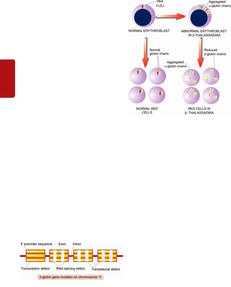
322 By deletion of a single α-chain gene causing heterozygous
α-thalassaemia trait called heterozygous α-thalassaemia.
CLINICAL FEATURES. α-thalassaemia trait due to two α-chain gene deletion is asymptomatic. It is suspected in a patient of refractory microcytic hypochromic anaemia in whom iron deficiency and β-thalassaemia minor have been excluded and the patient belongs to the high-risk ethnic group. One gene deletion α-thalassaemia trait is a silent carrier state.
|
LABORATORY FINDINGS. The patients of α-thalas- |
|
|
|
saemia trait may have the following haematological |
|
|
SECTION |
findings: |
|
|
3. MCV, MCH and MCHC may be slightly reduced. |
|
||
|
1. Haemoglobin level normal or mildly reduced. |
|
|
|
2. Blood film shows microcytic and hypochromic red cell |
|
|
|
morphology but no evidence of haemolysis or anaemia. |
|
|
II |
4. Haemoglobin electrophoresis reveals small amount of |
|
|
|
Hb-Bart’s in neonatal period (1-2% in α-thalassaemia 2 |
|
|
Haematology |
and 5-6% in α-thalassaemia 1) which gradually disappears |
|
|
by adult life. HbA2 is either normal or slightly decreased |
|
||
|
(contrary to the elevated HBA2 levels in β-thalassaemia |
|
|
|
trait). |
|
|
|
|
|
|
|
β-THALASSAEMIAS |
||
and |
MOLECULAR PATHOGENESIS. β-thalassaemias are |
||
caused by decreased rate of β-chain synthesis resulting in |
|||
|
|||
Lymphoreticular |
reduced formation of HbA in the red cells. The molecular |
||
absence of β-globin chain synthesis while β+ denotes partial |
|||
|
pathogenesis of the β-thalassaemias is more complex than |
||
|
that of α-thalassaemias. In contrast to α-thalassaemia, gene |
||
|
deletion rarely ever causes β-thalassaemia and is only seen |
||
|
in an entity called hereditary persistence of foetal haemoglobin |
||
|
(HPFH). Instead, most of β-thalassaemias arise from different |
||
|
types of mutations of β−globin gene resulting from single |
||
Tissues |
base changes. The symbol β° is used to indicate the complete |
||
synthesis of the β-globin chains. More than 100 such |
|||
|
|||
|
mutations have been described affecting the preferred sites |
||
|
in the coding sequences e.g. in promoter region, termination |
||
|
region, splice junctions, exons, introns. Some of the important |
||
|
ones having effects on β-globin chain synthesis are as under |
||
|
(Fig. 12.30): |
||
|
i) Transcription defect: Mutation affecting transcriptional |
||
|
promoter sequence causing reduced synthesis of β-globin |
||
|
chain. Hence the result is partially preserved synthesis i.e. |
||
|
β+ thalassaemia. |
||
Figure 12.30 
 Schematic representation of sites of β-globin gene mutation in chromosome 11 giving rise to β-thalassaemia.
Figure 12.31 
 Pathogenesis of β-thalassaemia major.
ii)Translation defect: Mutation in the coding sequence causing stop codon (chain termination) interrupting β-globin messenger RNA. This would result in no synthesis of β-globin chain i.e. β° thalassaemia.
iii)mRNA splicing defect: Mutation leads to defective mRNA processing forming abnormal mRNA that is degraded in the nucleus. Depending upon whether part of splice site remains
intact or is totally degraded, it may result in β+ thalassaemia
or β° thalassaemia.
Depending upon the extent of reduction in β-chain synthesis, there are 3 types of β-thalassaemia:
1. Homozygous form: β-Thalassaemia major. It is the most severe form of congenital haemolytic anaemia. It is further of 2 types (Fig. 12.31):
i)β° thalassaemia major characterised by complete absence of β-chain synthesis.
ii)β+ thalassaemia major having incomplete suppression of β-chain synthesis.
2.β-Thalassaemia intermedia: It is β-thalassaemia of intermediate degree of severity that does not require regular
blood transfusions. These cases are genetically heterozygous (β°/β or β+/β).
3.Heterozygous form: β-Thalassaemia minor (trait). It is a mild asymptomatic condition in which there is moderate suppression of β-chain synthesis.
Besides β-thalassaemia minor, a few uncommon globin chain combinations resulting in β-thalassaemia trait are as under:
i) δβ-thalassaemia minor in which there is total absence of both β and δ chain synthesis and is characterised by elevated HbF level but unlike β-thalassaemia minor there is normal or reduced HbA2 level.
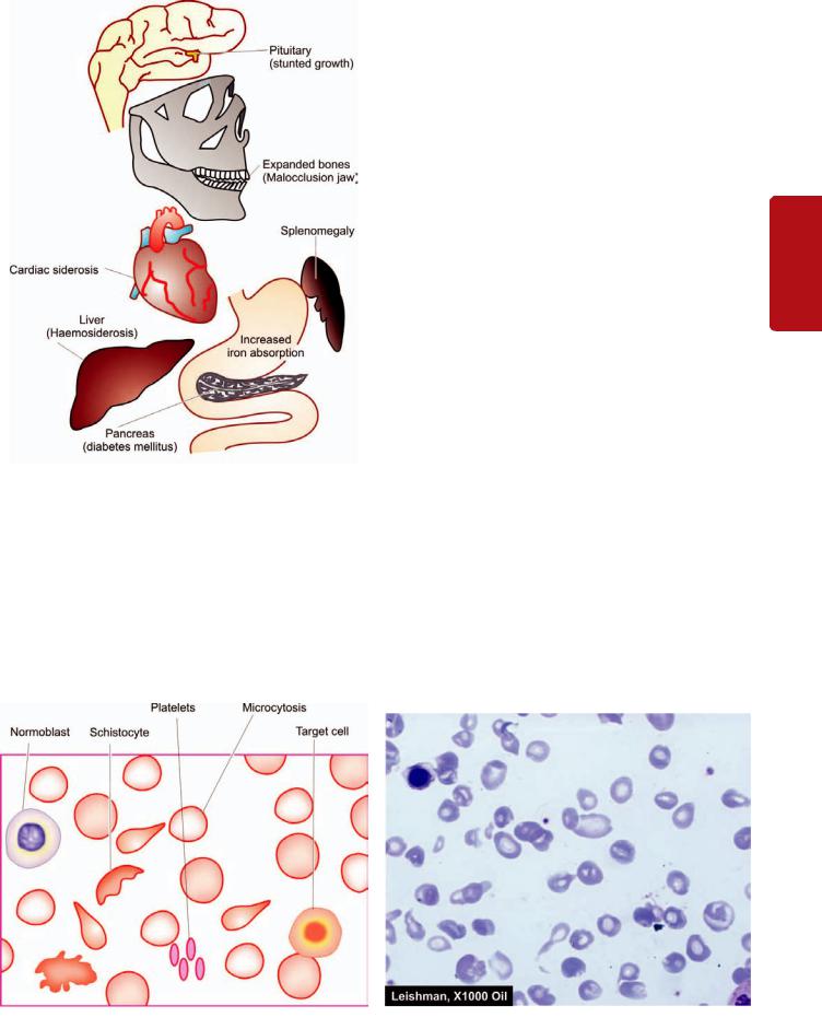
Figure 12.32 
 Major clinical features of β-thalassaemia major.
ii) Hb Lepore syndrome characterised by nonhomologous fusion of β- and δ-genes forming an abnormal haemoglobin called Hb Lepore (named after a family called Lepore). There is total absence of normal β-chain synthesis.
An individual may inherit one β-chain gene from each parent and produce heterozygous, homozygous, or double heterozygous states. Statistically, 25% of offsprings born to two heterozygotes (i.e. β-thalassaemia trait) will have the homozygous state i.e. β-thalassaemia major.
β-Thalassaemia Major |
323 |
||
β-thalassaemia major, also termed Mediterranean or Cooley’s |
|
||
anaemia is the most common form of congenital haemolytic |
|
||
anaemia. β-thalassaemia major is a homozygous state with |
|
||
either complete absence of β-chain synthesis (β° thalassaemia |
|
||
major) or only small amounts of β-chains are formed (β+ thalas- |
|
||
saemia major). These result in excessive formation of alternate |
|
||
haemoglobins, HbF (α2 γ2) and HbA2 (α2 δ2). |
|
||
CLINICAL FEATURES. Clinical manifestations appear |
|
||
insidiously and are as under (Fig. 12.32): |
|
||
1. Anaemia starts appearing within the first 4-6 months of |
CHAPTER |
||
life when the switch over from γ-chain to β-chain production |
|||
occurs. |
|||
|
|||
2. Marked hepatosplenomegaly occurs due to excessive red |
|
||
cell destruction, extramedullary haematopoiesis and iron |
|
||
overload. |
|
||
3. Expansion of bones occurs due to marked erythroid |
12 |
||
hyperplasia leading to thalassaemic facies and malocclusion |
|||
of the jaw. |
|
||
4. Iron overload due to repeated blood transfusions causes |
Introduction |
||
damage to the endocrine organs resulting in slow rate of |
|||
|
|||
growth and development, delayed puberty, diabetes mellitus |
|
||
and damage to the liver and heart. |
|
||
|
|
||
LABORATORY FINDINGS. The haematological |
|
|
|
investigations reveal the following findings: |
|
to |
|
1. Anaemia, usually severe. |
|
||
|
Haematopoietic |
||
2. Blood film shows severe microcytic hypochromic red cell |
|
||
|
|
||
morphology, marked anisopoikilocytosis, basophilic |
|
|
|
stippling, presence of many target cells, tear drop cells |
|
|
|
and normoblasts (Fig. 12.33). |
|
|
|
3. Serum bilirubin (unconjugated) is generally raised. |
|
|
|
4. Reticulocytosis is generally present. |
|
|
|
5. MCV, MCH and MCHC are significantly reduced. |
|
System |
|
6. WBC count is often raised with some shift to left of the |
|
||
neutrophil series, with presence of some myelocytes and |
|
||
metamyelocytes. |
|
||
|
|
Series Erythroid of Disorders and |
|
Figure 12.33 
 Peripheral blood film findings in β-thalassaemia major.
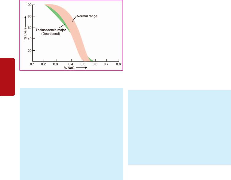
324
Tissues Lymphoreticular and Haematology II SECTION
Figure 12.34 
 Osmotic fragility testing β-thalassaemia major showing decreased fragility.
7.Platelet count is usually normal but may be reduced in patients with massive splenomegaly.
8.Osmotic fragility characteristically reveals increased resistance to saline haemolysis i.e. decreased osmotic fragility
(Fig. 12.34).
9.Haemoglobin electrophoresis shows presence of increased
amounts of HbF, increased amount of HbA2, and almost complete absence or presence of variable amounts of HbA.
The increased level of HbA2 has not been found in any other haemoglobin abnormality except β-thalassaemia.
The increased synthesis of HbA2 is probably due to increased activity at both δ-chain loci.
10.Bone marrow aspirate examination shows normoblastic erythroid hyperplasia with predominance of intermediate and late normoblasts which are generally smaller in size than normal. Iron staining demonstrates siderotic granules in the cytoplasm of normoblasts, increased reticuloendothelial iron but ring sideroblasts are only occasionally seen.
Treatment
Treatment of β-thalassaemia major is largely supportive.
1.Anaemia is generally severe and patients require regular blood transfusions (4-6 weekly) to maintain haemoglobin above 8 g/dl.
2.In order to maintain increased demand of hyperplastic marrow, folic acid supplement is given.
3.Splenectomy is beneficial in children over 6 years of age since splenic sequestration contributes to shortened red cell lifespan.
4.Prevention and treatment of iron overload is done by chelation therapy (desferrioxamine). Oral chelation with kelfer or deferiprone is also available now.
5.Bone marrow transplantation from HLA-matched donor that provides stem cells which can form normal haemoglobin is being done in many centres with fair success rate, especially when done at an early stage before end-organ damage has supervened..
6.Some workers have found success with cord blood transfusion.
7. Gene therapy of thalasaemia involving genetic manipulation in haematopoieitc stem cells may become an option for future.
Since these patients require multiple blood transfusions, they are at increased risk of developing AIDS. In general, patients with β -thalassaemia major have short life expectancy. The biggest problem is iron overload and consequent myocardial siderosis leading to cardiac arrhythmias, congestive heart failure, and ultimately death.
β-Thalassaemia Minor
The β-thalassaemia minor or β-thalassaemia trait, a heterozygous state, is a common entity characterised by moderate reduction in β-chain synthesis.
CLINICAL FEATURES. Clinically, the condition is usually asymptomatic and the diagnosis is generally made when the patient is being investigated for mild chronic anaemia. The spleen may be palpable.
LABORATORY FINDINGS. These are as under:
1.Mild anaemia; mean haemoglobin level is about 15% lower than in normal person for the age and sex.
2.Blood film shows mild anisopoikilocytosis, microcytosis and hypochromia, occasional target cells and basophilic stippling.
3.Serum bilirubin may be normal or slightly raised.
4.Mild reticulocytosis is often present.
5.MCV, MCH and MCHC may be slightly reduced.
6.Osmotic fragility test shows increased resistance to haemolysis i.e. decreased osmotic fragility.
7.Haemoglobin electrophoresis is confirmatory for the
diagnosis and shows about two-fold increase in HbA2 and a slight elevation in HbF (2-3%).
Treatment
Patients with β-thalassaemia minor do not require any treatment. But they should be explained about the genetic implications of the disorder, particularly to those of childbearing age. If the two subjects of β-thalassaemia trait marry, there is a 25% chance of developing thalassaemia major in offsprings. Patients with β-thalassaemia minor have normal life expectancy but those who are treated under the mistaken diagnosis of iron deficiency may develop siderosis and its complications.
Prevention of Thalassaemia
Finally, since thalassaemia is an inheritable disease, its prevention is possible by making an antenatal diagnosis. This is done by chorionic villous biopsy or cells obtained by amniocentesis and foetal DNA studied by PCR amplification technique for presence of genetic mutations of thalassaemias.
APLASTIC ANAEMIA AND OTHER PRIMARY BONE MARROW DISORDERS
‘Bone marrow failure’ is the term used for primary disorders of the bone marrow which result in impaired formation of the erythropoietic precursors and consequent anaemia. It includes the following disorders:
1. Aplastic anaemia, most importantly.
2. Other primary bone marrow disroders such as: myelophthisic anaemia, pure red cell aplasia, and myelodysplastic syndromes.
These disorders are examples of hypoproliferative anaemias in which anaemia is not the only finding but the major problem is pancytopenia i.e. anaemia, leucopenia and thrombocytopenia. While myelodysplastic syndromes are discussed in Chapter 14, other conditions are considered below.
APLASTIC ANAEMIA
Aplastic anaemia is defined as pancytopenia (i.e. simultaneous presence of anaemia, leucopenia and thrombocytopenia) resulting from aplasia of the bone marrow. The underlying defect in all cases appears to be sufficient reduction in the number of haematopoietic pluripotent stem cells which results in decreased or total absence of these cells for division and differentiation.
ETIOLOGY AND CLASSIFICATION. Based on the etiology, aplastic anaemia is classified into 2 main types: primary and secondary. Various causes that may give rise to both these types of aplastic anaemia are summarised in Table 12.13 and briefly considered below:
A. Primary aplastic anaemia. Primary aplastic anaemia includes 2 entities: a congenital form called Fanconi’s anaemia and an immunologically-mediated acquired form.
1.Fanconi’s anaemia. This has an autosomal recessive inheritance and is often associated with other congenital anomalies such as skeletal and renal abnormalities, and sometimes mental retardation.
2.Immune causes. In many cases, suppression of haematopoietic stem cells by immunologic mechanisms may cause aplastic anaemia. The observations in support of autoimmune mechanisms are the clinical response to immunosuppressive therapy and in vitro marrow culture experiments.
B. Secondary aplastic anaemia. Aplastic anaemia may occur secondary to a variety of industrial, physical, chemical, iatrogenic and infectious causes:
1. Drugs. A number of drugs are cytotoxic to the marrow and cause aplastic anaemia. The association of a drug with aplastic anaemia may be either predictably dose-related or an idiosyncratic reaction.
TABLE 12.13: Causes of Aplastic Anaemia.
A.PRIMARY APLASTIC ANAEMIA 1. Fanconi’s anaemia (congenital)
2. Immunologically-mediated (acquired)
B.SECONDARY APLASTIC ANAEMIA 1. Drugs
i) Dose-related aplasia e.g. with antimetabolites (methotrexate), mitotic inhibitors (daunorubicin), alkylating agents (busulfan), nitroso urea, anthracyclines.
ii)Idiosyncratic aplasia e.g. with chloramphenicol, sulfa drugs, oxyphenbutazone, phenylbutazone, chlorpromazine, gold salts.
2.Toxic chemicals e.g. benzene derivatives, insecticides, arsenicals.
3.Infections e.g. infectious hepatitis, EB virus infection, AIDS, other viral illnesses.
4.Miscellaneous e.g. association with SLE and therapeutic X-rays
TABLE 12.14: Causes of Pancytopenia.
I. Aplastic anaemia (Table 12.13)
II. Pancytopenia with normal or increased marrow cellularity e.g.
1.Myelodysplastic syndromes
2.Hypersplenism
3.Megaloblastic anaemia
III. Paroxysmal nocturnal haemoglobinuria (page 314) IV. Bone marrow infiltrations e.g.
1.Haematologic malignancies (leukaemias, lymphomas, myeloma)
2.Non-haematologic metastatic malignancies
3.Storage diseases
4.Osteopetrosis
5.Myelofibrosis
 Dose-related aplasia of the bone marrow occurs with antimetabolites (e.g. methotrexate), mitotic inhibitors (e.g. daunorubicin), alkylating agents (e.g. busulfan), nitroso urea and anthracyclines. In such cases, withdrawal of the drug usually allows recovery of the marrow elements.

 Idiosyncratic aplasia is depression of the bone marrow due to qualitatively abnormal reaction of an individual to a drug when first administered. The most serious and most common example of idiosyncratic aplasia is associated with chloramphenicol. Other such common drugs are sulfa drugs, oxyphenbutazone, phenylbutazone, chlorpromazine, gold salts etc.
2.Toxic chemicals. These include examples of industrial, domestic and accidental use of substances such as benzene derivatives, insecticides, arsenicals etc.
3.Infections. Aplastic anaemia may occur following viral hepatitis, Epstein-Barr virus infection, AIDS and other viral illnesses.
4.Miscellaneous. Lastly, aplastic anaemia has been reported in association with certain other illnesses such as SLE, and with therapeutic X-rays.
CLINICAL FEATURES. The onset of aplastic anaemia may occur at any age and is usually insidious. The clinical manifestations include the following:
1.Anaemia and its symptoms like mild progressive weakness and fatigue.
2.Haemorrhage from various sites due to thrombocytopenia such as from the skin, nose, gums, vagina, bowel, and occasionally in the CNS and retina.
3.Infections of the mouth and throat are commonly present.
4.The lymph nodes, liver and spleen are generally not enlarged.
LABORATORY FINDINGS. The diagnosis of aplastic anaemia is made by a thorough laboratory evaluation and excluding other causes of pancytopenia (Table 12.14). The following haematological features are found:
1.Anaemia. Haemoglobin levels are moderately reduced. The blood picture generally shows normocytic normochromic anaemia but sometimes macrocytosis may be present. The reticulocyte count is reduced or zero.
2.Leucopenia. The absolute granulocyte count is particularly low (below 1500/μl) with relative lymphocytosis. The neutrophils are morphologically normal but their alkaline phosphatase score is high.
325
Series Erythroid of Disorders and System Haematopoietic to Introduction 12 CHAPTER
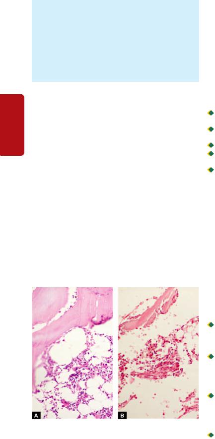
3263.Thrombocytopenia. Platelet count is always reduced.
4.Bone marrow examination. A bone marrow aspirate may yield a ‘dry tap’. A trephine biopsy is generally essential for making the diagnosis which reveals patchy cellular areas in a hypocellular or aplastic marrow due to replacement by fat. There is usually a severe depression of myeloid cells, megakaryocytes and erythroid cells so that the marrow chiefly consists of lymphocytes and plasma cells (Fig. 12.35). Haematopoieitc stem cells bearing CD34 marker are markedly reduced or absent.
|
Treatment |
|
SECTION |
The patients of mild aplasia may show spontaneous recovery, |
|
while the management of severe aplastic anaemia is a most |
||
|
||
|
challenging task. In general, younger patients show better |
|
|
response to proper treatment. The broad outlines of the |
|
|
treatment are as under: |
|
II |
A. General management: It consists of the following: |
|
1. Identification and elimination of the possible cause. |
||
|
||
Haematology |
2. Supportive care consisting of blood transfusions, platelet |
|
concentrates, and treatment and prevention of infections. |
||
2. Immunosuppressive therapy with agents such as anti- |
||
|
B. Specific treatment: The specific treatment has been |
|
|
attempted with varying success and includes the following: |
|
|
1. Marrow stimulating agents such as androgen may be |
|
|
administered orally. |
|
and |
thymocyte globulin and anti-lymphocyte serum has been |
|
tried with 40-50% success rate. Very high doses of |
||
TissuesLymphoreticular |
glucocorticoids or cyclosporine may yield similar responses. |
|
But splenectomy does not have any role in the management |
||
|
||
|
of aplastic anaemia. |
|
|
3. Bone marrow transplantation is considered in severe cases |
|
|
under the age of 40 years where the HLA and mixed culture- |
|
|
matched donor is available. (Other indications for bone |
Figure 12.35 
 Bone marrow trephine biopsy in aplastic anaemia contrasted against normal cellular marrow. A, normal marrow biopsy shows about 50% fatty spaces and about 50% is haematopoietic marrow which contains a heterogeneous mixture of myeloid, erythroid and lymphoid cells. B, In aplastic anaemia, the biopsy shows suppression of myeloid and erythroid cells and replacement of haematopoetic elements by fat. There are scanty foci of cellular components composed chiefly of lymphoid cells.
marrow transplantation are: acute leukaemias—AML in first remission and ALL in second remission, thalassaemia major and combined immunodeficiency disease). Complications of bone marrow transplantation such as severe infections, graft rejection and graft-versus-host disease (GVHD) may occur (Chapter 4).
Severe aplastic anaemia is a serious disorder terminating in death within 6-12 months in 50-80% of cases. Death is usually due to bleeding and/or infection.
MYELOPHTHISIC ANAEMIA
Development of severe anaemia may result from infiltration of the marrow termed as myelophthisic anaemia. The causes for marrow infiltrations include the following (Table12.14):  Haematologic malignancies (e.g. leukaemia, lymphoma, myeloma).

 Metastatic deposits from non-haematologic malignancies (e.g. cancer breast, stomach, prostate, lung, thyroid).

 Advanced tuberculosis.

 Primary lipid storage diseases (Gaucher’s and NiemannPick’s disease).
 Osteopetrosis and myelofibrosis may rarely cause myelophthisis.
The type of anaemia in myelophthisis is generally normocytic normochromic with some fragmented red cells, basophilic stippling and normoblasts in the peripheral blood. Thrombocytopenia is usually present but the leucocyte count is increased with slight shift-to-left of myeloid cells i.e. a picture of leucoerythroblastic reaction consisting of immature myeloid cells and normoblasts is seen in the peripheral blood. Treatment consists of reversing the underlying pathologic process.
PURE RED CELL APLASIA
Pure red cell aplasia (PRCA) is a rare syndrome involving a selective failure in the production of erythroid elements in the bone marrow but with normal granulopoiesis and megakaryocytopoiesis. Patients have normocytic normochromic anaemia with normal granulocyte and platelet count. Reticulocytes are markedly decreased or are absent.
PRCA exists in the following forms:

 Transient self-limited PRCA: It is due to temporary marrow failure in aplastic crisis in haemolytic anamias and in acute B19 parvovirus infection and in transient erythroblastopenia in normal children.
 Acquired PRCA: It is seen in middle-aged adults in association with some other diseases, most commonly thymoma; others are connective tissue diseases (SLE, rheumatoid arthritis), lymphoid malignancies (lymphoma, T-cell chronic lymphocytic leukaemia) and solid tumours.

 Chronic B19 parvovirus infections: PRCA may occur from chronic B19 parvovirus infection in children and is common and treatable. B19 parvovirus produces cytopathic effects on the marrow erythroid precursor cells and are characteristically seen as giant pronomoblasts.
 Congenital PRCA (Blackfan-Diamond syndrome) is a rare chronic disorder detected at birth or in early childhood. It occurs due to mutation in a ribosomal RNA processing gene termed as RPS19. The disorder is corrected by glucocorticoids and marrow transplantation.
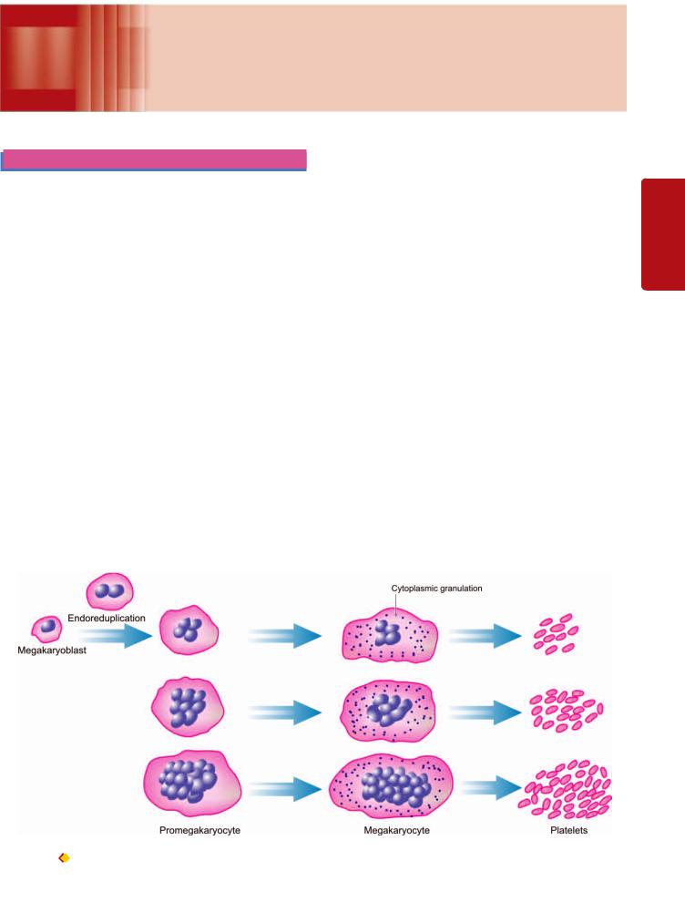
327
Disorders of Platelets, Chapter 13 Bleeding Disorders and
Basic Transfusion Medicine
THROMBOPOIESIS
As outlined in previous chapter and illustrated in Fig. 12.3, the trilineage myeloid stem cells in the bone marrow differentiate into erythroid progenitor, granulocytemonocyte progenitor, and megakaryocyte progenitor cells. Platelets are formed in the bone marrow by a process of fragmentation of the cytoplasm of megakaryocytes. Platelet production is under the control of thrombopoietin, the nature and origin of which are not yet established. The stages in platelet production are: megakaryoblast, promegakaryocyte, megakaryocyte, and discoid platelets (Fig. 13.1).
MEGAKARYOBLAST. The earliest precursor of platelets in the bone marrow is megakaryoblast. It arises from haematopoietic stem cell by a process of differentiation.
PROMEGAKARYOCYTE. A megakaryoblast undergoes endo-reduplication of nuclear chromatin i.e. nuclear chromatin replicates repeatedly in multiples of two without division of the cell. Ultimately, a large cell containing up to 32 times the normal diploid content of nuclear DNA (polyploidy) is formed when further nuclear replication ceases and cytoplasm becomes granular.
MEGAKARYOCYTE. A mature megakaryocyte is a large cell, 30-90 μm in diameter, and contains 4-16 nuclear lobes having coarsely clumped chromatin. The cytoplasm is abundant, light blue in colour and contains red-purple
granules. Platelets are formed from pseudopods of megakaryocyte cytoplasm which get detached into the blood stream. Each megakaryocyte may form up to 4000 platelets. The formation of platelets from the stem cell takes about 10 days.
PLATELETS. Platelets are small (1-4 μm in diameter), discoid, non-nucleate structures containing red-purple granules. The normal platelet count ranges from 150,000400,000/μl and the lifespan of platelets is 7-10 days. About 70% of platelets are in circulation while remaining 30% lie sequestered in the spleen. Newly-formed platelets spend 24-36 hours in the spleen before being released into circulation but splenic stasis does not cause any injury to the platelets normally. Factors such as stress, epinephrine and exercise stimulate platelet production.
The main functions of platelets is in haemostasis which includes two closely linked processes:
1. Primary haemostasis. This term is used for platelet plug formation at the site of injury. It is an immediate phenomenon appearing within seconds of injury and is responsible for cessation of bleeding from microvasculature. Primary haemostasis involves three steps: platelet adhesion, platelet granule release and platelet aggregation which are regulated by changes in membrane phospholipids, and calcium (Fig. 13.2). At molecular level, these important events are depicted diagrammatically in Fig. 13.3 and briefly outlined below:
Medicine Transfusion Basic and Disorders Bleeding Platelets, of Disorders 13 CHAPTER
Figure 13.1 |
Thrombopoiesis. |
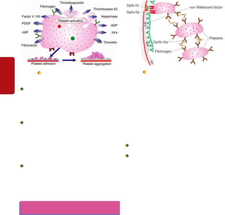
328
Tissues Lymphoreticular and Haematology II SECTION
Figure 13.2 
 Main events in primary haemostasis—platelet adhesion, release (activation) and aggregation.

 Platelet adhesion: Platelets adhere to collagen in the subendothelium due to presence of receptor on platelet
surface, glycoprotein (Gp) Ia-IIa which is an integrin. The adhesion to the vessel wall is further stabilised by von Willebrand factor, an adhesion glycoprotein. This is achieved by formation of a link between von Willebrand factor and another platelet receptor, GpIb-IX complex.

 Platelet release: After adhesion, platelets become activated and release three types of granules from their
cytoplasm: dense granules, α-granules and lysosomal vesicles. Important products released from these granules are: ADP, ATP, calcium, serotonin, platelet factor 4, factor V, factor VIII, thrombospondin, platelet-derived growth factor (PDGF), von Willebrand factor (vWF), fibronectin, fibrinogen, plasminogen activator inhibitor –1 (PAI-1) and thromboxane A2.

 Platelet aggregation: This process is mediated by fibrinogen which forms bridge between adjacent platelets via
glycoprotein receptors on platelets, GpIIb-IIIa.
2. Secondary haemostasis. This involves plasma coagulation system resulting in fibrin plug formation and takes several minutes for completion. This is discussed in detail in Chapter 5.
BLEEDING DISORDERS
(HAEMORRHAGIC DIATHESIS)
Bleeding disorders or haemorrhagic diatheses are a group of disorders characterised by defective haemostasis with abnormal bleeding. The tendency to bleeding may be spontaneous in the form of small haemorrhages into the skin and mucous membranes (e.g. petechiae, purpura, ecchymoses), or there may be excessive external or internal bleeding following trivial trauma and surgical procedure (e.g. haematoma, haemarthrosis etc).
The causes of haemorrhagic diatheses may or may not be related to platelet abnormalities. These causes are broadly divided into the following groups:
I. Haemorrhagic diathesis due to vascular abnormalities. II. Haemorrhagic diathesis related to platelet abnormalities. III. Disorders of coagulation factors.
Figure 13.3 |
Molecular mechanisms in platelet adhesion and release |
reaction (Gp = glycoprotein).
IV. Haemorrhagic diathesis due to fibrinolytic defects.
V. Combination of all these as occurs in disseminated intravascular coagulation (DIC).
Before discussing the bleeding disorders, it is considered desirable to briefly review the broad outlines of scheme of investigations to be carried out in such a case.
INVESTIGATIONS OF HAEMOSTATIC FUNCTION
Normal haemostatic mechanism and thrombogenesis were discussed in Chapter 5. In general, the haemostatic mechanisms have 2 primary functions:

 To promote local haemostasis at the site of injured blood vessel.
 To ensure that the circulating blood remains in fluid state while in the vascular bed i.e. to prevent the occurrence of
generalised thrombosis.
Formation of haemostatic plug is a complex mechanism and involves maintenance of a delicate balance among at least 5 components (Fig. 13.4): (i) blood vessel wall; (ii) platelets; (iii) plasma coagulation factors; (iv) inhibitors; and (v) fibrinolytic system.
Anything that interferes with any of these components results in defective haemostasis with abnormal bleeding. In order to establish a definite diagnosis in any case suspected to have abnormal haemostatic functions, the following scheme is followed:
A.Comprehensive clinical evaluation, including the patient’s history, family history and details of the site, frequency and character of haemostatic defect.
B.Series of screening tests for assessing the abnormalities in various components involved in maintaining haemostatic balance.
C.Specific tests to pinpoint the cause.
A brief review of general principles of tests used to
investigate haemostatic abnormalities is presented below and summarised in Table 13.1.
A. Investigation of Disordered Vascular Haemostasis
Disorders of vascular haemostasis may be due to increased vascular permeability, reduced capillary strength and failure
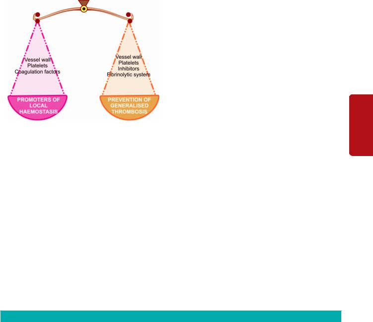
Figure 13.4 
 The haemostatic balance.
to contract after injury. Tests of defective vascular function are as under:
1.BLEEDING TIME. This simple test is based on the principle of formation of haemostatic plug following a standard incision on the volar surface of the forearm and the time the incision takes to stop bleeding is measured. The test is dependent upon capillary function as well as on platelet number and ability of platelets to adhere to form aggregates. Normal range is 3-8 minutes. A prolonged bleeding time may be due to following causes:
i)Thrombocytopenia.
ii)Disorders of platelet function.
iii)von Willebrand’s disease.
iv)Vascular abnormalities (e.g. in Ehlers-Danlos syndrome).
v)Severe deficiency of factor V and XI.
2.HESS CAPILLARY RESISTANCE TEST (TOURNIQUET TEST). This test is done by tying sphygmomanometer cuff to the upper arm and raising the pressure in it between
diastolic and systolic for 5 minutes. After deflation, the number of petechiae appearing in the next 5 minutes in 3 cm2 area over the cubital fossa are counted. Presence of more than 20 petechiae is considered a positive test. The test is positive in increased capillary fragility as well as in thrombocytopenia.
B. Investigation of Platelets and Platelet Function
Haemostatic disorders are commonly due to abnormalities in platelet number, morphology or function.
1. SCREENING TESTS. The screening tests carried out for assessing platelet-related causes are as under:
i)Peripheral blood platelet count.
ii)Skin bleeding time.
iii)Examination of fresh blood film to see the morphologic abnormalities of platelets.
2. SPECIAL TESTS. If these screening tests suggest a disorder of platelet function, the following platelet function tests may be carried out:
i)Platelet adhesion tests such as retention in a glass bead column, and other sophisticated techniques.
ii)Aggregation tests which are turbidometric techniques using ADP, collagen or ristocetin.
iii)Granular content of the platelets and their release can be assessed by electron microscopy or by measuring the substances released.
iv)Platelet coagulant activity is measured indirectly by prothrombin consumption index.
C. Investigation of Blood Coagulation
The normal blood coagulation system consists of cascade of activation of 13 coagulation factors. These form intrinsic, extrinsic and common pathways which culminate in formation of thrombin that acts on fibrinogen to produce fibrin. Fibrin clot so formed is strengthened by factor XIII
TABLE 13.1: Screening Tests for Haemostasis (Coagulation Tests).
|
Laboratory Test |
Factor/Function Measured |
Associated Disorders |
|
|
|
|
|
|
1. |
Bleeding time |
Platelet function, vascular integrity |
i) |
Qualitative disorders of platelets |
|
|
|
ii) |
von Willebrand’s disease |
|
|
|
iii) |
Quantitative disorders of platelets |
|
|
|
iv) |
Acquired vascular disorders |
2. |
Platelet count |
Quantification of platelets |
i) |
Thrombocytopenia |
|
|
|
ii) |
Thrombocytosis |
3. |
Prothrombin time |
Evaluation of extrinsic and common pathway |
i) |
Oral anticoagulant therapy |
|
|
(deficiency of factors I, II, V, VII, X) |
ii) |
DIC |
|
|
|
iii) |
Liver disease |
4. |
Partial thromboplastin time |
Evaluation of intrinsic and common pathway |
i) |
Parenteral heparin therapy |
|
|
(deficiency of factors I, II, V, VIII, IX, X, XI, XII) |
ii) |
DIC |
|
|
|
iii) |
Liver disease |
5. |
Thrombin time |
Evaluation of common pathway |
i) |
Afibrinogenaemia |
|
|
|
ii) |
DIC |
|
|
|
iii) |
Parenteral heparin therapy |
|
|
|
|
|
329
Medicine Transfusion Basic and Disorders Bleeding Platelets, of Disorders 13 CHAPTER
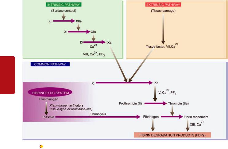
330
Tissues Lymphoreticular and Haematology II SECTION
Figure 13.5 |
Pathways of blood coagulation, fibrinolytic system and participation of platelets in activation of the cascade and their role in |
haemostatic plug formation.
which itself gets activated by thrombin. The process of fibrinolysis or clot dissolution and the role of platelets in activation of cascade and formation of haemostatic plug are illustrated in Fig. 13.5.
1. SCREENING TESTS. Tests for blood coagulation system include a battery of screening tests. These are as under:
I. Whole blood coagulation time. The estimation of whole blood coagulation time done by various capillary and tube methods is of limited value since it is an insensitive and nonspecific test. Normal range is 4-9 minutes at 37°C.
II. Activated partial thromboplastin time (APTT) or partial thromboplastin time with kaolin (PTTK). This test is used to measure the intrinsic system factors (VIII, IX, XI and XII) as well as factors common to both intrinsic and extrinsic systems (factors X, V, prothrombin and fibrinogen). The test consists of addition of 3 substances to the plasma—calcium, phospholipid and a surface activator such as kaolin. The normal range is 30-40 seconds. The common causes of a prolonged PTTK (or APTT) are as follows:
i)Parenteral administration of heparin.
ii)Disseminated intravascular coagulation.
iii)Liver disease.
iv)Circulating anticoagulants.
III. One-stage prothrombin time (PT). PT measures the extrinsic system factor VII as well as factors in the common pathway. In this test, tissue thromboplastin (e.g. brain extract) and calcium are added to the test. The normal PT in this test is 10-14 seconds. The common causes of prolonged one-stage PT are as under:
i)Administration of oral anticoagulant drugs.
ii)Liver disease, especially obstructive liver disease.
iii)Vitamin K deficiency.
iv)Disseminated intravascular coagulation.
IV. Measurement of fibrinogen. The screening tests for fibrinogen deficiency are semiquantitative fibrinogen titre and thrombin time (TT). The normal value of thrombin time is under 20 seconds, while a fibrinogen titre in plasma dilution up to 32 is considered normal. Following are the common causes for higher values in both these tests:
i)Hypofibrinogenaemia (e.g. in DIC).
ii)Raised concentration of FDP.
iii)Presence of heparin.
2. SPECIAL TESTS. In the presence of an abnormality in screening tests, detailed investigations for the possible cause are carried out. These include the following:
i) Coagulation factor assays. These bioassays are based on results of PTTK or PT tests and employ the use of substrate

plasma that contains all other coagulation factors except the one to be measured. The unknown level of the factor activity is compared with a standard control plasma with a known level of activity. Results are expressed as percentage of normal activity.
ii) Quantitative assays. The coagulation factors can be quantitatively assayed by immunological and other chemical methods.
D. Investigation of Fibrinolytic System
Increased levels of circulating plasminogen activator are present in patients with hyperfibrinolysis. Following screening tests are done to assess these abnormalities in fibrinolytic system:
1.Estimation of fibrinogen.
2.Fibrin degradation products (FDP) in the serum.
3.Ethanol gelation test.
4.Euglobin or whole blood lysis time.
More specific tests include: functional assays, immuno-
logical assays by ELISA, and chromogenic assays of plasminogen activators, plasminogen, plasminogen activator inhibitor, and FDP.
With this background knowledge on work up of a case of haemostatic abnormality, we now turn to discuss the common specific haemorrhagic disorders under the following headings:
I. Haemostatic diatheses due to vascular disorders. II. Haemostatic diatheses due to platelet disorders. III. Coagulation disorders.
IV. Haemostatic diatheses due to fibrinolytic defects. V. Disseminated intravascular coagulation (DIC)
HAEMORRHAGIC DIATHESES DUE TO
VASCULAR DISORDERS
Vascular bleeding disorders, also called non-thrombo- cytopenic purpuras or vascular purpuras, are normally mild and characterised by petechiae, purpuras or ecchymoses confined to the skin and mucous membranes. The pathogenesis of bleeding is poorly understood since majority of the standard screening tests of haemostasis including the bleeding time, coagulation time, platelet count and platelet function, are usually normal. Vascular purpuras arise from damage to the capillary endothelium, abnormalities in the subendothelial matrix or extravascular connective tissue that supports the blood vessels, or from formation of abnormal blood vessels.
Vascular bleeding disorders may be inherited or acquired.
A. Inherited Vascular Bleeding Disorders
A few examples of hereditary vascular disorders are given below:
1. Hereditary haemorrhagic telangiectasia (Osler-Weber- Rendu disease). This is an uncommon inherited autosomal dominant disorder. The condition begins in childhood and is characterised by abnormally telangiectatic (dilated) capillaries. These telangiectasias develop particularly in the skin, mucous membranes and internal organs and are the
source of frequent episodes of bleeding from the nose and gastrointestinal tract.
2. Inherited disorders of connective tissue matrix. These include Marfan’s syndrome, Ehlers-Danlos syndrome and pseudoxanthoma elasticum, all of which have inherited defect in the connective tissue matrix and, thus, have fragile skin vessels and easy bruising.
B. Acquired Vascular Bleeding Disorders
Several acquired conditions are associated with vascular purpuras. These are as under:
1.Henoch-Schönlein purpura. Henoch-Schönlein or anaphylactoid purpura is a self-limited type of hypersensitivity vasculitis occurring in children and young adults. Circulating immune complexes are deposited in the vessel wall consisting of IgA, C3 and fibrin, and in some cases, properdin suggesting activation of alternate complement pathway as the trigger event. The hypersensitivity vasculitis produces purpuric rash on the extensor surfaces of arms, legs and on the buttocks, as well as haematuria, colicky abdominal pain due to bleeding into the GIT, polyarthralgia and acute nephritis. In spite of these haemorrhagic features, all coagulation tests are normal.
2.Haemolytic-uraemic syndrome. Haemolytic-uraemic syndrome is a disease of infancy and early childhood in which there is bleeding tendency and varying degree of acute renal failure. The disorder remains confined to the kidney where hyaline thrombi are seen in the glomerular capillaries.
3.Simple easy bruising (Devil’s pinches). Easy bruising of unknown cause is a common phenomenon in women of child-bearing age group.
4.Infection. Many infections cause vascular haemorrhages either by causing toxic damage to the endothelium or by DIC. These are especially prone to occur in septicaemia and severe measles.
5.Drug reactions. Certain drugs form antibodies and produce hypersensitivity (or leucocytoclastic) vasculitis responsible for abnormal bleeding.
6.Steroid purpura. Long-term steroid therapy or Cushing’s syndrome may be associated with vascular purpura due to defective vascular support.
7.Senile purpura. Atrophy of the supportive tissue of cutaneous blood vessels in old age may cause senile atrophy, especially in the dorsum of forearm and hand.
8.Scurvy. Deficiency of vitamin C causes defective collagen synthesis which causes skin bleeding as well as bleeding into muscles, and occasionally into the gastrointestinal and genitourinary tracts.
HAEMORRHAGIC DIATHESES DUE TO
PLATELET DISORDERS
Disorders of platelets produce bleeding disorders by one of the following 3 mechanisms:
A. Due to reduction in the number of platelets i.e. various forms of thrombocytopenias.
331
Medicine Transfusion Basic and Disorders Bleeding Platelets, of Disorders 13 CHAPTER

332B. Due to rise in platelet count i.e. thrombocytosis. C. Due to defective platelet functions.
|
|
A. THROMBOCYTOPENIAS |
||
|
|
Thrombocytopenia is defined as a reduction in the peripheral |
||
|
|
blood platelet count below the lower limit of normal i.e. below |
||
|
|
150,000/μl. Thrombocytopenia is associated with abnormal |
||
|
|
bleeding that includes spontaneous skin purpura and |
||
|
|
mucosal haemorrhages as well as prolonged bleeding after |
||
|
|
trauma. However, spontaneous haemorrhagic tendency |
||
|
|
becomes clinically evident only after severe depletion of the |
||
SECTION |
|
platelet count to level below 20,000/μl. |
||
|
|
Thrombocytopenia may result from 4 main groups of |
||
causes: |
||||
|
||||
|
1. |
Impaired platelet production. |
||
|
2. |
Accelerated platelet destruction. |
||
II |
3. |
Splenic sequestration. |
||
4. |
Dilutional loss. |
|||
|
||||
|
|
|
A list of causes of thrombocytopenia is given in |
|
Haematology |
|
Table 13.2. Three of the common and important causes— |
||
|
drug-induced thrombocytopenia, idiopathic thrombocyto- |
|||
|
|
|||
|
|
penic purpura (ITP), and thrombotic thrombocytopenic |
||
|
|
purpura (TTP), are discussed below. |
||
|
|
Drug-induced Thrombocytopenia |
||
and |
|
Many commonly used drugs cause thrombocytopenia by |
||
|
depressing megakaryocyte production. In most cases, an |
|||
|
|
|||
Lymphoreticular |
|
immune mechanism by formation of drug-antibody comp- |
||
|
lexes is implicated in which the platelet is damaged as an |
|||
|
|
|||
|
|
‘innocent bystander’. Drug-induced thrombocytopenia is |
||
|
|
associated with many commonly used drugs and includes: |
||
|
|
chemotherapeutic agents (alkylating agents, anthracyclines, |
||
|
|
|
||
|
|
TABLE 13.2: Causes of Thrombocytopenia. |
||
Tissues |
|
I. |
IMPAIRED PLATELET PRODUCTION |
|
|
|
1. Generalised bone marrow failure e.g. |
||
|
|
Aplastic anaemia, leukaemia, myelofibrosis, megaloblastic |
||
|
|
anaemia, marrow infiltrations (carcinomas, lymphomas, multiple |
||
myeloma, storage diseases).
2. Selective suppression of platelet production e.g.
Drugs (quinine, quinidine, sulfonamides, PAS, rifampicin, anticancer drugs, thiazide diuretics), (heparin, diclofenac, acyclovir), alcohol intake.
II.ACCELERATED PLATELET DESTRUCTION
1.Immunologic thrombocytopenias e.g.
ITP (acute and chronic), neonatal and post-transfusion (isoimmune), drug-induced, secondary immune ‘thrombocytopenia (post-infection, SLE, AIDS, CLL, lymphoma).
2.Increased consumption e.g.
DIC, TTP, giant haemangiomas, microangiopathic haemolytic anaemia.
III. SPLENIC SEQUESTRATION Splenomegaly
IV. DILUTIONAL LOSS
Massive transfusion of old stored blood to bleeding patients.
antimetabolites), certain antibiotics (sulfonamides, PAS, rifampicin, penicillins), drugs used in cardiovascular diseases (digitoxin, thiazide diuretics), diclofenac, acyclovir, heparin and excessive consumption of ethanol.
Clinically, the patient presents with acute purpura. The platelet count is markedly lowered, often below 10,000/μl and the bone marrow shows normal or increased number of megakaryocytes.
The immediate treatment is to stop or replace the suspected drug with instruction to the patient to avoid taking the offending drug in future. Occasional patients may require temporary support with glucocorticoids, plasmapheresis or platelet transfusions.
Heparin-induced Thrombocytopenia
Thrombocytopenia due to administration of heparin is distinct from that caused by other drugs in following ways:
i)Thrombocytopenia is generally not so severe to fall to level below 20,000/μl.
ii)Unlike drug-induced thrombocytopenia, heparin-induced thrombocytopenia is not associated with bleeding but instead these patients are more prone to develop thrombosis.
The underlying mechanism of heparin-induced thrombocytopenia is formation of antibody against platelet factor 4 (PF-4)-heparin complex. This specific antibody activates the endothelial cells and initiates thrombus formation. It occurs in a small proportion of cases after the patient has received heparin for 5-10 days.
Diagnosis is made by a combination of laboratory and clinical features with 4 Ts: thrombocytopenia, thrombosis, time of fall of platelet count, absence of other causes of thrombocytopenia.
Immune Thrombocytopenic Purpura (ITP)
Idiopathic thrombocytopenic purpura or immune thrombocytopenic purpura (ITP), is characterised by immunologic destruction of platelets and normal or increased megakaryocytes in the bone marrow.
PATHOGENESIS. On the basis of duration of illness, ITP is classified into acute and chronic forms, both of which have different pathogenesis.
Acute ITP. This is a self-limited disorder, seen most frequently in children following recovery from a viral illness (e.g. hepatitis C, infectious mononucleosis, CMV infection, HIV infection) or an upper respiratory illness. The onset of acute ITP is sudden and severe thrombocytopenia but recovery occurs within a few weeks to 6 months. The mechanism of acute ITP is by formation of immune complexes containing viral antigens, and by formation of antibodies against viral antigens which crossreact with platelets and lead to their immunologic destruction.
Chronic ITP. Chronic ITP occurs more commonly in adults, particularly in women of child-bearing age (20-40 years). The disorder develops insidiously and persists for several years. Though chronic ITP is idiopathic, similar immunologic thrombocytopenia may be seen in association with SLE, AIDS and autoimmune thyroiditis. The pathogenesis of chronic ITP

333
Figure 13.6 
 Laboratory findings of ITP contrasted with those found in a normal individual. A, Peripheral blood in ITP shows presence of reduced number of platelets which are often large. B, Bone marrow in ITP shows characteristically increased number of megakaryocytes with single non-lobulated nuclei and reduced cytoplasmic granularity (inbox on right photomicrograph).
is explained by formation of anti-platelet autoantibodies, usually by platelet-associated IgG humoral antibodies synthesised mainly in the spleen. These antibodies are directed against target antigens on the platelet glycoproteins, Gp IIb-IIIa and Gp Ib-IX complex. Some of the antibodies directed against platelet surface also interfere in their function. The mechanism of platelet destruction is similar to that seen in autoimmune haemolytic anaemias. Sensitised platelets are destroyed mainly in the spleen and rendered susceptible to phagocytosis by cells of the reticuloendothelial system.
CLINICAL FEATURES. The clinical manifestation of ITP may develop abruptly in cases of acute ITP, or the onset may be insidious as occurs in majority of cases of chronic ITP. The usual manifestations are petechial haemorrhages, easy bruising, and mucosal bleeding such as menorrhagia in women, nasal bleeding, bleeding from gums, melaena and haematuria. Intracranial haemorrhage is, however, rare. Splenomegaly and hepatomegaly may occur in cases with chronic ITP but lymphadenopathy is quite uncommon in either type of ITP.
LABORATORY FINDINGS. The diagnosis of ITP can be suspected on clinical features after excluding the known causes of thrombocytopenia and is supported by the following haematologic findings:
1.Platelet count is markedly reduced, usually in the range of 10,000-50,000/μl.
2.Blood film shows only occasional platelets which are often large in size (Fig. 13.6,A).
3.Bone marrow shows increased number of megakaryocytes which have large non-lobulated single nuclei and may have reduced cytoplasmic granularity and presence of vacuoles (Fig. 13.6, B).
4.With sensitive techniques, anti-platelet IgG antibody can be demonstrated on platelet surface or in the serum of patients.
5.Platelet survival studies reveal markedly reduced platelet lifespan, sometimes less than one hour, as compared with normal lifespan of 7-10 days.
TREATMENT. Spontaneous recovery occurs in 90% cases of acute ITP, while only less than 10% cases of chronic ITP
Medicine Transfusion Basic and Disorders Bleeding Platelets, of Disorders 13 CHAPTER

334 recover spontaneously. Treatment is directed at reducing the level and source of autoantibodies and reducing the rate of destruction of sensitised platelets. This is possible by corticosteroid therapy, immunosuppressive drugs (e.g. vincristine, cyclophosphamide and azathioprine) and splenectomy. Beneficial effects of splenectomy in chronic ITP are due to both removal of the major site of platelet destruction and the major source of autoantibody synthesis. Platelet transfusions are helpful as a palliative measure only in patients with severe haemorrhage.
|
Thrombotic Thrombocytopenic Purpura (TTP) and |
|
SECTION |
Haemolytic-Uraemic Syndrome (HUS) |
|
Thrombotic thrombocytopenic purpura (TTP) and haemo- |
||
lytic-uraemic syndrome (HUS) are a group of thrombotic |
||
microangiopathies which are essentially characterised by |
||
triad of thrombocytopenia, microangiopathic haemolytic anaemia |
||
II |
and formation of hyaline fibrin microthrombi within the micro- |
|
|
vasculature throughout the body. These are often fulminant |
|
Haematology |
and lethal disorders occurring in young adults. The intra- |
|
different organs and tissues throughout the body. |
||
|
vascular microthrombi are composed predominantly of |
|
|
platelets and fibrin. The widespread presence of these platelet |
|
|
microthrombi is responsible for thrombocytopenia due to |
|
|
increased consumption of platelets, microangiopathic haemo- |
|
and |
lytic anaemia and protean clinical manifestations involving |
|
PATHOGENESIS. Unlike DIC, a clinicopathologically |
||
|
||
Lymphoreticular |
related condition, activation of the clotting system is not the |
|
primary event in formation of microthrombi. TTP is initiated |
||
|
||
|
by endothelial injury followed by release of von Willebrand |
|
|
factor and other procoagulant material from endothelial cells, |
|
|
leading to the formation of microthrombi. Trigger for the |
|
|
endothelial injury comes from immunologic damage by |
|
|
diverse conditions such as in pregnancy, metastatic cancer, |
|
Tissues |
high-doze chemotherapy, HIV infection, and mitomycin C. |
|
CLINICAL FEATURES. The clinical manifestations of TTP |
||
|
||
|
are due to microthrombi in the arterioles, capillaries and |
|
|
venules throughout the body. Besides features of thrombo- |
|
|
cytopenia and microangiopathic haemolytic anaemia, |
|
|
characteristic findings include fever, transient neurologic |
|
|
deficits and renal failure. The spleen may be palpable. |
LABORATORY FINDINGS. The diagnosis can be made from the following findings:
1.Thrombocytopenia.
2.Microangiopathic haemolytic anaemia with negative Coombs’ test.
3.Leucocytosis, sometimes with leukaemoid reaction.
4.Bone marrow examination reveals normal or slightly increased megakaryocytes accompanied with some myeloid hyperplasia.
5.Diagnosis is, however, established by examination of biopsy (e.g. from gingiva) which demonstrates typical microthrombi in arterioles, capillaries and venules, unassociated with any inflammatory changes in the vessel wall.
B. THROMBOCYTOSIS
Thrombocytosis is defined as platelet count in excess of 4,00,000/μl. While essential or primary thrombocytosis or thrombocythaemia is discussed under myeloproliferative disorders in the next chapter, secondary or reactive thrombocytosis can occur following massive haemorrhage, iron deficiency, severe sepsis, marked inflammation, disseminated cancers, haemolysis, or following splenectomy. Thrombocytosis causes bleeding or thrombosis but how it produces is not clearly known.
As such, transitory and secondary thrombocytisis does not require any separate treatment other than treating the cause.
C. DISORDERS OF PLATELET FUNCTIONS
Defective platelet function is suspected in patients who show skin and mucosal haemorrhages and have prolonged bleeding time but a normal platelet count. These disorders may be hereditary or acquired.
Hereditary Disorders
Depending upon the predominant functional abnormality, inherited disorders of platelet functions are classified into the following 3 groups:
1. DEFECTIVE PLATELET ADHESION. These are as under:
i)Bernard-Soulier syndrome is an autosomal recessive disorder with inherited deficiency of a platelet membrane glycoprotein which is essential for adhesion of platelets to vessel wall.
ii)In von Willebrand’s disease, there is defective platelet adhesion as well as deficiency of factor VIII (page 336).
2.DEFECTIVE PLATELET AGGREGATION. In thrombasthenia (Glanzmann’s disease), there is failure of primary platelet aggregation with ADP or collagen due to inherited deficiency of two of platelet membrane glycoproteins.
3.DISORDERS OF PLATELET RELEASE REACTION.
These disorders are characterised by normal initial aggregation of platelets with ADP or collagen but the subsequent release of ADP, prostaglandins and 5-HT is defective due to complex intrinsic deficiencies.
Acquired Disorders
Acquired defects of platelet functions include the following clinically significant examples:
1.ASPIRIN THERAPY. Prolonged use of aspirin leads to easy bruising and abnormal bleeding time. This is because aspirin inhibits the enzyme cyclooxygenase, and thereby suppresses the synthesis of prostaglandins which are involved in platelet aggregation as well as release reaction. The anti-platelet effect of aspirin is clinically applied in preventing major thromboembolic disease in recurrent myocardial infarction.
2.OTHERS. Several other acquired disorders are associated with various abnormalities in platelet functions at different
levels. These include: uraemia, liver disease, multiple myeloma, Waldenström’s macroglobulinaemia and various myeloproliferative disorders.
COAGULATION DISORDERS
The physiology of normal coagulation is described in Chapter 5 together with relatively more common coagulation disorders of arterial and venous thrombosis and embolism. A deficiency of each of the thirteen known plasma coagulation factors has been reported, which may be inherited or acquired. In general, coagulation disorders are less common as compared with other bleeding disorders. The type of bleeding in coagulation disorders is different from that seen in vascular and platelet abnormalities. Instead of spontaneous appearance of petechiae and purpuras, the plasma coagulation defects manifest more often in the form of large ecchymoses, haematomas and bleeding into muscles, joints, body cavities, GIT and urinary tract. For establishing the diagnosis, screening tests for coagulation (whole blood coagulation time, bleeding time, activated partial thromboplastin time and prothrombin time) are carried out, followed by coagulation factor assays as discussed already on page 330.
Disorders of plasma coagulation factors may have hereditary or acquired origin.
HEREDITARY COAGULATION DISORDERS. Most of the inherited plasma coagulation disorders are due to qualitative or quantitative defect in a single coagulation factor. Out of defects in various coagulation factors, two of the most common inherited coagulation disorders are the sex-(X)- linked disorders—classic haemophilia or haemophilia A (due to inherited deficiency of factor VIII), and Christmas disease or haemophilia B (due to inherited deficiency of factor IX). Another common and related coagulation disorder, von Willebrand’s disease (due to inherited defect of von Willebrand’s factor), is also discussed here.
ACQUIRED COAGULATION DISORDERS. The acquired coagulation disorders, on the other hand, are usually characterised by deficiencies of multiple coagulation factors. The most common acquired clotting abnormalities are: vitamin K deficiency, coagulation disorder in liver diseases, fibrinolytic defects and disseminated intravascular coagulation (DIC).
The more common of the hereditary and acquired coagulation disorders are discussed below.
Classic Haemophilia (Haemophilia A)
Classic haemophilia or haemophilia A is the second most common hereditary coagulation disorder next to von Willebrand’s disease occurring due to deficiency or reduced activity of factor VIII (anti-haemophilic factor). The disorder is inherited as a sex-(X-) linked recessive trait and, therefore, manifests clinically in males, while females are usually the carriers. However, occasional women carriers of haemophilia may produce factor VIII levels far below 50% and become symptomatic carriers, or rarely there may be true female haemophilics arising from consanguinity within the family (i.e. homozygous females). The chances of a proven carrier
mother passing on the abnormality to her children is 50:50 for each son and 50:50 for each daughter. A haemophilic father will have normal sons as they inherit his Y chromosome only that does not carry the genetic abnormality.
The disease has been known since ancient times but Schönlein in 1839 gave this bleeder’s disease its present name haemophilia. In 1952, it was found that haemophilia was not always due to deficiency of factor VIII as was previously considered but instead blood of some patients was deficient in factor IX (Christmas factor or plasma thromboplastin component). Currently, haemophilia A (classic haemophilia) is the term used for the disorder due to factor VIII deficiency, and haemophilia B (Christmas disease) for the disorder when factor IX is deficient.
The frequency of haemophilia varies in different races, the highest incidence being in populations of Britain, Northern Europe and Australia. Western literature reports give an overall incidence of haemophilia in 1 in 10,000 male births. Another interesting facet of the haemophilia which has attracted investigators and researchers is the occurrence of this disorder in the blood of royal families in Great Britain and some European countries.
PATHOGENESIS. Haemophilia A is caused by quantitative reduction of factor VIII in 90% of cases, while 10% cases have normal or increased level of factor VIII with reduced activity. Factor VIII is synthesised in hepatic parenchymal cells and regulates the activation of factor X in intrinsic coagulation pathway. Factor VIII circulates in blood complexed to another larger protein, von Willebrand’s factor (vWF), which comprises 99% of the factor VIII-vWF complex. The genetic coding, synthesis and functions of vWF are different from those of factor VIII and are considered separately below under von Willebrand’s disease. Normal haemostasis requires 25% factor VIII activity. Though occasional patients with 25% factor VIII level may develop bleeding, most symptomatic haemophilic patients have factor VIII levels below 5%.
CLINICAL FEATURES. Patients of haemophilia suffer from bleeding for hours or days after the injury. The clinical severity of the disease correlates well with plasma level of factor VIII activity. Haemophilic bleeding can involve any organ but occurs most commonly as recurrent painful haemarthroses and muscle haematomas, and sometimes as haematuria. Spontaneous intracranial haemorrhage and oropharyngeal bleeding are rare, but when they occur they are the most feared complications.
LABORATORY FINDINGS. The following tests are abnormal:
1.Whole blood coagulation time is prolonged in severe cases only.
2.Prothrombin time is usually normal.
3.Activated partial thromboplastin time (APTT or PTTK) is typically prolonged.
4.Specific assay for factor VIII shows lowered activity. The diagnosis of female carriers is made by the findings of about half the activity of factor VIII, while the manifest disease is associated with factor VIII activity below 25%.
335
Medicine Transfusion Basic and Disorders Bleeding Platelets, of Disorders 13 CHAPTER

336 TREATMENT. Symptomatic patients with bleeding episodes are treated with factor VIII replacement therapy, consisting of factor VIII concentrates or plasma cryoprecipitates. With the availability of this treatment, the life expectancy of even severe haemophilic patients was approaching normal but the occurrence of AIDS in multitransfused haemophilic patients has adversely affected the life expectancy.
|
Christmas Disease (Haemophilia B) |
|
|
Inherited deficiency of factor IX (Christmas factor or plasma |
|
|
thromboplastin component) produces Christmas disease or |
|
SECTION |
haemophilia B. Haemophilia B is rarer than haemophilia A; |
|
its estimated incidence is 1 in 100,000 male births. The |
||
|
||
|
inheritance pattern and clinical features of factor IX deficiency |
|
|
are indistinguishable from those of classic haemophilia but |
|
|
accurate laboratory diagnosis is critical since haemophilia B |
|
II |
requires treatment with different plasma fraction. The usual |
|
screening tests for coagulation are similar to those in classic |
||
|
||
|
haemophilia but bioassay of factor IX reveals lowered |
|
Haematology |
activity. |
|
the replacement therapy in factor IX deficiency may activate |
||
|
TREATMENT. Therapy in symptomatic haemophilia B |
|
|
consists of infusion of either fresh frozen plasma or a plasma |
|
|
enriched with factor IX. Besides the expected possibilities of |
|
|
complications of hepatitis, chronic liver disease and AIDS, |
|
and |
the coagulation system and cause thrombosis and embolism. |
|
von Willebrand’s Disease |
||
Lymphoreticular |
||
DEFINITION AND PATHOGENESIS. von Willebrand’s |
||
|
||
|
disease (vWD) is the most common hereditary coagulation |
|
|
disorder occurring due to qualitative or quantitative defect |
|
|
in von Willebrand’s factor (vWF). Its incidence is estimated |
|
|
to be 1 in 1,000 individuals of either sex. The vWF comprises |
|
|
the larger fraction of factor VIII-vWF complex which |
|
Tissues |
circulates in the blood. Though the two components of factor |
|
VIII-vWF complex circulate together as a unit and perform |
||
the important function in clotting and facilitate platelet |
||
adhesion to subendothelial collagen, vWF differs from factor |
||
|
VIII in the following respects: |
|
|
1. The gene for vWF is located at chromosome 12, while that |
|
|
of factor VIII is in X-chromosome. Thus, vWD is inherited as |
|
|
an autosomal dominant trait which may occur in either sex, |
|
|
while factor VIII deficiency (haemophilia A) is a sex (X-)- |
|
|
linked recessive disorder. |
|
|
2. The vWF is synthesised in the endothelial cells, mega- |
|
|
karyocytes and platelets but not in the liver cells, while the |
|
|
principal site of synthesis of factor VIII is the liver. |
|
|
3. The main function of vWF is to facilitate the adhesion of |
|
|
platelets to subendothelial collagen, while factor VIII is |
|
|
involved in activation of factor X in the intrinsic coagulation |
|
|
pathway. |
|
|
CLINICAL FEATURES. Clinically, the patients of vWD are |
|
|
characterised by spontaneous bleeding from mucous |
|
|
membranes and excessive bleeding from wounds. There are |
|
|
3 major types of vWD: |
Type I disease is the most common and is characterised by mild to moderate decrease in plasma vWF (50% activity). The synthesis of vWF is normal but the release of its multimers is inhibited.
Type II disease is much less common and is characterised by normal or near normal levels of vWF which is functionally defective.
Type III disease is extremely rare and is the most severe form of the disease. These patients have no detectable vWF activity and may have sufficiently low factor VIII levels.
Bleeding episodes in vWD are treated with cryoprecipitates or factor VIII concentrates.
LABORATORY FINDINGS. These are as under:
1.Prolonged bleeding time.
2.Normal platelet count.
3.Reduced plasma vWF concentration.
4.Defective platelet aggregation with ristocetin, an antibiotic.
5.Reduced factor VIII activity.
Vitamin K Deficiency
Vitamin K is a fat-soluble vitamin which plays important role in haemostasis since it serves as a cofactor in the formation of 6 prothrombin complex proteins (vitamin K- dependent coagulation factors) synthesised in the liver: factor II, VII, IX, X, protein C and protein S. Vitamin K is obtained from green vegetables, absorbed in the small intestine and stored in the liver (Chapter 9). Some quantity of vitamin K is endogenously synthesised by the bacteria in the colon.
Vitamin K deficiency may present in the newborn or in subsequent childhood or adult life:

 Neonatal vitamin K deficiency. Deficiency of vitamin K in the newborn causes haemorrhagic disease of the newborn. Liver cell immaturity, lack of gut bacterial synthesis of the vitamin and low quantities in breast milk, all contribute to vitamin K deficiency in the newborn and may cause haemorrhage on 2nd to 4th day of life. Routine administration of vitamin K to all newly born infants has led to disappearance of neonatal vitamin K deficiency.
 Vitamin K deficiency in children and adult. There are 3 major causes of vitamin K deficiency in childhood or adult
life:
1.Inadequate dietary intake.
2.Intestinal malabsorption.
3.Loss of storage site due to hepatocellular disease.
With the onset of vitamin K deficiency, the plasma levels
of all the 6 vitamin K-dependent factors (prothrombin complex proteins) fall. This, in turn, results in prolonged PT and PTTK. Parenteral administration of vitamin K rapidly restores vitamin K levels in the liver.
Coagulation Disorders in Liver Disease
Since liver is the major site for synthesis and metabolism of coagulation factors, liver disease often leads to multiple haemostatic abnormalities. The liver also produces inhibitors
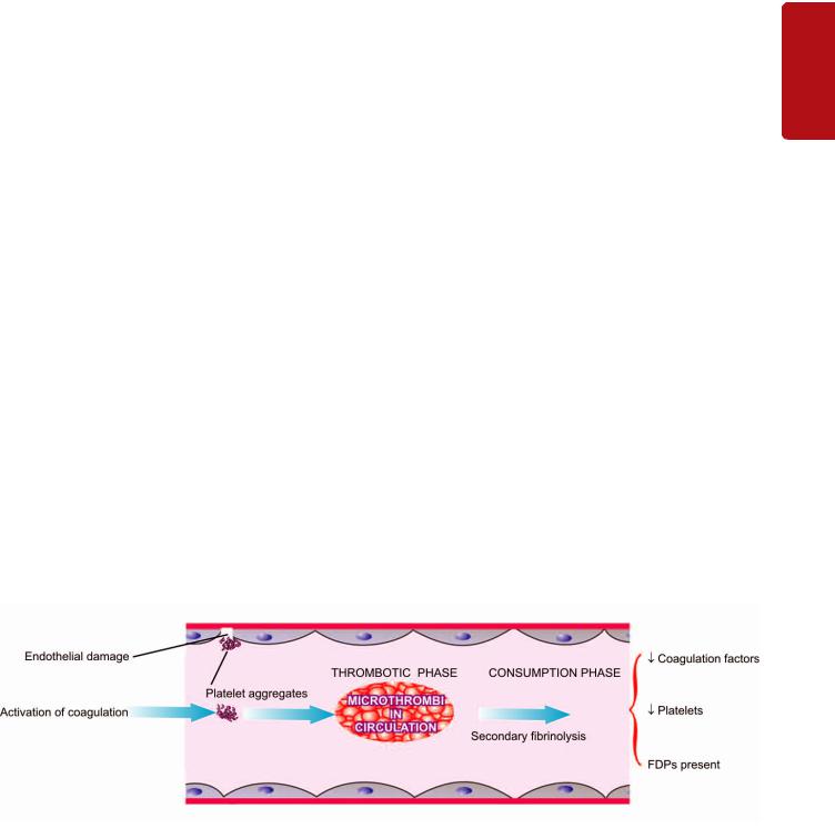
of coagulation such as antithrombin III and protein C and S and plays a role in the clearance of activated factors and fibrinolytic enzymes. Thus, patients with liver disease may develop hypercoagulability and are predisposed to develop DIC and systemic fibrinolysis.
The major causes of bleeding in liver diseases are as under:
A. Morphologic lesions:
1.Portal hypertension e.g. varices, splenomegaly with secondary thrombocytopenia.
2.Peptic ulceration.
3.Gastritis.
B. Hepatic dysfunctions:
1.Impaired hepatic synthesis of coagulation factors.
2.Impaired hepatic synthesis of coagulation inhibitors: protein C, protein S and antithrombin III.
3.Impaired absorption and metabolism of vitamin K.
4.Failure to clear activated coagulation factors causing DIC and systemic fibrinolysis.
C. Complications of therapy:
1.Following massive transfusion leading to dilution of platelets and coagulation factors.
2.Infusion of activated coagulation proteins.
3.Following heparin therapy.
Many a times, the haemostatic abnormality in liver
disease is complex but most patients have prolonged PT and PTTK, mild thrombocytopenia, normal fibrinogen level and decreased hepatic stores of vitamin K.
HAEMORRHAGIC DIATHESIS DUE TO
FIBRINOLYTIC DEFECTS
Normally, fibrinolysis consisting of plasminogen-plasmin and fibrin degradation products (FDPs) is an essential protective physiologic mechanism to limit the blood coagulation in the body. However, unchecked and excessive fibrinolysis may sometimes be the cause of bleeding. The
causes of primary pathologic fibrinolysis leading to haemorrhagic defects are as under:
1.Deficiency of α2-plasmin inhibitor following trauma or surgery.
2.Impaired clearance of tissue plasminogen activator such as in cirrhosis of liver.
At times, it may be difficult to distinguish primary pathologic fibrinolysis from secondary fibrinolysis accompanying DIC.
DISSEMINATED INTRAVASCULAR COAGULATION (DIC)
Disseminated intravascular coagulation (DIC), also termed defibrination syndrome or consumption coagulopathy, is a complex thrombo-haemorrhagic disorder (intravascular coagulation and haemorrhage) occurring as a secondary complication in some systemic diseases.
ETIOLOGY. Although there are numerous conditions associated with DIC, most frequent causes are listed below:
1.Massive tissue injury: in obstetrical syndromes (e.g. abruptio placentae, amniotic fluid embolism, retained dead foetus), massive trauma, metastatic malignancies, surgery.
2.Infections: especially endotoxaemia, gram-negative and meningococcal septicaemia, certain viral infections, malaria, aspergillosis.
3.Widespread endothelial damage: in aortic aneurysm, haemolytic-uraemic syndrome, severe burns, acute glomerulonephritis.
4.Miscellaneous: snake bite, shock, acute intravascular haemolysis, heat stroke.
PATHOGENESIS. Although in each case, a distinct triggering mechanism has been identified, the sequence of events, in general, can be summarised as under (Fig. 13.7):
1.Activation of coagulation. The etiologic factors listed above initiate widespread activation of coagulation pathway by release of tissue factor.
2.Thrombotic phase. Endothelial damage from the various thrombogenic stimuli causes generalised platelet aggregation and adhesion with resultant deposition of small thrombi and emboli throughout the microvasculature.
3.Consumption phase. The early thrombotic phase is followed by a phase of consumption of coagulation factors and platelets.
4.Secondary fibrinolysis. As a protective mechanism, fibrinolytic system is secondarily activated at the site of intravascular coagulation. Secondary fibrinolysis causes breakdown of fibrin resulting in formation of FDPs in the circulation.
Pathophysiology of DIC is summed up schematically in
Fig. 13.8.
337
Medicine Transfusion Basic and Disorders Bleeding Platelets, of Disorders 13 CHAPTER
Figure 13.7 
 The pathogenesis of disseminated intravascular coagulation.
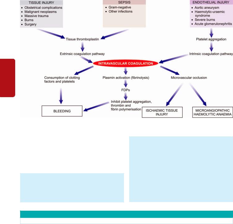
338
Tissues Lymphoreticular and Haematology II SECTION
Figure 13.8 
 Pathophysiology of disseminated intravascular coagulation.
CLINICAL FEATURES. There are 2 main features of DIC— bleeding as the most common manifestation, and organ damage due to ischaemia caused by the effect of widespread intravascular thrombosis such as in the kidney and brain. Less common manifestations include: microangiopathic haemolytic anaemia and thrombosis in larger arteries and veins.
LABORATORY FINDINGS. The laboratory investigations include the following:
1.The platelet count is low.
2.Blood film shows the features of microangiopathic haemolytic anaemia. There is presence of schistocytes and
fragmented red cells due to damage caused by trapping and passage through the fibrin thrombi.
3.Prothrombin time, thrombin time and activated partial thromboplastin time, are all prolonged.
4.Plasma fibrinogen levels are reduced due to consumption in microvascular coagulation.
5.Fibrin degradation products (FDPs) are raised due to secondary fibrinolysis.
A summary of important laboratory findings in common causes of haemostatic abnormalities is summed up in Table 13.3.
TABLE 13.3: Major Laboratory Findings in Common Haemostatic Disorders.
Disorder |
Platelet Count |
BT |
PT |
APTT |
TT |
FDPs |
F-VIII |
F-IX |
|
|
|
|
|
|
|
|
|
I.VASCULAR DISORDERS
Vascular purpuras |
N |
N |
N |
N |
N |
Absent |
N |
N |
II.PLATELET DISORDERS
1. |
ITP |
↓ |
↑ |
N |
N |
N |
Absent |
N |
N |
2. |
Heparin |
↓ |
↑ |
N |
↑ |
↑ |
Absent |
N |
N |
3. |
TTP |
↓ |
↑ |
N |
N |
N |
Absent |
N |
N |
III.COAGULATION DISORDERS
1. |
Haemophilia A |
N |
↑ |
N |
↑ |
↑ |
Absent |
↓ |
N |
2. |
Haemophilia B |
N |
↑ |
N |
↑ |
↑ |
Absent |
N |
↓ |
3. |
von Willebrand’s |
N |
↑ |
N |
↑ |
↑ |
Absent |
↓ |
N |
4. |
Vit. K deficiency |
N |
↑ |
↑ |
↑ |
↑ |
Absent |
N |
N |
5. |
Liver disease |
N |
↑ |
↑ |
↑ |
N |
Absent |
N |
N |
IV. DIC |
|
↓ |
↑ |
↑ |
↑ |
↑ |
Present |
↓ |
↓ |
|
|
|
|
|
|
|
|
|
|

BLOOD GROUPS AND BLOOD TRANSFUSION
BLOOD GROUP ANTIGENS AND ANTIBODIES
Karl Landsteiner described the existence of major human blood groups in 1900 and was awarded Nobel Prize in 1930. The term blood group is applied to any well-defined system of red blood cell antigens which are inherited characteristics. Over 20 blood group systems having approximately 400 blood group antigens are currently recognised. The ABO and Rhesus (Rh) blood group systems are of major clinical significance. Other minor and clinically less important blood group systems are: Lewis system, P system, I system, MNS system, Kell and Duffy system, and Luthern system.
Individuals who lack the corresponding antigen and have not been previously transfused have naturally-occurring antibodies in their serum. The most important are anti-A and anti-B antibodies, usually of IgM class. Immune antibodies, on the other hand, are acquired in response to transfusion and by transplacental passage during pregnancy. These are warm antibodies, usually of IgG class.
ABO SYSTEM. This system consists of 3 major allelic genes: A, B and O, located on the long arm of chromosome 9. These genes control the synthesis of blood group antigens A and B. The serum of an individual contains naturally-occurring antibodies to A and/or B antigen, whichever antigen is lacking in the person’s red cells (Table 13.4). Two subgroups of A—A1 and A2, and thus of AB also, A1B and A2B, are recognised but are of minor clinical significance. In routine practice, the ABO type is determined by testing the red blood cells with anti-A and anti-B and by testing the serum against A, B and O red blood cells.
Red blood cells of type O and A2 have large amounts of another antigen called H substance which is genetically different from ABO but is a precursor of A and B antigens. An O group individual who inherits A or B genes but fails to inherit H gene from either parent is called Oh phenotype or Bombay blood group. In such rare individual, despite the presence of all the three antibodies in serum (anti-A, anti-B and anti-H), the red cells are not agglutinated by the antisera.
RHESUS SYSTEM. The Rhesus (Rh) blood group system was first discovered on human red cells by the use of antisera prepared by immunising rabbits with red cells from a Rhesus monkey. The Rh allelic genes are C or c, D or d and E or e, located on chromosome 1. One set of 3 genes is inherited from each parent giving rise to various complex combinations. The corresponding antigens are similarly named Cc, Ee and only D since no d antigen exists.
TABLE 13.4: The ABO Blood Groups.
Blood |
Antigens on |
Naturally-Occurring |
Group |
Red Cells |
Serum Antibodies |
|
|
|
AB |
AB |
None |
A |
A |
Anti-B |
B |
B |
Anti-A |
O |
O |
Anti-A, Anti-B |
|
|
|
However, out of all these, D antigen is most strongly |
339 |
|
immunogenic and, therefore, clinically most important. In |
|
|
practice, Rh grouping is performed with anti-D antiserum. |
|
|
Individuals who are D-positive are referred to as Rh-positive |
|
|
and those who lack D antigen are termed Rh-negative. |
|
|
Practically, there are no naturally-occurring Rh anti- |
|
|
bodies. All Rh antibodies in Rh-negative individuals are |
|
|
acquired from immunisation such as by transfusion and |
|
|
during pregnancy, resulting in fatal haemolytic transfusion |
|
|
reaction and haemolytic disease of the newborn (described |
|
|
later). |
|
|
BLOOD TRANSFUSION |
CHAPTER |
|
A pre-transfusion compatibility testing is essential prior to |
||
|
||
any blood transfusion. The procedure consists of the |
|
|
following: |
|
|
1. ABO and Rh(D) grouping of the patient (recipient). |
13 |
|
2. Antibody screening of the patient’s serum to detect the |
||
presence of clinically significant antibodies. |
|
|
3. Selecting the donor blood of the same ABO and Rh group. |
Disorders |
|
4. Cross-matching the patient’s serum against donor red cells |
||
|
||
to confirm donor-recipient compatibility. |
|
|
The indications for blood transfusion are acute blood loss |
|
|
and various haematologic disorders considered already. In |
of |
|
addition to the whole blood transfusion, the modern blood- |
||
banking techniques have made it possible to transfuse blood |
Platelets, |
|
components such as packed red blood cells, platelets, white |
||
|
||
blood cell concentrates, plasma components and plasma- |
|
|
pheresis in specific situations. |
Bleeding |
|
Complications of Blood Transfusion |
||
|
||
A carefully prepared and supervised blood transfusion is |
|
|
quite safe. However, in 5-6% of transfusions, untoward |
|
|
complications occur, some of which are minor while others |
Disorders |
|
are more serious and at times fatal. |
||
|
||
Transfusion reactions are generally classified into 2 types: |
|
|
immune and non-immune. |
|
|
I. Immunologic transfusion reactions may be against red blood |
and |
|
cells (haemolytic reactions), leucocytes, platelets or |
||
|
||
immunoglobulins. |
Basic |
|
II. Non-immune transfusion reactions include circulatory |
||
|
||
overload, massive transfusion, or transmission of an |
|
|
infectious agent. |
Transfusion |
|
1. Haemolytic transfusion reactions. Haemolytic trans- |
||
These transfusion reactions are considered below. |
|
|
I. IMMUNOLOGIC TRANSFUSION REACTIONS. |
|
|
These are as under: |
|
|
fusion reaction may be immediate or delayed, intravascular or |
Medicine |
|
extravascular. |
||
|
||
Very rapid cell destruction associated with intravascular |
|
|
haemolysis is usually due to ABO incompatibility since both |
|
|
naturally-occurring antibodies, anti-A and anti-B, are capable |
|
|
of fixing complement. The symptoms include restlessness, |
|
|
anxiety, flushing, chest or lumbar pain, tachypnoea, |
|
|
tachycardia and nausea, followed by shock and renal failure. |
|

340 
 Extravascular haemolysis is more often due to immune antibodies of the Rh system. The clinical manifestations are
relatively less severe and usually consist of malaise and fever but shock and renal failure may rarely occur. Some patients develop delayed reactions in which the patient develops anaemia due to destruction of red cells in the RE system about a week after transfusion. Such delayed reactions are generally the result of previous transfusion or pregnancy (anamnestic reaction).
|
2. Transfusion-related acute lung injury (TRALI). This is |
|
|
an uncommon reaction resulting from transfusion of donor |
|
|
plasma containing high levels of anti-HLA antibodies which |
|
SECTION |
bind to leucocytes of recipient. These leucocytes then |
|
aggregate in pulmonary micromutation and release |
||
|
||
|
mediators of increased vascular permeability resulting in |
|
|
acute pulmonary oedema and signs and symptoms of |
|
|
respiratory failure. |
|
II |
3. Other allergic reactions. Besides haemolytic transfusion |
|
reaction, others reactions are as follows: |
||
|
||
Haematology |
i) Febrile reaction which is usually attributed to immunologic |
|
reaction against white blood cells, platelets, or IgA class |
||
iii) Allergic reactions such as urticaria may occur. |
||
|
immunoglobulins. |
|
|
ii) Patients with antibodies against IgA molecule sometimes |
|
|
develop anaphylactic shock on transfusion of blood from other |
|
|
human subjects. |
|
and |
iv) Transfusion-related graft-versus-host disease mediated by |
|
donor T lymphocytes may occur. |
||
Lymphoreticular |
II. NONIMMUNE TRANSFUSION REACTIONS. This |
|
|
||
|
category includes the following adverse effects: |
|
|
1. Circulatory overload. Circulatory overload resulting in |
|
|
pulmonary congestion and acute heart failure is the most |
|
|
important and most common complication that may result |
|
|
in death following transfusion. The risk of circulatory |
|
|
overload is particularly high in patients with chronic |
|
Tissues |
anaemia, and in infants and the elderly. The onset may be |
|
immediate, or may be delayed up to 24 hours. |
||
|
||
|
2. Massive transfusion. When the volume of stored blood |
|
|
transfused to bleeding patients exceeds their normal blood |
|
|
volume, it results in dilutional thrombocytopenia and |
|
|
dilution of coagulation factors. |
|
|
3. Transmission of infection. Many diseases can be |
|
|
transmitted by transfusion of an infected blood. These |
|
|
include: hepatitis (HBV, HCV), CMV infection, syphilis, |
|
|
malaria, toxoplasmosis, infectious mononucleosis, brucellosis |
|
|
and AIDS (HIV infection). The incidence increases in patients |
|
|
who receive multiple transfusions such as cases of |
|
|
haemophilia, thalassaemia major, acute leukaemias, acute |
|
|
severe haemorrhage etc. It has, therefore, been mandatory |
|
|
that prior to any human transfusion, every unit of blood must |
|
|
be screened for the serologic testing of HIV, HBV, HCV and |
|
|
syphilis and for the presence of malarial parasite. |
|
|
4. Air embolism. Air embolism is unlikely to occur if the |
|
|
blood transfusion is carried out with plastic bags with |
|
|
negative pressure as is the usual practice nowadays. A |
|
|
debilitated person may develop symptomatic air embolism |
even if a small volume (10-40 ml) makes its way into the circulation, whereas a healthy individual is at lesser risk.
5.Thrombophlebitis. The complication of thrombophlebitis is more commonly associated with venesection for blood transfusion, especially if it is done in the saphenous vein of the ankle rather than the veins of the arm. The risk of developing thrombophlebitis is further enhanced if the transfusion is continued longer than 12 hours at a single site.
6.Transfusion haemosiderosis. Post-transfusion iron overload with deposition of iron in the tissues of the body occurs after repeated transfusions in the absence of any blood loss e.g. in thalassaemia major and in severe chronic refractory anaemias. The body has no other means of getting rid of extra iron except iron excretion at the rate of 1 mg per day. A unit of whole blood (400 ml) contains about 250 mg of iron. After approximately 100 units, the liver, myocardium and endocrine glands are all damaged.
BLOOD COMPONENTS
Blood from donors is collected as whole blood in a suitable anticoagulant. Nowadays it is a common practice to divide whole blood into components which include: packed RBCs, platelets, fresh-frozen plasma (FFP) and cryoprecipitate.
The procedure consists of initial centrifugation at low speed to separate whole blood into two parts: packed RBCs and platelet-rich plasma (PRP). Subsequently, PRP is centrifuged at high speed to yield two parts: random donor platelets and FFP. Cryoprecipitates are obtained by thawing of FFP followed by centrifugation. Apheresis is the technique of direct collection of large excess of platelets from a single donor.
Applications of these blood components in clinical use are as under:
1.Packed RBCs. These are used to raise the oxygen-carrying capacity of blood and are used in normovolaemic patients of anaemia without cardiac disease. One unit of packed RBCs may raise haemoglobin by 1 g/dl.
2.Platelets. Transfusion of platelets is done in patients of thrombocytopenia who have haemorrhage. Optimally, platelet transfusions can be given to a patient with platelet count below 10,000/μl. Each unit of platelets can raise platelet count by 5,000 to 10,000/μl.
3.Fresh frozen plasma. FFP contains plasma proteins and coagulation factors that include albumin, protein C and S and antithrombin. FFP transfusion in indicated in patients of coagulation failure and TTP. Each unit of FFP raises coagulation factors by about 2%.
4.Cryoprecipitate. Cryoprecipitate is a source of insoluble plasma proteins, fibrinogen, factor VIII and vWF. Indications for transfusion of cryoprecipitate are for patients requiring fibrinogen, factor VIII and vWF. Transfusion of single unit of cryoprecipitate yields about 80 IU of factor VIII.
HAEMOLYTIC DISEASE OF NEWBORN
Haemolytic disease of the newborn (HDN) results from the passage of IgG antibodies from the maternal circulation

across the placenta into the circulation of the foetal red cells. Besides pregnancy, sensitisation of the mother may result from previous abortions and previous blood transfusion.
HDN can occur from incompatibility of ABO or Rh blood group system. ABO incompatibility is much more common but the HDN in such cases is usually mild, while Rh-D incompatibility results in more severe form of the HDN.
PATHOGENESIS. The pathogenesis of the two main forms of HDN is different.
HDN due to Rh-D incompatibility. Rh incompatibility occurs when a Rh-negative mother is sensitised to Rhpositive blood. This results most often from a Rh-positive foetus by passage of Rh-positive red cells across the placenta into the circulation of Rh-negative mother. Normally, during pregnancy very few foetal red cells cross the placenta but haemorrhage during parturition causes significant sensitisation of the mother. Sensitisation is more likely if the mother and foetus are ABO compatible rather than ABO incompatible. Though approximately 95% cases of Rh-HDN are due to anti-D, some cases are due to combination of anti- D with other immune antibodies of the Rh system such as anti-C and anti-E, and rarely anti-c alone.
It must be emphasised here that the risk of sensitisation of a Rh-negative woman married to Rh-positive man is small in first pregnancy but increases during successive pregnancies if prophylactic anti-D immunoglobulin is not given within 72 hours after the first delivery. If both the parents are Rh-D positive (homozygous), all the newborns will be Rh-D positive, while if the father is Rh-D positive (heterozygous), there is a 50% chance of producing a Rh-D negative child.
HDN due to ABO incompatibility. About 20% pregnancies with ABO incompatibility between the mother and the foetus develop the HDN. Naturally-occurring anti-A and anti-B antibodies’ which are usually of IgM class do not cross the
placenta, while immune anti-A and anti-B antibodies which are usually of IgG class may cross the placenta into foetal circulation and damage the foetal red cells. ABO HDN occurs most frequently in infants born to group O mothers who possess anti-A and/or anti-B IgG antibodies. ABO-HDN differs from Rh(D)-HDN, in that it occurs in first pregnancy, Coombs’ (antiglobulin) test is generally negative, and is less severe than the latter.
CLINICAL FEATURES. The HDN due to Rh-D incompatibility in its severest form may result in intrauterine death from hydrops foetalis. Moderate disease produces a baby born with severe anaemia and jaundice due to unconjugated hyperbilirubinaemia. When the level of unconjugated bilirubin exceeds 20 mg/dl, it may result in deposition of bile pigment in the basal ganglia of the CNS called kernicterus and result in permanent brain damage. Mild disease, however, causes only severe anaemia with or without jaundice.
LABORATORY FINDINGS. The haematologic findings in cord blood and mother’s blood are as under:
1.Cord blood shows variable degree of anaemia, reticulocytosis, elevated serum bilirubin and a positive direct Coombs’ test if the cord blood is Rh-D positive.
2.Mother’s blood is Rh-D negative with high plasma titre of anti-D.
COURSE AND PROGNOSIS. The course in HDN may range from death, to minimal haemolysis, to mental retardation. The practice of administration of anti-Rh immunoglobulin to the mother before or after delivery has reduced the incidence of HDN as well as protects the mother before the baby’s RBCs sensitise the mother’s blood. Exchange transfusion of the baby is done to remove the antibodies, remove red cells susceptible to haemolysis and also to lower the bilirubin level.
341
Medicine Transfusion Basic and Disorders Bleeding Platelets, of Disorders 13 CHAPTER

342
Chapter 14
Disorders of Leucocytes and
Lymphoreticular Tissues
Tissues Lymphoreticular and Haematology II SECTION
As illustrated in Fig. 12.3, pluripotent stem cells in the bone marrow divide to form two types of stem cells: myeloid trilineage stem cells and lymphoid stem cells. Myeloid trilineage stem cells further divide to form three types of cell lines: erythroid progenitor cells (discussed in Chapter 12), megakaryocytic progenitor cells (discussed in Chapter 13), and granulocyte-monocyte progenitor cells. In this chapter, we will discuss the abnormalities pertaining to the remaining two cell lines—granulocyte-monocyte cell line and the lymphoid cell lines, besides discussion on all haematopoietic neoplasms. Both these cell lines constitute leucocyte pool in the body at two distinct locations: in circulating blood and in the tissues. This concept holds more true for lymphoid cells in particular, which are present in circulation as well as are distributed in the lymphoid tissues of the body (lymph nodes, spleen, mucosa-associated lymphoid tissue—MALT, pharyngeal lymphoid tissue). B, T and NK lymphoid cells are formed after differentiation from lymphopoietic precursor cells in the bone marrow undergo further maturation in peripheral lymphoid organs and thymus (Chapter 4). Thus, relationship of haematopoietic tissues and lymphoreticluar tissues is quite close. In fact, current lymphoma-leukaemia classification system does not consider diseases of lymphocytes in the blood and in the lymphoid tissues as separate disorders but are seen to represent different stages of the same biologic process. Hence, in contemporary learning of these diseases, it is desirable to study diseases of leucocytes together with diseases of lymphoreticular tissues of the body.
LYMPH NODES: NORMAL AND REACTIVE
NORMAL STRUCTURE
The lymph nodes are bean-shaped or oval structures varying in length from 1 to 2 cm and form the part of lymphatic network distributed throughout the body. Each lymph node is covered by a connective tissue capsule. At the convex surface of the capsule several afferent lymphatics enter which drain into the peripheral subcapsular sinus, branch into the lymph node and terminate at the concavity (hilum) as a single efferent lymphatic vessel. These lymphatic vessels are lined by mononuclear phagocytic cells.
The inner structure of the lymph node is divided into a peripheral cortex and central medulla. The cortex consists of several rounded aggregates of lymphocytes called lymphoid follicles. The follicle has a pale-staining germinal centre surrounded by small dark-staining lymphocytes called the mantle zone. The deeper region of the cortex or paracortex is
the zone between the peripheral cortex and the inner medulla. The medulla is predominantly composed of cords of plasma cells and some lymphocytes. The capsule and the structure within the lymph node are connected by supportive delicate reticulin framework (Fig. 14.1,A).
Functionally, the lymph node is divided into T and B lymphocyte zones:
 B-cell zone lies in the follicles in the cortex, the mantle zone and the interfollicular space, while plasma cells are also present in the interfollicular zone.
 T-cell zone is predominantly present in the medulla. There are two main functions of the lymph node—to
mount immune response in the body, and to perform the function of active phagocytosis for particulate material. Besides T and B-cells, the follicular centre has dendritic histiocytes and antigen-presenting Langerhans’ histiocytes
(formerly together called tingible body macrophages due to engulfment of particulate material by them) and endothelial cells. The follicular centre is a very active zone where lymphocytes from peripheral blood continuously enter and leave, interact with macrophage-histiocytes and endothelial cells and undergo maturation and transformation. Lymphocytes and endothelial cells have surface molecules which interact and serve as ‘addresses’ so that endothelial cells can direct the lymphocytes; these molecules are appropriately termed as addressins or homing receptors. Peripheral blood B and T lymphocytes on entering the lymph node are stimulated immunologically which transforms them to undergo cytoplasmic and nuclear maturation which may be in the follicular centre or paracortex as per following sequence and schematically depicted in Fig. 14.1,B:
i)Follicular centre, small non-cleaved cells or centroblasts
ii)Follicular centre, small cleaved cells or centrocytes
iii)Follicular centre, large cleaved cells
iv)Follicular centre, large non-cleaved cells
v)Immunoblasts (in paracortex)
vi)Convoluted cells or lymphoblasts (in paracortex)
vii)Plasma cells.
Lymph nodes are secondarily involved in a variety of systemic diseases, local injuries and infections, and are also the site for some important primary neoplasms. Many of these diseases such as tuberculosis, sarcoidosis, histoplasmosis, typhoid fever, viral infections etc. have been considered elsewhere in the textbook along with description of these primary diseases. Reactive lymphadenitis is discussed below while the subject of lymphoid neoplasms including plasma cell disorders and Langerhans’ cell histiocytosis is discussed under haematologic neoplasms.
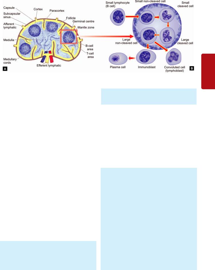
Figure 14.1 
 Normal lymph node. A, The anatomic structure and functional zones of a lymph node. B, Maturation of lymphoid cells in the follicle.
REACTIVE LYMPHADENITIS
Lymph nodes undergo reactive changes in response to a wide variety of stimuli which include microbial infections, drugs, environmental pollutants, tissue injury, immune-complexes and malignant neoplasms. However, the most common causes of lymph node enlargement are inflammatory and immune reactions, aside from primary malignant neoplasms and metastatic tumour deposits. Those due to primary inflammatory reaction are termed reactive lymphadenitis, and those due to primary immune reactions are referred to as
lymphadenopathy.
Reactive lymphadenitis is a nonspecific response and is categorised into acute and chronic types, each with a few variant forms.
Acute Nonspecific Lymphadenitis
All kinds of acute inflammations may cause acute nonspecific lymphadenitis in the nodes draining the area of inflamed tissue. Most common causes are microbiologic infections or their breakdown products, and foreign bodies in the wound or into the circulation etc. Most frequently involved lymph nodes are: cervical (due to infections in the oral cavity), axillary (due to infection in the arm), inguinal (due to infection in the lower extremities), and mesenteric (due to acute appendicitis, acute enteritis etc).
Acute lymphadenitis is usually mild and transient but occasionally it may be more severe. Acutely inflamed nodes are enlarged, tender, and if extensively involved, may be fluctuant. The overlying skin is red and hot. After control of infection, majority of cases heal completely without leaving any scar. If the inflammation does not subside, acute lymphadenitis changes into chronic lymphadenitis.
MORPHOLOGIC FEATURES. Grossly, the affected lymph nodes are enlarged 2-3 times their normal size and may show abscess formation if the involvement is extensive.
Microscopically, the sinusoids are congested, widely dilated and oedematous and contain numerous neutro-
phils. The lymphoid follicles are prominent with presence of many mitoses and phagocytosis. In more severe cases, necrosis may occur and neutrophil abscesses may form.
Chronic Nonspecific Lymphadenitis
Chronic nonspecific lymphadenitis, commonly called reactive lymphoid hyperplasia, is a common form of inflammatory reaction of draining lymph nodes as a response to antigenic stimuli such as repeated attacks of acute lymphadenitis and lymph from malignant tumours.
Depending upon the pattern in chronic nonspecific lymphadenitis, three types are distinguished, each having its own set of causes. These are: follicular hyperplasia,
paracortical hyperplasia and sinus histiocytosis. However, mixed patterns may also be seen in which case one of the patterns predominates over the others.
MORPHOLOGIC FEATURES. Grossly, the affected lymph nodes are usually enlarged, firm and non-tender. Microscopically, the features of 3 patterns of reactive lymphoid hyperplasia are as under:
1. Follicular hyperplasia is the most frequent pattern, particularly encountered in children. Besides nonspecific stimulation, a few specific causes are: rheumatoid arthritis, toxoplasmosis, syphilis and AIDS. The microscopic features are as follows (Fig. 14.2):
i)There is marked enlargement and prominence of the germinal centres of lymphoid follicles (proliferation of B- cell areas) due to the presence of numerous mitotically active lymphocytes and proliferation of phagocytic cells containing phagocytosed material.
ii)Parafollicular and medullary regions are more cellular and contain plasma cells, histiocytes, and some neutrophils and eosinophils.
iii)There is hyperplasia of mononuclear phagocytic cells lining the lymphatic sinuses in the lymph node.
 Angiofollicular lymphoid hyperplasia or Castleman’s disease is a clinicopathologic variant of follicular
343
Tissues Lymphoreticular and Leucocytes of Disorders 14 CHAPTER
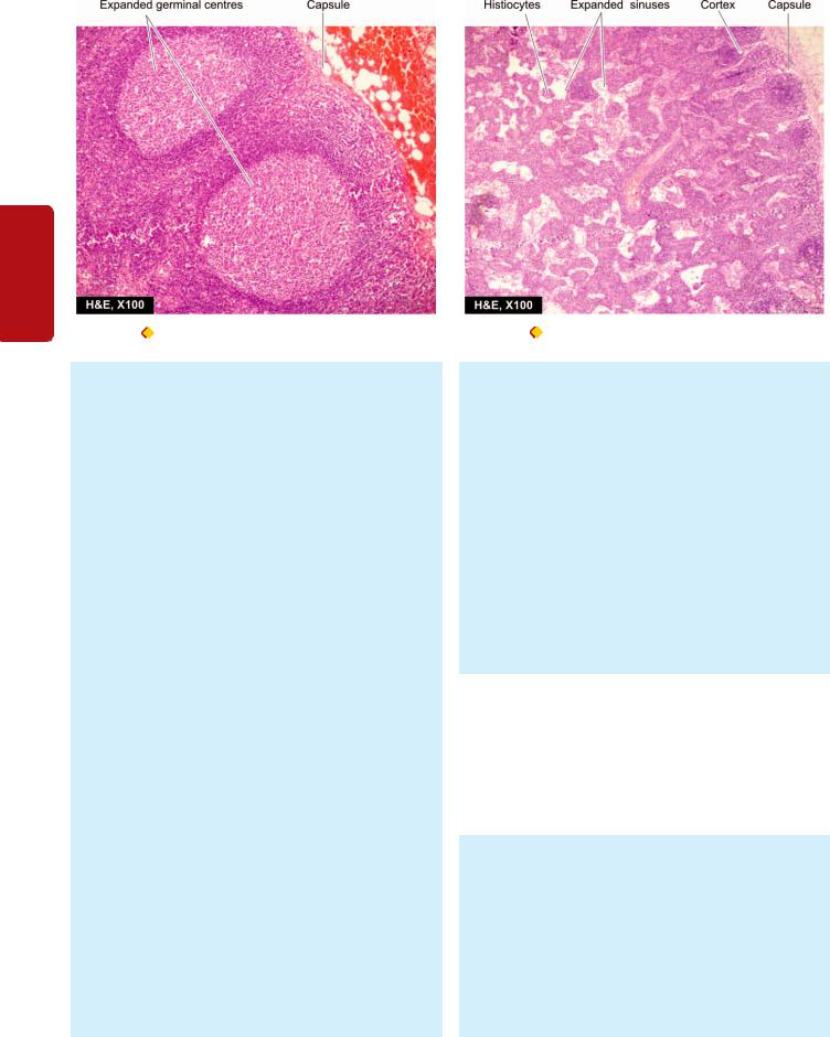
344
II SECTION
Figure 14.2 |
Reactive lymphadenitis, follicular hyperplasia type. |
Figure 14.3 |
Reactive lymphadenitis, sinus histiocytosis type. |
Tissues Lymphoreticular and Haematology
hyperplasia. The condition may occur at any age and possibly has an association with Epstein-Barr virus infection. Two histologic forms are distinguished:
i)Hyaline-vascular type is more common (90% cases) and is characterised by the presence of hyalinised arterioles in small lymphoid follicles and proliferation of vessels in the interfollicular area.
ii)Plasma cell form is less common and is characterised by plasma cell hyperplasia and vascular proliferation in the interfollicular region.
2. Paracortical lymphoid hyperplasia is due to hyper-
plasia of T-cell-dependent area of the lymph node. Amongst the important causes are immunologic reactions caused by drugs (e.g. dilantin), vaccination, viruses (e.g. infectious mononucleosis) and autoimmune disorders. Its histologic features are:
i)Expansion of the paracortex (T-cell area) with increased number of T-cell transformed immunoblasts.
ii)Encroachment by the enlarged paracortex on the lymphoid follicles, sometimes resulting in their effacement.
iii)Hyperplasia of the mononuclear phagocytic cells in the lymphatic sinuses.
Variants of paracortical lymphoid hyperplasia are angio-immunoblastic lymphadenopathy, dermatopathic lymphadenopathy, dilantin lymphadenopathy and postvaccinial lymphadenopathy.
 Angioimmunoblastic lymphadenopathy is characterised by diffuse hyperplasia of immunoblasts rather than paracortical hyperplasia only, and there is proliferation of blood vessels. The condition occurs in elderly patients with generalised lymph node enlargement and hypergammaglobulinaemia.
 Dermatopathic lymphadenopathy occurs in lymph node draining an area of skin lesion. Besides the hyperplastic paracortex, there is presence of dark melanin pigment within the macrophages in the lymph node.
3. Sinus histiocytosis or sinus hyperplasia is a very common type found in regional lymph nodes draining inflammatory lesions, or as an immune reaction of the host to a draining malignant tumour or its products. The hallmark of histologic diagnosis is the expansion of the sinuses by proliferating large histiocytes containing phagocytosed material (Fig. 14.3). The presence of sinus histiocytosis in the draining lymph nodes of carcinoma such as in breast carcinoma has been considered by some workers to confer better prognosis in such patients due to good host immune response.
 Sinus histiocytosis with massive lymphadenopathy is characterised by marked enlargement of lymph nodes, especially of the neck, in young adolescents. It is associated with characteristic clinical features of painless but massive lymphadenopathy with fever and leucocytosis and usually runs a benign and self-limiting course.
HIV-related Lymphadenopathy
HIV infection and AIDS have already been discussed in Chapter 4; here one of the frequent finding in early cases of AIDS, persistent generalised lymphadenopathy (PGL), is described. The presence of enlarged lymph nodes of more than 1 cm diameter at two or more extra-inguinal sites for more than 3 months without any other obvious cause is frequently the earliest symptom of primary HIV infection.
Histologically, the findings at biopsy of involved lymph node vary depending upon the stage of HIV infection: 1. In the early stage marked follicular hyperplasia is the dominant finding and reflects the polyclonal B-cell proliferation.
2 In the intermediate stage, there is a combination of follicular hyperplasia and follicular involution. However, adenopathic form of Kaposi’s sarcoma too may develop at this stage (page 414).
3. In the last stage, there is decrease in the lymph node size indicative of prognostic marker of disease
progression. Microscopic findings of node at this stage reveal follicular involution and lymphocyte depletion. At this stage, other stigmata of AIDS in the lymph node may also appear e.g. lymphoma, mycobacterial infection, toxoplasmosis, systemic fungal infections etc.
WHITE BLOOD CELLS:
NORMAL AND REACTIVE
The leucocytes of the peripheral blood are of 2 main varieties, distinguished by the presence or absence of granules: granulocytes and nongranular leucocytes. The granulocytes, according to the appearance of nuclei, are subdivided into polymorphonuclear leucocytes and monocytes. Further, depending upon the colour of granules, polymorphonuclear leucocytes are of 3 types: neutrophils, eosinophils and basophils. The nongranular leucocytes are 3 types of lymphocytes: T, B and natural killer (NK) cells.
GRANULOPOIESIS
Site of Formation and Kinetics
All forms of granulocytes are produced in the bone marrow and are termed,‘myeloid series’. Myeloid series include maturing stages: myeloblast (most primitive precursor), promyelocyte, myelocyte, metamyelocyte, band forms and segmented granulocyte (mature form). The myeloblast, promyelocyte and myelocyte form a ‘proliferative or mitotic pool’, while the remainder (i.e. metamyelocyte, band forms and segmented granulocytes) make up a ‘mature or postmitotic pool’. It takes about 12 days for formation of mature granulocytes from the myeloblast. Normally the bone marrow contains more myeloid cells than the erythroid cells in the ratio of 2:1 to 15:1 (average 3:1), the largest proportion being that of metamyelocytes, band forms and segmented neutrophils.
Normally, the bone marrow storage compartment contains about 10-15 times the number of granulocytes found in the peripheral blood. Following their release from the bone marrow, granulocytes spend about 10 hours in the circulation before they move into the tissues, where they perform their respective functions. The blood pool of granulocytes consists of 2 components of about equal size—the circulating pool that is included in the blood count, and the marginating pool that is not included in the blood count. Granulocytes spend about 4-5 days in the tissues before they are either destroyed during phagocytosis or die due to senescence. To control the various compartments of granulocytes, a ‘feed-back system’ exists between the circulating and tissue granulocytes on one side, and the marrow granulocytes on the other. The presence of a humoral regulatory substance, ‘granulopoietin’ analogous to erythropoietin has also been identified by in vitro studies of colony-forming units (CFU) and is characterised as G-CSF (granulocyte colony-stimulating factor) and GM-CSF (granulocyte-monocyte colony-stimulating factor).
The kinetics of monocytes is less well understood than that of other myeloid cells. Monocytes spend about 20-40 hours in the circulation after which they leave the blood to
enter extravascular tissues where they perform their main function of active phagocytosis. The extravascular lifespan of tissue macrophages which are the transformed form of blood monocytes, may vary from a few months to a few years.
Myeloid Series
The development of myeloid cells from myeloblast takes place in the following sequence (Fig. 14.4):
1.MYELOBLAST. The myeloblast is the earliest recognisable precursor of the granulocytes, normally comprising about 2% of the total marrow cells. The myeloblast varies considerably in size (10-18 μm in diameter), having a large round to oval nucleus nearly filling the cell, has fine nuclear chromatin and contains 2-5 well-defined pale nucleoli. The thin rim of cytoplasm is deeply basophilic and devoid of granules. The myeloblasts of acute myeloid leukaemia may, however, show the presence of rod-like cytoplasmic inclusions called Auer’s rods which represent abnormal derivatives of primary azurophilic granules.
The nuclei of successive stages during their development from myeloblast become progressively coarser and lose their nucleoli and the cytoplasm loses its blue colour. As the cells become mature lysosomal granules appear; firstly nonspecific primary or azurophilic granules appear which are followed by specific or secondary granules that differentiate the neutrophils, eosinophils and basophils.
2.PROMYELOCYTE. The promyelocyte is slightly larger than the myeloblast (12-18 μm diameter). It possesses a round to oval nucleus, having fine nuclear chromatin which is slightly condensed around the nuclear membrane. The nucleoli are present but are less prominent and fewer than those in the myeloblast. The main distinction of promyelocyte from myeloblast is in the cytoplasm which contains azurophilic (primary or non-specific) granules.
3.MYELOCYTE. The myelocyte is the stage in which specific or secondary granules appear in the cytoplasm, and accordingly, the cell can be identified at this stage as belonging to the neutrophilic, eosinophilic or basophilic myelocyte. Primary granules also persist at this stage but formation of new primary granules stops. The nucleus of myelocyte is eccentric, round to oval, having somewhat coarse nuclear chromatin and no visible nucleoli. The myeloid cells up to the myelocyte stage continue to divide and, therefore, comprise mitotic or proliferative pool.
4.METAMYELOCYTE. The metamyelocyte stage is 10-18 μm in diameter and is characterised by a clearly indented or horseshoe-shaped nucleus without nucleoli. The nuclear chromatin is dense and clumped. The cytoplasm contains both primary and secondary granules. The metamyelocytes are best distinguished from the monocytes by the clumped nuclear chromatin while the latter have fine chromatin.
5.BAND FORMS. Band form is juvenile granulocyte, 10-
16μm in diameter, characterised by further condensation of nuclear chromatin and transformation of nuclear shape into band configuration of uniform thickness.
345
Tissues Lymphoreticular and Leucocytes of Disorders 14 CHAPTER
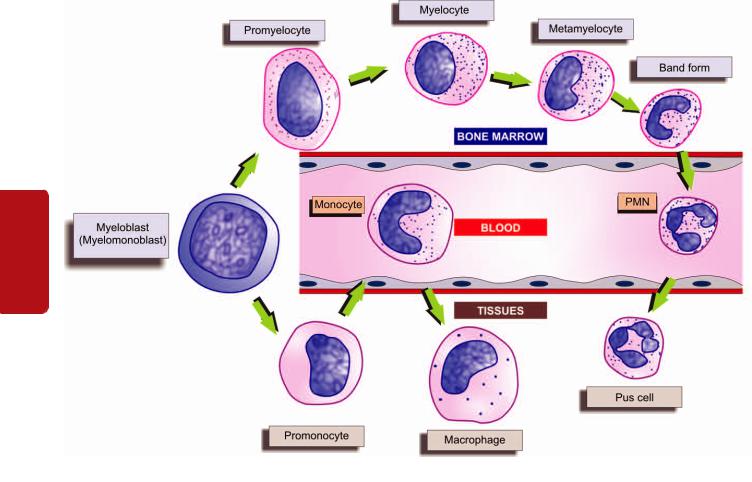
346
Tissues Lymphoreticular and Haematology II SECTION
Figure 14.4 
 Granulopoiesis and the cellular compartments of myeloid cells in the bone marrow, blood and tissues.
6. SEGMENTED GRANULOCYTES. The mature polymorphonuclear leucocytes, namely: the neutrophils, eosinophils and basophils, are described separately below.
Common surface markers for all stages of myeloid series of cells are CD33, CD13 and CD15. However, stages from myelocytes to mature neutrophils also carry CD11b and CD14. Band forms and mature neutrophils have further CD 10 and CD16.
Monocyte-Macrophage Series
The monocyte-macrophage series of cells, though comprise a part of myeloid series alongwith other granulocytic series, but are described separately here in view of different morphologic stages in their maturation (Fig. 14.4).
1.MONOBLAST. The monoblast is the least mature of the recognisable cell of monocyte-macrophage series. It is very similar in appearance to myeloblasts except that it has ground-glass cytoplasm with irregular border and may show phagocytosis as indicated by the presence of engulfed red cells in the cytoplasm. However, differentiation from myeloblast at times may be difficult even by electron microscopy and, therefore, it is preferable to call the earliest precursor of granulocytic series as myelomonoblast.
2.PROMONOCYTE. The promonocyte is a young monocyte, about 20 μm in diameter and possesses a large indented nucleus containing a nucleolus. The cytoplasm is basophilic
and contains no azurophilic granules but may have fine granules which are larger than those in the mature monocyte.
3. MONOCYTE. The mature form of monocytic series is described below, while the transformed stages of these cells in various tissues (i.e. macrophages) are a part of RE system discussed in Chapter 4.
Monocyte-macrophage series having specialised function of phagocytosis secrete active products such as lysozyme, neutral proteases, acid hydrolases, components of complement, tranferrin, fibronectin, nucleosides and several cytokines (TNF-α, IL-1, IL-8, IL-12, IL-18). They express lineage-specific molecules CD 14, cell surface LPS receptors etc.
LYMPHOPOIESIS
Sites of Formation and Kinetics
The lymphocytes and the plasma cells are immunocompetent cells of the body. In man, the bone marrow and the thymus are the primary lymphopoietic organs where lymphoid stem cells undergo spontaneous division independent of antigenic stimulation. The secondary or reactive lymphoid tissue is comprised by the lymph nodes, spleen and gut-associated lymphoid tissue (GALT). These sites actively produce lymphocytes from the germinal centres of lymphoid follicles as a response to antigenic stimulation. Lymphocytes pass
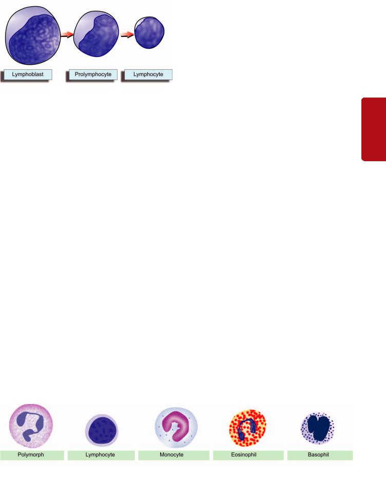
Figure 14.5 
 The formation of lymphoid series of cells.
through a series of developmental changes in the course of their evolution into lymphocyte subpopulations and subsets. It includes migration of immature lymphocytes to other organs such as the thymus where locally-produced factors act on them.
Functionally, the lymphocytes are divided into T, B and natural killer (NK) cells depending upon whether they are immunologically active in cell-mediated immunity (T cells), in humoral antibody response (B cells) or form part of the natural or innate immunity and act as killer of some viruses (NK cells). In man, the B cells are derived from the bone marrow stem cells, while in birds they mature in the bursa of Fabricius. After antigenic activation, B cells proliferate and mature into plasma cells which secrete specific immunoglobulin antibodies. The T cells are also produced in the bone marrow and possibly in the thymus. NK cells do not have B or T cell markers, nor are these cells dependent upon thymus for development. The concept of T, B and NK cells along with lymphocyte subpopulations and their functions is discussed in Chapter 4.
Lymphoid Series
The maturation stages in production of lymphocytes are illustrated in Fig. 14.5 and are as under:
1. LYMPHOBLAST. The lymphoblast is the earliest identifiable precursor of lymphoid cells and is a rapidly dividing cell. It is a large cell, 10-18 μm in diameter, containing a large round to oval nucleus having slightly clumped or stippled nuclear chromatin. The nuclear membrane is denser and the number of nucleoli is fewer (1-2) as compared with those in myeloblast (2-5). The cytoplasm is scanty, basophilic and nongranular.
The distinguishing morphologic features between the myeloblast and lymphoblast are summarised in Table 14.1.
TABLE 14.1: Morphologic Characteristics of the Blast Cells |
347 |
|||
|
in Romanowsky Stains. |
|
|
|
|
Feature |
Myeloblast |
Lymphoblast |
|
1. |
Size |
10-18 μm |
10-18 μm |
|
2. |
Nucleus |
Round or oval |
Round or oval |
|
3. |
Nuclear chromatin |
Fine meshwork |
Slightly clumped |
|
4. |
Nuclear membrane |
Very fine |
Fairly dense |
|
5. |
Nucleoli |
2-5 |
1-2 |
|
6. |
Cytoplasm |
Scanty, blue, |
Scanty, clear blue, |
|
|
|
agranular, Auer |
agranular |
|
|
|
rods may be seen |
|
|
|
|
|
|
|
2. PROLYMPHOCYTE. This stage is an intermediate stage |
CHAPTER |
|
|
||
between the lymphoblast and mature lymphocyte. These |
|
|
young lymphocytes are 9-18 μm in diameter, contain round |
|
|
to indented nucleus with slightly stippled or coarse |
|
|
chromatin and may have 0-1 nucleoli. |
14 |
|
3. LYMPHOCYTE. The mature lymphocytes are described |
||
|
||
below. |
Disorders |
|
Normally, only mature leucocytes namely: polymorphs, |
||
MATURE LEUCOCYTES IN HEALTH AND |
|
|
REACTION IN DISEASE |
|
|
lymphocytes, monocytes, eosinophils and basophils, are |
of |
|
found in the peripheral blood. The normal range of total and |
||
Leucocytes |
||
differential leucocyte count (TLC and DLC expressed |
||
|
||
sequentially as P, L, M, E, B) in health in adults and children |
|
|
is given in Table 14.2. White cell count tends to be higher in |
|
|
infants and children than in adults. It also normally |
|
|
undergoes minor degree of diurnal variation with a slight |
and |
|
rise in the afternoon. The total white cell count is normally |
||
|
||
high in pregnancy and following delivery, usually returning |
Lymphoreticular |
|
to normal within a week. The pathological variations in white |
||
|
||
cell values together with brief review of their morphology |
|
|
and functions are considered below (Fig. 14.6): |
|
|
Polymorphs (Neutrophils) |
|
|
MORPHOLOGY. A polymorphonuclear neutrophil (PMN), |
|
|
commonly called polymorph or neutrophil, is 12-15 μm in |
|
|
diameter. It consists of a characteristic dense nucleus, having |
Tissues |
|
several enzymes and are of 2 types: |
||
2-5 lobes and pale cytoplasm containing numerous fine |
|
|
violet-pink granules. These lysosomal granules contain |
|
Primary or azurophilic granules are large and coarse and appear early at the promyelocyte stage. These granules
Figure 14.6 
 Morphology of normal mature leucocytes in peripheral blood.

348
Tissues Lymphoreticular and Haematology II SECTION
TABLE 14.2: Normal White Blood Cell Counts in Health.
|
Absolute Count |
|
|
TLC |
|
Adults |
4,000–11,000/μl |
Infants (Full term, at birth) |
10,000–25,000/μl |
Infants (1 year) |
6,000–16,000/μl |
Children (4–7 years) |
5,000–15,000/μl |
Children (8–12 years) |
4,500–13,500/μl |
DLC IN ADULTS |
|
Polymorphs (neutrophils) 40–75% |
2,000–7,500/μl |
Lymphocytes 20–50% |
1,500–4,000/μl |
Monocytes 2–10% |
200–800/μl |
Eosinophils 1–6% |
40–400/μl |
Basophils <l% |
10–100/μl |
|
|
contain hydrolases, elastase, myeloperoxidase, cathepsin-G, cationic proteins, permeability increasing protein, and microbicidal protein called defensins.
Secondary or specific granules are smaller and more numerous. These appear later at myelocyte stage, are MPOnegative and contain lactoferrin, NADPH oxidase, histaminase, vitamin B12 binding protein, and receptors for chemoattractants and for laminin.
The normal functions of neutrophils are as under:
1.Chemotaxis or cell mobilisation in which the cell is attracted towards bacteria or at the site of inflammation.
2.Phagocytosis in which the foreign particulate material of tiny sizes is phagocytosed by actively motile neutrophils; thus PMNs act as microphages compared to function of monocytes as macrophages.
3.Killing of the microorganism is mediated by oxygendependent and oxygen-independent pathways (Chapter 3).
PATHOLOGIC VARIATIONS. Pathologic variations in neutrophils include variations in count, morphology and defective function.
Variation in count. An increase in neutrophil count (neutrophil leucocytosis or neutrophilia) or a decrease in count
(neutropenia) may occur in various diseases.
Neutrophil leucocytosis. An increase in circulating neutrophils above 7,500/μl is the commonest type of leucocytosis and occurs most commonly as a response to acute bacterial infections. Some common causes of neutrophilia are as under:
1.Acute infections, local or generalised, especially by cocci but also by certain bacilli, fungi, spirochaetes, parasites and some viruses. For example: pneumonia, cholecystitis, salpingitis, meningitis, diphtheria, plague, peritonitis, appendicitis, actinomycosis, poliomyelitis, abscesses, furuncles, carbuncles, tonsillitis, otitis media, osteomyelitis etc.
2.Other inflammations e.g. tissue damage resulting from burns, operations, ischaemic necrosis (such as in MI), gout, collagen-vascular diseases, hypersensitivity reactions etc.
3.Intoxication e.g. uraemia, diabetic ketosis, eclampsia, poisonings by chemicals and drugs.
4.Acute haemorrhage, internal or external.
5.Acute haemolysis.
6.Disseminated malignancies.
7.Myeloproliferative disorders e.g. myeloid leukaemia, polycythaemia vera, myeloid metaplasia.
8.Miscellaneous e.g. following corticosteroid therapy, idiopathic neutrophilia.
Neutropenia. When the absolute neutrophil count falls below 2,500/μl, the patient is said to have neutropenia and is prone to develop recurrent infections. Some common causes of neutropenia (and hence leucopenia) are as follows:
1.Certain infections e.g. typhoid, paratyphoid, brucellosis, influenza, measles, viral hepatitis, malaria, kala-azar etc.
2.Overwhelming bacterial infections especially in patients with poor resistance e.g. miliary tuberculosis, septicaemia.
3.Drugs, chemicals and physical agents which induce aplasia of the bone marrow cause neutropenia, e.g. antimetabolites, nitrogen mustards, benzene, ionising radiation. Occasionally, certain drugs produce neutropenia due to individual sensitivity such as: anti-inflammatory (amidopyrine, phenylbutazone), antibacterial (chloramphenicol, cotrimoxazole), anticonvulsants, antithyroids, hypoglycaemics and antihistaminics.
4.Certain haematological and other diseases e.g. pernicious anaemia, aplastic anaemia, cirrhosis of the liver with splenomegaly, SLE, Gaucher’s disease.
5.Cachexia and debility.
6.Anaphylactoid shock.
7.Certain rare hereditary, congenital or familial disorders e.g. cyclic neutropenia, primary splenic neutropenia, idiopathic benign neutropenia.
VARIATIONS IN MORPHOLOGY. Some of the common variations in neutrophil morphology are shown in Fig. 14.7. These are as under:
1.Granules. Heavy, dark staining, coarse toxic granules are characteristic of bacterial infections.
2.Vacuoles. In bacterial infections such as in septicaemia, cytoplasmic vacuolation may develop.
3.Döhle bodies. These are small, round or oval patches, 2-3 μm in size, in the cytoplasm. They are mostly seen in bacterial infections.
4.Nuclear abnormalities. These include the following:
i)Sex chromatin is a normal finding in 2-3% of neutrophils in female sex. It consists of a drumstick appendage of chromatin, about 1 μm across, and attached to one of the nuclear lobes by a thin chromatin strand. Their presence in more than 20% of PMNs is indicative of female sex chromosomes (Chapter 10).
ii)A ‘shift-to-left’ is the term used for appearance of neutrophils with decreased number of nuclear lobes in the peripheral blood e.g. presence of band and stab forms and a few myelocytes in the peripheral blood. It is seen in severe infections, leucoerythroblastic reaction or leukaemia.
iii)A ‘shift-to-right’ is appearance of hypersegmented (more than 5 nuclear lobes) neutrophils in the peripheral blood such
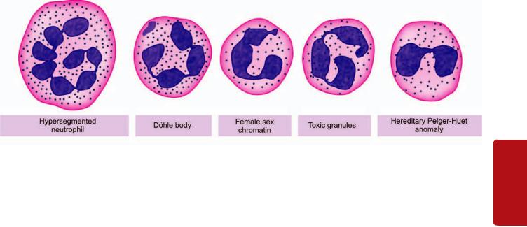
Figure 14.7 
 Common variations in neutrophil morphology.
as in megaloblastic anaemia, uraemia, and sometimes in leukaemia.
iv) Pelger-Huët anomaly is an uncommon autosomal dominant inherited disorder in which nuclei in majority of neutrophils are distinctively bilobed (spectacle-shaped) and coarsely staining chromatin. Acquired pseudo-Pelger-Huët abnormality may occur in acute infections or in MDS. However, the physiologic role of multilobed nucleus of neutrophils is unknown and the bilobed anomaly is an innocuous condition.
DEFECTIVE FUNCTIONS. The following abnormalities in neutrophil function may sometimes be found:
1.Defective chemotaxis e.g. in a rare congenital abnormality called lazy-leucocyte syndrome; following corticosteroid therapy, aspirin ingestion, alcoholism, and in myeloid leukaemia.
2.Defective phagocytosis due to lack of opsonisation e.g. in hypogammaglobulinaemia, hypocomplementaemia, after splenectomy, in sickle cell disease.
3.Defective killing e.g. in chronic granulomatous disease, Chédiak-Higashi syndrome, myeloid leukaemias.
Lymphocytes
MORPHOLOGY. Majority of lymphocytes in the peripheral blood are small (9-12 μm in diameter) but large lymphocytes (12-16 μm in diameter) are also found. Both small and large lymphocytes have round or slightly indented nucleus with coarsely-clumped chromatin and scanty basophilic cytoplasm. Plasma cells are derived from B lymphocytes under the influence of appropriate stimuli. The nucleus of plasma cell is eccentric and has cart-wheel pattern of clumped nuclear chromatin. The cytoplasm is characteristically deeply basophilic with a pale perinuclear zone. Plasma cells are normally not present in peripheral blood but their pathological proliferation occurs in myelomatosis. Reactive lymphocytes (or Turk cells or plasmacytoid lymphocytes) are seen in certain viral infections and have sufficiently basophilic cytoplasm that they resemble plasma cells.
As discussed in Chapter 4, functionally, there are 3 types of lymphocytes and possess distinct surface markers called clusters of differentiation (CD) which aid in identification of stage of their differentiation:
T lymphocytes i.e. thymus-dependent lymphocytes, which mature in the thymus and are also known as thymocytes. They are mainly involved in direct action on antigens and are therefore involved in cell-mediated immune (CMI) reaction by its subsets such as cytotoxic (killer) T cells (CD3+), CD8+
T cells, and delayed hypersensitivity reaction by CD4+ T cells.
B lymphocytes i.e. bone marrow-dependent or bursaequivalent lymphocytes as well as their derivatives, plasma cells, are the source of specific immunoglobulin antibodies. They are, therefore, involved in humoral immunity (HI) or
circulating immune reactions.
NK cells i.e. natural killer cells are those lymphocytes which morphologically have appearance of lymphocytes but do not possess functional features of T or B cells. As the name indicates they are identified with ‘natural’ or innate immunity and bring about direct ‘killing’ of microorganisms (particularly certain viruses) or lysis of foreign body.
PATHOLOGIC VARIATIONS. A rise in the absolute count of lymphocytes exceeding the upper limit of normal (above 4,000/μm) is termed lymphocytosis, while absolute lymphocyte count below 1,500/μm is referred to as lymphopenia.
Lymphocytosis. Some of the common causes of lymphocytosis are as under:
1.Certain acute infections e.g. pertussis, infectious mononucleosis, viral hepatitis, infectious lymphocytosis.
2.Certain chronic infections e.g. brucellosis, tuberculosis, secondary syphilis.
3.Haematopoietic disorders e.g. lymphocytic leukaemias, lymphoma, heavy chain disease.
4.Relative lymphocytosis is found in viral exanthemas, convalescence from acute infections, thyrotoxicosis, conditions causing neutropenia.
Lymphopenia. Lymphopenia is uncommon and occurs in the following conditions:
1.Most acute infections.
2.Severe bone marrow failure.
3.Corticosteroid and immunosuppressive therapy.
4.Widespread irradiation.
Monocytes
MORPHOLOGY. The monocyte is the largest mature leucocyte in the peripheral blood measuring 12-20 μm in
349
Tissues Lymphoreticular and Leucocytes of Disorders 14 CHAPTER

350
Tissues Lymphoreticular and Haematology II SECTION
diameter. It possesses a large, central, oval, notched or indented or horseshoe-shaped nucleus which has characteristically fine reticulated chromatin network. The cytoplasm is abundant, pale blue and contains many fine dust-like granules and vacuoles.
The main functions of monocytes are as under:
1.Phagocytosis of antigenic material or microorganisms.
2.Immunologic function as antigen-presenting cells and present the antigen to lymphocytes to deal with further.
3.As mediator of inflammation, they are involved in release of prostaglandins, stimulation of the liver to secrete acute phase reactants.
Tissue macrophages of different types included in RE system are derived from blood monocytes (Chapter 4).
PATHOLOGIC VARIATIONS. A rise in the blood monocytes above 800/μl is termed monocytosis. Some common causes of monocytosis are as follows:
1.Certain bacterial infections e.g. tuberculosis, subacute bacterial endocarditis, syphilis.
2.Viral infections.
3.Protozoal and rickettsial infections e.g. malaria, typhus, trypanosomiasis, kala-azar.
4.Convalescence from acute infection.
5.Haematopoietic disorders e.g. monocytic leukaemia, lymphomas, myeloproliferative disorders, multiple myeloma, lipid storage disease.
6.Malignancies e.g. cancer of the ovary, stomach, breast.
7.Granulomatous diseases e.g. sarcoidosis, inflammatory bowel disease.
8.Collagen-vascular diseases.
Eosinophils
MORPHOLOGY. Eosinophils are similar to segmented neutrophils in size (12-15 μm in diameter), and have coarse, deep red staining granules in the cytoplasm and have usually two nuclear lobes. Granules in eosinophils contain basic protein and stain more intensely for peroxidase than granules in the neutrophils. In addition, eosinophils also contain cell adhesion molecules, cytokines (IL-3, IL-5), and a protein that precipitates Charcot-Leyden crystals in lung tissues in asthmatic patients.
Eosinophils are involved in reactions to foreign proteins and to antigen-antibody reactions.
PATHOLOGIC VARIATIONS. An increase in the number of eosinophilic leucocytes above 400/μl is referred to as eosinophilia and below 40/μl is termed as eosinopenia.
Eosinophilia. The causes are as under:
1.Allergic disorders e.g. bronchial asthma, urticaria, angioneurotic oedema, hay fever, drug hypersensitivity.
2.Parasitic infestations e.g. trichinosis, echinococcosis, intestinal parasitism.
3.Skin diseases e.g. pemphigus, dermatitis herpetiformis, erythema multiforme.
4.Löeffler’s syndrome.
5.Pulmonary infiltration with eosinophilia (PIE) syndrome.
6.Tropical eosinophilia.
7.Haematopoietic diseases e.g. CML, polycythaemia vera, pernicious anaemia, Hodgkin’s disease, following splenectomy.
8.Malignant diseases with metastases.
9.Irradiation.
10.Miscellaneous disorders e.g. polyarteritis nodosa, rheumatoid arthritis, sarcoidosis.
Eosinopenia. Adrenal steroids and ACTH induce eosinopenia in man.
Basophils
MORPHOLOGY. Basophils resemble the other mature granulocytes but are distinguished by coarse, intensely basophilic granules which usually fill the cytoplasm and often overlie and obscure the nucleus.
The granules of circulating basophils (as well as their tissue counterparts as mast cells) contain heparin, histamine and 5-HT. Mast cells or basophils on degranulation are associated with histamine release.
PATHOLOGIC VARIATIONS. Basophil leucocytosis or basophilia refers to an increase in the number of basophilic leucocytes above 100/μl. Basophilia is unusual and is found in the following conditions:
1.Chronic myeloid leukaemia
2.Polycythaemia vera
3.Myelosclerosis
4.Myxoedema
5.Ulcerative colitis
6.Following splenectomy
7.Hodgkin’s disease
8.Urticaria pigmentosa.
INFECTIOUS MONONUCLEOSIS
Infectious mononucleosis (IM) or glandular fever is a benign, self-limiting lymphoproliferative disease caused by EpsteinBarr virus (EBV), one of the herpesviruses. Infection may occur from childhood to old age but the classical acute infection is more common in teenagers and young adults. The infection is transmitted by person-to-person contact such as by kissing with transfer of virally-contaminated saliva. Groups of cases occur particularly in young people living together in boarding schools, colleges, camps and military institutions. Primary infection in childhood is generally asymptomatic, while 50% of adults develop clinical manifestations. The condition is so common that by the age of 40, most people have been infected and developed antibodies. It may be mentioned here that EBV is oncogenic as well and is strongly implicated in the African (endemic) Burkitt’s lymphoma and nasopharyngeal carcinoma as discussed in Chapter 8.
Pathogenesis
EBV, the etiologic agent for IM, is a B lymphotropic herpesvirus. The disease is characterised by fever, generalised lymphadenopathy, hepatosplenomegaly, sore throat,
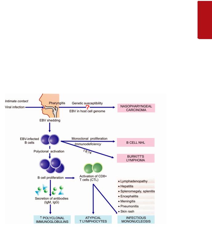
and appearance in blood of atypical ‘mononucleosis cells’. The pathogenesis of these pathologic features is outlined below:
1.In a susceptible sero-negative host who lacks antibodies, the virus in the contaminated saliva invades and replicates within epithelial cells of the salivary gland and then enters B cells in the lymphoid tissues which possess receptors for EBV. The infection spreads throughout the body via bloodstream or by infected B cells.
2.Viraemia and death of infected B cells cause an acute febrile illness and appearance of specific humoral antibodies which peak about 2 weeks after the infection and persist throughout life. The appearance of antibodies marks the
disappearance of virus from the blood.
3. Though the viral agent has disappeared from the blood, the EBV-infected B cells continue to be present in the circulation as latent infection. EBV-infected B cells undergo polyclonal activation and proliferation. These cells perform two important roles which are the characteristic diagnostic features of IM:
i)They secrete antibodies—initially IgM but later IgG class antibodies appear. IgM antibody is the heterophile antisheep antibody used for diagnosis of IM while IgG antibody persists for life and provides immunity against re-infection.
ii)They activate CD8+ T lymphocytes— also called cytotoxic T cells (or CTL) or suppressor T cells. CD8+ T cells bring about killing of B cells and are pathognomonic atypical lymphocytes seen in blood in IM.
4.The proliferation of these cells is responsible for generalised lymphadenopathy and hepatosplenomegaly.
5.The sore throat in IM may be caused by either necrosis of B cells or due to viral replication within the salivary epithelial cells in early stage.
Besides the involvement of EBV in the pathogenesis of IM, its role in neoplastic transformation in nasopharyngeal carcinoma and Burkitt’s lymphoma is discussed in Chapter 8 and diagrammatically depicted in Fig. 14.8.
Clinical Features
The incubation period of IM is 30-50 days in young adults, while children have shorter incubation period. A prodromal period of 3-5 days is followed by Frank clinical features lasting for 1-3 weeks, and subsequently complete recovery occurs after 2 months. The usual clinical features are as under:
1.During prodromal period (first 3-5 days), the symptoms are mild such as malaise, myalgia, headache and fatigue.
2.Frank clinical features (next 7-21 days) commonly are fever, sore throat and bilateral cervical lymphadenopathy. Less commonly, splenomegaly (50% patients), hepatomegaly (10% cases), transient erythematous maculopapular eruption on the trunk and extremities, and neurologic manifestations are found. Pneumonia and cardiac involvement are infrequent. One of the complications of IM is proneness for splenic rupture due to splenitis.
351
Tissues Lymphoreticular and Leucocytes of Disorders 14 CHAPTER
Figure 14.8 
 The role of EBV in the pathogenesis of infectious mononucleosis, nasopharyngeal carcinoma and Burkitt’s lymphoma.
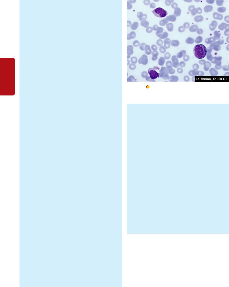
352Laboratory Findings
The diagnosis of IM is made by characteristic haematologic and serologic findings.
1. HAEMATOLOGIC FINDINGS. Major abnormalities in blood are as under:
i) TLC: There is a moderate rise in total white cell count (10,000-20,000/μl) during 2nd to 3rd week after infection.
ii) DLC: There is an absolute lymphocytosis. The lymphocytosis is due to rise in normal as well as atypical T lymphocytes. There is relative neutropenia.
|
iii) Atypical T cells: Out of the total lymphocytes in the |
|
SECTION |
peripheral blood, essential to the diagnosis of IM is the |
|
presence of at least 10-12% atypical T cells (or mononucleosis |
||
|
||
|
cells) (Fig. 14.9). The mononucleosis cells are variable in |
|
|
appearance and are classed as Downey type I, II and III, |
|
|
of which Downey type I are found most frequently. These |
|
II |
atypical T lymphocytes are usually of the size of large |
|
lymphocytes (12-16 μm diameter). The nucleus, rather |
||
|
||
Haematology |
than the usual round configuration, is oval, kidney-shaped |
|
or slightly lobate due to indentation of nuclear membrane |
||
and contains relatively fine chromatin without nucleoli, |
||
|
||
|
suggesting an immature pattern but short of leukaemic |
|
|
features. The cytoplasm is more abundant, basophilic and |
|
|
finely granular and may contain vacuoles. The greatest |
|
|
number of atypical lymphocytes is found between 7th to |
|
and |
10th day of the illness and these cells may persist in the |
|
blood for up to 2 months. |
||
|
||
Lymphoreticular |
iv) CD 4+ and CD8+ T cell counts. There is reversal of |
|
CD4+/CD8+ T cell ratio. There is marked decrease in |
||
|
||
|
CD4+ T cells while there is substantial rise in CD8+ T cells. |
|
|
v) Platelets. There is generally thrombocytopenia in the |
|
|
first 4 weeks of illness. |
|
|
2. SEROLOGIC DIAGNOSIS. The second characteristic |
|
|
laboratory finding is the demonstration of antibodies in |
|
Tissues |
the serum of infected patient. These are as under: |
|
of IM. In this test, patient’s serum is absorbed with guinea |
||
|
i) Test for heterophile antibodies. Heterophile antibody |
|
|
test (Paul-Bunnell test) is used for making the diagnosis |
|
|
pig kidney. Serum dilutions are prepared which are used |
|
|
for agglutination of red cells of sheep, horse or cow and |
|
|
are reported as heterophile titer of test serum. A high |
|
|
serum titer of 40 or more times is diagnostic of acute IM |
|
|
infection in symptomatic case in the first week. |
|
|
Heterophile antibodies peak during the 3rd week in 80- |
|
|
90% cases. The test remains positive for about 3 months |
|
|
after the illness started. Thus, the test has to be repeatedly |
|
|
performed. Similar antibody is also produced in patients |
|
|
suffering from serum sickness and has to be distinguished |
|
|
by differential absorption studies. Heterophile antibodies |
|
|
are not demonstrable in children under 5 years of age or |
|
|
in quite elderly. Currently, more sensitive and rapid kit- |
|
|
based test for heterophile antibodies, monospot, is also |
|
|
available. |
|
|
ii) EBV-specific antibody tests. Specific antibodies against |
|
|
the viral capsid and nucleus of EBV can be demonstrated |
|
|
in patients who are negative for heterophile antibody test: |
Figure 14.9 Peripheral blood film showing atypical lymphocytes in
infectious mononucleosis.
a)Specific antibody against EBV capsid antigen show elevated titers in over 90% cases during acute infection. IgM class antibody appears early and is thus most useful for diagnosis of acute infection. IgG class antibody appears later and persists throughout life; thus it does not have diagnostic value but is instead used for assessing the past exposure to EBV infection.
b)Antibodies against EBV nuclear antigen are detected 3 to 6 weeks after infection and, like IgG class antibodies, persist throughout life.
c)Antibodies to early antigens may be elevated but are less useful for making diagnosis of IM. However, titers of these antibodies remain elevated for 3 to 6 months and their levels are high in cases of nasopharyngeal carcinoma and African Burkitt’s lymphoma.
d)IgA antibodies to EBV antigen are seen in patients of nasopharyngeal carcinoma or those who are at high risk of developing this EBV-induced cancer.
iii) EBV antigen detection. Detection of EBV DNA or proteins can be done in blood or CSF by PCR method.
3. LIVER FUNCTION TESTS. In addition, abnormalities of the liver function test are found in about 90% of cases. These include elevated serum levels of transaminases (SGOT and SGPT), rise in serum alkaline phosphatase and mild elevation of serum bilirubin.
LEUKAEMOID REACTIONS
Leukaemoid reaction is defined as a reactive excessive leucocytosis in the peripheral blood resembling that of leukaemia in a subject who does not have leukaemia. In spite of confusing blood picture, the clinical features of leukaemia such as splenomegaly, lymphadenopathy and haemorrhages are usually absent and the features of underlying disorder causing the leukaemoid reaction are generally obvious.
Leukaemoid reaction may be myeloid or lymphoid; the former is much more common.
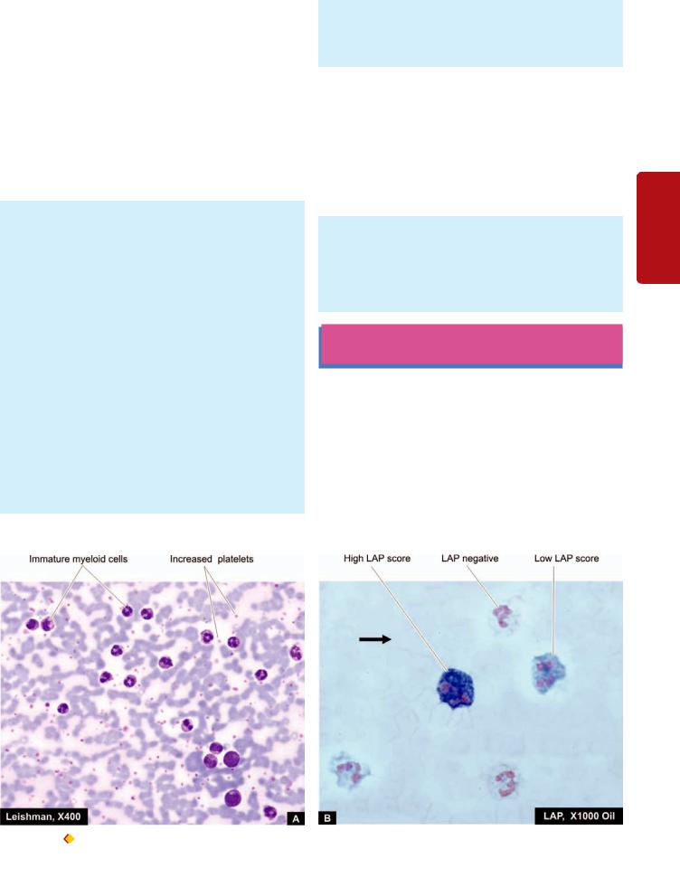
Myeloid Leukaemoid Reaction
CAUSES. Majority of leukaemoid reactions involve the granulocyte series. It may occur in association with a wide variety of diseases. These are as under:
1.Infections e.g. staphylococcal pneumonia, disseminated tuberculosis, meningitis, diphtheria, sepsis, endocarditis, plague, infected abortions etc.
2.Intoxication e.g. eclampsia, mercury poisoning, severe burns.
3.Malignant diseases e.g. multiple myeloma, myelofibrosis, Hodgkin’s disease, bone metastases.
4.Severe haemorrhage and severe haemolysis.
LABORATORY FINDINGS. Myeloid leukaemoid reaction is characterised by the following laboratory features:
1.Leucocytosis, usually moderate, not exceeding 100,000/ μl.
2.Proportion of immature cells mild to moderate, comprised by metamyelocytes, myelocytes (5-15%), and blasts fewer than 5% i.e. the blood picture simulates somewhat with that of CML (Fig. 14.10, A).
3.Infective cases may show toxic granulation and Döhle bodies in the cytoplasm of neutrophils.
4.Neutrophil (or Leucocyte) alkaline phosphatase (NAP or LAP) score in the cytoplasm of mature neutrophils in leukaemoid reaction is characteristically high and is very useful to distinguish it from chronic myeloid leukaemia in doubtful cases (Fig. 14.10, B).
5.Cytogenetic studies may be helpful in exceptional cases which reveal negative Philadelphia chromosome in myeloid leukaemoid reaction but positive in cases of CML.
6.Additional features include anaemia, normal-to-raised platelet count, myeloid hyperplasia of the marrow and
absence of infiltration by immature cells in organs and tissues.
Table 14.3 sums up the features to distinguish myeloid leukaemoid reaction from chronic myeloid leukaemia.
Lymphoid Leukaemoid Reaction
CAUSES. Lymphoid leukaemoid reaction may be found in the following conditions:
1.Infections e.g. infectious mononucleosis, cytomegalovirus infection, pertussis (whooping cough), chickenpox, measles, infectious lymphocytosis, tuberculosis.
2.Malignant diseases may rarely produce lymphoid leukaemoid reaction.
LABORATORY FINDINGS. The blood picture is characterised by the following findings:
1.Leucocytosis not exceeding 100,000/μl.
2.The differential white cell count reveals mostly mature lymphocytes simulating the blood picture found in cases of CLL.
HAEMATOLOGIC NEOPLASMS
(LEUKAEMIAS-LYMPHOMAS): GENERAL
CLASSIFICATION: CURRENT CONCEPTS
Neoplastic proliferations of white blood cells— leukaemias and lymphomas, are the most important group of leucocyte disorders.
Historically, leukaemias have been classified on the basis of cell types predominantly involved into myeloid and lymphoid, and on the basis of natural history of the disease, into acute and chronic. Thus, the main types of leukaemias
have been: acute myeloblastic leukaemia and acute lymphoblastic
353
Tissues Lymphoreticular and Leucocytes of Disorders 14 CHAPTER
Figure 14.10 |
Leukaemoid reaction. A, Peripheral blood film showing marked neutrophilic leucocytosis accompanied with late precursors of |
myeloid series. B, Neutrophil (or leucocyte) alkaline phosphatase (NAP or LAP) activity is higher as demonstrated by this cytochemical stain.

354
Tissues Lymphoreticular and Haematology II SECTION
TABLE 14.3: Contrasting Features of Leukaemoid Reaction and Chronic Myeloid Leukaemia.
Feature |
Leukaemoid Reaction |
CML |
|
1. |
TLC |
25,000-100,000/μl |
> 100,000/μl |
2. |
DLC |
i) Dominant cells PMN’s |
i) All maturation stages |
|
|
ii) Immature cells predominantly metamyelocytes |
ii) Immature cells all stages, |
|
|
and myelocytes (5-15%), myeloblasts and |
myeloblasts and promyelocytes |
|
|
promyelocytes > 5% |
< 10% |
|
|
iii) Basophils normal |
iii) Basophilia present |
3. |
NAP score |
Elevated |
Reduced |
4. |
Philadelphia chromosome |
Absent |
Present |
5. |
ABL-BCR fusion gene |
Absent |
Present |
6. |
Major etiology |
Infections, intoxication, disseminated malignancy, |
RNA viruses, HTLV oncogenesis, |
|
|
severe haemorrhage |
genetic factors, radiations, certain drugs |
|
|
|
and chemicals |
7. |
Additional haematologic findings |
i) Anaemia |
Anaemia |
|
|
ii) Normal to raised platelet count |
Normal to raised platelet count |
|
|
iii) Myeloid hyperplasia in bone marrow |
Myeloid hyperplasia in bone marrow |
8. |
Organ infiltration |
Absent |
May be present |
9. |
Massive splenomegaly |
Absent |
Present |
|
|
|
|
leukaemia (AML and ALL), and chronic myeloid leukaemia and chronic lymphocytic leukaemias (CML and CLL); besides there are some other uncommon variants. In general, acute leukaemias are characterised by predominance of undifferentiated leucocyte precursors or leukaemic blasts and have a rapidly downhill course. Chronic leukaemias, on the other hand, have easily recognisable late precursor series of leucocytes circulating in large number as the predominant leukaemic cell type and the patients tend to have more indolent behaviour. The incidence of both acute and chronic leukaemias is higher in men than in women. ALL is primarily a disease of children and young adults, whereas AML occurs at all ages. CLL tends to occur in the elderly, while CML is found in middle age.
Similary, over the years, lymphomas which are malignant tumours of lymphoreticular tissues have been categorised into two distinct clinicopathologic groups: Hodgkin’s lymphoma or Hodgkin’s disease (HD) characterised by pathognomonic presence of Reed-Sternberg cells, and a heterogenous group of non-Hodgkin’s lymphomas (NHL).
In the last 50 years, several classification systems have been proposed for leukaemias and lymphomas—clinicians favouring an approach based on clinical findings while pathologists have been interested in classifying them on morphologic features. More recent classification schemes have been based on cytochemistry, immunophenotyping, cytogenetics and molecular markers which have become available to pathologists and haematologists. The most recent classification scheme proposed by the World Health Organisation (WHO) in 2002 combines all tumours of haematopoietic and lymphoid tissues together. The basis of the WHO classification is the cell type of the neoplasm as identified by combined approach of clinical features and morphologic, cytogenetic and molecular characteristics, rather than location of the neoplasm (whether in blood or in tissues) because of the fact that haematopoietic cells are present in circulation as well as in tissues in general, and lymphoreticular tissues in particular.
Currently, neoplasms of haematopoietic and lymphoid tissues are considered as a unified group and are divided into 3 broad categories:
I. Myeloid neoplasms: This group includes neoplasms of myeloid cell lineage and therefore includes neoplastic proliferations of red blood cells, platelets, granulocytes and monocytes. There are 5 categories under myeloid series of neoplasms: myeloproliferative disorders, myeloproliferative/myelodysplastic diseases, myelodysplastic syndromes (MDS), and acute myeloid leukaemia (AML), acute biphenotypic leukaemias.
II. Lymphoid neoplasms: Neoplasms of lymphoid lineage include leukaemias and lymphomas of B, T or NK cell origin. This group thus includes B cell neoplasms (including plasma cell disorders), T cell neoplasms, NK cell neoplasms and Hodgkin’s disease.
III. Histiocytic neoplasms: This group is of interest mainly due to neoplastic proliferations of histiocytes in Langerhans cell histiocytisis.
Besides the WHO classification, the FAB (French- American-British) Cooperative Group classification of lymphomas and leukaemias based on morphology and cytochemistry is also widely used.
These as well as other classification schemes have been tabulated and discussed later under separate headings of myeloid and lymphoid malignancies.
ETIOLOGY
Like in most cancers, the exact etiology of leukaemias and lymphomas is not known. However, a number of factors have been implicated:
1. HEREDITY. There is evidence to suggest that there is role of family history, occurrence in identical twins and predisposition of these malignancies in certain genetic syndromes:
i)Identical twins: There is high concordance rate among identical twins if acute leukaemia develops in the first year of life. Hodgkin’s disease is 99 times more common in identical twin of an affected case compared with general population, implicating genetic origin strongly.
ii)Family history. Families with excessive incidence of leukaemia have been identified.
iii)Genetic disease association. Acute leukaemia occurs with increased frequency with a variety of congenital disorders such as Down’s, Bloom’s, Klinefelter’s and Wiskott-Aldrich’s syndromes, Fanconi’s anaemia and ataxia telangiectasia. Hodgkin’s disease has familial incidence and with certain HLA type.
2. INFECTIONS. There is evidence to suggest that certain infections, particularly viruses, are involved in development of lymphomas and leukaemias (Chapter 8):
i)Human T cell leukaemia-lymphoma virus I (HTLV-I) implicated in etiology of adult T cell leukaemia-lymphoma (ATLL).
ii)HTLV II for T cell variant of hairy cell leukaemia.
iii)Epstein-Barr virus (EBV) implicated in the etiology of Hodgkin’s disease (mixed cellularity type and nodular sclerosis type), endemic variety of Burkitt’s lymphoma, posttransplant lymphoma,
iv)HIV in diffuse large B-cell lymphoma and Burkitt’s lymphoma.
v)Hepatitis C virus (HCV) in lymphoplasmacytic lymphoma.
vi)Human herpes virus 8 (HHV-8) in primary effusion lymphoma.
vii)Helicobacter pylori bacterial infection of gastric mucosa in MALT lymphoma of the stomach.
3.ENVIRONMENTAL FACTORS. Certain environmental factors are known to play a role in the etiology of leukaemias and lymphomas:
i)Ionising radiation. Damage due to radiation exposure has been linked to development of leukaemias and lymphomas. Individuals exposed to occupational radiation exposure, patients receiving radiation therapy, and Japanese survivors of the atomic bomb explosions have been found to be at higher risk of developing haematopoietic malignancies, particularly prone to development of CML, AML and ALL but not to CLL or hairy cell leukaemia.
ii)Chemical carcinogens. Benzene, tobacco smoking, alcohol, use of certain hair dyes and exposure to agriculture chemicals are associated with increased risk of development of haematopoietic malignancies.
iii)Certain drugs. Long-term exposure to certain drugs such as phenytoin, alkylating agents and other chemotherapeutic agents is associated with increased incidence of leukaemias and lymphomas. Patients treated for Hodgkin’s disease can develop NHL.
4.ASSOCIATION WITH DISEASES OF IMMUNITY.
Since lymphoid cells are the immune cells of the body, diseases with derangements of the immune system have higher incidence of haematopoietic malignancies:
i)Immunodeficiency diseases. Various inherited and acquired immunodeficiency diseases including AIDS and iatrogenic immunosuppression induced by chemotherapy or radiation, are associated with subsequent development of lymphomatous transformation.
ii)Autoimmune disease association. A few autoimmune diseases such as Sjögren’s syndrome, nontropical sprue, rheumatoid arthritis and SLE are associated with higher incidence of NHL.
PATHOGENESIS
It needs to be emphasised that since haematopoietic cells have a rapid turnover, they are more vulnerable to chromosomal damages and cytogenetic changes under influence of various etiologic factors listed above.
1.Genetic damage to single clone of target cells. Leukaemias and lymphomas arise following malignant transformation of a single clone of cells belonging to myeloid or lymphoid series, followed by proliferation of the transformed clone. Basic mechanism of malignant transformation is genetic damage to the DNA of the target white cells followed by proliferation, disrupting normal growth and differentiation. The heritable genetic damage may be induced by various etiologic agents listed above (e.g. RNA viruses HTLV-I, EBV etc) and causes insertional mutagenesis for which oncogenes may play a role (Chapter 8). The evolution of leukaemia is multi-step process, and in many cases, acute leukaemia may develop after a pre-existing myelodysplastic or myeloproliferative disorder.
2.Chromosomal translocations. A number of cytogenetic abnormalities have been detected in cases of leukaemiaslymphomas, most consistent of which are chromosomal translocations. In NHL, translocation involving antigen receptor genes, immunoglobulin genes, or overexpression of BCL-2 protein may be seen. The most consistent chromosomal abnormality in various forms of acute and chronic leukaemias is Philadelphia (Ph) chromosome seen in 70-90% cases with CML, involving reciprocal translocation of parts of long arm of chromosome 22 to the long arm of chromosome 9 i.e. t(9;22) (Fig. 14.11).
3.Maturation defect. In acute leukaemia, the single most prominent characteristic of the leukaemic cells is a defect in maturation beyond the myeloblast or promyelocyte level in AML, and the lymphoblast level in ALL. It needs to be emphasised that it is the maturation defect in leukaemic blasts rather than rapid proliferation of leukaemic cells responsible for causing acute leukaemia. In fact, the generation time of leukaemic blasts is somewhat prolonged rather than shortened.
4.Myelosuppression. As the leukaemic cells accumulate in the bone marrow, there is suppression of normal haematopoietic stem cells, partly by physically replacing the normal marrow precursors, and partly by inhibiting normal haematopoiesis via cell-mediated or humoral mechanisms. This is based on the observation that some patients with acute leukaemia have a hypocellular marrow indicating that
355
Tissues Lymphoreticular and Leucocytes of Disorders 14 CHAPTER
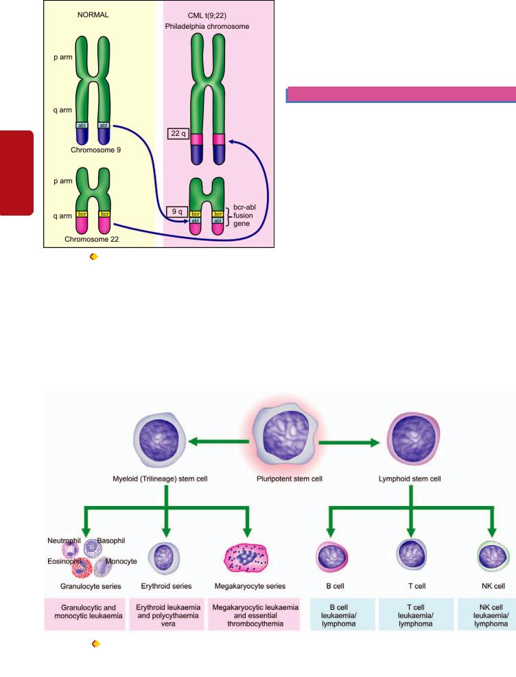
356
Tissues Lymphoreticular and Haematology II SECTION
Figure 14.11 |
The Philadelphia (Ph) chromosome. There is reciprocal |
translocation of the part of the long arms of chromosome 22 to the long arms of chromosome 9 written as t(9;22).
marrow failure is not due to overcrowding by leukaemic cells alone. Nevertheless, some normal haematopoietic stem cells do remain in the marrow which are capable of proliferating and restoring normal haematopoiesis after effective antileukaemic treatment.
5. Organ infiltration. The leukaemic cells proliferate primarily in the bone marrow, circulate in the blood and
infiltrate into other tissues such as lymph nodes, liver, spleen, skin, viscera and the central nervous system.
6. Cytokines. Presence of reactive inflammatory cells in the Hodgkin’s disease is due to secretion of cytokines from the Reed Sternberg cells e.g. IL-5 (growth factor for eosinophils), IL-13 (for autocrine stimulation of RS cells) and transforming growth factor-β (for fibrogenesis).
MYELOID NEOPLASMS
Based on the cell line of differentiation of the pluripotent stem cell, the WHO classification (2002) recognises all haematopoieitc neoplasms into 2 groups: myeloid and lymphoid neoplasms. Since myeloid trilineage stem cells further differentiate into 3 series of progenitor cells: erythroid, granulocyte-monocyte, and megakaryocytic series, therefore all examples of myeloid neoplasms fall into these three categories of cell-lines. Based on this concept, the WHO classification of myeloid neoplasms includes following 5 groups (Fig. 14.12):
I. Myeloproliferative diseases
II. Myelodysplastic/myeloproliferative diseases III. Myelodysplastic syndrome (MDS)
IV. Acute myeloid leukaemia (AML) V. Acute biphenotypic leukaemia
Each of these groups is subclassifed into further types as shown in Table 14.4. Important examples within each group are discussed below.
MYELOPROLIFERATIVE DISEASES
The myeloproliferative disorders are a group of neoplastic proliferation of multipotent haematopoietic stem cells. Besides their common stem cell origin, these disorders are closely related, occasionally leading to evolution of one entity into another during the course of the disease.
Figure 14.12 Maturation stages of myeloid cells and stages in development of myeloid neoplasms.

TABLE 14.4: WHO Classification of Myeloid Neoplasms.
I.MYELOPROLIFERATIVE DISEASES
1.Chronic myeloid leukaemia (CML), {Ph chromosome t(9;22) (q34;11), BCR/ABL-positive}
2.Chronic neutrophilic leukaemia
3.Chronic eosinophilic leukaemia/ hypereosinophilic syndrome
4.Chronic idiopathic myelofibrosis
5.Polycythaemia vera (PV)
6.Essential thrombocythaemia (ET)
7.Chronic myeloproliferative disease, unclassifiable
II.MYELODYSPLASTIC/MYELOPROLIFERATIVE DISEASES 1. Chronic myelomonocytic leukaemia (CMML)
III.MYELODYSPLASTIC SYNDROME (MDS)
1.Refractory anaemia (RA)
2.Refractory anaemia with ring sideroblasts (RARS)
3.Refractory cytopenia with multilineage dysplasia (RCMD)
4.RCMD with ringed sideroblasts (RCMD-RS)
5.Refractory anaemia with excess blasts (RAEB-1)
6.RAEB-2
7.Myelodysplastic syndrome unclassified (MDS-U)
8.MDS with isolated del 5q
IV. ACUTE MYELOID LEUKAEMIA (AML)
1.AML with recurrent cytogenetic abnormalities
i)AML with t(8;21)(q22;q22)
ii)AML with abnormal bone marrow eosinophils {inv(16) (p13q22)}
iii)Acute promyelocytic leukaemia {t(15;17)(q22;q12)}
iv)AML with 11q23 abnormalities (MLL)
2.AML with multilineage dysplasia
i)With prior MDS
ii)Without prior MDS
3.AML and MDS, therapy-related
i)Alkylating agent-related
ii)Topoisomerase type II inhibitor-related
iii)Other types
4.AML, not otherwise categorised
i)AML, minimally differentiated
ii)AML without maturation
iii)AML with maturation
iv)Acute myelomonocytic leukaemia (AMML)
v)Acute monoblastic and monocytic leukaemia
vi)Acute erythroid leukaemia
vii)Acute megakaryocytic leukaemia
viii)Acute basophilic leukaemia
ix)Acute panmyelosis with myelofibrosis
x)Myeloid sarcoma
V. ACUTE BIPHENOTYPIC LEUKAEMIA
The WHO classification of myeloproliferative disorders includes 7 types as shown in Table 14.4. Classic and common examples are chronic myeloid leukaemia (CML), polycythaemia vera (PV), and essential thrombocytosis (ET), each one representing corresponding excess of granulocytes, red blood cells, and platelets, respectively. The group as a whole has slow and insidious onset of clinical features and indolent clinical behaviour.
CHRONIC MYELOID LEUKAEMIA (CML)
Definition and Pathophysiology
By WHO definition, CML is established by identification of the clone of haematopoietic stem cell that possesses the
reciprocal translocation between chromosomes 9 and 22, forming Philadelphia chromosome. The t(9;22) involves fusion of BCR (breakpoint cluster region) gene on chromosome 22q11 with ABL (named after Abelson murine leukaemia virus) gene located on chromosome 9q34. The fusion product so formed is termed “Ph chromosome t(9;22) (q34;11), BCR/ABL” which should be positive for making the diagnosis of CML. This identification may be done by microsatellite PCR or by FISH. The underlying pathophysiologic mechanism of human CML is based on the observation that BCR/ABL fusion product proteins are capable of transforming haematopoietic progenitor cells in vitro and form malignant clone. BCR/ABL fusion product brings about following functional changes:
i) ABL protein is activated to function as a tyrosine kinase enzyme that in turn activates other kinases which inhibits
apoptosis.
ii)Ability of ABL to act as DNA-binding protein is altered.
iii)Binding of ABL to actin microfilaments of the cytoskeleton is increased.
Exact mechanism of progression of CML to the blastic
phase is unclear but following mechanisms may be involved:
i)Structural alterations in tumour suppressor p53 gene.
ii)Structural alterations in tumour suppressor Rb gene.
iii)Alterations in RAS oncogene.
iv)Alterations in MYC oncogene.
v)Release of cytokine IL-1β.
vi)Functional inactivation of tumour suppressor protein,
phosphatase A2.
Clinical Features
Chronic myeloid (myelogenous, granulocytic) leukaemia comprises about 20% of all leukaemias and its peak incidence is seen in 3rd and 4th decades of life. A distinctive variant of CML seen in children is called juvenile CML. Both sexes are affected equally. The onset of CML is generally insidious. Some of the common presenting manifestations are as under:
1.Features of anaemia such as weakness, pallor, dyspnoea and tachycardia.
2.Symptoms due to hypermetabolism such as weight loss, lassitude, anorexia, night sweats.
3.Splenomegaly is almost always present and is frequently massive. In some patients, it may be associated with acute pain due to splenic infarction.
4.Bleeding tendencies such as easy bruising, epistaxis, menorrhagia and haematomas may occur.
5.Less common features include gout, visual disturbance, neurologic manifestations and priapism.
6.Juvenile CML is more often associated with lymph node enlargement than splenomegaly. Other features are frequent infections, haemorrhagic manifestations and facial rash.
Laboratory Findings
The diagnosis of CML is generally possible on blood picture alone. However, bone marrow, cytochemical stains and other investigations are of help.
I. BLOOD PICTURE. The typical blood picture in a case of CML at the time of presentation shows the following features (Fig. 14.13):
357
Tissues Lymphoreticular and Leucocytes of Disorders 14 CHAPTER
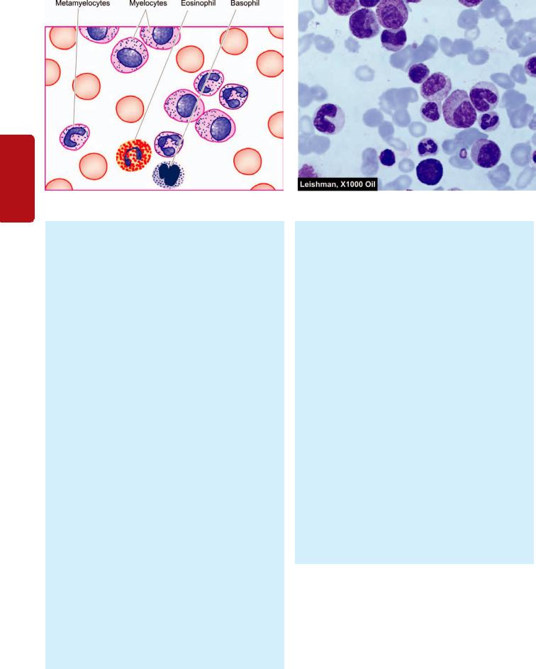
358
Tissues Lymphoreticular and Haematology II SECTION
Figure 14.13 
 PBF findings in chronic myeloid leukaemia (CML).
1.Anaemia. Anaemia is usually of moderate degree and is normocytic normochromic in type. Occasional normoblasts may be present.
2.White blood cells. Characteristically, there is marked leucocytosis (approximately 200,000/μl or more at the time of presentation). The natural history of CML consists of 3 phases—chronic, accelerated, and blastic.
 Chronic phase of CML begins as a myeloproliferative disorder and consists of excessive proliferation of myeloid cells of intermediate grade (i.e. myelocytes and metamyelocytes) and mature segmented neutrophils. Myeloblasts usually do not exceed 10% of cells in the peripheral blood and bone marrow. An increase in the proportion of basophils up to 10% is a characteristic feature of CML. A rising basophilia is indicative of impending blastic transformation. An accelerated phase of CML is also described in which there is progressively rising leucocytosis associated with thrombocytosis or thrombocytopenia and splenomegaly.
 Accelerated phase is defined as increasing degree of anaemia, blast count in blood or marrow between 10-20%, marrow basophils 20% or more, and platelet count falling below 1,00,000/μl.
 Blastic phase or blast crisis in CML fulfills the definition of acute leukaemia in having blood or marrow blasts >20%. These blast cells may be myeloid, lymphoid, erythroid or undifferentiated and are established by morphology, cytochemistry, or immunophenotyping. Myeloid blast crisis in CML is more common and resembles AML. However, unlike AML, Auer rods are not seen in myeloblasts of CML in blast crisis.
3. Platelets. Platelet count may be normal but is raised in about half the cases.
II. BONE MARROW EXAMINATION. Examination of marrow aspiration yields the following results:
1. Cellularity. Generally, there is hypercellularity with total or partial replacement of fat spaces by proliferating myeloid cells.
2.Myeloid cells. The myeloid cells predominate in the bone marrow with increased myeloid-erythroid ratio. The differential counts of myeloid cells in the marrow show similar findings as seen in the peripheral blood with predominance of myelocytes.
3.Erythropoiesis. Erythropoiesis is normoblastic but there is reduction in erythropoietic cells.
4.Megakaryocytes. Megakaryocytes are conspicuous but are usually smaller in size than normal.
5.Cytogenetics. Cytogenetic studies on blood and bone marrow cells show the characteristic chromosomal abnormality called Philadelphia (Ph) chromosome seen in 90-95% cases of CML. Ph chromosome is formed by reciprocal balanced translocation between part of long arm of chromosome 22 and part of long arm of chromosome 9{(t(9;22) (q34;11)} forming product of fusion gene, BCR/
ABL (see Fig. 14.11).
III. CYTOCHEMISTRY. The only significant finding on cytochemical stains is reduced scores of neutrophil alkaline phosphatase (NAP) which helps to distinguish CML from myeloid leukaemoid reaction in which case NAP scores are elevated (see Fig. 14.10,B, and Table 14.3). However, NAP scores in CML return to normal with successful therapy, corticosteroid administration and in infections.
IV. OTHER INVESTIGATIONS. A few other accompanying findings are seen in CML:
1.Elevated serum B12 and vitamin B12 binding capacity.
2.Elevated serum uric acid (hyperuricaemia).
Treatment and Complications
Insight into molecular mechanism of CML has brought about major changes in its therapy. The approach of modern therapy in CML is targetted at removal of all malignant clones of cells bearing BCR/ABL fusion protein, so that patient reverts back to prolonged non-clonal haematopoiesis i.e. molecular remission from disease. This is achievable by the following approaches:

1.Imatinib oral therapy: The basic principle underlying imatinib oral treatment is to competitively inhibit ATP binding site of the ABL kinase, which in turn, inhibits signal transduction BCR/ABL fusion protein. Imatinib induces apoptosis in BCR/ABL-positive cells and thus eliminates them. Imatinib is found more effective in newly diagnosed cases of CML. Complete haematologic remission is achieved for 18 months in 97% cases treated with imatinib.
2.Allogenic bone marrow (stem cell) transplantation.
Although this treatment modality offers proven cure, it is complicated with mortality due to procedure and development of post-transplant graft-versus-host disease (GVHD) and, therefore, post-transplant immunosuppressive treatment has to be continued.
3.Interferon-α. Prior to imatinib and allogenic transplantation, chronic phase of CML used to be treated with interferon-α was the drug of choice.
4.Chemotherapy. Chemotherapeutic agents are used in treatment of CML for lowering the total population of WBCs. These include use of busulfan, cyclophosphamide (melphalan) and hydroxyurea.
5.Others. Besides above, other forms of treatment include splenic irradiation, splenectomy and leucopheresis.
The most common cause of death (in 80% cases) in CML is disease acceleration and blastic transformation.
POLYCYTHAEMIA VERA
Definition and Pathophysiology
Polycythaemia vera (PV) is a clonal disorder characterised by increased production of all myeloid elements resulting in pancytosis (i.e increased red cells, granulocytes, platelets) in the absence of any recognisable cause. The term ‘polycythaemia vera’ or ‘polycythaemia rubra vera’ is used
for primary or idiopathic polycythaemia only and is the most common of all the myeloproliferative disorders. Secondary polycythaemia or erythrocytosis, on the other hand, may occur secondary to several causes e.g. high altitude, cardiovascular disease, pulmonary disease with alveolar hypoventilation, heavy smoking, inappropriate increase in erythropoietin (renal cell carcinoma, hydronephrosis, hepatocellular carcinoma, cerebellar haemangioblastoma, massive uterine leiomyoma); sometimes relative or spurious polycythaemia may result from plasma loss such as in burns and in dehydration from vomiting or water deprivation. None of the secondary causes of polycythaemia is associated with splenic enlargement or increased leucocytes and platelets which are typical of PV.
The exact etiology of PV is not known but about a third of cases show inconsistent and varied chromosomal abnormalities such as 20q, trisomy 8 and 9p. Major pathogenetic mechanism is a tyrosine kinase JAK2 mutation which removes the autoinhibitory control and activates the kinases.
Clinical Features
PV is a disease of late middle life and is slightly more common in males. The disease generally runs a chronic but
slowly progressive course. Clinical features are the result of hyperviscosity, hypervolaemia, hypermetabolism and decreased cerebral perfusion. These are as under:
1.Headache, vertigo, tinnitus, visual alterations syncope or even coma.
2.Increased risk of thrombosis due to accelerated atherosclerosis.
3.Increased risk of haemorrhages due to increased blood volume and intrinsic platelet dysfunction e.g. epistaxis, peptic ulcer disease.
4.Splenomegaly producing abdominal fullness.
5.Pruritus, especially after a bath.
6.Increased risk of urate stones and gout due to hyperuricaemia.
Laboratory Findings
PV is diagnosed by the following haematologic findings:
1.Raised haemoglobin concentration (above 17.5 g/dl in males and 15.5 g/dl in females).
2.Erythrocytosis (above 6 million/μl in males and 5.5 million/μl in females).
3.Haematocrit (PCV) above 55% in males and above 47% in females.
4.Mild to moderate leucocytosis (15,000-25,000/μl) with basophilia and raised neutrophil alkaline phosphatase scores.
5.Thrombocytosis with defective platelet function.
6.Bone marrow examination reveals erythroid hyperplasia or panhyperplasia.
7.Cytogenetic abnormalities such as 20q, trisomy 8 and 9p are found in 30% cases of PV.
8.In PV, unlike secondary polycythaemia, erythropoietin levels in serum and urine are reduced.
Treatment and Complications
Since PV runs an indolent course, therapy is aimed at maintaining normal blood counts and relieve the patient of symptoms.
1.Phlebotomy (venesection) by blood letting is done at regular interval to reduce total blood cell mass and to induce a state of iron deficiency.
2.Anticoagulant therapy is adminstered in case thrombosis has occurred.
3.Chemotherapy may be indicated to induce myelosuppresion.
4.Hyperuricaemia is treated with uricosuric drugs.
5.Interferon-α is associated with good results because it reduces JAK2 expression in these patients which is the underlying cytogenetic abnormality.
Patients receiving phlebotomy alone may survive for 1012 years. About 25% patients progress to myelofibrosis. A small proportion of patients develop secondary haematologic malignancies such as AML, non-Hodgkin’s lymphoma and multiple myeloma. Major complication and cause of death in PV is vascular thrombosis.
359
Tissues Lymphoreticular and Leucocytes of Disorders 14 CHAPTER

360 |
ESSENTIAL THROMBOCYTHAEMIA |
|
|
Definition and Pathophysiology |
|
|
Essential thrombocythaemia (ET), also termed essential |
|
|
thrombocytosis or primary (idiopathic) thrombocythaemia |
|
|
is a clonal disorder characterised by markedly elevated |
|
|
platelet count in the absence of any recognisable stimulus. |
|
|
Secondary or reactive thrombocytosis, on the other hand, occurs |
|
|
in response to known stimuli such as: chronic infection, |
|
|
haemorrhage, postoperative state, chronic iron deficiency, |
|
|
malignancy, rheumatoid arthritis and postsplenectomy. |
|
|
ET is an uncommon disorder and represents an |
|
SECTION |
overproduction of platelets from megakaryocyte colonies |
|
other cell lines may also be involved in the expansion of |
||
|
without any added stimulus but no clonal marker is available |
|
|
to distinguish primary from secondary thrombocytosis. |
|
|
Though an elevated platelet count is the dominant feature, |
|
II |
neoplastic clone. |
|
|
The underlying pathophysiologic mechanism in ET is the |
|
Haematology |
absence of control by thrombopoietin that regulates |
|
Clinical Features |
||
|
endomitosis in the megakaryocytes to produce platelets. This |
|
|
results in uncontrolled proliferation of not only mega- |
|
|
karyocytes but also the platelets. There is probably role of |
|
|
heredity in ET since families with ET have been reported. |
|
and |
The condition has an insidious onset and is more frequent in |
|
|
||
Lymphoreticular |
older people. Haemorrhagic and thrombotic events are |
|
common. These include the following: |
||
|
||
|
1. Arterial or venous thrombosis. |
|
|
2. Easy bruisability following minor trauma. |
|
|
3. Spontaneous bleeding. |
|
|
4. Transient ischaemic attack or frank stroke due to platelet |
|
Tissues |
aggregation in microvasculature of the CNS. |
|
Laboratory Findings |
||
|
||
|
The prominent laboratory features pertain to platelets. |
|
|
These include the following: |
1.Sustained elevation in platelet count (above 400,000 μl).
2.Blood film shows many large platelets, megakaryocyte fragments and hypogranular forms.
3.Consistently abnormal platelet functions, especially abnormality in platelet aggregation.
4.Bone marrow examination reveals a large number of hyperdiploid megakaryocytes and variable amount of increased fibrosis.
Treatment and Complications
ET runs a benign course and may not require any therapy. Treatment is given only if platelet count is higher than one million. Complications of ET are occurrence of acquired von Willebrand’s disease and bleeding but incidence of thrombosis is not higher than matched controls.
CHRONIC IDIOPATHIC MYELOFIBROSIS
Definition and Pathophysiology
Chronic idiopathic myelofibrosis (IMF), also called agnogenic (of unknown origin) myeloid metaplasia, primary myelofibrosis and myelosclerosis, is a clonal disorder characterised by proliferation of neoplastic stem cells at multiple sites outside the bone marrow (i.e. extramedullary haematopoiesis), especially in the liver and spleen, without an underlying etiology. Secondary myelofibrosis, on the other hand, develops in association with certain well-defined marrow disorders, or it is the result of toxic action of chemical agents or irradiation.
The exact etiology is not known. Several chromosomal abnormalities have been reported but without a specific cytogenetic abnormality. Fibrosis in the bone marrow is due to overproduction of transforming growth factor-β, osteosclerosis of the bone is related to osteonectin and marrow angiogenesis is due to increased production of vascular endothelial growth factor (VEGF).
Clinical Features
The disease begins in the late middle life and is gradual in onset. Both sexes are affected equally. The symptomatology includes the following:
1.Anaemia with constitutional symptoms such as fatigue, weakness and anorexia.
2.Massive splenomegaly producing abdominal discomfort, pain and dyspnoea.
3.Hepatomegaly is present in half the cases.
4.Petechial and other bleeding problems are found in about 20% cases.
5.Less common findings are lymphadenopathy, jaundice, ascites, bone pain and hyperuricaemia.
Laboratory Findings
1.Mild anaemia is usual except in cases where features of polycythaemia vera are coexistent.
2.Leucocytosis at the time of presentation but later there may be leucopenia.
3.Thrombocytosis initially but advanced cases show thrombocytopenia.
4.Peripheral blood smear shows bizarre red cell shapes, tear drop poikilocytes, basophilic stippling, nucleated red cells, immature leucocytes (i.e. leucoerythroblastic reaction), basophilia and giant platelet forms.
5.Bone marrow aspiration is generally unsuccessful and yields ‘dry tap’. Examination of trephine biopsy shows focal areas of hypercellularity and increased reticulin network and variable amount of collagen in which clusters of megakaryocytes are seen well preserved.
6.Extramedullary haematopoiesis can be documented by liver biopsy or splenic aspiration.
Treatment and Complications
Chronic idiopathic myelofibrosis does not require any specific therapy. Anaemia and ineffective erythropoiesis

cannot be readily treated and do not respond to erythropoietin, androgens. Splenectomy may be necessary in some cases. In general, chronic idiopathic myelofibrosis has poorer outcome compared with PV and ET.
MYELODYSPLASTIC SYNDROMES
Definition and Classification
Myelodysplastic syndromes (MDS) are a heterogeneous group of haematopoietic clonal stem cell disorders having abnormal development of different marrow elements (i.e. dysmyelopoiesis), usually characterised by cytopenias, associated with cellular marrow and ineffective blood cell formation. These conditions are, therefore, also termed as preleukaemic syndromes or dysmyelopoietic syndromes.
There have been two main classification schemes for MDS:
FAB CLASSIFICATION OF MDS (1983): FAB (French- American-British) Cooperative Group classified MDS into the following 5 groups:
1.Refractory anaemia (RA).
2.Refractory anaemia with ringed sideroblasts (primary acquired sideroblastic anaemia) (RARS).
3.Refractory anaemia with excess blasts (RAEB).
4.Chronic myelomonocytic leukaemia (CMML).
5.Refractory anaemia with excess of blasts in transformation (RAEB-t).
As per FAB classification, the marrow may contain <30% myeloblasts in MDS and this was considered as the dividing line for distinguishing cases of AML (blasts >30%) from MDS.
WHO CLASSIFICATION OF MDS (2002): According to the WHO classification, patients with blast count of 20-30% and labelled as RAEB-t (group 5 above) in FAB classification have prognosis similar to patients with blast count above 30% (i.e. AML cases). Thus, as per WHO classification, marrow blast count for making the diagnosis of AML has been revised and brought down to 20%. Thus, patients with FAB category of RAEB-t (i.e. group 5 above) are currently considered and treated as cases of AML and therefore the term RAEB-t stands excluded from WHO MDS classification. Besides, the WHO classification excludes CMML (FAB category 4) from MDS and puts in the hybrid category of myelodysplastic/ myeloproliferative disorder since CMML behaves like a myeloproliferative disorder; moreover refractory anaemia in it is not due to erythroid lineage which was considered as an inclusion criteria for MDS. Thus, the WHO classification of MDS consists of following 8 categories:
1.Refractory anaemia (RA): Same as FAB type 1 MDS. Incidence 5-10%; characterised by anaemia without any blasts in blood; marrow may show <5% blasts.
2.Refractory anaemia with ringed sideroblasts (RARS):
Same as FAB type 2 MDS. Incidence 10-12%; all findings in blood and marrow similar to RA above but in addition marrow may show >15% ringed sideroblasts.
3.Refractory cytopenia with multilineage dysplasia (RCMD): New entity. Incidence 24%; blood shows cytopenia
of 2 or 3 cell lineage and monocytosis but no blasts; marrow shows <5% blasts.
4.RCMD with ringed sideroblasts (RCMD-RS): New entity. Incidence 15%; all blood and marrow findings similar to RCMD plus in addition >15% ringed sideroblasts in marrow.
5.Refractory anaemia with excess blasts (RAEB-1): RAEB of FAB category 3 divided into 2 subtypes with combined incidence of 40%. RAEB –1 has blood cytopenia with <5% blasts and monocytosis; marrow blast count 5-9%.
6.RAEB-2: Findings of blood and marrow similar to RAEB-1 but marrow blast count is 10-19%.
7.Myelodysplastic syndrome unclassified (MDS-U): New entity in MDS. Blood cytopenia without any blasts; marrow shows dysplasia of myeloid and thrombocytic cell lineage and marrow blast count <5%.
8.MDS with isolated del (5q): New entity in MDS. Anaemia in blood and blasts <5%; marrow blasts <5%, normal or increased megakaryocytes and characteristic isolated deletion of 5q.
Pathophysiology
Primary MDS is idiopathic but factors implicated in etiology are radiation exposure and benzene carcinogen. Secondary (therapy-related) MDS may occur following earlier anticancer treatment, aplastic anaemia treated with immunosuppressive therapy and in Fanconi’s anaemia. Several cytogenetic abnormalities are seen in about 50% of MDS which include trisomy, translocations and deletions. Cases evolving into leukaemia more often have aneuploidy. At molecular level, mutations are seen in N-RAS oncogene and p53 anti-oncogene, which together with suppressed immunity, accelerate apoptosis in the bone marrow. Mutations in the mitochondrial genes probably account for occurrence of sideroblastic anaemia in MDS and consequently disordered iron metabolism and ineffective erythropoiesis.
Clinical Features
In general, MDS is found more frequently in older people past 6th decade of life, with slight male preponderance. Therapy-related MDS is generally not age-related and may occur about a decade after anti-cancer therapy. Clinical features are quite non-specific and MDS may be discovered during routine CBC examination done for some other cause. At presentation the patient may have following features:
1.Anaemia appreciated by pallor, fatigue and weakness.
2.Fever.
3.Weight loss.
4.Sweet syndrome having neutrophilic dermatosis seen in some cases.
5.Splenomegaly seen in 20% cases of MDS.
Laboratory Findings
Various combinations of features are seen in different types of MDS. In general, laboratory findings are as under:
BLOOD FINDINGS. There is cytopenia affecting two (bi-) or all the three blood cell lines (pancytopenia):
361
Tissues Lymphoreticular and Leucocytes of Disorders 14 CHAPTER

3621. Anaemia: Generally macrocytic or dimorphic.
2.TLC: Usually normal; cases of CMML may have high TLC but these cases in WHO classification of myeloid neoplasms have been put in a separate group of myelodysplastic/myeloproliferative diseases and not in MDS.
3.DLC: Neutrophils are hyposegmented and hypogranulated. Myeloblasts may be seen in PBF and their number correlates with marrow blasts count.
4.Platelets: Thrombocytopenia with large agranular platelets.
|
BONE MARROW FINDINGS. There is constellation of |
II SECTION |
findings in the marrow as under: |
1. Cellularity: Normal to hypercellular to hypocellular. |
|
2. Erythroid series: Dyserythropoiesis as seen by |
|
abnormally appearing nuclei and ring sideroblasts. |
|
Megaloblasts may be seen. |
|
3. Myeloid series: Hypogranular and hyposegmented |
|
|
myeloid precursor cells. Myeloblasts increased depending |
|
upon the type of MDS. |
ACUTE MYELOID LEUKAEMIA
Definition and Pathophysiology
Acute myeloid leukaemia (AML) is a heterogeneous disease characterised by infiltration of malignant myeloid cells into the blood, bone marrow and other tissues. AML is mainly a disease of adults (median age 50 years), while children and older individuals may also develop it.
AML develops due to inhibition of maturation of myeloid stem cells due to mutations. These mutations may be induced by several etiologic factors—heredity, radiation, chemical carcinogens (tobacco smoking, rubber, plastic, paint, insecticides etc) and long-term use of anti-cancer drugs but viruses do not appear to have role in the etiology of AML. The defect induced by mutations causes accumulation of precursor myeloid cells of the stage at which the myeloid maturation and differentiation is blocked. A few important examples of chromosomal mutations in AML are translocations {t(8;21)(q22q22) and t(15;17)(q22;q12)} and inversions { inv(16)(p13;q22)}.
Tissues Lymphoreticular and Haematology
4. Megakaryocyte series: Reduced in number and having abnormal nuclei.
Treatment and Complications
MDS is difficult to treat and may not respond to cytotoxic chemotherapy. Stem cell transplantation offers cure and longer survival. Survival rates vary depending upon the type of MDS: cases of refractory anaemia with or without sideroblasts (RA and RARS) and 5q syndrome survive for years, while cases of refractory anaemia with excess blasts (RAEB-1 and 2) have poor survival for a few months only. Patients generally either succumb to infections or develop into acute myeloid leukaemia.
Classification
Currently, two main classification schemes for AML are followed:
FAB CLASSIFICATION. According to revised FAB clasification system, a leukaemia is acute if the bone marrow consists of more than 30% blasts. Based on morphology and cytochemistry, FAB classification divides AML into 8 subtypes (M0 to M7) (Table 14.5).
WHO CLASSIFICATION (2002). WHO classification for AML differs from revised FAB classification in the following 2 ways:
1. It places limited reliance on cytochemistry for making the diagnosis of subtype of AML but instead takes into
TABLE 14.5: Revised FAB Classification of Acute Myeloblastic Leukaemias.
FAB Class |
Percent Cases |
Morphology |
Cytochemistry |
|
|
|
|
|
|
M0: |
Minimally |
2 |
Blasts lack definite cytologic and cytochemical |
Myeloperoxidase – |
|
differentiated AML |
|
features but have myeloid lineage antigens |
|
M1: |
AML without |
20 |
Myeloblasts predominate; few if any |
Myeloperoxidase + |
|
maturation |
|
granules or Auer rods |
|
M2: |
AML with |
30 |
Myeloblasts with promyelocytes predominate; |
Myeloperoxidase +++ |
|
maturation |
|
Auer rods may be present |
|
M3: |
Acute promyelocytic |
5 |
Hypergranular promyelocytes; often with |
Myeloperoxidase +++ |
|
leukaemia |
|
multiple Auer rods per cell |
|
M4: |
Acute myelomonocytic |
30 |
Mature cells of both myeloid and monocytic series |
Myeloperoxidase ++ |
|
leukaemia (Naegeli type) |
|
in peripheral blood; myeloid cells resemble M2 |
Non-specific esterase + |
M5: |
Acute monocytic |
10 |
Two subtypes: M5a shows poorly-differentiated |
Non-specific esterase ++ |
|
leukaemia |
|
monoblasts, M5b shows differentiated |
|
|
(Schilling type) |
|
promonocytes and monocytes |
|
M6: |
Acute erythroleukaemia |
<5 |
Erythroblasts predominate (>50%); myeloblasts |
Erythroblasts:PAS + |
|
(Di Guglielmo’s |
|
and promyelocytes also increased |
Myeloblasts: |
|
syndrome) |
|
|
myeloperoxidase + |
M7: |
Acute megakaryocytic |
<5 |
Pleomorphic undifferentiated blasts predominate; |
Platelet peroxidase + |
|
leukaemia |
|
react with antiplatelet antibodies |
|
|
|
|
|
|

consideration clinical, cytogenetic and molecular abnormalities in different types. These features can be studied by multiparametric flow cytometry.
2. WHO classification for AML has revised and lowered the cut off percentage of marrow blasts to 20% from 30% in the FAB classification for making the diagnosis of AML. WHO classifcation of AML is included in Table 14.4.
Both FAB as well as WHO classification schemes for AML are followed in different settings depending upon the laboratory facilities available in various centres. Moreover, most of the current clinical and laboratory data are based on FAB grouping and hence detailed morphologic and cytochemical features of various AML groups are given in
Table 14.5.
Clinical Features
AML and ALL share many clinical features and the two are difficult to distinguish on clinical features alone. In approximately 25% of patients with AML, a preleukaemic syndrome with anaemia and other cytopenias may be present for a few months to years prior to the development of overt leukaemia.
Clinical manifestations of AML are divided into 2 groups: those due to bone marrow failure, and those due to organ
infiltration.
I. DUE TO BONE MARROW FAILURE. These are as under:
1.Anaemia producing pallor, lethargy, dyspnoea.
2.Bleeding manifestations due to thrombocytopenia causing spontaneous bruises, petechiae, bleeding from gums and other bleeding tendencies.
3.Infections are quite common and include those of mouth, throat, skin, respiratory, perianal and other sites.
4.Fever is generally attributed to infections in acute leukaemia but sometimes no obvious source of infection can be found and may occur in the absence of infection.
II. DUE TO ORGAN INFILTRATION. The clinical manifestations of AML are more often due to replacement of the marrow and other tissues by leukaemic cells. These features are as under:
1.Pain and tenderness of bones (e.g. sternal tenderness) are due to bone infarcts or subperiosteal infiltrates by leukaemic cells.
2.Lymphadenopathy and enlargement of the tonsils may occur.
3.Splenomegaly of moderate grade may occur. Splenic infarction, subcapsular haemorrhages, and rarely, splenic rupture may occur.
4.Hepatomegaly is frequently present due to leukaemic infiltration but the infiltrates usually do not interfere with the function of the liver.
5.Leukaemic infiltration of the kidney may be present and ordinarily does not interfere with its function unless secondary complications such as haemorrhage or blockage of ureter supervene.
6.Gum hypertrophy due to leukaemic infiltration of the gingivae is a frequent finding in myelomonocytic (M4) and monocytic (M5) leukaemias.
7.Chloroma or granulocytic sarcoma is a localised tumourforming mass occurring in the skin or orbit due to local infiltration of the tissues by leukaemic cells. The tumour is greenish in appearance due to the presence of myeloperoxidase.
8.Meningeal involvement manifested by raised intracranial pressure, headache, nausea and vomiting, blurring of vision and diplopia are seen more frequently in ALL during haematologic remission. Sudden death from massive intracranial haemorrhage as a result of leucostasis may occur.
9.Other organ infiltrations include testicular swelling and mediastinal compression.
Laboratory Findings
The diagnosis of AML is made by a combination of routine blood picture and bone marrow examination, coupled with cytochemical stains and other special laboratory investigations.
I. BLOOD PICTURE. Findings of routine haematologic investigations are as under (Fig. 14.14):
1.Anaemia. Anaemia is almost always present in AML. It is generally severe, progressive and normochromic. A moderate reticulocytosis up to 5% and a few nucleated red cells may be present.
2.Thrombocytopenia. The platelet count is usually moderately to severely reduced (below 50,000/μl) but occasionally it may be normal. Bleeding tendencies in AML are usually correlated with the level of thrombocytopenia but most serious spontaneous haemorrhagic episodes develop in patients with fewer than 20,000/μl platelets. Acute promyelocytic leukaemia (M3) may be associated with a serious coagulation abnormality, disseminated intravascular coagulation (DIC).
3.White blood cells. The total WBC count ranges from subnormal-to-markedly elevated values. In 25% of patients, the total WBC count at presentation is reduced to 1,000-4,000 /μl. More often, however, there is progressive rise in white cell count which may exceed 100,000/μl in more advanced disease. Majority of leucocytes in the peripheral blood are blasts and there is often neutropenia due to marrow infiltration by leukaemic cells. The basic morphologic features of myeloblasts and lymphoblasts are summed up in Table 14.1. Typical characteristics of different forms of AML (M0 to M7) are given in Table
14.5.In some instances, the identification of blast cells is greatly aided by the company they keep i.e. by more mature and easily identifiable leucocytes in the company of blastic cells of myeloid series. Some ‘smear cells’ in the peripheral blood representing degenerated leucocytes may be seen.
II. BONE MARROW EXAMINATION. An examination of bone marrow aspirate or trephine reveals the following features:
1. Cellularity. Typically, the marrow is hypercellular but sometimes a ‘blood tap’ or ‘dry tap’ occurs. A dry tap in AML may be due to pancytopenia, but sometimes even
363
Tissues Lymphoreticular and Leucocytes of Disorders 14 CHAPTER
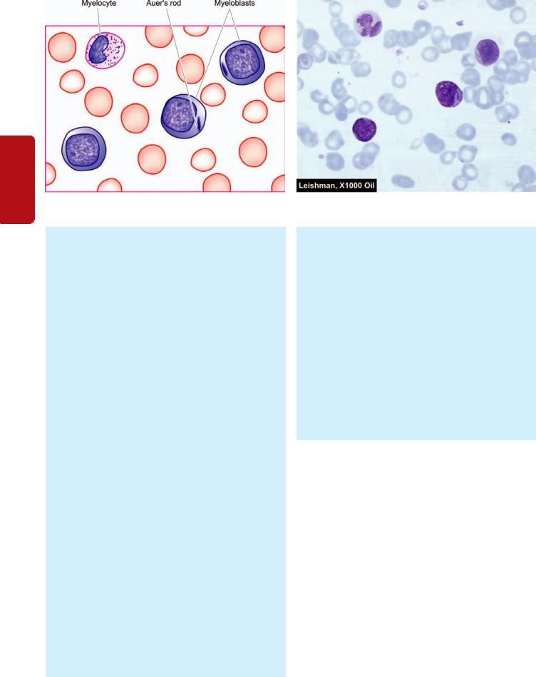
364
Tissues Lymphoreticular and Haematology II SECTION
Figure 14.14 
 PBF findings in a case of acute myeloblastic leukaemia (AML).
when the marrow is so much filled with leukaemic cells that they cannot be aspirated because the cells are adhesive and enmeshed in reticulin fibres. In such cases, trephine biopsy should be done.
2.Leukaemic cells. The bone marrow is generally tightly packed with leukaemic blast cells. The diagnosis of the type of leukaemic cells, according to FAB classification, is generally possible with routine Romanowsky stains but cytochemical stains may be employed as an adjunct to Romanowsky staining for determining the type of leukaemia. The essential criteria for diagnosis of AML, as per FAB classification, was the presence of at least 30% blasts in the bone marrow. However, as per WHO classification, these criteria have been revised and lowered to 20% blasts in the marrow for labelling and treating a case as AML.
3.Erythropoiesis. Erythropoietic cells are reduced. Dyserythropoiesis, megaloblastic features and ring sideroblasts are commonly present.
4.Megakaryocytes. They are usually reduced or absent.
5.Cytogenetics. Chromosomal analysis of dividing leukaemic cells in the marrow shows karyotypic abnormalities in 75% of cases which may have a relationship to prognosis. WHO classification emphasises on the categorisation of AML on the basis of cytogenetic abnormalities. Two of the most consistent cytogenetic abnormalities in specific FAB groups are as under:
i)M3 cases have t(15;17)(q22;q12).
ii)M4E0 (E for abnormal eosinophils in the bone marrow) cases have inv(16)(p13q22).
6. Immunophenotyping. AML cells express CD13 and CD33 antigens. M7 shows CD41 and CD42 positivity.
III. CYTOCHEMISTRY. Some of the commonly employed cytochemical stains, as an aid to classify the type of AML are as under (also see Table 14.5):
1. Myeloperoxidase: Positive in immature myeloid cells containing granules and Auer rods but negative in M0 myeloblasts.
2.Sudan Black: Positive in immature cells in AML.
3.Periodic acid-Schiff (PAS): Positive in immature lymphoid cells and in erythroleukaemia (M6).
4.Non-specific esterase (NSE): Positive in monocytic series (M4 and M5).
5.Acid phosphatase: Focal positivity in leukaemic blasts in ALL and diffuse reaction in monocytic cells (M4 and M5).
IV. BIOCHEMICAL INVESTIGATIONS. These may be of some help:
1.Serum muramidase. Serum levels of lysozyme (i.e. muramidase) are elevated in myelomonocytic (M4) and monocytic (M5) leukaemias.
2.Serum uric acid. Because of rapidly growing number of leukaemic cells, serum uric acid level is frequently increased. The levels are further raised after treatment with cytotoxic drugs because of increased cell breakdown.
Treatment and Complications
The management of acute leukaemia involves the following aspects:
I. TREATMENT OF ANAEMIA AND HAEMORRHAGE.
Anaemia and haemorrhage are managed by fresh blood transfusions and platelet concentrates. Patients with severe thrombocytopenia (platelet count below 20,000/μl) require regular platelet transfusions since haemorrhage is an important cause of death in these cases.
II. TREATMENT AND PROPHYLAXIS OF INFECTION.
Neutropenia due to bone marrow replacement by leukaemic blasts and as a result of intensive cytotoxic therapy renders these patients highly susceptible to infection. The infections are predominantly bacterial but viral, fungal, and protozoal infections also occur. For prophylaxis against infection in such cases, the patient should be isolated and preferably placed in laminar airflow rooms. Efforts are made to reduce the gut and other commensal flora which are the usual source
of infection. This is achieved by bowel sterilisation and by topical antiseptics. If these fail to achieve the desired results, systemic antibiotics and leucocyte concentrates are considered for therapy.
III.CYTOTOXIC DRUG THERAPY. The aims of cytotoxic therapy are firstly to induce remission, secondly to continue therapy to reduce the hidden leukaemic cell population by repeated courses of therapy. Most commonly, cyclic combinations of 2, 3 and 4 drugs are given with treatmentfree intervals to allow the bone marrow to recover.
The most effective treatment of AML is a combination of 3 drugs: cytosine arabinoside, anthracyclines (daunorubicin, adriamycin) and 6-thioguanine. Another addition is amsacrine (m-AMSA) administered with cytosine arabinoside, with or without 6-thioguanine. Following remission-induction therapy, various drug combinations are given intermittently for maintenance. However, promyelocytic leukaemia (M3) is treated with tretinoin orally that reduces the leukaemic cells bearing t(15;17)(q22;q21) but devlopment of DIC due to liberation of granules of dying cells is a problem.
IV.BONE MARROW TRANSPLANTATION. Bone marrow (or stem cell) transplantation from suitable allogenic or autologous donor (HLA and mixed lymphocytes culturematched) is increasingly being used for treating young adults with AML in first remission. The basic principle of marrow transplantation is to reconstitute the patient’s haematopoietic system after total body irradiation and intensive chemotherapy have been given so as to kill the remaining leukaemic cells. Bone marrow transplantation has resulted in cure in about half the cases.
Remission rate with AML is lower (50-70%) than in ALL, often takes longer to achieve remission, and disease-free intervals are shorter. AML is most malignant of all leukaemias; median survival with treatment is 12-18 months.
LYMPHOID NEOPLASMS
Lymphoid cells constitute the immune system of the body. These cells circulate in the blood and also lie in the lymphoid tissues and undergo differentiation and maturation in these organs. The haematopoietic stem cells which form myeloid and lymphoid series, undergo further differentiation of lymphoid cells into B cells (including formation of plasma cells), T cells and NK cells. Lymphoid malignancies can be formed by malignant transformation of each of these cell lines. These lymphoid malignancies can range from indolent to highly aggressive human cancers.
Conventionally, malignancies of lymphoid cells in blood have been termed as lymphatic leukaemias and those of lymphoid tissues as lymphomas. Just like myeloid leukaemias discussed earlier, lymphoid leukaemias have been classified on the basis of survival and biologic course, into chronic and acute (CLL and ALL). Similarly, two clinicopathologically distinct groups of lymphomas are distinguished: Hodgkin’s lymphoma or Hodgkin’s disease (HD)
and non-Hodgkin’s lymphomas (NHL).
However, while HD can be identified by the pathognomonic presence of Reed-Sternberg cells, there have been controversies and confusion in classification of other lymphoid cancers (i.e. NHL and lymphoid leukaemias). In order to resolve the issue, over the years several classification schemes have emerged for lymphoid cancers due to following two main reasons:
1.Biologic course of lymphoma-leukaemia. While some of the lymphoid malignancies initially present as leukaemias (i.e. in the blood and bone marrow), many others present as solid masses in the lymphoid tissues or in various other tissues, especilly in the spleen, liver, bone marrow and other tissues. Still others may have initial presentation as either leukaemia or lymphomas. In fact, the line of demarcation for lymphoid malignancies is so blurred that during the biologic course of the disease, lymphoid leukaemia or lymphoma may spill over and transform to the other.
2.Technological advances. In recent times, modern diagnostic tools have become available to pathologists and haematologists which go much beyond making the diagnosis of lymphomas and leukaemias on clinical grounds combined with morphology and cytochemical stains alone. This includes methods for immunophenotyping, cytogenetics and molecular markers for the stage of differentiation of the cell of origin rather than location of the cell alone.
These aspects form the basis of current concept for WHO classification of malignancies of lymphoid cells of blood and lymphoreticular tissues as ‘lymphoid neoplasms’ as a unified group. However, it needs to be appreciated that in several centres in developing countries of the world, limited laboratory facilities are available. Thus, judiciously speaking, some of the older classification schemes for lymphoid malignancies need to be retained, while others can be dumped as historical. In view of this, a balanced approach of middle path of retaining old and including new classification schemes of lymphoid malignancies is proposed to be followed for discussion below:
I. HISTORICAL CLASSIFICATIONS. These classifications can be traced as under:
Morphologic classification. Rappaport classification (1966) proposed a clinically relevant morphologic classification based on two main features: low-power microscopy of the overall pattern of the lymph node architecture, and high-power microscopy revealing the cytology of the neoplastic cells. Based on these two features, Rappaport divided NHL into two major subtypes:
1.Nodular or follicular lymphomas which retain some of the features of normal lymph node in that the neoplastic cells form lymphoid ‘nodules’ rather than lymphoid follicles with germinal centres.
2.Diffuse lymphomas, on the other hand, are characterised by effacement of the normal lymph node architecture and there may be infiltration of neoplastic cells outside the capsule of the involved lymph node.
NHL was further classified according to the degree of differentiation of neoplastic cells into: well-differentiated,
365
Tissues Lymphoreticular and Leucocytes of Disorders 14 CHAPTER

366
Tissues Lymphoreticular and Haematology II SECTION
TABLE 14.6: FAB Classification of ALL.
A. MORPHOLOGIC CRITERIA
Fab Class |
Percent Cases |
Morphology |
|
Cytochemistry |
|
|
|
|
|
|
|
L1: |
Childhood-ALL |
More common |
Homogeneous small lymphoblasts; scanty |
PAS ± |
|
|
(B-ALL, and T-ALL) |
in children |
cytoplasm, regular round nuclei, |
|
Acid phosphatase ± |
|
|
|
inconspicuous nucleoli |
|
|
L2: |
Adult-ALL |
More frequent |
Heterogeneous lymphoblasts; variable amount |
PAS ± |
|
|
(mostly T-ALL) |
in adults |
of cytoplasm, irregular or cleft nuclei, large nucleoli |
Acid phosphatase ± |
|
L3: |
Burkitt type-ALL |
Uncommon |
Large homogeneous lymphoblasts; round nuclei, |
PAS – |
|
|
(B-ALL) |
|
prominent nucleoli, cytoplasmic vacuolation |
Acid phosphatase – |
|
B. CYTOGENETIC AND IMMUNOLOGIC CRITERIA |
|
|
|
||
|
|
|
|
|
|
Subtype |
Incidence |
Markers |
FAB Subtype |
Cytogenetic Abnormalities |
|
|
|
|
|
|
|
Pre-B ALL |
75% |
CD10+ (90%), TdT+ |
L1, L2 |
t(9;22) i.e. Philadelphia+ALL |
|
B cell ALL |
5% |
CD10+ (50%), TdT- |
L3 |
t(8;14) (Burkitt’s leukaemia) |
|
T cell ALL |
20% |
CD10+ (30%), TdT+ |
L1, L2 |
14q11 |
|
(TdT= terminal deoxynucleotidyl transferase)
poorly-differentiated, and histiocytic (large cells) types of both nodular and diffuse lymphomas.
Immunologic classifications. Lukes-Collins classification (1974) was proposed to correlate the type of NHL with the immune system because the identification of T and B-cells and their subpopulations had become possible in early 70s. Its subsequent modification was Kiel classification (1981). Both these classifications employed immunologic markers for tumour cells, and divided all malignant lymphomas into either B-cell or T-cell origin, and rarely of macrophages. The B and T-cell tumours were further subdivided on the basis of their light microscopic characteristics. The majority of NHL were B lymphocyte derivatives and arise from follicular centre cells (FCC). The FCC in the germinal centre undergo transformation to become large immunoblasts and pass through the four stages—small cleaved cells and large cleaved
cells, small non-cleaved cells and large non-cleaved cells.
Though these classification schemes were immunologically correct, they were unclear about varying prognosis of different clinical types of NHL of either B-cell or T-cell origin.
II. OLD CLINICOPATHOLOGIC CLASSIFICATIONS. In view of the objections to above pure morphologic and immunologically correct classifications, following two clinically relevant classifications were proposed which cannot be readily abandoned:
FAB classification of lymphoid leukaemia. Although old, FAB classification for lymphoid leukaemia was initially based on morphology and cytochemistry into 3 types of ALL (L1 to L3), but was subsequently revised to include cytogenetic and immunologic features as well (Table 14.6). FAB classification is still followed in many centres where both pathologists and clinicians stick to labelling lymphoid leukaemia separate from lymphomas.
Working Formulations for Clinical Usage (1982). This classification proposed by a panel of experts from National Cancer Institute of the US incorporates the best features of
all previous classification systems, and as the name implies, has strong clinical relevance. Based on the natural history of disease and long-term survival studies, Working Formulations divides all NHLs into following 3 prognostic groups:

 Low-grade NHL: 5-year survival 50-70%

 Intermediate-grade NHL: 5-year survival 35-45%

 High-grade NHL: 5-year survival 25-35%.
In this classification, no attempt is made to determine whether the tumour cells have origin from B-cells, T-cells or macrophages. Each prognostic group includes a few morphologic subtypes, and lastly, a miscellaneous group is also described.
Working Formulations still has many takers in several centres and is retained in Table 14.7.
REAL classification (1994). International Lymphoma Study Group (Harris et al) proposed another classification called
TABLE 14.7: Classiifcation of NHL-Working Formulations for Clinical Usage (1982).
I.LOW-GRADE
A)Small lymphocytic
B)Follicular, predominantly small cleaved cell
C)Follicular, mixed small and large cleaved cell
II.INTERMEDIATE-GRADE
D)Follicular, predominantly large cell
E)Diffuse, small cleaved cell
F)Diffuse, mixed small and large cell
G)Diffuse, large, cell
III.HIGH-GRADE
H)Large cell, immunoblastic
I)Lymphoblastic
J)Small non-cleaved cell (Burkitt’s)
IV. MISCELLANEOUS
1.Adult T-cell leukaemia/lymphoma
2.Cutaneous T-cell lymphoma
3.Histiocytic (Histiocytic medullary reticulosis)
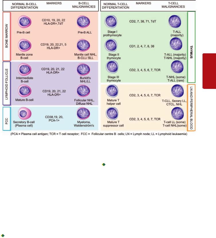
Figure 14.15 
 Schematic representation of WHO-REAL classification of lymphoid neoplasms. Various immunophenotypes of B and T-cell malignancies are correlated with normal immunophenotypic differentiation/maturation stages of B and T-cells in the bone marrow, lymphoid tissue, peripheral blood and thymus.
revised European-American classification of lymphoid neoplasms abbreviated as REAL classification. This classification was based on the hypothesis that all forms of lymphoid malignancies (NHLs as well as lymphoblastic leukaemias) represent malignant counterparts of normal population of immune cells (B-cells, T-cells and histiocytes) present in the lymph node and bone marrow. It is believed that lymphoid malignancies arise due to arrest at the various differentiation stages of B and T-cells since tumours of histiocytic origin are quite uncommon. Accordingly, it is considered essential to understand and correlate the differentiation stages of B and T-cells with various lymphoid malignancies (Fig. 14.15). REAL classification divides all lymphoid malignancies into two broad groups, each having further subtypes:

 Leukaemias and lymphomas of B-cell origin: B-cell derivation comprises 80% cases of lymphoid leukaemias and 90% cases of NHLs. Based upon these phenotypic and genotypic features, B-cell neoplasms are of pre-B and mature B-cell origin. Based on their biologic behaviour, B-cell malignancies are further subclassified into indolent and aggressive. All these tumours express Pan-B (CD19) antigen besides other markers.

 Leukaemias and lymphomas of T-cell origin: T-cell malignancies comprise the remainder 20% cases of lymphoid leukaemia and 10% cases of NHLs. T-cell malignancies reflect the stages of T-cell ontogeny. Like B-cell malignancies, T- cell derivatives too are further categorised into indolent and aggressive T-cell malignancies. The most widely expressed T-cell antigens are CD2 and CD7.
REAL classification subsequently merged into WHO classification described below.
III. WHO CLASSIFICATION OF LYMPHOID NEOPLASMS (1999): In view of confusion surrounding the classification schemes of lymphoid cancer, Harris et al, who described REAL classification, evolved a consensus international classification of all lymphoid neoplasms together as a unified group (lymphoid leukaemias-lymphomas) under the aegis of the WHO. Although this classification has many similarities with REAL classification as regards identification of B and T cell types (Fig. 14.15), WHO classification has more classes. WHO classification takes into account morphology, clinical features, immunophenotyping, and cytogenetic of the tumour cells. Hence, on this basis, it is possible to know the stage of maturity of the neoplastic cell and thus has a better clinical and therapeutic relevance.
367
Tissues Lymphoreticular and Leucocytes of Disorders 14 CHAPTER

368 TABLE 14.8: WHO Classification of Lymphoid Malignancies (1999).
Tissues Lymphoreticular and Haematology II SECTION
Hodgkin’s Disease |
B Cell Malignancies |
T Cell Malignancies |
|
|
|
Nodular lymphocytic predominant HD Classic HD
1.Nodular sclerosis HD
2.Lymphocytic rich classic HD
3.Mixed cellularity
4.Lymphocytic depletion HD
Precursor (Immature) B-cell malignancies
Precursor B lymphoblastic leukaemia/ lymphoma (precursor B-cell ALL)
Peripheral (Mature) B-cell malignancies
1.B-cell CLL/SLL (chronic lymphocytic leukaemia/small lymphocytic lymphoma)
2.B-cell prolymphocytic leukaemia
3.Lymphoplasmacytic lymphoma
4.Splenic marginal zone B-cell lymphoma
5.Hairy cell leukaemia
6.Plasma cell myeloma/plasmacytoma
7.Extranodal marginal zone B-cell lymphoma, MALT type
8.Mantle cell lymphoma
9.Follicular lymphoma
10.Nodal marginal zone B-cell lymphoma (Monocytoid B-cell lymphoma)
11.Diffuse large B-cell lymphoma
12.Burkitt’s lymphoma/Burkitt cell leukaemia
Precursor (Immature) T-cell malignancies
Precursor T lymphoblastic lymphoma/ leukaemia (Precursor T-cell ALL)
Peripheral (Mature) T-cell and NK-cell malignancies
1.T-cell prolymphocytic leukaemia
2.T-cell granular lymphocytic leukaemia
3.Aggressive NK cell leukaemia
4.Adult T-cell lymphoma/leukaemia (HTLV-I +)
5.Extranodal NK/T-cell lymphoma, nasal type
6.Enteropathy-type T-cell lymphoma
7.Hepatosplenic T-cell lymphoma
8.Subcutaneous panniculitis like T-cell lymphoma
9.Mycosis fungoides/Sézary syndrome
10.Anaplastic large T lymphoma, primary cutaneous type
11.Peripheral T-cell lymphoma, not otherwise specified
12.Angioimmunoblastic T-cell lymphoma
13.Anaplastic large T-cell lymphoma (ALCL), primary systemic type
As per WHO classification scheme (Table 14.8), all lymphoid neoplasms (i.e. lymphoid leukaemias and lymphomas) fall into following 5 categories:
I. Hodgkin’s disease
II. Precursor (Immature) B-cell malignancies III. Peripheral (Mature) B-cell malignancies IV. Precursor (Immature) T-cell malignancies
V. Peripheral (Mature) T-cell and NK–cell malignancies
Thus, in the WHO classification of lymphoid neoplasms, Hodgkin’s disease stands distinctive; remaining four categories listed above fall into 2 groups:
1)Precursor or immature lymphoid malignancies of B or T cell origin, meaning blastic type of leukaemias-lymphomas (mainly B or T-cell ALL); and
2)Peripheral or mature malignancies of B or T cell origin (meaning CLL and other lymphomas).
GENERAL COMMENTS ON LYMPHOID MALIGNANCIES
Before plunging into discussion of various common examples in the WHO classification system, a few general aspects on lymphoid neoplasms need to be understood:
1. Overall frequency: Five major forms of lymphoid malignancies and their relative frequency are as under:
i)NHL= 62%, most common lymphoma
ii)HD= 8%
iii)Plasma cell disorders = 15%
iv)CLL= 9%, most common lymphoid leukaemia
v)ALL= 4%
Relative frequency of subtypes within various NHLs listed in Table 14.8 is as under:
i)Diffuse large B cell lymphoma = 31%
ii)Follicular lymphoma = 22%
iii)MALT lymphoma = 8%
iv)Mature T cell lymphoma = 8%
v)Small lymphocytic lymphoma (SLL) = 7%
vi)Mantle cell lymphoma = 6% vii)Mediastinal large B cell lymphoma = 2.5%
viii)Anaplastic large cell lymphoma (ALCL) = 2.5%
ix)Burkitt’s lymphoma = 2.5%
x)Others = ~10%
2.Incidence of B, T, NK cell malignancies: Majority of lymphoid malignancies are of B cell origin (75% of lymphoid leukaemias and 90% of lymphomas) while remaining are T cell malignancies; NK-cell lymphomas-leukaemias are rare.
3.Diagnosis: The diagnosis of lymphoma (both Hodgkin’s and non-Hodgkin’s) can only be reliably made on examination of lymph node biopsy. While the initial diagnosis of ALL and CLL can be made on CBC examination, bone marrow biopsy is done for genetic and immunologic studies. Subsequently, clinical chemistry, electrophoresis and tests for organ involvement including CSF examination if CNS involvement is suspected, need to be carried out.
4.Staging: In both HD and NHL, Ann Arbor staging is done for proper evaluation and planning treatment.
5.Ancillary studies: CT scan, PET scan and gallium scan are additional imaging modalities which can be used in staging HD and NHL cases.

6. Immune abnormalities: Since lymphoid neoplasms arise from immune cells of the body, immune derangements pertaining to the cell of origin may accompany these cancers. This is particularly so in B-cell malignancies and include occurrence of autoimmune haemolytic anaemia, autoimmune thrombocytopenia and hypogammaglobulinaemia.
With this background, we now turn to discussion of some common and important examples of lymphoid malignancies.
HODGKIN’S DISEASE
Hodgkin’s disease (HD) primarily arises within the lymph nodes and involves the extranodal sites secondarily. This group comprises about 8% of all cases of lymphoid neoplasms. The incidence of the disease has bimodal peaks— one in young adults between the age of 15 and 35 years and the other peak after 5th decade of life. The HD is more prevalent in young adult males than females. The classical diagnostic feature is the presence of Reed-Sternberg (RS) cell (or Dorothy-Reed-Sternberg cell) (described later).
Classification
The diagnosis of HD requires accurate microscopic diagnosis by biopsy, usually from lymph node, and occasionally from other tissues. Unlike NHL, there is only one universally accepted classification of HD i.e. Rye classification adopted since 1966. Rye classification divides HD into the following 4 subtypes:
1.Lymphocyte-predominance type
2.Nodular-sclerosis type
3.Mixed-cellularity type
4.Lymphocyte-depletion type.
However, the WHO classification of lymphoid neoplasms
divides HD into 2 main groups:
I. Nodular lymphocyte-predominant HD (a new type).
II. Classic HD (includes all the 4 above subtypes in the Rye classification).
Central to the diagnosis of HD is the essential identification of Reed-Sternberg cell though this is not the sole criteria (see below).
The salient features of the 4 histologic subtypes of HD are summarised in Table 14.9.
Reed-Sternberg Cell
The diagnosis of Hodgkin’s disease rests on identification of RS cells, though uncommonly similar cells can occur in infectious mononucleosis and other forms of lymphomas. Therefore, additional cellular and architectural features of the biopsy must be given due consideration for making the histologic diagnosis.
There are several morphologic variants of RS cells which characterise different histologic subtypes of HD (Fig. 14.16):
1.Classic RS cell is a large cell which has characteristically a bilobed nucleus appearing as mirror image of each other but occasionally the nucleus may be multilobed. Each lobe of the nucleus contains a prominent, eosinophilic, inclusionlike nucleolus with a clear halo around it, giving an owl-eye appearance. The cytoplasm of cell is abundant and amphophilic.
2.Lacunar type RS cell is smaller and in addition to above features has a pericellular space or lacuna in which it lies, which is due to artefactual shrinkage of the cell cytoplasm. It is characteristically found in nodular sclerosis variety of HD.
3.Polyploid type (or popcorn or lymphocytic-histiocytic i.e. L and H) RS cells are seen in lymphocyte predominance type of HD. This type of RS cell is larger with lobulated nucleus in the shape of popcorn.
4.Pleomorphic RS cells are a feature of lymphocyte depletion type. These cells have pleomorphic and atypical nuclei.
The nature and origin of RS cells, which are the real neoplastic cells in HD, have been a matter of considerable debate. One main reason for this difficulty in their characterisation is that in HD, unlike most other malignancies, the number of neoplastic cells (i.e. RS cells) is very small (less than 5%) which are interspersed in the predominant reactive cells. In general, the number of RS cells is inversely proportional to
TABLE 14.9: Modified WHO Classification of Hodgkin’s Disease.
Histologic Subtype |
Incidence |
Main Pathology |
RS Cells |
Prognosis |
|
|
|
|
|
I. CLASSIC HD |
|
|
|
|
Lymphocyte-predominance |
5% |
Proliferating lymphocytes, |
Few, classic and polyploid |
Excellent |
|
|
a few histiocytes |
type, CD15–, CD30–, CD20+ |
|
Nodular sclerosis |
70% |
Lymphoid nodules, |
Frequent, lacunar type, |
Very good |
|
|
collagen bands |
CD15+, CD30+ |
|
Mixed cellularity |
22% |
Mixed infiltrate |
Numerous, classic type, |
Good |
|
|
|
CD15+, CD30+ |
|
Lymphocyte-depletion |
1% |
Scanty lymphocytes, |
Numerous, |
Poor |
(Diffuse fibrotic and |
|
atypical histiocytes, fibrosis |
pleomorphic type, |
|
reticular variants) |
|
|
CD15+, CD30+ |
|
II. NODULAR LYMPHOCYTE-PREDOMINANT HD |
|
|
|
|
|
2% |
Proliferation of small lympho- |
Sparse number of RS cells, |
Chronic |
|
|
cytes, nodular pattern of growth |
CD45+, EMA+, CD15-, CD30- |
relapsing, may |
|
|
|
|
transform into |
|
|
|
|
large B cell NHL |
|
|
|
|
|
369
Tissues Lymphoreticular and Leucocytes of Disorders 14 CHAPTER
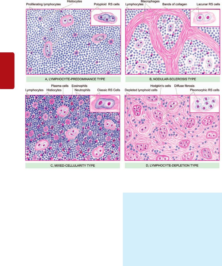
370
Tissues Lymphoreticular and Haematology II SECTION
Figure 14.16 
 Microscopic features of 4 forms of Hodgkin’s disease of lymph node. The inset on right side of each type shows the morphologic variant of RS cell seen more often in particular histologic type.
the number of lymphocytes in a particular histologic subtype of HD.
Immunophenotyping of RS cells reveals monoclonal lymphoid cell origin of RS cell from B-cells of the germinal centre in most subtypes of Hodgkin’s disease. RS cells in all types of Hodgkin’s diseases, except in lymphocyte predominance type, express immunoreactivity for CD15 and CD30 (Fig. 14.17). RS cells in lymphocyte predominance type, however, are negative for both CD15 and CD30, but positive for CD20.
RS cells are invariably accompanied by variable number of atypical Hodgkin cells which are believed to be precursor RS cells but are not considered diagnostic of HD. Hodgkin cells are large mononuclear cells (rather than mirror image nuclei) having nuclear and cytoplasmic similarity to that of RS cell.
MORPHOLOGIC FEATURES. Grossly, the gross appearance of Hodgkin’s and non-Hodgkin’s lymphoma is much the same. Any lymph node group may be involved but most commonly affected are the cervical, supraclavicular and axillary groups. Initially, the lymph nodes are discrete and separate from one another but later the lymph nodes form a large matted mass due to infiltration into the surrounding connective tissue. Extranodal involvements produce either a discrete tumour or diffuse enlargement of the affected organ. The sectioned surface of the involved lymph nodes or extranodal organ involved appears grey-white and fishflesh-like. Nodular sclerosis type HD may show formation of nodules due to scarring while mixed cellularity and lymphocyte depletion types HD may show abundance of necrosis. Lympho-
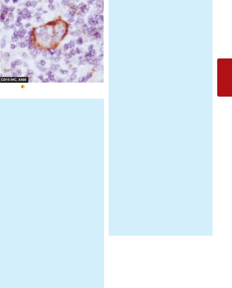
Figure 14.17 RS cells showing positive immunostaining for CD15,
a B-cell marker.
matous involvement of the liver, spleen and other organs may be diffuse or may form spherical masses similar to metastatic carcinoma.
Microscopically, the criteria for diagnosis of histologic subtypes of HD are as under:
I. CLASSIC HD
As per WHO classification, classic group of HD includes 4 types of HD of older Rye classification:
1. Lymphocyte-predominance type (Fig. 14.16,A). The lymphocyte-predominance type of HD is characterised by proliferation of small lymphocytes admixed with a varying number of histiocytes forming nodular or diffuse pattern.
i)Nodular form is characterised by replacement of nodal architecture by numerous large neoplastic nodules.
ii)Diffuse form does not have discernible nodules but instead there is diffuse proliferation of cells.
However, currently nodular form of lymphocyte predominent HD has been categorised separately due to its distinct immunophenotyping features and prognosis (discussed below).
For making the diagnosis, definite demonstration of RS cells is essential which are few in number, requiring a thorough search. In addition to typical RS cells, polyploid variant having polyploid, and twisted nucleus (popcornlike) may be found in some cases. This type of HD usually does not show other cells like plasma cells, eosinophils and neutrophils, nor are necrosis or fibrosis seen.
2.Nodular-sclerosis type (Fig. 14.16,B). Nodular sclerosis is the most frequent type of HD, seen more commonly in women than in men. It is characterised by two essential features (Fig. 14.18, A):
i)Bands of collagen: Variable amount of fibrous tissue is characteristically present in the involved lymph nodes. Occasionally, the entire lymph node may be replaced by dense hyalinised collagen.
ii)Lacunar type RS cells: Characteristic lacunar type of RS cells with distinctive pericellular halo are present. These cells appear lacunar due to the shrinkage of cytoplasm in formalin-fixed tissue. The pericellular halo is not seen if the tissue is fixed in Zenker’s fluid.
In addition to these 2 characteristics, the nodules between the fibrous septa consist predominantly of lymphocytes and macrophages, sometimes with foci of necrosis.
3.Mixed-cellularity type (Fig. 14.16,C). This form of HD generally replaces the entire affected lymph nodes by heterogeneous mixture of various types of apparently normal cells. These include proliferating lymphocytes, histiocytes, eosinophils, neutrophils and plasma cells. Some amount of fibrosis and focal areas of necrosis are generally present. Typical RS cells are frequent (Fig. 14.18,
B).
4.Lymphocyte-depletion type (Fig. 14.16,D). In this type of HD, the lymph node is depleted of lymphocytes. There are two variants of lymphocyte-depletion HD:
i)Diffuse fibrotic variant is hypocellular and the entire lymph node is replaced by diffuse fibrosis, appearing as homogeneous, fibrillar hyaline material. The area of hyalinosis contains some lymphocytes, atypical histiocytes (Hodgkin cells), and numerous typical and atypical (pleomorphic) RS cells.
ii)Reticular variant is much more cellular and consists of large number of atypical pleomorphic histiocytes, scanty lymphocytes and a few typical RS cells.
II. NODULAR LYMPHOCYTE-PREDOMINANT HD
This is a newly described entity which is distinct from the classic HD described above. This type was previously included in lymphocyte predominant type of HD. Its peculiarities are as under:
i)These cases of HD have a nodular growth pattern (similar to nodular sclerosis type).
ii)Like lymphocyte-predominant pattern of classic type, there is predominance of small lymphocytes with sparse number of RS cells.
iii)These cases of HD have distinctive immuno- phenotyping—CD45 positive, epithelial membrane antigen (EMA) positive but negative for the usual markers for RS cells (CD15 and CD30 negative).
iv)Though generally it has a chronic relapsing course, but some cases of this type of HD may transform into large B-cell NHL.
Clinical Features
Hodgkin’s disease is particularly frequent among young and middle-aged adults. All histologic subtypes of HD, except the nodular sclerosis variety, are more common in males. The disease usually begins with superficial lymph node enlargement and subsequently spreads to other lymphoid and non-lymphoid structures.
1. Most commonly, patients present with painless, movable and firm lymphadenopathy. The cervical and mediastinal
371
Tissues Lymphoreticular and Leucocytes of Disorders 14 CHAPTER
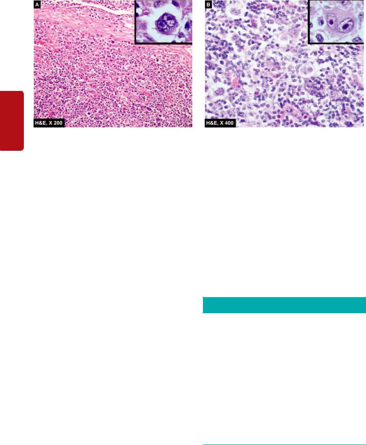
372
Tissues Lymphoreticular and Haematology II SECTION
Figure 14.18 
 Hodgkin’s disease. A, Nodular sclerosis type. There are bands of collagen forming nodules and characteristic lacunar RS cells
(inbox in left figure) . B, Mixed cellularity type. There is admixture of mature lymphocytes, plasma cells, neutrophils and eosinophils and classic RS cells in the centre of the field (inbox in right figure).
lymph nodes are involved most frequently. Other lymph node groups like axillary, inguinal and abdominal are involved sometimes.
2.Approximately half the patients develop splenomegaly during the course of the disease. Liver enlargement too may occur.
3.Constitutional symptoms (type B symptoms) are present in 25-40% of patients. The most common is low-grade fever with night sweats and weight loss. Other symptoms include fatigue, malaise, weakness and pruritus.
Other Laboratory Findings
Besides clinical and pathologic findings, there are some haematologic and immunologic abnormalities in HD.
Haematologic abnormalities:
1.A moderate, normocytic and normochromic anaemia is often present.
2.Serum iron and TIBC are low but marrow iron stores are normal or increased.
3.Marrow infiltration by the disease may produce marrow failure with leucoerythroblastic reaction.
4.Routine blood counts reveal moderate leukaemoid reaction. Cases with pruritus frequently show peripheral eosinophilia. Advanced disease is associated with absolute lymphopenia.
5Platelet count is normal or increased.
6.ESR is invariably elevated.
Immunologic abnormalities:
1.There is progressive fall in immunocompetent T-cells with defective cellular immunity. There is reversal of CD4: CD8 ratio and anergy to routine skin tests.
2.Humoral antibody production is normal in untreated patients until late in the disease.
Staging
Following biopsy and histopathologic classification of HD, the extent of involvement of the disease (i.e. staging) is
studied in order to select proper treatment and assess the prognosis. Ann Arbor staging classification takes into account both clinical and pathologic stage of the disease.
The suffix A or B are added to the above stages depending upon whether the three constitutional symptoms (fever, night sweats and unexplained weight loss exceeding 10% of normal) are absent (A) or present (B). The suffix E or S are used for extranodal involvement and splenomegaly respectively (Table 14.10).
For complete staging, a number of other essential diagnostic studies are recommended. These are as under:
1.Detailed physical examination including sites of nodal involvement and splenomegaly.
2.Chest radiograph to exclude mediastinal, pleural and lung parenchymal involvement.
3.CT scan of abdomen and pelvis.
TABLE 14.10: Ann Arbor Staging Classification of Hodgkin’s Disease.
Stage I |
I |
Involvement of a single lymph node region. |
(A or B) |
IE |
Involvement of a single extra-lymphatic organ |
|
|
or site. |
Stage II |
II |
Involvement of two or more lymph node |
(A or B) |
|
regions on the same side of the diaphragm. |
|
IIE |
(or) with localised contiguous involvement of an |
|
|
extranodal organ or site. |
Stage III |
III |
Involvement of lymph node regions on both sides |
(A or B) |
|
of the diaphragm. |
|
IIIE |
(or) with localised contiguous involvement of an |
|
|
extranodal organ or site. |
|
IIIS |
(or) with involvement of spleen. |
|
IllES |
(or) both features of IIIE and IIIS. |
Stage IV |
IV |
Multiple or disseminated involvement of one |
(A or B) |
|
or |
|
|
more extra-lymphatic organs or tissues with or |
|
|
without lymphatic involvement. |
(A = Asymptomatic; B = Presence of constitutional symptoms; E = Extranodal involvement; S = Splenomegaly).

4.Documentation of constitutional symptoms (B symptoms).
5.Laboratory evaluation of complete blood counts, liver and kidney function tests.
6.Bilateral bone marrow biopsy.
7.Finally, histopathologic documentation of the type of Hodgkin’s disease.
More invasive investigations include lymphangiography of lower extremities and staging laparotomy. Staging laparotomy includes biopsy of selected lymph nodes in the retroperitoneum, splenectomy and wedge biopsy of the liver.
Prognosis
With use of aggressive radiotherapy and chemotherapy, the outlook for Hodgkin’s disease has improved significantly. Although several factors affect the prognosis, two important considerations in evaluating its outcome are the extent of
involvement by the disease (i.e. staging) and the histologic subtype.

 With appropriate treatment, the overall 5 years survival rate for stage I and II A is as high as about 100%, while the advanced stage of the disease may have upto 50% 5-year survival rate.
 Patients with lymphocyte-predominance type of HD tend to have localised form of the disease and have excellent prognosis.

 Nodular sclerosis variety too has very good prognosis but those patients with larger mediastinal mass respond poorly to both chemotherapy and radiotherapy.
 Mixed cellularity type occupies intermediate clinical position between the lymphocyte predominance and the lymphocyte-depletion type, but patients with disseminated disease and systemic manifestations do poorly.

 Lymphocyte-depletion type is usually disseminated at the time of diagnosis and is associated with constitutional symptoms. These patients usually have the most aggressive form of the disease.
The salient features to distinguish Hodgkin’s disease and non-Hodgkin’s lymphoma are summarised in
Table 14.11.
PRECURSOR (IMMATURE) B- AND
T-CELL LEUKAEMIA/LYMPHOMA
(SYNONYM: ACUTE LYMPHOBLASTIC LEUKAEMIA)
Lymphoid malignancy originating from precursor series of B or T cell (i.e. pre-B and pre-T) is the most common form of cancer of children under 4 years of age, together constituting 4% of all lymphoid malignancies. Pre-B cell ALL constitutes 90% cases while pre-T cell lymphoid malignancies comprise the remaining 10%. This group of lymphoid malignancies arise from more primitive stages of B or T cells but the stage of differentiation is not related to aggressiveness. Because of morphologic similarities, both these are presented together.
Clinical Features
PRECURSOR B-CELL LYMPHOBLASTIC LEUKAEMIA/ LYMPHOMA. Most often, it presents as ALL in children; rarely presentation may be in the form of lymphoma in
TABLE 14.11: Contrasting Features of Hodgkin’s Disease |
|
373 |
||||
|
and Non-Hodgkin’s Lymphoma. |
|
|
|
||
|
|
|
|
|
|
|
|
Feature |
Hodgkin’s |
Non-Hodgkin’s |
|
|
|
1. |
Cell derivation |
B-cell mostly |
90% B |
|
||
|
|
|
10% T |
|
||
2. |
Nodal involve- |
Localised, may |
Disseminated nodal |
|
||
|
ment |
spread to |
spread |
|
||
|
|
contiguous nodes |
|
|
|
|
3. |
Extranodal |
Uncommon |
Common |
|
||
|
spread |
|
|
|
|
|
4. |
Bone marrow |
Uncommon |
Common |
|
||
|
involvement |
|
|
|
CHAPTER |
|
5. |
Constitutional |
Common |
Uncommon |
|||
|
||||||
|
symptoms |
|
|
|
|
|
6. |
Chromosomal |
Aneuploidy |
Translocations, |
|
||
|
defects |
|
deletions |
|
||
7. |
Spill-over |
Never |
May spread to blood |
|
||
8. |
Prognosis |
Better |
Bad |
14 |
||
|
|
(75-85% cure) |
(30-40% cure) |
|||
|
|
|
||||
|
|
|
|
|
Disorders |
|
children or adults and it rapidly transforms into leukaemia. |
||||||
|
||||||
In cases having leukaemic presentation, extranodal site |
|
|||||
involvement is early such as lymphadenopathy accompanied |
|
|||||
with hepatomegaly, splenomegaly, CNS infiltration, |
of |
|||||
testicular enlargement, and at times cutaneous infiltration. |
||||||
Infections due to cytopenia are present. |
Leucocytes |
|||||
PRECURSOR T-CELL LYMPHOBLASTIC LEUKAEMIA/ |
||||||
|
||||||
LYMPHOMA. As the name implies, these cases may present |
|
|||||
as ALL or as lymphoma. Since the precursor T-cells |
|
|||||
differentiate in the thymus, this tumour often presents as |
and |
|||||
mediastinal mass and pleural effusion and progresses rapidly |
||||||
|
||||||
to develop leukaemia in the blood and bone marrow. |
Lymphoreticular |
|||||
Clinically, features of bone marrow failure are present which |
||||||
|
||||||
include anaemia, neutropenia and thrombocytopenia. |
|
|||||
Lymphadenopathy, hepatosplenomegaly and CNS |
|
|||||
involvement are frequent. |
|
|
|
|||
|
Precursor T-cell lymphoma-leukaemia is, however, more |
|
||||
aggressive than its B-cell counterpart. |
|
|||||
MORPHOLOGIC FEATURES |
|
|
Tissues |
|||
Precursor B and T-cell ALL/lymphoma are indistinguish- |
||||||
|
||||||
able on routine morphology. The diagnosis is made by following investigations:
1. Blood examination. Peripheral blood generally shows anaemia and thrombocytopenia, and may show leucopenia-to-normal TLC-to-leucocytosis. DLC shows large number of circulating lymphoblasts having round to convoluted nuclei, high nucleo-cytoplasmic ratio and absence of cytoplasmic granularity. It is important to distinguish AML from ALL; the morphologic features of myeloblasts and lymphoblasts are contrasted in Table 14.1 for comparison (Fig. 14.19). Typical characteristics of different forms of ALL (L1 to L3) are given in Table 14.6. It is usual to find some ‘smear cells’ in the peripheral blood which represent degenerated leucocytes.
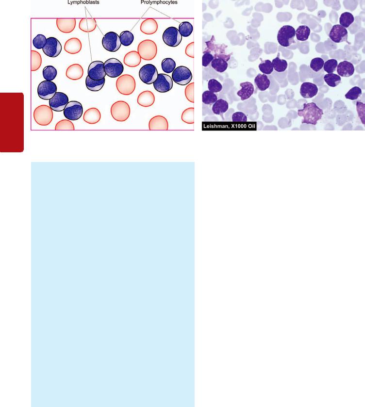
374
II SECTION
Figure 14.19 
 PBF findings in acute lymphoblastic leukaemia (ALL). The cells are large, with round to convoluted nuclei having high N/C ratio and no cytoplasmic granularity.
Tissues Lymphoreticular and Haematology
2.Bone marrow examination. Marrow examination shows malignant undifferentiated cells of precursor B or T cell origin as demonstrated by immunophenotyping. Megakaryocytes are usually reduced or absent.
3.Cytochemistry. Cytochemical stains may be employed as an adjunct to Romanowsky staining for determining the type of leukaemia. Some of the commonly employed cytochemical stains in characterisation of leukaemic blasts in ALL are as under:
i)Periodic acid-Schiff (PAS): Positive in immature lymphoid cells in ALL.
ii)Acid phosphatase: Focal positivity in leukaemic blasts in ALL.
iii)Myeloperoxidase: Negative in immature cells in ALL.
iv)Sudan Black: Negative in immature cells in ALL.
v)Non-specific esterase (NSE): Negative in ALL.
Immunophenotyping. TdT (terminal deoxynucleotidyl transferase) is expressed by the nuclei of both pre-B and pre-T stages of differentiation of lymphoid cells. Specific diagnosis is eastablished by following immunophenotyping:
Pre-B-cell type: Typically positive for pan-B cell markers CD19, CD10, CD9a.
Pre-T-cell type: Typically positive for CD1, CD2, CD3, CD5, CD7.
Cytogenetic analysis: Leukaemic blasts in pre-B-cell ALL show characteristic cytogenetic abnormality of t(9;22) i.e. Philadelphia positive-ALL.
Treatment
Treatment plan for children with pre-B or pre-T cell ALL is intensive remission induction with combination therapy. Patients presenting with pre-B or pre-T cell lymphoma are treated as a case of ALL.
CHEMOTHERAPY. The most useful drugs in the treatment of ALL are combination of vincristine, prednisolone, anthracyclines (daunorubicin, adriamycin) and L-aspara- ginase. Other agents used are cytosine arabinoside and methotrexate. Though 90% of children with ALL show remission with this therapy, patients with T cell ALL and those with meningeal involvement carry a less favourable prognosis. CNS involvement is beyond the reach of most of the cytotoxic drugs used in the therapy of ALL. CNS prophylaxis in such cases is considered after the initial remission has been obtained and includes cranial irradiation and course of intrathecal methotrexate or cytosine arabinoside.
BONE MARROW TRANSPLANTATION. Bone marrow (Stem cell) transplantation from suitable allogenic or autologous donor (HLA and mixed lymphocytes culturematched) is used in preB and pre-T cell ALL in adults with relapses. Bone marrow transplantation has resulted in cure in about half the cases.
Prognosis and disease-free survival of children with both pre-B cell an dpre-T cell ALL is better than in adults. Mean survival with treatment in children without CNS prophylaxis is 33 months, while with CNS prophylaxis is 60 months or more. Adult pre-T cell ALL, however, is as grave as AML and median survival is 12-18 months. Patients having limited disease confined to lymph nodes in both pre-B and pre-T cell type have a higher cure rate and better prognosis.
The salient differences between the two main forms of acute leukaemia (AML and ALL) are summarised in
Table 14.12.
PERIPHERAL(MATURE) B-CELL MALIGNANCIES
Peripheral or mature B-cell cancers are the most common lymphoid malignancies. These arise from the stage of lymphoid cells at which they become committed to B cell development, acquire surface characteristics and begin to secrete immunoglobulins. It includes following common examples.
TABLE 14.12: Contrasting Features of AML and ALL.
|
Feature |
AML |
ALL |
|
|
|
|
1. |
Common age |
Adults between 15-40 years; com- |
Children under 15 years; comprise 80% of childhood |
|
|
prise 20% of childhood leukaemias |
leukaemias |
2. |
Physical findings |
Splenomegaly + |
Splenomegaly ++ |
|
|
Hepatomegaly + |
Hepatomegaly ++ |
|
|
Lymphadenopathy + |
Lymphadenopathy ++ |
|
|
Bony tenderness + |
Bony tenderness + |
|
|
Gum hypertrophy + |
CNS involvement + |
3. |
Laboratory findings |
Low-to-high TLC; predominance of |
Low-to-high TLC, predominance of lymphoblasts in blood |
|
|
myeloblasts and promyelocytes in |
and bone marrow; thrombocytopenia moderate to severe. |
|
|
blood and bone marrow; throm- |
|
|
|
bocytopenia moderate to severe. |
|
4. |
Diagnostic criteria |
FAB types M0-M7 |
FAB types L1-L3, WHO types Pre B (90%) Pre T (10%) |
|
|
WHO criteria = >20% blasts |
WHO criteria = >20% blasts |
5. |
Cytochemical stains |
Myeloperoxidase +, Sudan black +, |
PAS +, acid phosphatase (focal) + |
|
|
NSE + in M4 and M5, acid phos- |
|
|
|
phatase (diffuse) + in M4 and M5 |
|
6. |
Specific therapy |
Cytosine arabinoside, |
Vincristine, prednisolone, anthracyclines and |
|
|
anthracyclines (daunorubicin, |
L-asparaginase |
|
|
adriamycin) and 6-thioguanine |
|
7. |
Immunophenotyping |
CD13, 33, 41, 42 |
Both B and T cell ALL TdT +ve |
|
|
|
Pre B: CD19, 20 |
|
|
|
Pre T: CD1, 2, 3, 5, 7 |
8. |
Cytogenetics |
M3: t(15;17) |
Pre B: t(9;21) |
|
|
M4: in(16) |
|
9. |
Response to therapy |
Remission rate low, duration of |
Remission rate high, duration of remission prolonged |
|
|
remission shorter |
|
10.Median survival |
12-18 months |
Children without CNS prophylaxis 33 months, with CNS |
|
|
|
|
prophylaxis 60 months; adults 12-18 months |
|
|
|
|
B-CELL CLL/SLL (CHRONIC LYMPHOCYTIC LEUKAEMIA/ SMALL LYMPHOCYTIC LYMPHOMA)
As the name implies, this subtype may present as leukaemia or lymphoma constituting 9% of all lymphoid neoplasms. As lymphoid leukaemia (CLL), this is the most common form while as SLL it constitutes 7% of all NHLs. B-cell CLL/SLL occurs more commonly in middle and older age groups (over 50 years of age) with a male preponderance (male-female ratio 2:1).
Clinical Features
The condition may remain asymptomatic, or may have an insidious onset and may present with nonspecific clinical features. Common presenting manifestations are as under:
1.Features of anaemia such as gradually increasing weakness, fatigue and dyspnoea.
2.Enlargement of superficial lymph nodes is a very common finding. The lymph nodes are usually symmetrically enlarged, discrete and non-tender.
3.Splenomegaly and hepatomegaly are usual.
4.Haemorrhagic manifestations are found in case of CLL with thrombocytopenia.
5.Susceptibility to Infections, particularly of respiratory tract, are common in CLL.
6.Less common findings are: mediastinal pressure, tonsillar enlargement, disturbed vision, and bone and joint pains.
MORPHOLOGIC FEATURES
The diagnosis of CLL can usually be made on the basis of physical findings and blood smear examination
(Fig. 14.20):
I. BLOOD PICTURE. The findings of routine blood picture are as under:
1.Anaemia. Anaemia is usually mild to moderate and normocytic normochromic in type. Mild reticulocytosis may be present. About 20% cases develop a Coombs’- positive autoimmune haemolytic anaemia.
2.White blood cells. Typically, there is marked leucocytosis but less than that seen in CML (50,000-200,000/ μl). Usually, more than 90% of leucocytes are mature small lymphocytes. Smudge or basket cells (degenerated forms) are present due to damaged nuclei of fragile malignant lymphocytes. The absolute neutrophil count is, however, generally within normal range. Granulocytopenia occurs when disease is fairly advanced.
3.Platelets. The platelet count is normal or moderately reduced as an autoimmune phenomenon.
II. BONE MARROW EXAMINATION. The typical findings are as under:
1.Increased lymphocyte count (25-95%).
2.Reduced myeloid precursors.
3.Reduced erythroid precursors.
375
Tissues Lymphoreticular and Leucocytes of Disorders 14 CHAPTER
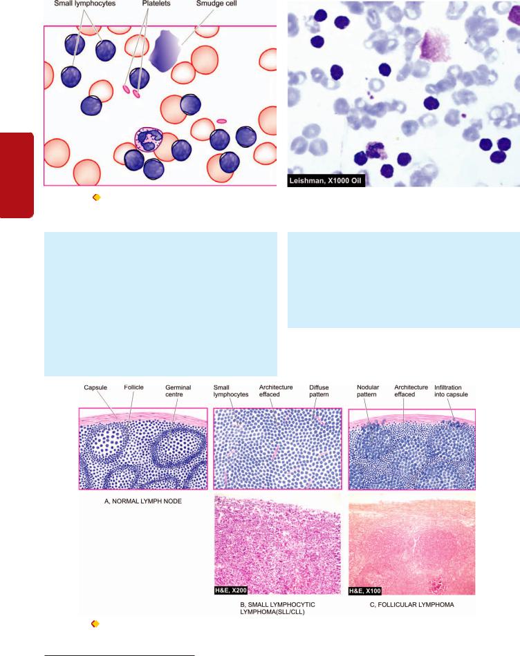
376
II SECTION
Figure 14.20 |
PBF in chronic lymphocytic leukaemia (CLL). There is large excess of mature and small differentiated lymphocytes and some |
degenerated forms appearing as bare smudged nuclei.
Tissues Lymphoreticular and Haematology
III. LYMPH NODE BIOPSY. Cases with lymphadenopathy at presentation show replacement of the lymph node by diffuse proliferation of well-differentiated, mature, small and uniform lymphocytes without any cytologic atypia or significant mitoses (Fig. 14.21,B). These cells are of monoclonal B-cell origin having immunologic features of mantle zone B-cells.
IV. OTHER INVESTIGATIONS. These include the following:
1. Erythrocyte rosette test with mouse red cells is positive in more than 95% of cases indicating that CLL is a monoclonal B cell neoplasm.
2.Positive for B-cell markers e.g. typically CD5 positive; other pan-B cell markers are CD19, CD20, CD23, surface immunoglobulins of various classes, monoclonal light chains (λ or κ type).
3.Serum immunoglobulin levels are generally reduced.
4.Coombs’ test is positive in 20% cases.
5.Cytogenetic abnormalities, most commonly trisomy 12 seen in about 25% cases.
Treatment and Prognosis
Unlike other leukaemias, none of the available drugs and radiation therapy are capable of eradicating CLL and induce
Figure 14.21 |
Prototypes of non-Hodgkin’s |
lymphoma—small lymphocytic lymphoma SLL/
CLL (B) and follicular lymphoma (C) contrasted with structure of normal lymph node (A).

true complete remission. Treatment is, therefore, palliative and symptomatic, and with optimal management patient can usually lead a relatively normal life for several years. These approaches include: alkylating drugs (e.g. chlorambucil, cyclophosphamide), corticosteroids and radiotherapy. Splenectomy is indicated in cases of CLL with autoimmune haemolytic anaemia.
Prognosis of CLL/SLL is generally better than CML since blastic transformation seldom occurs. Prognosis generally correlates with the stage of disease as under:
Stage A: characterised by lymphocytosis alone, or with limited lymphadenopathy, has a good prognosis (median survival more than 10 years).
Stage B: having lymphocytosis with associated significant lymphadenopathy and hepatosplenomegaly has intermediate prognosis (median survival about 5 years).
Stage C: having lymphocytosis with associated anaemia and thrombocytopenia has a worse prognosis (median survival of less than 2 years).
Generally, the course is indolent. However, some cases of SLL may transform into more aggressive diffuse large B-cell lymphoma, or may be associated with occurrence of an IgM monoclonal gammopathy called Waldenström’s macroglobulinaemia (page 384).
FOLLICULAR LYMPHOMA
In the earlier classification schemes, follicular lymphoma was known as nodular (poorly-differentiated) or follicular lymphoma (predominantly small/large cleaved cell type). Follicular lymphomas comprise approximately 22% of all NHLs. Follicular lymphomas occur in older individuals, most frequently presenting with painless peripheral lymphadenopathy which is usually waxing and waning type. In contrast to diffuse lymphomas, extranodal involvement is also infrequent.
MORPHOLOGIC FEATURES. Following features are seen:
Lymph node biopsy: As the name suggests, follicular lymphoma is characterised by follicular or nodular pattern of growth. The nuclei of tumour cells may vary from predominantly small cleaved (or indented) to predominantly large cleaved variety (Fig. 14.21,C). The former is more common, has infrequent mitoses and the rate of growth slow (low grade), while the patients with large cell lymphoma have high proliferation and progress rapidly (high grade). In all follicular lymphomas, the tumour cells are positive for pan-B markers such as CD19 and CD20 along with expression of BCL-2 protein (for distinction from normal germinal centre which is BCL-2 negative). Cytogenetic studies show characteristic translocation t(14;18) in tumour cells.
Blood and bone marrow: Peripheral blood involvement as occurs in SLL is uncommon in this variety. Infiltration in the bone marrow is typically paratrabecular.
About half the cases of low-grade follicular lymphomas, during their indolent biologic course, may evolve into diffuse
large B-cell lymphoma. Median survival for patients with low grade follicular lymphoma is 7-9 years.
Diffuse Large B-cell Lymphoma
Diffuse large B-cell lymphoma, earlier termed as diffuse poorly-differentiated lymphocytic lymphoma or follicular centre cell diffuse large, cleaved/non-cleaved lymphoma, is the most common comprising about 31% of all NHLs. It occurs in older patients with mean age of 60 years. It may present primarily as a lymph node disease or at extranodal sites. About half the cases have extranodal involvement at the time of presentation, particularly in the bone marrow and the alimentary tract. Primary diffuse large B-cell lymphoma of CNS may also occur.
A few subtypes of diffuse large B-cell lymphoma are described with distinct clinicopathologic settings:

 Epstein-Barr virus (EBV) infection has been etiologically implicated in diffuse large B-cell lymphoma in immunosuppressed patients of AIDS and organ transplant cases.

 Human herpes virus type 8 (HHV-8) infection along with presence of immunosuppression is associated with a subtype of diffuse large B-cell lymphoma presenting with effusion,
termed primary effusion lymphoma.

 Mediastinal large B-cell lymphoma is diagnosed in patients with prominent involvement of mediastinum, occurs in young females and frequently spreads to CNS and abdominal viscera.
MORPHOLOGIC FEATURES. This variety is the diffuse counterpart of follicular large cleaved cell lymphoma i.e. it is composed of large cleaved cells spread in a diffuse pattern. The cytoplasm in these tumour cells is pale and abundant while the nuclei have prominent 1-2 nucleoli. Immunophenotypic markers for pan-B cells (CD19, CD20) are positive, besides overexpression of surface immunoglobulins (IgM, IgG and light chains) and of BCL-2 protein.
In general, diffuse large B-cell lymphomas are aggressive tumours and disseminate widely.
BURKITT’S LYMPHOMA/LEUKAEMIA
Burkitt’s lymphoma/leukaemia is an uncommon tumour in adults but comprises about 30% of childhood NHLs. Burkitt’s leukaemia corresponds to L3 ALL of FAB grouping and is uncommon. Three subgroups of Burkitt’s lymphoma are recognised: African endemic, sporadic and immunodeficiency-
associated.:

 African endemic Burkitt’s lymphoma was first described in African children, predominantly presenting as jaw tumour that spreads to extranodal sites such as the bone marrow and meninges. The relationship of this tumour with oncogenic virus, Epstein-Barr virus (EBV), has been discussed in Chapter 8.

 Sporadic Burkitt’s lymphoma is a related tumour in which the tumour cells are similar to those of Burkitt’s lymphoma but are more pleomorphic and may sometimes be multinucleated. Sporadic variety has a propensity to infiltrate the CNS and is more aggressive than true Burkitt’s lymphoma.

 Immunodeficiency-associated Burkitt’s lymphoma includes cases seen in association with HIV infection.
377
Tissues Lymphoreticular and Leucocytes of Disorders 14 CHAPTER
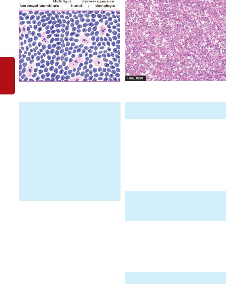
378
II SECTION
Figure 14.22 
 Burkitt’s lymphoma. The tumour shows uniform cells having high mitotic rate. Scattered among the tumour cells are benign macrophages surrounded by a clear space giving ‘starry sky’ appearance.
Tissues Lymphoreticular and Haematology
MORPHOLOGIC FEATURES. Histologically all three types of Burkitt’s lymphoma are similar. Tumour cells are intermediate in size, non-cleaved, and homogeneous in size and shape. The nuclei are round to oval and contain 2-5 nucleoli. The cytoplasm is basophilic and contains lipid vacuolation. The tumour cells have a very high mitotic rate, and therefore high cell death. This feature accounts for presence of numerous macrophages in the background of this tumour containing phagocytosed tumour debris giving it a ‘starry sky’ appearance (Fig. 14.22).
Burkitt’s leukaemia is identified by classical appearance of monomorphic medium-sized cells having round nuclei, frequent mitoses, multiple nucleoli, and basophilic cytoplasm with vacuoles.
Immunophenotypically, the tumour cells are positive for CD19 and CD10 and surface immunoglobulin IgM. Typical cytogenetic abnormalities in the tumour cells are t(8;14) and t(8;22) involving MYC gene on chromosome 8, with overexpression of MYC protein having transforming activity.
Burkitt’s lymphoma is a high-grade tumour and is a very rapidly progressive human tumour.
EXTRANODAL MARGINAL ZONE B-CELL LYMPHOMA OF MALT TYPE (SYNONYM: MALTOMA)
This type comprises about 8% of all NHLs. In the earlier classification, it was included under SLL, but in the WHO scheme it is categorised separately for 2 reasons: etiologic association with H. pylori infection and occurrence at extranodal sites. Most frequent is gastric lymphoma of MALT type with its characteristic etiologic association with H. pylori; other extranodal sites for this subtype of NHL are intestine, orbit, lung, thyroid, salivary glands and CNS. About half the cases of gastric MALT lymphoma show Genetic mutation t(11;18). Median age for this form of NHL is 60 years and often remains localised to the organ of origin but may infiltrate the regional lymph nodes.
MORPHOLOGIC FEATURES. It is characterised by diffuse infiltration by monoclonal small B lymphocytes which are negative for CD5.
MALT lymphoma has a good prognosis. Rarely, it may be more aggressive and may metastasise, or transform into diffuse large B-cell lymphoma.
MANTLE CELL LYMPHOMA
This subtype of NHL comprises about 8% of all NHLs. It was earlier included in SLL but has been identified as a separate subtype due to characteristic chromosomal translocation, t(11;14) and overexpression of BCL-1 and surface immunoglobulins IgM and IgD protein. However, both SLL and mantle cell NHL are positive for CD5 antigen. Patients of mantle cell lymphoma are generally older males. The disease involves bone marrow, spleen, liver and bowel.
MORPHOLOGIC FEATURES. Mantle cell lymphoma arises from B-cells of mantle zone of normal lymphoid follicle. The tumour cells show diffuse or nodular pattern of involvement in the lymph node and have somewhat indented nuclei.
It is more aggressive than other types of SLLs.
HAIRY CELL LEUKAEMIA
Hairy cell leukaemia (HCL) is an unusual and uncommon form of B-cell malignancy characterised by presence of hairy cells in the blood and bone marrow and splenomegaly. It occurs in the older males. HCL is characterised clinically by the manifestations due to infiltration of reticuloendothelial organs (bone marrow, liver and spleen) and, hence, its
previous name as leukaemic reticuloendotheliosis. Patients have susceptibility to infection with M. avium intercellulare.
MORPHOLOGIC FEATURES. Laboratory diagnosis is made by the presence of pancytopenia due to marrow
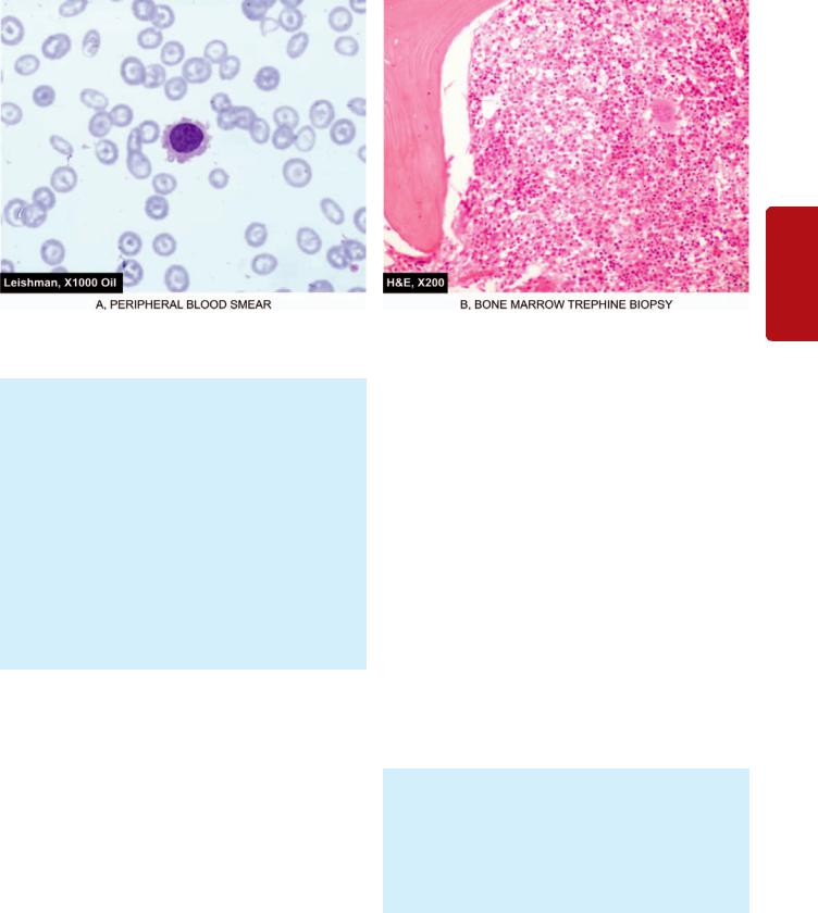
Figure 14.23 
 Hairy cell leukaemia. A, Peripheral blood shows presence of a leukaemic cells with hairy cytoplasmic projections B, Trephine biopsy shows replacement of marrow spaces with abnormal mononuclear cells.
failure and splenic sequestration, and identification of characteristic hairy cells in the blood and bone marrow. Hairy cells are abnormal mononuclear cells with hairy cytoplasmic projections which are seen in the bone marrow, peripheral blood and spleen. These cells are best recognised under phase contrast microscopy but may also be visible in routine blood smears (Fig. 14.23). These leukaemic ‘hairy cells’ have characteristically positive cytochemical staining for tartrate-resistant acid phosphatase
(TRAP).
The controversy on the origin of hairy cells whether these cells represent neoplastic T cells, B cells or monocytes, is settled with the molecular analysis of these cells which assigns them B cell origin expressing CD19, CD20 and CD22 antigen. In addition to B cell markers, hairy cells are also positive for CD11, CD25 and CD103.
The disease often runs a chronic course requiring supportive care. The mean survival is 4-5 years. Patients respond to splenectomy, α-interferon therapy and 2- chlorodeoxyadenosine (2-CDA).
OTHER B-CELL MALIGNANCIES
Brief mention is made below on the other types of B-cell malignant tumours given in the WHO classification in Table 14.8:
i)B-cell prolymphocytic leukaemia is involvement of blood and bone marrow by large B lymphocytes having prominent nucleoli. These patients have leucocytosis with splenomegaly and lymphadenopathy.
ii)Splenic marginal zone lymphoma is another uncommon B- cell neoplasm in which the splenic white pulp is infiltrated by small monoclonal B lymphocytes. The condition has a slow and indolent behaviour.
iii)Lymphoplasmacytic lymphoma is tissue manifestation of Waldenström’s macroglobulinaemia, discussed later in this
chapter. There is infiltration by IgM-secreting monoclonal lymphoplasmacytic cells into lymph nodes, spleen, bone marrow, and sometimes in the peripheral blood. It behaves like an indolent B-cell lymphoma. Etiologic association of this form of lymphoma with hepatitis C virus infection has also been proposed.
iv) Nodal marginal zone lymphoma (monocytoid B-cell lymphoma) is another uncommon subtype of aggressive NHL. At presentation, the patients often have disseminated disease, involving bone marrow and leukaemic picture.
PERIPHERAL(MATURE) T-CELL MALIGNANCIES
Peripheral or mature T-cell lymphoid malignancies are relatively less common compared to mature B cell cancers. These arise at the stage when the lymphoid cells migrate to thymus and become committed to T-cell differentiation by acquiring T cell antigen receptor genes. A few common examples are discussed below.
MYCOSIS FUNGOIDES/SÉZARY SYNDROME
Mycosis fungoides is a slowly evolving cutaneous T-cell lymphoma occurring in middle-aged adult males.
MORPHOLOGICAL FEATURES. The condition is often preceded by eczema or dermatitis for several years (premycotic stage). This is followed by infiltration by CD4+T- cells in the epidermis and dermis as a plaque (plaque stage) and eventually as tumour stage. The disease may spread to viscera and to peripheral blood as a leukaemia characterised by Sézary cells having cerebriform nuclei termed as Sézary syndrome.
Mycosis fungoides/Sézary syndrome is an indolent NHL and has a median survival of 8 to 9 years.
ADULT T-CELL LYMPHOMA/LEUKAEMIA (ATLL)
This is an uncommon T-cell malignancy but has gained much prominence due to association with retrovirus, human T-cell
379
Tissues Lymphoreticular and Leucocytes of Disorders 14 CHAPTER

380lymphotropic virus-I (HTLV-I) (Chapter 8). The infection is acquired by blood transfusion, breast milk, sexual route or transplacentally. ATLL is common in Japan, the Caribbean and parts of the US but is rare in rest of the world.
MORPHOLOGICAL FEATURES. The involved lymph nodes have proliferation of CD4 positive large atypical T- cells with indented nuclei, called ‘flower cells’, most prominent in the paracortical zone. The blood also shows large pleomorphic T-cell leukaemia.
|
The patients have usually widespread lymphadenopathy |
|
|
with leukaemia, hepatosplenomegaly and involvement of |
|
SECTION |
skin and leptomeninges. This disease runs a fulminant |
|
course. |
||
|
||
|
ANAPLASTIC LARGE T/NK CELL LYMPHOMA |
|
II |
This relatively newer entity is the T-cell counterpart of diffuse |
|
large B-cell lymphoma and was previously included under |
||
|
||
|
malignant histiocytosis or diagnosed as anaplastic carcinoma. |
|
Haematology |
|
|
MORPHOLOGICAL FEATURES. There is diffuse |
||
|
||
|
infiltration of lymph nodes by anaplastic T-cells/null cells |
|
|
positive for CD30 (Ki-1). Cytogenetic abnormality consists |
|
|
of t(2;5). Involvement of the skin occurs frequently and |
|
|
produces an indolent cutaneous large T/null cell |
|
|
lymphoma. |
|
and |
|
|
PERIPHERAL T-CELL LYMPHOMAS |
||
|
||
Lymphoreticular |
This group includes a variety of aggressive T-cell lymphomas |
|
i) Angioimmunoblastic T-cell lymphoma is relatively more |
||
|
which are morphologically heterogeneous but have common |
|
|
immunotypic features of mature T-cells (CD4+, CD8+, or |
|
|
both). These are more common in young adults and often |
|
|
have bone marrow involvement at presentation. Subtypes |
|
|
of peripheral T-cell lymphomas include the following |
|
|
syndromes: |
|
Tissues |
common subtype, comprising about 20% of all T-cell NHLs. |
|
The patients have profound constitutional symptoms (fever, |
||
|
||
|
weight loss, skin rash), generalised lymphadenopathy and |
|
|
polyclonal hypergammaglobulinaemia. |
|
|
ii) Extranodal T/NK cell lymphoma of nasal type is involvement |
|
|
of the upper airways by the monoclonal T-cells. The condition |
|
|
is quite aggressive and was earlier called as lethal midline |
|
|
granuloma or angiocentric lymphoma. The patients have |
|
|
haemophagocytic syndrome. During the course of the |
|
|
disease, blood and bone marrow may be involved producing |
|
|
leukaemic picture. |
|
|
iii) Enteropathy type T-cell lymphoma is a rare aggressive |
|
|
lymphoma seen in association with untreated cases of gluten- |
|
|
sensitive enteropathy. |
|
|
iv) Hepatosplenic T-cell lymphoma, unlike other lymphomas |
|
|
which occur as tumour masses, is characterised by sinusoidal |
|
|
infiltration of the liver, spleen and bone marrow by |
|
|
monoclonal T-cells. Systemic features are often present. |
|
|
Staging of NHL |
|
|
The Ann Arbor staging system developed for Hodgkin’s |
|
|
disease (see Table 14.10, page 372) is used for staging NHL |
too. This staging system depends upon the number and location of nodal and extranodal sites involved, and presence or absence of constitutional (B) symptoms. But the concept of staging is much less helpful in NHL than in Hodgkin’s disease because the prognosis is not correlated with the anatomic site of involvement of the disease.
PLASMA CELL DISORDERS
The plasma cell disorders are characterised by abnormal proliferation of immunoglobulin-producing cells and result in accumulation of monoclonal immunoglobulin in serum and urine. The group as a whole is known by various
synonyms such as plasma cell dyscrasias, paraproteinaemias,
dysproteinaemias and monoclonal gammopathies. The group comprises the following six disease entities:
1.Multiple myeloma
2.Localised plasmacytoma
3.Lymphoplasmacytic lymphoma (discussed above)
3.Waldenström’s macroglobulinaemia
4.Heavy chain disease
5.Primary amyloidosis (Chapter 4)
6.Monoclonal gammopathy of undetermined significance (MGUS).
The feature common to all plasma cell disorders is the neoplastic proliferation of cells derived from B-lymphocyte lineage. These disorders constitute 16% of all B-cell malignancies. Normally B lymphocytes have surface immunoglobulin molecules of both M and G heavy chains. Under normal circumstances, B-cells are stimulated by exposure to surface immunoglobulin-specific antigen and mature to form IgG-producing plasma cells. However, in plasma cell disorders, the control over this process is lost and results in abnormal production of immunoglobulin that appears in the blood and urine. These disorders differ from other B-cell lymphoid malignancies in having monoclonal synthesis of immunoglobulins and lack of prominent lymphadenopathy. In addition to the rise in complete immunoglobulins, plasma cell disorders synthesise excess of light chains (kappa or lambda), or heavy chains of a single class (alpha, gamma, or mu). Bence Jones proteins are free light chains present in blood and excreted in urine of some plasma cell disorders.
After these brief general comments, we now turn to the discussion of the specific plasma cell disorders.
MULTIPLE MYELOMA
Multiple myeloma is a multifocal malignant proliferation of plasma cells derived from a single clone of cells (i.e. monoclonal). The terms multiple myeloma is used interchangeably with myeloma. The tumour, its products (M component), and the host response result in the most important and most common syndrome in the group of plasma cell disorders that produces osseous as well as extraosseous manifestations. Multiple myeloma primarily affects the elderly (peak incidence in 5th-6th decades) and increases in incidence with age. It is rare under the age of 40. Myeloma is more common in males than females.
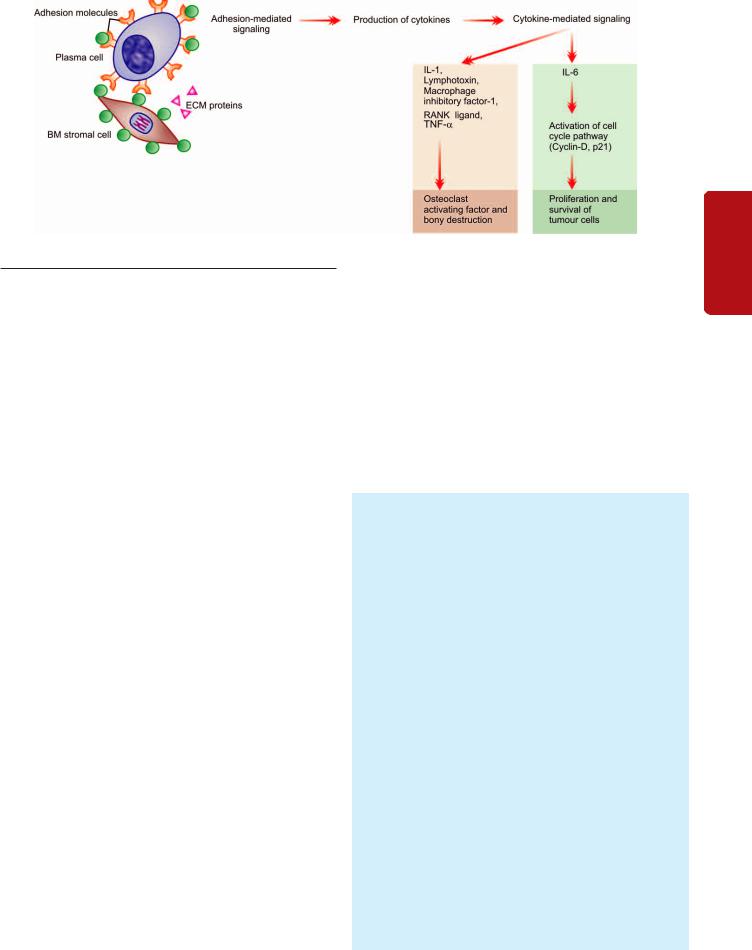
Figure 14.24 
 Schematic diagram showing molecular pathogenesis of multiple myeloma and its major manifestations.
Etiology and Pathogenesis
Myeloma is a monoclonal proliferation of B-cells. The etiology of myeloma remains unknown. However, following factors and abnormalities have been implicated:
1.Radiation exposure. Large dose exposure to radiation with a long latent period has been seen in myeloma. For instance, survivors of nuclear attack in World War-II developed myeloma about 20 years later.
2.Epidemiologic factors. Myeloma has higher incidence in blacks. Occupational exposure to petroleum products has been associated with higher incidence. Certain occupations such as farmers, wood workers and leather workers are more prone.
3.Karyotypic abnormalities. Several chromosomal alterations have been observed in cases of myeloma, which include following translocations and deletions:
i)Translocations t(11;14)(q13;q32) and t(4;14)(p16;q32).
ii)Deletion of 13q.
4.Oncogenes-antioncogenes. Overexpresion and mutations in following genes have been noted in proliferation of tumour cells in myeloma:
i)Overexpression of MYC and RAS growth promoting oncogenes in some cases.
ii)Mutation in p53 and RB growth-suppressing antioncogene in some cases.
Based on above, the molecular pathogenesis of multiple myeloma and its major manifestations can be explained as under and is schematically illustrated in Fig. 14.24:
1.Cell-surface adhesion molecules bind myeloma cells to bone marrow stromal cells and extracellular matrix proteins.
2.This binding triggers adhesion-mediated signaling and mediates production of several cytokines by fibroblasts and macrophages of the marrow. These include: IL-6, VEGF, TGFβ, TNF-α IL-1, lymphotoxin, macrophage inhibitory factor-1α (MIP-1α) and receptor activator of nuclear factor-κB (RANK) ligand.
3.Adhesion-mediated signaling affects the cell cycle via cyclin-D and p21 causing abnormal production of myeloma
(M) proteins.
4.IL-6 cytokine plays a central role in cytokine-mediated signaling and causes proliferation as well as cell survival of tumour cells via its antiapoptotic effects on tumour cells.
5.Certain cytokines produced by myeloma cells bring about bony destruction by acting as osteoclast-activating factor (OAF).
These are: IL-1, lymphotoxin, VEGF, macrophage inhibitory factor-1α (MIP-1α), receptor activator of NF-κB ligand, and tumour necrosis factor (TNF).
6.Other effects of adhesion-mediated and cytokine-mediated signaling are development of drug resistance and migration of tumour cells in the bone marrow milieu.
MORPHOLOGIC FEATURES. Myeloma affects principally the bone marrow though during the course of the disease other organs are also involved. Therefore, the pathologic findings are described below under two headings—osseous (bone marrow) lesions and extraosseous
lesions.
A. OSSEOUS (BONE MARROW) LESIONS. In more than 95% of cases, multiple myeloma begins in the bone marrow. In majority of cases, the disease involves multiple bones. By the time the diagnosis is made, most of the bone marrow is involved. Most commonly affected bones are those with red marrow i.e. skull, spine, ribs and pelvis, but later long bones of the limbs are also involved (Fig. 14.25). The lesions begin in the medullary cavity, erode the cancellous bone and ultimately cause destruction of the bony cortex. Radiographically, these lesions appear as punched out, rounded, 1-2 cm sized defects in the affected bone.
Grossly, the normal bone marrow is replaced by soft, gelatinous, reddish-grey tumours. The affected bone usually shows focal or diffuse osteoporosis.
Microscopically, the diagnosis of multiple myeloma can be usually established by examining bone marrow aspiration from an area of bony rarefaction. However, if the bone marrow aspiration yields dry tap or negative results, biopsy of radiologically abnormal or tender site is usually diagnostic. The following features characterise a case of myeloma:
381
Tissues Lymphoreticular and Leucocytes of Disorders 14 CHAPTER
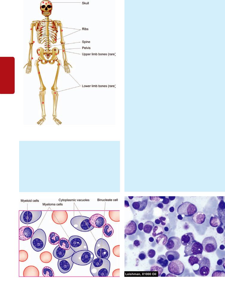
382
Tissues Lymphoreticular and Haematology II SECTION
Figure 14.25 
 The major sites of lesions in multiple myeloma.
i)Cellularity: There is usually hypercellularity of the bone marrow.
ii)Myeloma cells: Myeloma cells constitute >10% of the marrow cellularity. These cells may form clumps or sheets, or may be scattered among the normal haematopoietic cells. Myeloma cells may vary in size from small, differentiated cells resembling normal plasma cells to large, immature and undifferentiated cells. Binucleate and multinucleate cells are sometimes present. The nucleus of myeloma cell is commonly eccentric similar to plasma
cells but usually lacks the cart-wheel chromatin pattern seen in classical plasma cells. Nucleoli are frequently present. The cytoplasm of these cells is abundant and basophilic with perinuclear halo, vacuolisation and contains Russell bodies consisting of hyaline globules composed of synthesised immunoglobulin (Fig. 14.26).
In addition to neoplastic proliferation of plasma cells in multiple myeloma, reactive plasmacytosis in the bone marrow can occur in some other disorders; these include: aplastic anaemia, rheumatoid arthritis, SLE, cirrhosis of liver, metastatic cancer and chronic inflammation and infections such as tuberculosis. However, in all these conditions the plasma cells are mature and they do not exceed 10% of the total marrow cells.
B. EXTRAOSSEOUS LESIONS. Late in the course of disease, lesions at several extraosseous sites become evident. Some of the commonly involved sites are as under:
1.Blood. Approximately 50% of patients with multiple myeloma have a few atypical plasma cells in the blood. Other changes in the blood in myeloma are the presence of anaemia (usually normocytic normochromic type), marked red cell rouleaux formation due to hyperviscosity of blood, and an elevated ESR.
2.Myeloma kidney. Renal involvement in myeloma called myeloma nephrosis occurs in many cases (Chapter 22). The main mechanism of myeloma kidney is by filtration of light chain proteins (Bence Jones proteins) which are precipitated in the distal convoluted tubules in combination with Tamm-Horsfall proteins as tubular casts. The casts may be surrounded by some multinucleate giant-cells and a few inflammatory cells.
3.Myeloma neuropathy. Infiltration of the nerve trunk roots by tumour cells produces nonspecific polyneuropathy. Pathologic fractures, particularly of the vertebrae, may occur causing neurologic complications.
4.Systemic amyloidosis. Systemic primary generalised amyloidosis (AL amyloid) may occur in 10% cases of multiple myeloma and involve multiple organs and systems.
Figure 14.26 
 Bone marrow aspirate in myeloma showing numerous plasma cells, many with abnormal features.
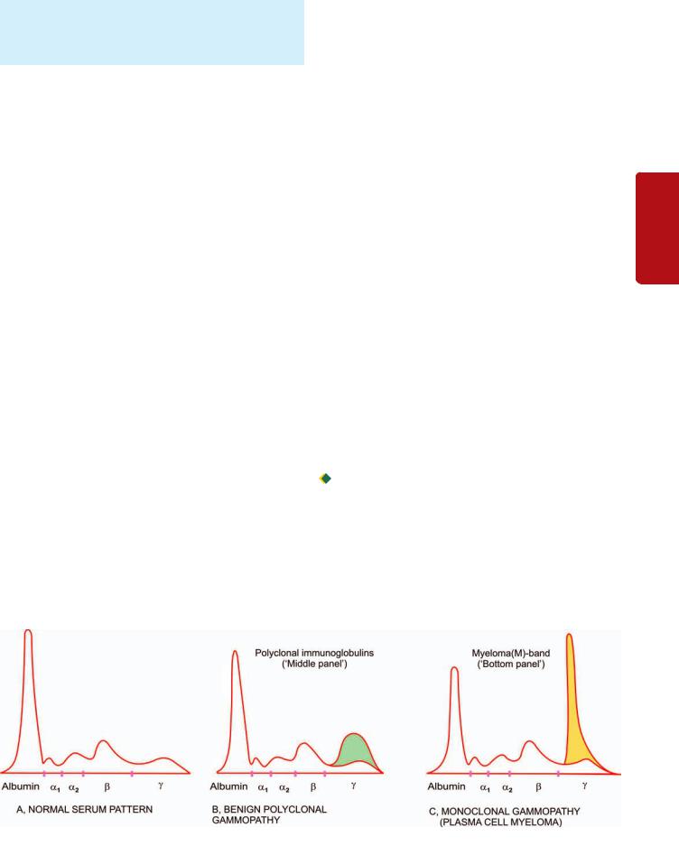
5. Liver, spleen involvement. Involvement of the liver and spleen by myeloma cells sufficient to cause hepatomegaly, and splenomegaly occurs in a small percentage of cases.
Clinical Features
The clinical manifestations of myeloma result from the effects of infiltration of the bones and other organs by neoplastic plasma cells and from immunoglobulin synthesis. The principal clinical features are as under:
1.Bone pain is the most common symptom. The pain usually involves the back and ribs. Pathological fractures may occur causing persistent localised pain. Bone pain results from the proliferation of tumour cells in the marrow and activation of osteoclasts which destroy the bones.
2.Susceptibility to infections is the next most common clinical feature. Particularly common are bacterial infections such as pneumonias and pyelonephritis. Increased susceptibility to infection is related mainly to hypogammaglobulinaemia, and partly to granulocyte dysfunction and neutropenia.
3.Renal failure occurs in about 25% of patients, while renal pathology occurs in 50% of cases. Causes of renal failure in myeloma are hypercalcaemia, glomerular deposits of amyloid, hyperuricaemia and infiltration of the kidney by myeloma cells.
4.Anaemia occurs in about 80% of patients of myeloma and is related to marrow replacement by the tumour cells (myelophthisis) and inhibition of haematopoiesis.
5.Bleeding tendencies may appear in some patients due to thrombocytopenia, deranged platelet function and interaction of the M component with coagulation factors.
6.Hyperviscosity syndrome owing to hyperglobulinaemia may produce headache, fatigue, visual disturbances and haemorrhages.
7.Neurologic symptoms occur in a minority of patients and are explained by hyperviscosity, cryoglobulins and amyloid deposits.
8.Biochemical abnormalities. These include the following:
i)hypercalcaemia due to destruction of bone;
ii)hyperuricaemia from necrosis of tumour mass and from uraemia related to renal failure; and
iii) increased β-2 microglobulins and other globulins in urine and serum.
9. POEMS syndrome is seen in about 1% cases of myeloma and includes simultaneous manifestations of polyneuropathy, organomegaly, endocrinopathy, multiple myeloma and skin changes.
Diagnosis
The diagnosis of myeloma is made by classic triad of features:
1.Marrow plasmacytosis of more than 10%
2.Radiologic evidence of lytic bony lesions
3.Demonstration of serum and/or urine M component. There is rise in the total serum protein concentration due
to paraproteinaemia but normal serum immunoglobulins (IgG, IgA and IgM) and albumin are depressed. Paraproteins are abnormal immunoglobulins or their parts circulating in plasma and excreted in urine. About two-third cases of myeloma excrete Bence Jones (light chain) proteins in the urine, consisting of either kappa (κ) or lambda (λ) light chains, along with presence of Bence Jones paraproteins in the serum. On serum electrophoresis, the paraprotein usually appears as a single narrow homogeneous M-band component, most commonly in the region of γ-globulin (Fig. 14.27). Most frequent paraprotein is IgG seen in about 50% cases of myeloma, IgA in 25%, and IgD in 1%, while about 20% patients have only light chains in serum and urine (light chain myeloma). Non-secretory myeloma is absence of M-band on serum and/or electrophoresis but presence of other two features out of triad listed above. Though the commonest cause of paraproteinaemias is multiple myeloma, certain other conditions which may produce serum paraproteins need to be distinguished. These are as under:
 Monoclonal gammopathy of undetermined significance (MGUS)

 Waldenström’s macroglobulinaemia

 Benign monoclonal gammopathy

 B-cell lymphomas

 CLL
CLL

 Light chain disease

 Heavy chain disease
 Cryoglobulinaemia.
383
Tissues Lymphoreticular and Leucocytes of Disorders 14 CHAPTER
Figure 14.27 
 Serum electrophoresis showing normal serum pattern (A), as contrasted with that in benign polyclonal gammopathy (B) and in monoclonal gammopathy (C), typical of plasma cell myeloma.

384 Treatment and Prognosis
|
Treatment of multiple myeloma consists of systemic |
|
|
chemotherapy in the form of alkylating agents and |
|
|
symptomatic supportive care. Autologous stem cell |
|
|
transplantation and interferon-therapy are the other modern |
|
|
treatment modalities offered. Poor prognostic predictors for |
|
|
lower survival are: secretion of λ-light chain than those |
|
|
secreting κ-light chain, larger number of cytogenetic |
|
|
abnormalities, and increased β-2 microglobulin level. The |
|
|
median survival is 2 years after the diagnosis is made. The |
|
|
terminal phase is marked by the development of |
|
|
pancytopenia, severe anaemia and sepsis. |
|
SECTION |
LOCALISED PLASMACYTOMA |
|
Two variants of myeloma which do not fulfil the criteria of |
||
classical triad are the localised from of solitary bone |
||
plasmacytoma and extramedullary plasmacytoma. Both these are |
||
II |
associated with M component in about a third of cases and |
|
|
occur in young individuals. Solitary bone plasmacytoma is |
|
Haematology |
a lytic bony lesion without marrow plasmacytosis. |
|
Extramedullary plasmacytoma involves most commonly the |
||
submucosal lymphoid tissue of nasopharynx or paranasal |
||
|
||
|
sinuses. Both variants have better prognosis than the classic |
|
|
multiple myeloma. Plasma cell granuloma, on the other hand, |
|
|
is an inflammatory condition having admixture of other |
|
|
inflammatory cells with mature plasma cells, which can be |
|
and |
easily distinguished by a discernible observer. |
|
WALDENSTRÖM’S MACROGLOBULINAEMIA |
||
Lymphoreticular |
||
Waldenström’s macroglobulinaemia is an uncommon |
||
|
||
|
malignant proliferation of monoclonal B lymphocytes which |
|
|
secrete IgM paraproteins called macroglobulins as they have |
|
|
high molecular weight. The condition is more common in |
|
|
men over 50 years of age and behaves clinically like a slowly |
|
|
progressive lymphoma. |
|
|
The exact etiology is not known but a possible relation- |
|
Tissues |
ship of IgM macroglobulin with myelin-associated |
|
glycoprotein which is lost in degenerating diseases has been |
||
|
||
|
suggested. The clinical evidence in favour is the appearance |
|
|
of peripheral neuropathy before the occurrence of macro- |
|
|
globulinaemia in some patients. |
|
|
|
|
|
MORPHOLOGIC FEATURES. Pathologically, the disease |
|
|
can be regarded as the hybrid between myeloma and small |
|
|
lymphocytic lymphoma. |
|
|
Like myeloma, the disease involves the bone marrow, |
|
|
but unlike myeloma it usually does not cause extensive |
|
|
bony lesions or hypercalcaemia. The bone marrow shows |
|
|
pleomorphic infiltration by lymphocytes, plasma cells, |
|
|
lymphocytoid plasma cells, mast cells and histiocytes. Like |
|
|
in myeloma, serum M component is present. |
|
|
Unlike myeloma and more like small lymphocytic |
|
|
lymphoma, enlargement of lymph nodes, spleen and liver |
|
|
due to infiltration by similar type of cells is present more |
|
|
frequently. |
|
|
|
|
|
CLINICAL FEATURES. The clinical features of the disease |
|
|
are due to both infiltration by the disease and paraproteins |
|
|
in the blood. |
1.Hyperviscosity syndrome is the major clinical manifestation. It results in visual disturbances, weakness, fatiguability, weight loss and nervous system symptoms. Raynaud’s phenomenon may occur.
2.Moderate organomegaly in the form of lymphadenopathy, hepatomegaly and splenomegaly are frequently seen.
3.Anaemia due to bone marrow failure may be present.
4.Bleeding tendencies may occur due to interaction of macroglobulins with platelets and coagulation factors.
DIAGNOSIS. Unlike myeloma, there are no characteristic radiologic findings. The diagnosis rests on laboratory data as under:
1.Pleomorphic bone marrow infiltration
2.Raised total serum protein concentration
3.Raised serum monoclonal M component which is due to IgM paraprotein
4.Elevated ESR
5.Normocytic normochromic anaemia.
The management of the patients is similar to that of myeloma. Patients respond to chemotherapy with a median survival of 3-5 years.
HEAVY CHAIN DISEASES
Heavy chain diseases are rare malignant proliferations of B- cells accompanied by monoclonal excess of one of the heavy chains. Depending upon the type of excessive heavy chain, three types—γ , α and μ, of heavy chain diseases are distinguished:
GAMMA HEAVY CHAIN DISEASE. Also called Franklin’s disease, it is characterised by excess of mostly γ1-paraprotein, both in the serum and urine and is demonstrated as M component. Clinically, the condition may develop at any age and present with lymphadenopathy, splenomegaly, hepatomegaly, involvement of pharyngeal lymphoid tissue (Waldeyer’s ring) and fever. Patients have rapidly downhill course due to severe and fatal infection.
ALPHA HEAVY CHAIN DISEASE. This is the commonest of heavy chain diseases characterised by α-heavy chains in the plasma which are difficult to demonstrate in electrophoresis due to rapid polymerisation. The patients present with bowel symptoms such as chronic diarrhoea, malabsorption and weight loss and may have enlargement of abdominal lymph nodes. Chemotherapy may induce long-term remissions.
MU HEAVY CHAIN DISEASE. This is the rarest heavy chain disease. The neoplastic B-cells produce μ heavy chains which donot appear in the urine but κ light chains which are also produced appear in the urine. Another feature that distinguishes this type of heavy chain disease from the others is the presence of vacuoles in the malignant B lymphocytes. The course and prognosis are like those of leukaemia or lymphoma.
MONOCLONAL GAMMOPATHY OF UNDETERMINED SIGNIFICANCE (MGUS)
A relatively recently desribed entity, monoclonal gammopathy of undetermined significance (MGUS), is increasingly
diagnosed in asymptomatic healthy aging population—1% at 50 years of age and in 10% individuals older than 75 years. This makes it the most common form of plasma cell dyscrasia. The defining criteria for MGUS are as under:
i)M-protein in serum <3 gm/dl
ii)Marrow plasmacytosis <10%
iii)No evidence of other B-cell proliferative disorder
iv)Absence of myeloma-related end-organ tissue damage (i.e. absence of lytic bone lesions, high calcium level, anaemia).
The condition needs to be cautiously distinguished from myeloma and long term follow-up is required. In fact, MGUS has been considered as a forerunner of multiple myeloma in many studies because MGUS has been found to have the same chromosomal abnormalities as in myeloma.
LYMPH NODE METASTATIC TUMOURS
The regional lymph nodes draining the site of a primary malignant tumour are commonly enlarged. This enlargement
may be due to benign reactive hyperplasia or metastatic tumour deposits.
1.Benign reactive hyperplasia, as already discussed (page 343), is due to immunologic reaction by the lymph node in response to tumour-associated antigens. It may be expressed as sinus histiocytosis, follicular hyperplasia, plasmacytosis and occasionally may show non-caseating granulomas.
2.Metastatic deposits in regional lymph nodes occur most commonly from carcinomas and malignant melanoma. Sarcomas often disseminate via haematogenous route but uncommonly may metastasise to the regional lymph nodes. Metastatic tumour cells from the primary malignant tumour are drained via lymphatics into the subcapsular sinuses initially but subsequently the lymph node stroma is also invaded. The pushing margins of advancing metastatic tumour in stroma of lymph node is characteristically well demarcated. Areas of necrosis are frequent in metastatic carcinomas (Fig. 14.28).
The morphologic features of primary malignant tumour are recapitulated in metastatic tumour in lymph nodes.
HISTIOCYTIC NEOPLASMS:
LANGERHANS’ CELL HISTIOCYTOSIS
Langerhans’ cell histiocytosis (LCH) is a group of rare malignant proliferations of dendritic cells or macrophages and includes three clinicopathologically related conditions ocurring in children: eosinophilic granuloma, Hand-Schüller- Christian disease and Letterer-Siwe syndrome. Earlier, this group was referred to as histiocytosis-X but now following facts about this group are known:
 Firstly, histiocytosis-X are not proliferations of unknown origin (X-for unknown) but proliferating cells are actually Langerhans’ cells of marrow origin. Langerhans’ cells are normally present mainly in the epidermis of the skin but also in some other organs.

 Secondly, the three conditions included under histio- cytosis-X are actually different expression of the same basic disorder. This concept has emerged from 2 features:
Figure 14.28 |
Metastatic carcinomatous deposits in the matted mass |
of lymph nodes. There are areas of necrosis in the circumscribed nodular areas.
i)Demonstration of common antigens on these cells by immunohistochemical stains for S-100 protein, CD1a and HLADR.
ii)Electron microscopic demonstration of histiocytosis-X bodies or Birbeck granules in the cytoplasm. These are rod-shaped structures having dilated tennis-racket like terminal end. Their function is not known but they arise from receptormediated endocytosis of langerin found in human epidermal cells, a protein involved in Birbeck granule biosynthesis.
The three disorders included in the group are briefly considered below.
Eosinophilic Granuloma
Unifocal eosinophilic granuloma is more common (60%) than the multifocal variety which is often a component of Hand- Schüller-Christian disease (described below). Most of the patients are children and young adults, predominantly males. The condition commonly presents as a solitary osteolytic lesion in the femur, skull, vertebrae, ribs and pelvis. The diagnosis requires biopsy of the lytic bone lesion.
Microscopically, the lesion consists largely of closelypacked aggregates of macrophages admixed with variable number of eosinophils (Fig. 14.29). The macrophages contain droplets of fat or a few granules of brown pigment indicative of phagocytic activity. A few multinucleate macrophages may also be seen. The cytoplasm of these macrophages may contain rod-shaped inclusions called histiocytosis-X bodies or Birbeck granules, best seen by electron microscopy.
Clinically, unifocal eosinophilic granuloma is a benign disorder. The bony lesion remains asymptomatic until the erosion of the bone causes pain or fracture. Spontaneous fibrosis or healing may occur in some cases, while others may require curettage or radiotherapy.
Hand-Schüller-Christian Disease
A triad of features consisting of multifocal bony defects, diabetes insipidus and exophthalmos is termed Hand-Schüller-Christian disease. The disease develops in children under 5 years of
385
Tissues Lymphoreticular and Leucocytes of Disorders 14 CHAPTER
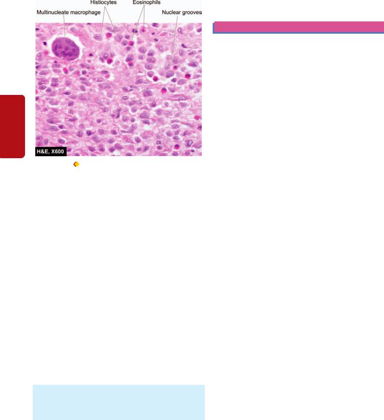
386
II SECTION |
|
|
|
Haematology |
Figure 14.29 |
Langerhans cell histiocytosis—eosinophilic |
|
granuloma. Bone biopsy shows presence of infiltrate by collections of |
|||
|
|||
|
histiocytes having vesicular nuclei admixed with eosinophils. |
||
|
age. The multifocal lytic bony lesions may develop at any |
||
|
site. Orbital lesion causes exophthalmos, while involvement |
||
|
of the hypothalamus causes diabetes insipidus. Multiple |
||
and |
spherical lesions in the lungs are frequently present. Half |
||
the patients have involvement of the liver, spleen and lymph |
|||
|
|||
Lymphoreticular |
nodes. |
|
|
|
|||
unifocal eosinophilic granuloma. The lesions may resolve |
|||
|
Microscopically, the lesions are indistinguishable from |
||
|
those of unifocal eosinophilic granuloma. |
||
|
Clinically, the affected children frequently have fever, |
||
|
skin lesions, recurrent pneumonitis and other infections. |
||
|
Though the condition is benign, it is more disabling than the |
||
Tissues |
spontaneously or may require chemotherapy or radiation. |
||
Letterer-Siwe Disease |
|||
|
|||
Letterer-Siwe disease is an acute disseminated form of LCH occurring in infants and children under 2 years of age. The disease is characterised by hepatosplenomegaly, lymphadenopathy, thrombocytopenia, anaemia and leucopenia. There is generalised hyperplasia of tissue macrophages in various organs.
Microscopically, the involved organs contain aggregates of macrophages which are pleomorphic and show nuclear atypia. The cytoplasm of these cells contains vacuoles and
rod-shaped histiocytosis-X bodies.
Clinically, the child has acute symptoms of fever, skin rash, loss of weight, anaemia, bleeding disorders and enlargement of lymph nodes, liver and spleen. Cystic bony lesions may be apparent in the skull, pelvis and long bones. Intense chemotherapy helps to control Letterer-Siwe disease but intercurrent infections result in fatal outcome in many
cases. The condition is currently regarded as an unusual form of malignant lymphoma.
SPLEEN
NORMAL STRUCTURE
The spleen is the largest lymphoid organ of the body. Under normal conditions, the average weight of the spleen is about 150 gm in the adult. Normally, the organ lies well protected by the 9th, 10th and 11th ribs in the upper left quadrant. The surface of the spleen is covered by a layer of peritoneum underneath which the organ is ensheathed by a thin capsule. From the capsule extend connective tissue trabeculae into the pulp of the organ and serve as supportive network. Blood enters the spleen by the splenic artery which divides into branches that penetrate the spleen via trabeculae. From the trabeculae arise small branches called central arterioles. Blood in the central arterioles empties partly into splenic venules and from there into splenic vein, but largely into vascular sinuses of the red pulp and thence into the splenic venous system.
Grossly, the spleen consists of homogeneous, soft, dark red mass called the red pulp and long oval grey-white nodules called the white pulp (malpighian bodies).
Microscopically, the red pulp consists of a network of thinwalled venous sinuses and adjacent blood spaces. The blood spaces contain blood cells, lymphocytes and macrophages and appear to be arranged in cords called splenic cords or cords of Billroth. The white pulp is made up of lymphocytes surrounding an eccentrically placed arteriole. The periarteriolar lymphocytes are mainly T-cells, while at other places the lymphocytes have a germinal centre composed principally of B-cells surrounded by densely packed lymphocytes.
The spleen is a lymphoreticular organ that performs at least the following four functions:
1. Like other lymphoid tissues, it is an organ of the immune system where B and T lymphocytes multiply and help in
immune responses.
2.The spleen plays an active role in sequestering and removing normal and abnormal blood cells.
3.The vasculature of the spleen plays a role in regulating
portal blood flow.
4. Under pathologic conditions, the spleen may become the
site of extramedullary haematopoiesis.
The spleen is rarely the primary site of disease. Being the largest lymphoreticular organ, it is involved secondarily in a wide variety of systemic disorders which manifest most commonly as splenic enlargement (splenomegaly) described below. A few other systemic involvements such as splenic infarcts and chronic venous congestion (CVC) of spleen have already been considered in Chapter 5 of General Pathology.
SPLENOMEGALY
Enlargement of the spleen termed splenomegaly, occurs in a wide variety of disorders which increase the cellularity and vascularity of the organ. Many of the causes are exaggerated

forms of normal splenic function. Splenic enlargement may occur as a result of one of the following pathophysiologic mechanisms:
I. Infections
II. Disordered immunoregulation III. Altered splenic blood flow
IV. Lymphohaematogenous malignancies V. Diseases with abnormal erythrocytes VI. Storage diseases
VII. Miscellaneous causes.
Based on these mechanisms, an abbreviated list of causes of splenomegaly is given in Table 14.13. Most of these conditions have been discussed elsewhere.
The degree of splenomegaly varies with the disease entity:
 Mild enlargement (upto 5 cm) occurs in CVC of spleen in CHF, acute malaria, typhoid fever, bacterial endocarditis, SLE, rheumatoid arthritis and thalassaemia minor.

 Moderate enlargement (upto umbilicus) occurs in hepatitis, cirrhosis, lymphomas, infectious mononucleosis, haemolytic anaemia, splenic abscesses and amyloidosis.

 Massive enlargement (below umbilicus) occurs in CML, myeloid metaplasia with myelofibrosis, storage diseases, thalassaemia major, chronic malaria, leishmaniasis and portal vein obstruction.
Mild to moderate splenomegaly is usually symptomless, while a massively enlarged spleen may cause dragging sensation in the left hypochondrium. Spleen becomes palpable only when it is enlarged.
Grossly, an enlarged spleen is heavy and firm. The capsule is tense and thickened. The sectioned surface of the organ is firm with prominent trabeculae.
Microscopically, there is dilatation of sinusoids with prominence of splenic cords. The white pulp is atrophic while the trabeculae are thickened. Long-standing congestion may produce haemorrhages and Gamna-Gandy bodies resulting in fibrocongestive splenomegaly, also called
Banti’s spleen ( see Figs. 5.13 and 5.14)
HYPERSPLENISM
The term hypersplenism is used for conditions which cause excessive removal of erythrocytes, granulocytes or platelets from the circulation. The mechanism for excessive removal could be due to increased sequestration of cells in the spleen by altered splenic blood flow or by production of antibodies against respective blood cells. The criteria for hypersplenism are as under:
1.Splenomegaly.
2.Splenic destruction of one or more of the cell types in the peripheral blood causing anaemia, leucopenia, thrombocytopenia, or pancytopenia.
3.Bone marrow cellularity is normal or hyperplastic.
4.Splenectomy is followed by improvement in the severity of blood cytopenia.
TABLE 14.13: Causes of Splenomegaly.
I. INFECTIONS
1.Malaria
2.Leishmaniasis
3.Typhoid
4.Infectious mononucleosis
5.Bacterial septicaemia
6.Bacterial endocarditis
7.Tuberculosis
8.Syphilis
9.Viral hepatitis
10.AIDS
II.DISORDERS OF IMMUNOREGULATION
1.Rheumatoid arthritis
2.SLE
3.Immune haemolytic anaemias
4.Immune thrombocytopenias
5.Immune neutropenias
III.ALTERED SPLENIC BLOOD FLOW
1.Cirrhosis of liver
2.Portal vein obstruction
3.Splenic vein obstruction
4.Congestive heart failure
IV. LYMPHO-HAEMATOGENOUS MALIGNANCIES
1.Hodgkin’s disease
2.Non-Hodgkin’s lymphomas
3.Multiple myeloma
4.Leukaemias
5.Myeloproliferative disorders (e.g. CML, polycythaemia vera, myeloid metaplasia with myelofibrosis)
V. DISEASES WITH ABNORMAL ERYTHROCYTES
1.Thalassaemias
2.Spherocytosis
3.Sickle cell disease
4.Ovalocytosis
VI. STORAGE DISEASES
1.Gaucher’s disease
2.Niemann-Pick’s disease
VII. MISCELLANEOUS
1.Amyloidosis
2.Primary and metastatic splenic tumours
3.Idiopathic splenomegaly
EFFECTS OF SPLENECTOMY
In view of the prominent role of normal spleen in sequestration of blood cells, splenectomy in a normal individual is followed by significant haematologic alterations. Induction of similar haematologic effects is made use in the treatment of certain pathologic conditions. For example, in autoimmune haemolytic anaemia or thrombocytopenia, the respective blood cell counts are increased following splenectomy. The blood changes following splenectomy are as under:
1. Red cells: There is appearance of target cells in the blood film. Howell-Jolly bodies are present in the red cells as they are no longer cleared by the spleen. Osmotic fragility test shows increased resistance to haemolysis. There may be appearance of normoblasts.
387
Tissues Lymphoreticular and Leucocytes of Disorders 14 CHAPTER

3882. White cells: There is leucocytosis reaching its peak in 1-
2days after splenectomy. There is shift-to-left of the myeloid cells with appearance of some myelocytes.
3.Platelets: Within hours after splenectomy, there is rise in platelet count upto 3-4 times normal.
|
SPLENIC RUPTURE |
||
|
The most common cause of splenic rupture or laceration is |
||
|
blunt trauma. The trauma may be direct or indirect. Non- |
||
|
traumatic or spontaneous rupture occurs in an enlarged |
||
|
spleen but almost never in a normal spleen. In acute |
||
SECTION |
infections, the spleen can enlarge rapidly to 2 to 3 times its |
||
normal size causing acute splenic enlargement termed acute |
|||
splenic tumour e.g. in pneumonias, septicaemia, acute |
|||
endocarditis etc. Some of the other common causes of |
|||
spontaneous splenic rupture are splenomegaly due to chronic |
|||
malaria, infectious mononucleosis, typhoid fever, splenic |
|||
II |
abscess, thalassaemia and leukaemias. |
||
|
|
Rupture of spleen is an acute surgical emergency due to |
|
Haematology |
rapid blood loss and haemoperitoneum. Sometimes |
||
only notable benign tumours are haemangiomas and |
|||
|
fragments of splenic tissue are autotransplanted within the |
||
|
peritoneal cavity and grow into tiny spleens there (splenosis). |
||
|
TUMOURS |
||
|
|
Primary tumours of the spleen are extremely rare. The |
|
and |
lymphangioma, while examples of primary malignant |
||
neoplasms of haematopoietic system i.e. Hodgkin’s disease |
|||
Lymphoreticular |
and non-Hodgkin’s lymphomas. Non-haematopoietic |
||
tumours of the spleen such as angiosarcoma are rare. |
|||
|
|||
|
|
Secondary tumours occur late in the course of disease |
|
|
and represent haematogenous dissemination of the |
||
|
malignant tumour. Splenic metastases appear as multiple |
||
|
nodules. The most frequent primary sites include: lung, |
||
|
breast, prostate, colon and stomach. Rarely, direct extension |
||
Tissues |
from an adjacent malignant neoplasm may occur. |
||
|
|
||
|
THYMUS |
||
|
|||
|
|
||
|
NORMAL STRUCTURE |
||
|
The thymus gland is a complex lymphoreticular organ lying |
||
|
buried within the mediastinum. At birth, the gland weighs |
||
|
10-35 gm and grows in size upto puberty, following which |
||
|
there is progressive involution in the elderly. In the adult, |
||
|
thymus weighs 5-10 gm. |
||
|
|
The gland consists of right and left encapsulated lobes, |
|
|
joined together by fibrous connective tissue. Connective |
||
|
tissue septa pass inwards from the capsule and subdivide |
||
|
the lobe into large number of lobules. The histologic structure |
||
|
of the lobule shows outer cortex and inner medulla. Both cortex |
||
|
and medulla contain two types of cells: epithelial cells and |
||
|
lymphocytes (thymocytes). |
||
|
|
Epithelial cells are similar throughout the thymus gland. |
|
|
These cells have elongated cytoplasmic processes forming |
||
|
network in which thymocytes and macrophages are found. |
||
Hassall’s corpuscles are distinctive structures within the medulla composed of onion skin-like concentrically arranged epithelial cells having central area of keratinisation.
 Thymocytes are predominantly present in the cortex. These cells include immature T lymphocytes in the cortex and mature T lymphocytes in the medulla. Well-developed B-cell lymphoid follicles with germinal centres are rare in thymus gland.
The main function of the thymus is in the cell-mediated immunity by T-cells and by secretion of thymic hormones
such as thymopoietin and thymosin-α1.
Thymic lesions are associated with diverse conditions which may be immunologic, haematologic or neoplastic. These can be broadly categorised into thymic hypoplasia and agenesis, thymic hyperplasia and thymoma, while thymic involvement in myasthenia gravis is discussed in Chapter 28.
THYMIC HYPOPLASIA AND AGENESIS
Thymic hypoplasia and agenesis are acquired and congenital disorders respectively in which the gland is either unusually small or absent. These conditions are various types of hereditary (primary) immunodeficiency diseases such as DiGeorge’s syndrome, severe combined immunodeficiency and reticular dysgenesis. Acquired hypoplasia occurs as an ageing phenomenon or may occur in the young due to severe stress, malnutrition, irradiation, therapy with cytotoxic drugs and glucocorticoids.
THYMIC HYPERPLASIA
Enlargement of the thymus or failure to involute produces thymic hyperplasia. Hyperplasia is usually associated with appearance of lymphoid follicles in the medulla of the thymus and is called thymic follicular hyperplasia. Most common cause of follicular hyperplasia of the thymus is myasthenia gravis. Less common causes are: Addison’s disease, Graves’ disease, rheumatoid arthritis, SLE, scleroderma and cirrhosis of liver.
THYMOMA
Most common primary tumour present in the anterosuperior mediastinum is thymoma. Although thymus is a lymphoepithelial organ, the term thymoma is used for the tumour of epithelial origin. Most of the patients are adults. In about half the cases, thymoma remains asymptomatic and is accidentally discovered in X-rays. Other patients have associated conditions like myasthenia gravis or local symptoms such as cough, dyspnoea and chest pain.
MORPHOLOGIC FEATURES. Grossly, the tumour is spherical, measuring 5-10 cm in diameter with an average weight of 150 gm. Sectioned surface is soft, yellowish, lobulated and may be either homogeneous or contain cysts due to the presence of haemorrhage and necrosis.
Microscopically, the tumour has a thick fibrous capsule from which extend collagenous septa into the tumour dividing it into lobules. The histology of lobule shows

various patterns. The tumour consists of neoplastic epithelial cells and variable number of non-neoplastic lymphocytes. Thymoma may be of following types:
Benign thymoma is more common. It consists of epithelial cells which are similar to the epithelial cells in the medulla of thymus and hence also called as medullary thymoma.
Malignant thymoma is less common and is further of 2 types:
Type 1 is cytologically benign looking but aggressive and invades the mediastinal structures locally.
Type 2 is also called thymic carcinoma and has cytologic |
|
389 |
|
|
|
features of cancer. Further subtypes of epithelial |
|
|
malignancy may be squamous cell type (most common) |
|
|
and lymphoepithelial type. |
|
|
|
|
|
Thymomas are known for their association with |
|
|
paraneoplastic syndrome. These include: myasthenia gravis |
|
|
(most common), hypogammaglobulinaemia, erythroid |
|
|
hypoplasia (pure red cell aplasia), peripheral T cell leukae- |
|
|
mia/lymphoma, multiple myeloma, other autoimmune |
|
|
disease associations and other systemic malignancies. |
|
|
CHAPTER 14
of Disorders and Leucocytes
Tissues Lymphoreticular

390
Section III
Chapter 15
SYSTEMIC PATHOLOGY
The Blood Vessels
and Lymphatics
Pathology Systemic III SECTION
The blood vessels are closed circuits for the transport of blood from the left heart to the metabolising cells, and then back to the right heart. The blood containing oxygen, nutrients and metabolites is routed through arteries, arterioles, capillaries, venules and veins. These blood vessels differ from each other in their structure and function.
ARTERIES
NORMAL STRUCTURE
Depending upon the calibre and certain histologic features, arteries are divided into 3 types: large (elastic) arteries, medium-sized (muscular) arteries and the smallest arterioles.
Histologically, all the arteries of the body have 3 layers in their walls: the tunica intima, the tunica media and the tunica adventitia. These layers progressively decrease with diminution in the size of the vessels.
1. Tunica intima. This is the inner coat of the artery. It is composed of the lining endothelium, subendothelial connective tissue and bounded externally by internal elastic lamina.

 Endothelium is a layer of flattened cells adjacent to the flowing blood. Narrow junctions exist between the adjoining endothelial cells through which certain materials pass. The integrity of the endothelial layer is of paramount importance in maintenance of vascular functions since damage to it is the most important event in the initiation of thrombus formation at the site.
 Subendothelial tissue consists of loose meshwork of connective tissue that includes myointimal cells, collagen, proteoglycans, elastin and matrix glycoproteins.

 Internal elastic lamina is a layer of elastic fibres having minute fenestrations.
2. Tunica media. Tunica media is the middle coat of the arterial wall, bounded internally by internal elastic lamina and externally by external elastic lamina. This layer is the thickest and consists mainly of smooth muscle cells and elastic fibres. The external elastic lamina consisting of condensed elastic tissue is less well defined than the internal
elastic lamina.
3. Tunica adventitia. The outer coat of arteries is the tunica adventitia. It consists of loose mesh of connective tissue and some elastic fibres that merge with the adjacent tissues. This layer is rich in lymphatics and autonomic nerve fibres.
The layers of arterial wall receive nutrition and oxygen from 2 sources:
1.Tunica intima and inner third of the media are nourished by direct diffusion from the blood present in the lumen.
2.Outer two-thirds of the media and the adventitia are supplied by vasa vasora (i.e. vessels of vessels), the nutrient vessels arising from the parent artery.
As the calibre of the artery decreases, the three layers progressively diminish. Thus, there are structural variations in the three types of arteries:

 Large, elastic arteries such as the aorta, innominate, common carotid, major pulmonary, and common iliac arteries have very high content of elastic tissue in the media and thick elastic laminae and hence the name.
 Medium-sized, muscular arteries are the branches of elastic arteries. All the three layers of arterial wall are thinner than in the elastic arteries. The internal elastic lamina appears as a single wavy line while the external elastic lamina is less prominent. The media primarily consists of smooth muscle cells and some elastic fibres (Fig. 15.1).

 Arterioles are the smallest branches with internal diameter 20-100 μm. Structurally, they consist of the three layers as in muscular arteries but are much thinner and cannot be distinguished. The arterioles consist of a layer of endothelial cells in the intima, one or two smooth muscle cells in the media and small amount of collagen and elastic tissue comprising the adventitia. The elastic laminae are virtually lost.
 Capillaries are about the size of an RBC (7-8 μm) and have a layer of endothelium but no media. Blood from capillaries returns to the heart via post-capillary venules and thence into venules and then veins.
In the following pages, diseases of arteries are discussed under 3 major headings: arteriosclerosis, arteritis (vasculitis) and aneurysms. This is followed by brief account of diseases of veins and lymphatics, while the vascular tumours are described at the end of the chapter.
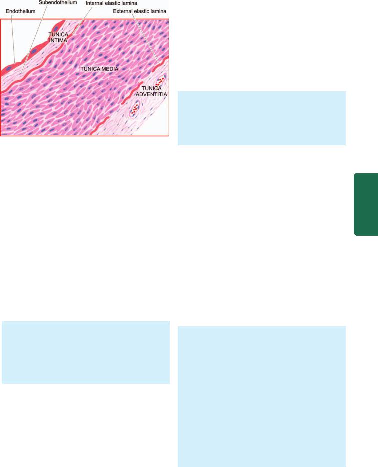
Figure 15.1 
 The structure of a medium-sized muscular artery.
ARTERIOSCLEROSIS
Arteriosclerosis is a general term used to include all conditions with thickening and hardening of the arterial walls. The following morphologic entities are included under arteriosclerosis:
I. Senile arteriosclerosis
II. Hypertensive arteriolosclerosis III. Mönckeberg’s arteriosclerosis
(Medial calcific sclerosis) IV. Atherosclerosis
The last-named, atherosclerosis, is the most common and most important form of arteriosclerosis; if not specified, the two terms are used interchangeably with each other.
SENILE ARTERIOSCLEROSIS
Senile arteriosclerosis is the thickening of media and intima of the arteries seen due to aging. The changes are nonselective and affect most of the arteries. These are possibly induced by stress and strain on vessel wall during life.
MORPHOLOGIC FEATURES. The changes are as under:
1.Fibroelastosis: The intima and media are thickened due to increase in elastic and collagen tissue.
2.Elastic reduplication: The internal elastic lamina is split or reduplicated so that two wavy layers are seen.
Eventually, the fibrotic changes result in age-related elevation of systolic blood pressure.
HYPERTENSIVE ARTERIOLOSCLEROSIS
Hypertension is the term used to describe an elevation in blood pressure. Pathology of 3 forms of hypertension— systemic, pulmonary and portal, is discussed in detail with diseases of the kidneys (Chapter 22), lungs (Chapter 17) and liver (Chapter 21) respectively.
Arteriolosclerosis is the term used to describe 3 morphologic forms of vascular disease affecting arterioles and small muscular arteries. These are: hyaline arteriolosclerosis,
hyperplastic arteriolosclerosis and necrotising arteriolitis. All the three types are common in hypertension but may occur due to other causes as well.
Hyaline Arteriolosclerosis
Hyaline sclerosis is a common arteriolar lesion that may be seen physiologically due to aging, or may occur pathologically in benign nephrosclerosis in hypertensives and as a part of microangiopathy in diabetics; the subject is discussed again in Chapter 22.
MORPHOLOGIC CHANGES. The visceral arterioles are particularly involved. The vascular walls are thickened and the lumina narrowed or even obliterated.
Microscopically, the thickened vessel wall shows structureless, eosinophilic, hyaline material in the intima and media (Fig. 15.2,A).
PATHOGENESIS. The exact pathogenesis is not known. However, the following hypotheses have been proposed:
i)The lesions result most probably from leakage of components of plasma across the vascular endothelium. This is substantiated by the demonstration of immunoglobulins, complement, fibrin and lipids in the lesions. The permeability of the vessel wall is increased, due to haemodyanamic stress in hypertension and metabolic stress in diabetes, so that these plasma components leak out and get deposited in the vessel wall.
ii)An alternate possibility is that the lesions may be due to immunologic reaction.
iii)Some have considered it to be normal aging process that is exaggerated in hypertension and diabetes mellitus.
Hyperplastic Arteriolosclerosis
The hyperplastic or proliferative type of arteriolosclerosis is a characteristic lesion of malignant hypertension; other causes include haemolytic-uraemic syndrome, scleroderma and toxaemia of pregnancy.
MORPHOLOGIC FEATURES. The morphologic changes affect mainly the intima, especially of the interlobular arteries in the kidneys. Three types of intimal thickening may occur.
i)Onion-skin lesion consists of loosely-placed concentric layers of hyperplastic intimal smooth muscle cells like the bulb of an onion. The basement membrane is also thickened and reduplicated (Fig. 15.2, B).
ii)Mucinous intimal thickening is the deposition of amorphous ground substance, probably proteoglycans, with scanty cells.
iii)Fibrous intimal thickening is less common and consists of bundles of collagen, elastic fibres and hyaline deposits in the intima.
Severe intimal sclerosis results in narrowed or obliterated lumen. With time, the lesions become more and more fibrotic.
391
Lymphatics and Vessels Blood The 15 CHAPTER
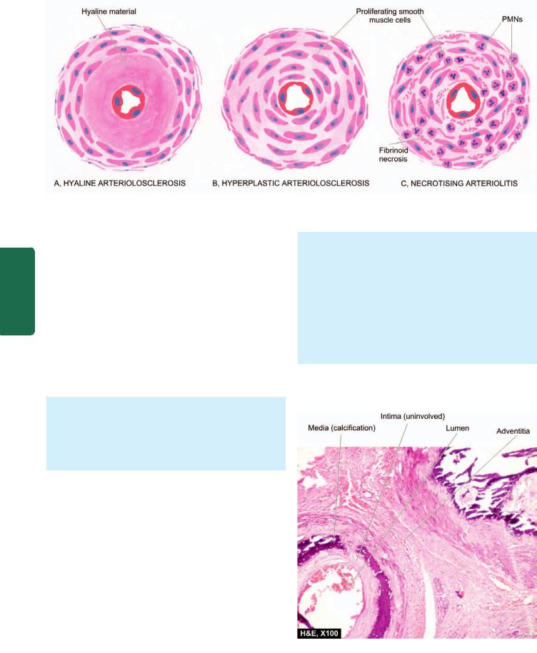
392
Figure 15.2 
 Diagrammatic representation of three forms of arteriolosclerosis, commonly seen in hypertension.
Pathology Systemic III SECTION
PATHOGENESIS. The pathogenesis of hyperplastic intimal thickening is unclear. Probably, the changes result following endothelial injury from systemic hypertension, hypoxia or immunologic damage leading to increased permeability. A healing reaction occurs in the form of proliferation of smooth muscle cells with fibrosis.
Necrotising Arteriolitis
In cases of severe hypertension and malignant hypertension, parts of small arteries and arterioles show changes of hyaline sclerosis and parts of these show necrosis, or necrosis may be superimposed on hyaline sclerosis. However, hyaline sclerosis may not be always present in the vessel wall.
MORPHOLOGIC FEATURES. Besides the changes of hyaline sclerosis, the changes of necrotising arteriolitis include fibrinoid necrosis of vessel wall, acute inflammatory infiltrate of neutrophils in the adventitia. Oedema and haemorrhages often surround the affected vessels (Fig. 15.2,C).
PATHOGENESIS. Since necrotising arteriolitis occurs in vessels in which there is sudden and great elevation of pressure, the changes are said to result from direct physical injury to the vessel wall.
MÖNCKEBERG’S ARTERIOSCLEROSIS (MEDIAL CALCIFIC SCLEROSIS)
Mönckeberg’s arteriosclerosis is calcification of the media of large and medium-sized muscular arteries, especially of the extremities and of the genital tract, in persons past the age of 50. The condition occurs as an age-related degenerative process, and therefore, an example of dystrophic calcification, and has little or no clinical significance. However, medial calcification also occurs in some pathological states like pseudoxanthoma elasticum and in idiopathic arterial calcification of infancy.
MORPHOLOGIC FEATURES. Medial calcification is often an incidental finding in X-rays of the affected sites having muscular arteries. The deposition of calcium salts in the media produces pipestem-like rigid tubes without causing narrowing of the lumen.
Microscopically, Mönckeberg’s arteriosclerosis is characterised by deposits of calcium salts in the media without associated inflammatory reaction while the intima and the adventitia are spared (Fig. 15.3). Often, coexistent changes of atherosclerosis are present altering the histologic appearance.
PATHOGENESIS. Pathogenesis of this condition is not known but it is considered as an age-related physiologic change due to prolonged effect of vasoconstriction.
Figure 15.3 
 Monckeberg’s arteriosclerosis (medial calcific sclerosis). There is calcification exclusively in the tunica media unassociated with any significant inflammation.

ATHEROSCLEROSIS
Definition
Atherosclerosis is a specific form of arteriosclerosis affecting primarily the intima of large and medium-sized muscular arteries and is characterised by fibrofatty plaques or atheromas. The term atherosclerosis is derived from athero- (meaning porridge) referring to the soft lipid-rich material in the centre of atheroma, and sclerosis (scarring) referring to connective tissue in the plaques. Atherosclerosis is the commonest and the most important of the arterial diseases. Though any large and medium-sized artery may be involved in atherosclerosis, the most commonly affected are the aorta, the coronary and the cerebral arterial systems. Therefore, the major clinical syndromes resulting from ischaemia due to atherosclerosis pertain to the heart (angina and myocardial infarcts or heart attacks), and the brain (transient cerebral ischaemia and cerebral infarcts or strokes); other sequelae are peripheral vascular disease, aneurysmal dilatation due to weakened arterial wall, chronic ischaemic heart disease, ischaemic encephalopathy and mesenteric arterial occlusion.
Etiology
Atherosclerosis is widely prevalent in industrialised countries. However, majority of the data on etiology are based on the animal experimental work and epidemiological studies. The incidences for atherosclerosis quoted in the literature are based on the major clinical syndromes produced by it, the most important interpretation being that death from myocardial infarction is related to underlying atherosclerosis. Cardiovascular disease, mostly related to atherosclerotic coronary heart disease or ischaemic heart disease (IHD) is the most common cause of premature death in the developed countries of the world. It is estimated that by the year 2020, cardiovascular disease, mainly atherosclerosis, will become the leading cause of total global disease burden.
Systematic large scale studies of investigations on living populations have revealed a number of risk factors which are associated with increased risk of developing clinical atherosclerosis. Often, they are acting in combination rather than singly. These risk factors are divided into two groups
(Table 15.1):
TABLE 15.1: Risk Factors in Atherosclerosis.
I. |
MAJOR RISK FACTORS |
II. |
EMERGING RISK FACTORS |
|
A) |
Modifiable |
1. |
Environmental influences |
|
|
1. |
Dyslipidaemia |
2. |
Obesity |
|
2. |
Hypertension |
3. |
Hormones:oestrogen defi- |
|
3. |
Diabetes mellitus |
|
ciency, oral contraceptives |
|
4. |
Smoking |
4. |
Physical inactivity |
B) |
Constitutional |
5. |
Stressful life |
|
|
1. |
Age |
6. |
Homocystinuria |
|
2. |
Sex |
7. |
Role of alcohol |
|
3. |
Genetic factors |
8. |
Prothrombotic factors |
|
4. |
Familial and racial factors |
9. |
Infections (C.pneumoniae, |
|
|
|
|
Herpesvirus, CMV) |
|
|
|
10. |
High CRP |
|
|
|
|
|
I. Major risk factors. These are further considered under 2 393 headings:
A)Major risk factors modifiable by life style and/or therapy: This includes major risk factors which can be controlled by modifying life style and/or by pharmacotherapy and includes: dyslipidaemias, hypertension, diabetes mellitus and smoking.
B)Constitutional risk factors: These are non-modifiable major risk factors that include: increasing age, male sex, genetic abnormalities, and familial and racial predisposition.
II. Non-traditional emerging risk factors. This includes a host of factors whose role in atherosclerosis is minimal, and in some cases, even uncertain.
Apparently, a combination of etiologic risk factors have
additive effect in producing the lesions of atherosclerosis. |
|
|
MAJOR RISK FACTORS MODIFIABLE BY LIFE STYLE |
|
|
AND/OR THERAPY |
|
|
There are four major risk factors in atherogenesis—lipid |
|
|
disorders, hypertension, cigarette smoking and diabetes |
|
|
mellitus. |
CHAPTER |
|
1. DYSLIPIDAEMIAS. Virchow in 19th century first |
||
|
||
identified cholesterol crystals in the atherosclerotic lesions. |
|
|
Since then, extensive information on lipoproteins and their |
|
|
role in atherosclerotic lesions has been gathered. |
|
|
Abnormalities in plasma lipoproteins have been firmly |
15 |
|
established as the most important major risk factor for |
||
atherosclerosis. It has been firmly established that hyper- |
|
|
cholesterolaemia has directly proportionate relationship with |
The |
|
atherosclerosis and IHD. The following evidences are cited |
||
|
||
in support of this: |
Blood |
|
i) The atherosclerotic plaques contain cholesterol and |
||
cholesterol esters, largely derived from the lipoproteins in |
||
the blood. |
Vessels |
|
ii) The lesions of atherosclerosis can be induced in |
||
|
||
experimental animals by feeding them with diet rich in |
|
|
cholesterol. |
and |
|
iii) Individuals with hypercholesterolaemia due to various |
||
|
||
causes such as in diabetes mellitus, myxoedema, nephrotic |
Lymphatics |
|
syndrome, von Gierke’s disease, xanthomatosis and familial |
||
|
||
hypercholesterolaemia have increased risk of developing |
|
|
atherosclerosis and IHD. |
|
|
iv) Populations having hypercholesterolaemia have higher |
|
|
mortality from IHD. Dietary regulation and administration |
|
|
of cholesterol-lowering drugs have beneficial effect on |
|
|
reducing the risk of IHD. |
|
|
The concentration of total cholesterol in the serum reflects |
|
|
the concentrations of different lipoproteins in the serum. The |
|
|
lipoproteins are divided into classes according to the density |
|
|
of solvent in which they remain suspended on centrifugation |
|
|
at high speed. The major classes of lipoprotein particles are |
|
|
chylomicrons, very-low density lipoproteins (VLDL), low-density |
|
|
lipoproteins (LDL), and high-density lipoproteins (HDL). Lipids |
|
|
are insoluble in blood and therefore are carried in circulation |
|
|
and across the cell membrane by carrier proteins called |
|
|
apoproteins. Apoprotein surrounds the lipid for carrying it, |
|

394 TABLE 15.2: Fractions of Lipoproteins in Serum.
|
Classes |
Sites of Synthesis |
Normal Serum Levels |
Role in Atherosclerosis |
|
|
|
|
|
1. |
HDL cholesterol |
Liver, intestine |
> 60 mg/dl |
Protective |
2. |
LDL cholesterol |
Liver |
< 130 mg/dl |
Maximum |
3. |
VLDL triglycerides |
Intestine, liver |
< 160 mg/dl |
Less marked |
4. |
Chylomicrons |
Liver, intestine, macrophage |
— |
Indirect |
|
|
|
|
|
Pathology Systemic III SECTION
different apoproteins being named by letter A, B, C, D etc while their subfractions are numbered serially.
The major fractions of lipoproteins tested in blood lipid profile and their varying effects on atherosclerosis and IHD are as under (Table 15.2):
i)Total cholesterol: Desirable normal serum level is 140-200 mg/dl, while levels of borderline high are considered between 200-240 mg/dl. An elevation of total serum cholesterol levels above 260 mg/dl in men and women between 30 and 50 years of age has three times higher risk of developing IHD as compared with people with total serum cholesterol levels within normal limits.
ii)Triglycerides: Normal serum level is below 160 mg/dl.
iii)Low-density lipoproteins (LDL) cholesterol: Normal optimal serum level is <130 mg/dl. LDL is richest in cholesterol and has the maximum association with atherosclerosis.
iv)Very-low-density lipoprotein (VLDL): VLDL carries much of the triglycerides and its blood levels therefore parallel with that of triglycerides; VLDL has less marked effect than LDL.
v)High-density lipoproteins (HDL) cholesterol: Normal desirable serum level is <60 mg/dl. HDL is protective (‘good cholesterol’) against atherosclerosis.
Many studies have demonstrated the harmful effect of diet containing larger quantities of saturated fats (e.g. in eggs, meat, milk, butter etc) and trans fats (i.e. unsaturated fats produced by artificial hydrogenation of polyunsaturated fats) which raise the plasma cholesterol level. This type of diet is consumed more often by the affluent societies who are at greater risk of developing atherosclerosis. On the contrary, a diet low in saturated fats and high in poly-unsaturated fats and having omega-3 fatty acids (e.g. in fish, fish oils etc) lowers the plasma cholesterol levels. Aside from lipid-rich diet, high intake of the total number of calories from carbohydrates, proteins, alcohol and sweets has adverse effects.
Besides above, familial hypercholesterolaemia, an autosomal codominant disorder, is characterised by elevated LDL cholesterol and normal triglycerides and occurrence of xanthomas and premature coronary artery disease. It occurs due to mutations in LDL receptor gene.
Currently, management of dyslipidaemia is directed at lowering LDL in particular and total cholesterol in general by use of statins, and for raising HDL by weight loss, exercise and use of nicotinic acid. Thus presently, preferred term for hyperlipidaemia is dyslipidaemia because one risky plasma lipoprotein (i.e. LDL) is elevated and needs to be brought
down, while the other good plasma lipoprotein (i.e. HDL) when low requires to be raised.
How hypercholesterolaemia and various classes of lipoproteins produce atherosclerosis is described under ‘pathogenesis’.
2.HYPERTENSION. Hypertension is a risk factor for all clinical manifestations of atherosclerosis. Hypertension doubles the risk of all forms of cardiovascular disease. It acts probably by mechanical injury to the arterial wall due to increased blood pressure. Elevation of systolic pressure of over 160 mmHg or a diastolic pressure of over 95 mmHg is associated with five times higher risk of developing IHD than in people with blood pressure within normal range (140/90 mmHg or less).
3.SMOKING. The extent and severity of atherosclerosis are much greater in smokers than in non-smokers. Cigarette smoking is associated with higher risk of atherosclerotic IHD and sudden cardiac death. Men who smoke a pack of cigarettes a day are 3-5 times more likely to die of IHD than non-smokers. The increased risk and severity of atherosclerosis in smokers is due to reduced level of HDL, deranged coagulation system and accumulation of carbon monoxide in the blood that produces carboxyhaemoglobin and eventually hypoxia in the arterial wall favouring atherosclerosis.
4.DIABETES MELLITUS. Clinical manifestations of atherosclerosis are far more common and develop at an early age in people with both type 1 and type 2 diabetes mellitus. In particular, association of type 2 diabetes mellitus characterised by metabolic (insulin resistance) syndrome and abnormal lipid profile termed ‘diabetic dyslipidaemia’ is common and heightens the risk of cardiovascular disease. The risk of developing IHD is doubled, tendency to develop cerebrovascular disease is high, and frequency to develop gangrene of foot is about 100 times increased. The causes of increased severity of atherosclerosis are complex and numerous which include endothelial dysfunction, increased aggregation of platelets, increased LDL and decreased HDL.
CONSTITUTIONAL RISK FACTORS
Age, sex and genetic influences do affect the appearance of lesions of atherosclerosis.
1. AGE. Atherosclerosis is an age-related disease. Though early lesions of atherosclerosis may be present in childhood, clinically significant lesions are found with increasing age. Fully-developed atheromatous plaques usually appear in the
4th decade and beyond. Evidence in support comes from the high death rate from IHD in this age group.
2.SEX. The incidence and severity of atherosclerosis are more in men than in women and the changes appear a decade earlier in men (>45 years) than in women (>55 years). The prevalence of atherosclerotic IHD is about three times higher in men in 4th decade than in women and the difference slowly declines with age but remains higher at all ages in men. The lower incidence of IHD in women, especially in premenopausal age, is probably due to high levels of oestrogen and high-density lipoproteins, both of which have anti-atherogenic influence.
3.GENETIC FACTORS. Genetic factors play a significant role in atherogenesis. Hereditary genetic derangements of lipoprotein metabolism predispose the individual to high blood lipid level and familial hypercholesterolaemia.
4.FAMILIAL AND RACIAL FACTORS: The familial predisposition to atherosclerosis may be related to other risk factors like diabetes, hypertension and hyperlipoproteinaemia. Racial differences too exist; Blacks have generally less severe atherosclerosis than Whites.
EMERGING RISK FACTORS
There are a number of nontraditional newly emerging risk factors for which the role in the etiology of atherosclerosis is yet not fully supported. These factors are as under:
1.Higher incidence of atherosclerosis in developed countries and low prevalence in underdeveloped countries, suggesting the role of environmental influences.
2.Obesity, if the person is overweight by 20% or more, is associated with increased risk.
3.Use of exogenous hormones (e.g. oral contraceptives) by women or endogenous oestrogen deficiency (e.g. in postmenopausal women) has been shown to have an increased risk of developing myocardial infarction or stroke.
4.Physical inactivity and lack of exercise are associated with the risk of developing atherosclerosis and its complications.
5.Stressful life style, termed as ‘type A’ behaviour pattern, characterised by aggressiveness, competitive drive, ambitiousness and a sense of urgency, is associated with enhanced risk of IHD compared with ‘type B’ behaviour of relaxed and happy-go-lucky type.
6.Patients with homocystinuria, an uncommon inborn error of metabolism, have been reported to have early atherosclerosis and coronay artery disease.
7.There are some reports which suggest that moderate consumption of alcohol has slightly beneficial effect by raising the level of HDL cholesterol.
8.Prothrombotic factors and elevated fibrinogen levels favour formation of thrombi which is the gravest complication of atherosclerosis.
9.Role of infections, particularly of Chlamydia pneumoniae and viruses such as herpesvirus and cytomegalovirus, has been found in coronary atherosclerotic lesions by causing inflammation. Possibly, infections may be acting in combination with some other factors.
10. Markers of inflammation such as elevated C reactive protein, an acute phase reactant, correlate with risk of developing atherosclerosis.
Pathogenesis
As stated above, atherosclerosis is not caused by a single etiologic factor but is a multifactorial process whose exact pathogenesis is still not known. Since the times of Virchow, a number of theories have been proposed.

 Insudation hypothesis. The concept hypothesised by Virchow in 1856 that atherosclerosis is a form of cellular proliferation of the intimal cells resulting from increased imbibing of lipids from the blood came to be called the ‘lipid theory’. Modified form of this theory is currently known as
‘response to injury hypothesis’ and is now-a-days the most widely accepted theory.

 Encrustation hypothesis. The proposal put forth by
Rokitansky in 1852 that atheroma represented a form of encrustation on the arterial wall from the components in the blood forming thrombi composed of platelets, fibrin and leucocytes, was named as ‘encrustation theory’ or ‘thrombogenic theory’. Since currently it is believed that encrustation or thrombosis is not the sole factor in atherogenesis but the components of thrombus (platelets, fibrin and leucocytes) have a role in atheromatous lesions, this theory has now been incorporated into the response-to-injury hypothesis mentioned above.
Though, there is no consensus regarding the origin and progression of lesion of atherosclerosis, the role of four key factors—arterial smooth muscle cells, endothelial cells, blood monocytes and dyslipidaemia, is accepted by all. However, the areas of disagreement exist in the mechanism and sequence of events involving these factors in initiation, progression and complications of disease. Currently, pathogenesis of atherosclerosis is explained on the basis of the following two theories:
1.Reaction-to-injury hypothesis, first described in 1973, and modified in 1986 and 1993 by Ross.
2.Monoclonal theory, based on neoplastic proliferation of smooth muscle cells, postulated by Benditt and Benditt in 1973.
1. REACTION-TO-INJURY HYPOTHESIS. This theory is most widely accepted and incorporates aspects of two older historical theories of atherosclerosis—the lipid theory of Virchow and thrombogenic (encrustation) theory of Rokitansky.

 The original response to injury theory was first described in 1973 according to which the initial event in atherogenesis was considered to be endothelial injury followed by smooth muscle cell proliferation so that the early lesions, according to this theory, consist of smooth muscle cells mainly.

 The modified response-to-injury hypothesis described subsequently in 1993 implicates lipoprotein entry into the intima as the initial event followed by lipid accumulation in the macrophages (foam cells now) which according to modified theory, are believed to be the dominant cells in early lesions.
395
Lymphatics and Vessels Blood The 15 CHAPTER
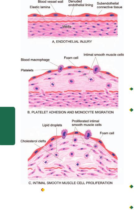
396
III SECTION |
|
|
Systemic |
|
|
Pathology |
atherosclerosis as explained by ‘reaction-to-injury’ hypothesis. A, Endo- |
|
|
Figure 15.4 |
Diagrammatic representation of pathogenesis of |
thelial injury. B, Adhesion of platelets and migration of blood monocytes from blood stream. C, Smooth muscle cell proliferation into the intima and ingrowth of new blood vessels.
Both these theories—original and modified, have attracted support and criticism. However, following is the generally accepted role of key components involved in atherogenesis, diagrammatically illustrated in Fig. 15.4.
i) Endothelial injury. It has been known for many years that endothelial injury is the initial triggering event in the development of lesions of atherosclerosis. Actual endothelial denudation is not an essential requirement, but endothelial dysfunction may initiate the sequence of events. Numerous causes ascribed to endothelial injury in experimental animals are: mechanical trauma, haemodynamic forces, immunological and chemical mechanisms, metabolic agent as chronic dyslipidaemia, homocystine, circulating toxins from systemic infections, viruses, hypoxia, radiation, carbon monoxide and tobacco products.
In man, two of the major risk factors which act together to produce endothelial injury are: haemodynamic stress from
hypertension and chronic dyslipidaemia. The role of haemodynamic forces in causing endothelial injury is further supported by the distribution of atheromatous plaques at points of bifurcation or branching of blood vessels which are under greatest shear stress.
ii) Intimal smooth muscle cell proliferation. Endothelial injury causes adherence, aggregation and platelet release reaction at the site of exposed subendothelial connective tissue and infiltration by inflammatory cells. Proliferation of intimal smooth muscle cell and production of extracellular matrix are stimulated by various cytokines such as IL-1 and TNF-α released from invading monocyte-macrophages and by activated platelets at the site of endothelial injury. These cytokines lead to local synthesis of following growth factors having distinct roles in plaque evolution:

 Platelet-derived growth factor (PDGF) and fibroblast growth factor (FGF) stimulate proliferation and migration of smooth muscle cells from their usual location in the media into the intima.
 Transforming growth factor-β (TGF-β) and interferon- (IFN-γ) derived from activated T lymphocytes within lesions regulate the synthesis of collagen by smooth muscle cells.
Smooth muscle cell proliferation is also facilitated by biomolecules such as nitric oxide and endothelin released from endothelial cells. Intimal proliferation of smooth muscle cells is accompanied by synthesis of matrix proteins—collagen, elastic fibre proteins and proteoglycans.
iii) Role of blood monocytes. Though blood monocytes do not possess receptors for normal LDL, LDL does appear in the monocyte cytoplasm to form foam cell by mechanism illustrated in Fig. 15.5. Plasma LDL on entry into the intima undergoes oxidation. The ‘oxidised LDL’ formed in the intima performs the following all-important functions on monocytes and endothelium:

 For monocytes: Oxidised LDL acts to attract, proliferate, immobilise and activate them as well as is readily taken up by scavenger receptor on the monocyte to transform it to a lipid-laden foam cell.

 For endothelium: Oxidised LDL is cytotoxic.
Death of foam cell by apoptosis releases lipid to form lipid core of plaque.
iv)Role of dyslipidaemia. As stated already, chronic dyslipidaemia in itself may initiate endothelial injury and dysfunction by causing increased permeability. In particular, hypercholesterolaemia with increased serum concentration of LDL promotes formation of foam cells, while high serum concentration of HDL has anti-atherogenic effect.
v)Thrombosis. As apparent from the foregoing, endothelial injury exposes subendothelial connective tissue resulting in formation of small platelet aggregates at the site and causing proliferation of smooth muscle cells. This causes mild inflammatory reaction which together with foam cells is incorporated into the atheromatous plaque. The lesions enlarge by attaching fibrin and cells from the blood so that thrombus becomes a part of atheromatous plaque.
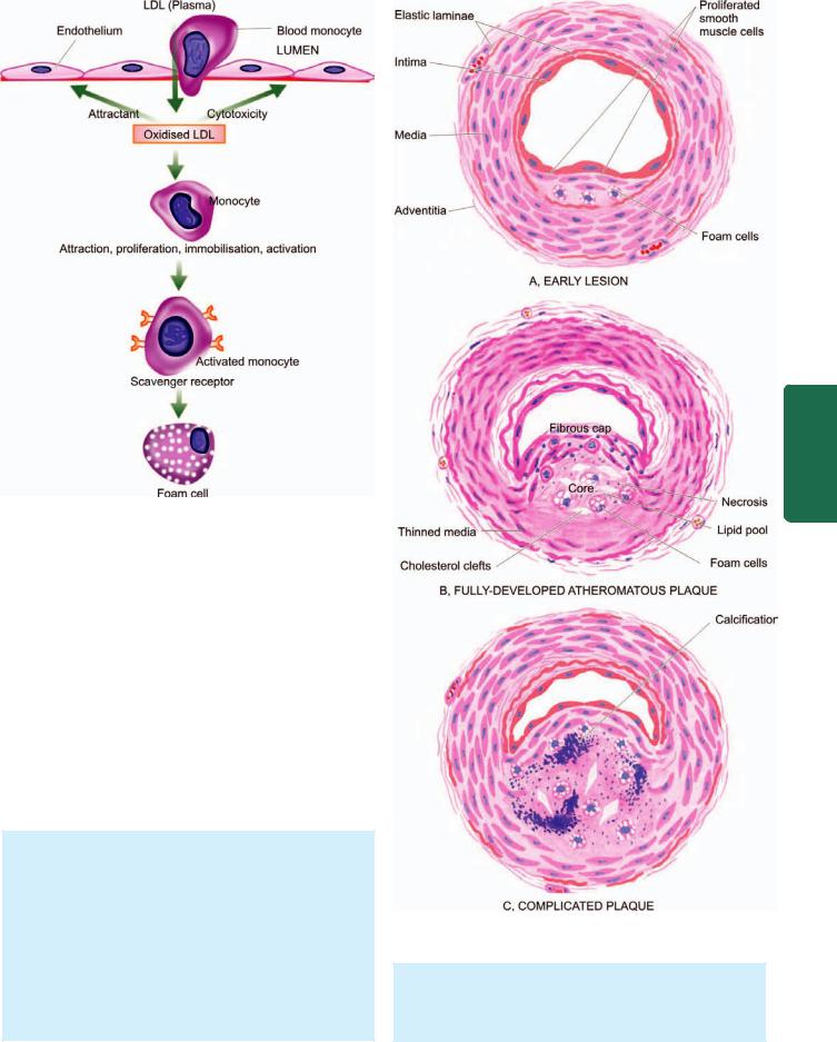
Figure 15.5 
 Mechanism of foam cell formation.
2. MONOCLONAL HYPOTHESIS. This hypothesis is based on the postulate that proliferation of smooth muscle cells is the primary event and that this proliferation is monoclonal in origin similar to cellular proliferation in neoplasms (e.g. in uterine leiomyoma, Chapter 8). The evidence cited in support of monoclonal hypothesis is the observation on proliferated smooth muscle cells in atheromatous plaques which have only one of the two forms of glucose-6-phosphate dehydrogenase (G6PD) isoenzymes, suggesting monoclonality in origin. The monoclonal proliferation of smooth muscle cells in atherosclerosis may be initiated by mutation caused by exogenous chemicals (e.g. cigarette smoke), endogenous metabolites (e.g. lipoproteins) and some viruses (e.g. Marek’s disease virus in chickens, herpesvirus).
MORPHOLOGIC FEATURES
Early lesions in the form of diffuse intimal thickening, fatty streaks and gelatinous lesions are often the forerunners in the evolution of atherosclerotic lesions. However, the clinical disease states due to luminal narrowing in atherosclerosis are caused by fully developed atheromatous plaques and complicated plaques (Fig. 15.6).
1. FATTY STREAKS AND DOTS. Fatty streaks and dots on the intima by themselves are harmless but may be the precursor lesions of atheromatous plaques. They are seen in all races of the world and begin to appear in
Figure 15.6 
 Schematic evolution of lesions in atherosclerosis.
the first year of life. However, they are uncommon in older persons and are probably absorbed. They are especially prominent in the aorta and other major arteries, more often on the posterior wall than the anterior wall.
397
Lymphatics and Vessels Blood The 15 CHAPTER
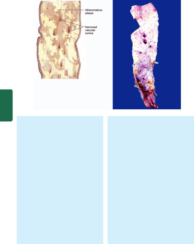
398
Pathology Systemic III SECTION
Figure 15.7  Structure of a fully-developed atheroma. The opened up inner surface of the abdominal aorta shows a variety of atheromatous lesions. While some are raised yellowish-white lesions raised above the surface, a few have ulcerated surface. Orifices of some of the branches coming out of the wall are narrowed by the atherosclerotic process.
Grossly, the lesions may appear as flat or slightly elevated and yellow. They may be either in the form of small, multiple dots, about 1 mm in size, or in the form of elongated, beaded streaks.
Microscopically, fatty streaks lying under the endothelium are composed of closely-packed foam cells, lipidcontaining elongated smooth muscle cells and a few lymphoid cells. Small amount of extracellular lipid, collagen and proteoglycans are also present.
2.GELATINOUS LESIONS. Gelatinous lesions develop in the intima of the aorta and other major arteries in the first few months of life. Like fatty streaks, they may also be precursors of plaques. They are round or oval, circumscribed grey elevations, about 1 cm in diameter.
Microscopically, gelatinous lesions are foci of increased ground substance in the intima with thinned overlying endothelium.
3.ATHEROMATOUS PLAQUES. A fully developed atherosclerotic lesion is called atheromatous plaque, also
called fibrous plaque, fibrofatty plaque or atheroma. Unlike fatty streaks, atheromatous plaques are selective in different geographic locations and races and are seen in advanced age. These lesions may develop from progression of early lesions of the atherosclerosis described above. Most often and most severely affected is the abdominal aorta, though smaller lesions may be seen in descending thoracic aorta and aortic arch. The major branches of the aorta around the ostia are often severely involved, especially the iliac, femoral, carotid, coronary, and cerebral arteries.
Grossly, atheromatous plaques are white to yellowishwhite lesions, varying in diameter from 1-2 cm and raised on the surface by a few millimetres to a centimetre in thickness (Fig. 15.7). Cut section of the plaque reveals the luminal surface as a firm, white fibrous cap and a central core composed of yellow to yellow-white, soft, porridgelike material and hence the name atheroma.
Microscopically, the appearance of plaque varies depending upon the age of the lesion. However, the following features are invariably present (Fig. 15.8):
 Superficial luminal part of the fibrous cap is covered by endothelium, and is composed of smooth muscle cells, dense connective tissue and extracellular matrix containing proteoglycans and collagen.
 Cellular area under the fibrous cap is comprised by a mixture of macrophages, foam cells, lymphocytes and a few smooth muscle cells which may contain lipid.
 Deeper central soft core consists of extracellular lipid material, cholesterol clefts, fibrin, necrotic debris and lipidladen foam cells.
 In older and more advanced lesions, the collagen in the fibrous cap may be dense and hyalinised, smooth muscle cells may be atrophic and foam cells are fewer.
4. COMPLICATED PLAQUES. Various pathologic changes that occur in fully-developed atheromatous plaques are called the complicated lesions. These account for the most serious harmful effects of atherosclerosis and even death. These changes include calcification, ulceration, thrombosis, haemorrhage and aneurysmal dilatation. It is not uncommon to see more than one form of complication in a plaque.
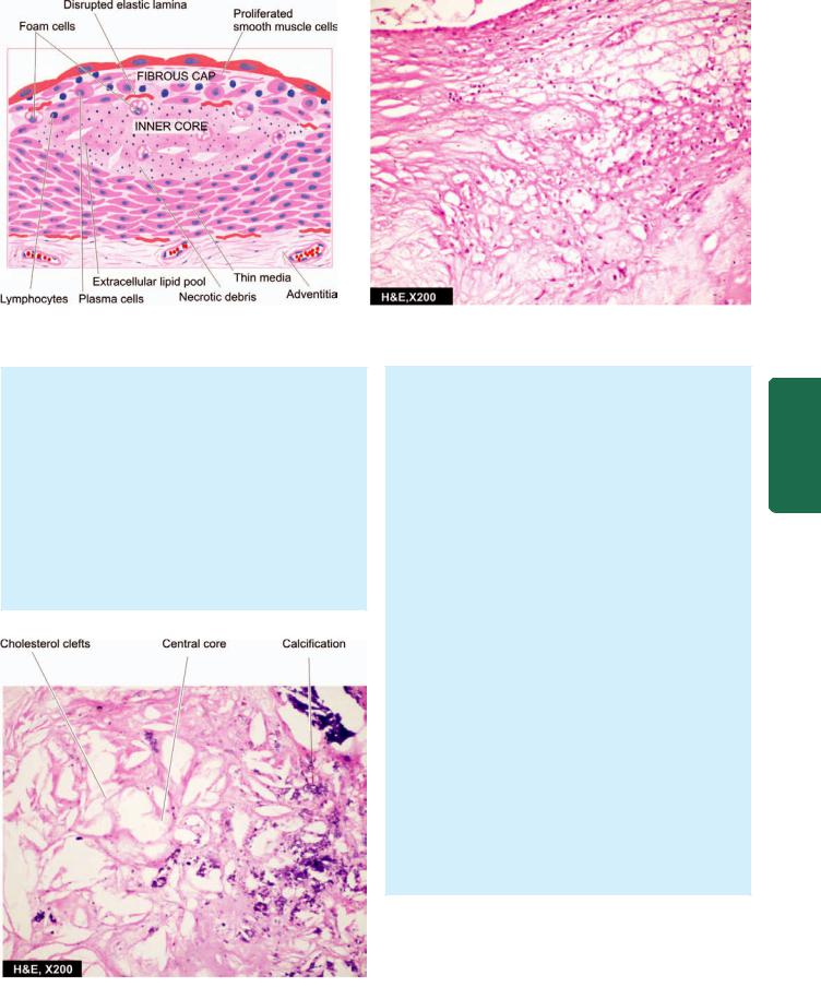
399
Figure 15.8 
 Histologic appearance of a fully-developed atheroma.
i)Calcification. Calcification occurs more commonly in advanced atheromatous plaques, especially in the aorta and coronaries. The diseased intima cracks like an eggshell when the vessel is incised and opened.
Microscopically, the calcium salts are deposited in the vicinity of necrotic area and in the soft lipid pool deep in the thickened intima (Fig. 15.9). This form of atherosclerotic intimal calcification differs from Mönckeberg’s medial calcific arteriosclerosis that affects only the tunica media (page 392).
ii)Ulceration. The layers covering the soft pultaceous material of an atheroma may ulcerate as a result of haemodynamic forces or mechanical trauma. This results
Figure 15.9 
 Complicated atheromatous plaque lesion. There is narrowing of the lumen of coronary due to fully developed atheromatous plaque which has dystrophic calcification in its core.
in discharge of emboli composed of lipid material and debris into the blood stream, leaving a shallow, ragged ulcer with yellow lipid debris in the base of the ulcer. Occasionally, atheromatous plaque in a coronary artery may suddenly rupture into the arterial lumen forcibly and cause thromboembolic occlusion.
iii)Thrombosis. The ulcerated plaque and the areas of endothelial damage are vulnerable sites for formation of superimposed thrombi. These thrombi may get dislodged to become emboli and lodge elsewhere in the circulation, or may get organised and incorporated into the arterial wall as mural thrombi. Mural thrombi may become occlusive thrombi which may subsequently recanalise.
iv)Haemorrhage. Intimal haemorrhage may occur in an atheromatous plaque either from the blood in the vascular lumen through an ulcerated plaque, or from rupture of thin-walled capillaries that vascularise the atheroma from adventitial vasa vasorum. Haemorrhage is particularly a common complication in coronary arteries. The haematoma formed at the site contains numerous haemosiderin-laden macrophages.
v)Aneurysm formation. Though atherosclerosis is primarily an intimal disease, advanced lesions are associated with secondary changes in the media and adventitia. The changes in media include atrophy and thinning of the media and fragmentation of internal elastic lamina. The adventitia undergoes fibrosis and some inflammatory changes. These changes cause weakening in the arterial wall resulting in aneurysmal dilatation.
Clinical Effects
The clinical effects of atherosclerosis depend upon the size and type of arteries affected. In general, the clinical effects result from the following:
1.Slow luminal narrowing causing ischaemia and atrophy.
2.Sudden luminal occlusion causing infarction necrosis.
Lymphatics and Vessels Blood The 15 CHAPTER
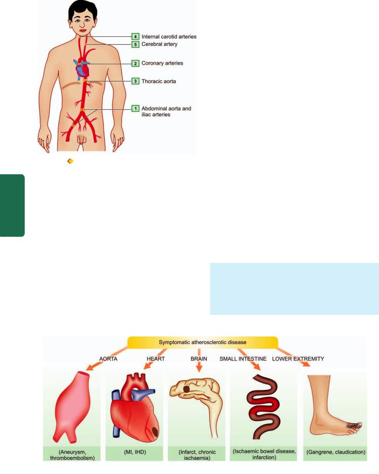
400
Figure 15.10 |
Major sites of atherosclerosis (serially numbered) in |
descending order of frequency. |
|
SECTION |
3. Propagation of plaque by formation of thrombi and |
|
|
|
emboli. |
|
4. Formation of aneurysmal dilatation and eventual rupture. |
|
Large arteries affected most often are the aorta, renal, |
III |
mesenteric and carotids, whereas the mediumand small- |
sized arteries frequently involved are the coronaries, |
|
|
cerebrals and arteries of the lower limbs. Accordingly, the |
Systemic |
symptomatic atherosclerotic disease involves most often the |
are listed below (Fig. 15.11): |
|
|
heart, brain, kidneys, small intestine and lower extremities |
|
(Fig. 15.10). The effects pertaining to these organs are |
|
described in relevant chapters later while the major effects |
Pathology |
i) Aorta—Aneurysm formation, thrombosis and |
infarction. |
|
|
embolisation to other organs. |
|
ii) Heart—Myocardial infarction, ischaemic heart disease. |
|
iii) Brain—Chronic ischaemic brain damage, cerebral |
iv)Small intestine—Ischaemic bowel disease, infarction.
v)Lower extremities—Intermittent claudication, gangrene.
ARTERITIS
Arteritis, angiitis and vasculitis are the common terms used for inflammatory process in an artery or an arteriole. It may occur following invasion of the vessel by infectious agents, or may be induced by non-infectious injuries such as chemical, mechanical, immunologic and radiation injury. The non-infectious group is more important than the infectious type. A classification of arteritis based on this is given in
Table 15.3.
I.INFECTIOUS ARTERITIS
Direct invasion of the artery by infectious agents, especially bacteria and fungi, causes infectious arteritis. It may be found in the vicinity of an infected focus like in tuberculosis, pneumonia, abscesses, etc. or less frequently may arise from haematogenous spread of infection such as in infective endocarditis, septicaemia, etc. Some common types are described below.
Endarteritis Obliterans
Endarteritis obliterans is not a disease entity but a pathologic designation used for non-specific inflammatory response of arteries and arterioles to a variety of irritants. It is commonly seen close to the lesions of peptic ulcers of the stomach and duodenum, tuberculous and chronic abscesses in the lungs, chronic cutaneous ulcers, chronic meningitis, and in post-partum and post-menopausal uterine arteries.
Grossly, the affected vessels may appear unaltered externally but on cross-section show obliteration of their lumina.
Microscopically, the obliteration of the lumen is due to concentric and symmetric proliferation of cellular fibrous tissue in the intima. Though the condition has suffix—itis
Figure 15.11 
 Major forms of symptomatic atherosclerotic disease.
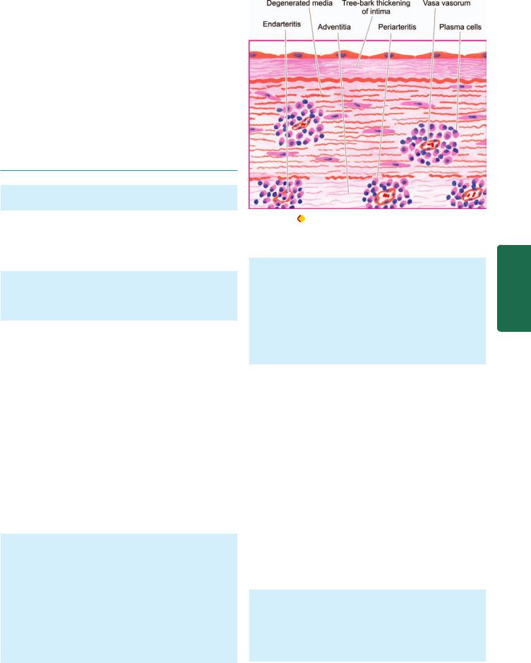
TABLE 15.3: Classification of Vasculitis. |
401 |
|
|
I.INFECTIOUS ARTERITIS
1.Endarteritis obliterans
2.Non-syphilitic infective arteritis
3.Syphilitic arteritis
II.NON-INFECTIOUS ARTERITIS
1.Polyarteritis nodosa (PAN)
2.Hypersensitivity (allergic, leucocytoclastic) vasculitis
3.Wegener’s granulomatosis
4.Temporal (giant cell) arteritis
5.Takayasu’s arteritis (pulseless disease)
6.Kawasaki’s disease
7.Buerger’s disease (thromboangiitis obliterans)
8.Miscellaneous vasculitis
attached to it, there is minimal or no inflammatory cell infiltrate.
Non-syphilitic Infective Arteritis
Various forms of invasions of the artery by bacteria, fungi, parasites or viruses, either directly or by haematogenous route, cause non-syphilitic infective arteritis.
Microscopically, the inflammatory infiltrate is present in the vessel wall. The vascular lumen may get occluded by thrombi and result in ischaemic necrosis of the affected tissue.
Syphilitic Arteritis
Syphilitic or luetic vascular involvement occurs in all stages of syphilis but is more prominent in the tertiary stage. The changes that are found in the syphilitic arteritis are seen within the arterial tissue (syphilitic endarteritis) and in the periarterial tissues (syphilitic periarteritis). Manifestations of the disease are particularly prominent at two sites—the aorta and the cerebral arteries.
SYPHILITIC AORTITIS. Syphilitic involvement of the ascending aorta and the aortic arch is the commonest manifestation of cardiovascular syphilis. It occurs in about 80% cases of tertiary syphilis. Preferential involvement of the arch of aorta may be due to involvement of mediastinal lymph nodes in secondary syphilis through which the treponemes spread to the lymphatics around the aortic arch. The lesions diminish in severity in descending thoracic aorta and disappear completely at the level of the diaphragm.
Grossly, the affected part of the aorta may be dilated, and its wall somewhat thickened and adherent to the neighbouring mediastinal structures. Longitudinally opened vessels show intimal surface studded with pearlywhite thickenings, varying from a few millimeters to a centimeter in diameter. These lesions are separated by wrinkled normal intima, giving it characteristic tree-bark appearance. Cut section of the lesion shows more firm and fibrous appearance than the atheromatous plaques. However, superimposed atherosclerotic lesions may be present.
Figure 15.12 |
Syphilitic aortitis. There is endarteritis and periarteritis |
of the vasa vasorum in the media and adventitia. There is perivascular infiltrate of plasma cells, lymphocytes and macrophages.
Microscopically, the conspicuous features are as under
(Fig. 15.12):
 Endarteritis and periarteritis of the vasa vasorum located in the media and adventitia.
 Perivascular accumulation of plasma cells, lymphocytes and macrophages that may form miliary gummas which undergo necrosis and are replaced by scar tissue.
 Intimal thickenings consist of dense avascular collagen that may undergo hyalinisation and calcification.
The effects of syphilitic aortitis may vary from trivial to catastrophic. These are as follows:
a)Aortic aneurysm may result from damage to the aortic wall (page 406).
b)Aortic valvular incompetence used to be considered an important sequela of syphilis but now-a-days rheumatic disease is considered more important cause for this. The aortic incompetence results from spread of the syphilitic process to the aortic valve ring.
c)Stenosis of coronary ostia is seen in about 20% cases of syphilitic aortitis and may lead to progressive myocardial fibrosis, angina pectoris and sudden death.
The features distinguishing syphilitic aortitis from aortic atheroma are given in Table 15.4.
CEREBRAL SYPHILITIC ARTERITIS (HEUBNER’S ARTERITIS). Syphilitic involvement of small and mediumsized cerebral arteries occurs during the tertiary syphilis. The changes may accompany syphilitic meningitis.
Grossly, the cerebral vessels are white, rigid and thickwalled.
Microscopically, changes of endarteritis and periarteritis similar to those seen in syphilitic aortitis are found. There is atrophy of muscle in the media and replacement by fibrosis. This results in ischaemic atrophy of the brain.
Lymphatics and Vessels Blood The 15 CHAPTER

402 TABLE 15.4: Contrasting Features of Syphilitic Aortitis and Aortic Atheroma.
|
Feature |
Syphilitic Aortitis |
Aortic Atheroma |
|
|
|
|
1. |
Sites |
Ascending aorta, aortic arch; |
Progressive increase from the arch to abdominal |
|
|
absent below diaphragm |
aorta, more often at the bifurcation |
2. |
Macroscopy |
Pearly-white intimal lesions resembling |
Yellowish-white intimal plaques with fat in the core; |
|
|
tree-bark without fat in the core; ulceration |
ulceration and calcification in plaques common |
|
|
and calcification often not found |
|
3. Microscopy |
Endarteritis and periarteritis of vasa |
|
vasorum, perivascular infiltrate of plasma |
|
cells and lymphocytes |
Fibrous cap with deeper core containing foam cells, cholesterol clefts and soft lipid
4. Effects |
Thoracic aortic aneurysm, incompetence |
Abdominal aortic aneurysm, aortic valve stenosis, |
|
of the aortic valve, stenosis of coronary ostia |
stenosis of abdominal branches |
|
|
|
Pathology Systemic III SECTION
II. NON-INFECTIOUS ARTERITIS
This group consists of most of the important forms of vasculitis, more often affecting arterioles, venules and capillaries, and hence also termed as small vessel vasculitis. Their exact etiology is not known but available evidence suggests that many of them have immunologic origin. Serum from many of patients with vasculitis of immunologic origin show the presence of following immunologic features:
1.Anti-neutrophil cytoplasmic antibodies (ANCAs).
Patients with immunologic vasculitis have autoantibodies in their serum against the cytoplasmic antigens of the neutrophils, macrophages and endothelial cells; these are called ANCAs. Neutrophil immunofluorescence is used to demonstrate their presence, of which two distinct patterns of ANCAs are seen:

 Cytoplasmic ANCA (c-ANCA) pattern is specific for proteinase-3 (PR-3), a constituent of neutrophilic granules; this is seen in cases with active Wegener’s granulomatosis.

 Perinuclear ANCA (p-ANCA) pattern is specific for myeloperoxidase enzyme; this is noted in patients with microscopic polyarteritis nodosa and primary glomerular disease.
2.Anti-endothelial cell antibodies (AECAs). These antibodies are demonstable in cases of SLE, Kawasaki disease and Buerger’s disease.
3.Pauci-immune vasculitis. While most cases of immunologic vasculitis have immune complex deposits in the vessel wall, there are some cases which do not have such immune deposits and are termed as cases of pauci-immune vasculitis (similar to pauci-immune glomerulonephritis, Chapter 22). Pathogenesis of lesions in these cases is explained by other mechanisms.
Polyarteritis Nodosa
Polyarteritis nodosa (PAN) is a necrotising vasculitis involving small and medium-sized muscular arteries of multiple organs and tissues. ‘Polyarteritis’ is the preferred nomenclature over ‘periarteritis’ because inflammatory involvement occurs in all the layers of the vessel wall.
The disease occurs more commonly in adult males than females. Most commonly affected organs, in descending order of frequency of involvement, are the kidneys, heart,
liver, gastrointestinal tract, muscle, pancreas, testes, nervous system and skin. The syndrome of PAN presents with varied symptoms pertaining to different organs. However, some usual clinical features are fever, malaise, weakness, weight loss, renal manifestations (albuminuria, haematuria and renal failure), vascular lesions in the alimentary tract (abdominal pain and melaena), peripheral neuritis and hypertension. The condition is believed to result from deposition of immune complexes and tumour-related antigens.
Grossly, the lesions of PAN involve segments of vessels, especially at the bifurcations and branchings, as tiny beaded nodules.
Microscopically, there are 3 sequential stages in the evolution of lesions in PAN:
i)Acute stage—There is fibrinoid necrosis in the centre of the nodule located in the media. An acute inflammatory response develops around the focus of fibrinoid necrosis. The inflammatory infiltrate is present in the entire circumference of the affected vessel (periarteritis) and consists chiefly of neutrophils and eosinophils, and some mononuclear cells. The lumen may show thrombi and the weakened wall may be the site of aneurysm formation.
ii)Healing stage—This is characterised by marked fibroblastic proliferation producing firm nodularity. The inflammatory infiltrate now consists mainly of lymphocytes, plasma cells and macrophages.
iii)Healed stage—In this stage, the affected arterial wall is markedly thickened due to dense fibrosis. The internal elastic lamina is fragmented or lost. Healed stage may contain haemosiderin-laden macrophages and organised thrombus.
However, it may be mentioned here that various stages of the disease may be seen in different vessels and even within the same vessel.
Hypersensitivity Vasculitis
Hypersensitivity vasculitis, also called as allergic or leucocytoclastic vasculitis or microscopic polyarteritis, is a group of clinical syndromes differing from PAN in having inflammatory involvement of venules, capillaries and arterioles. The tissues and organs most commonly involved are the skin, mucous membranes, lungs, brain, heart,
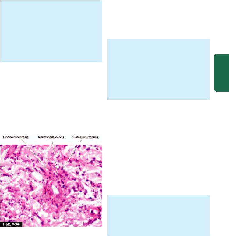
gastrointestinal tract, kidneys and muscle. The condition results from immunologic response to an identifiable antigen that may be bacteria (e.g. streptococci, staphylococci, mycobacteria), viruses (e.g. hepatitis B virus, influenza virus, CMV), malarial parasite, certain drugs and chemicals. Hypersensitivity vasculitis includes clinicopathologic entities such as serum sickness, Henoch-Schönlein purpura, mixed cryoglobulinaemia, vasculitis associated with malignancy, and vasculitis associated with connective tissue diseases like rheumatoid arthritis and SLE.
Microscopically, the lesions characteristically involve smallest vessels, sparing medium-sized and larger arteries. Two histologic forms are described:
i)Leucocytoclastic vasculitis, characterised by fibrinoid necrosis with neutrophilic infiltrate in the vessel wall. Many of the neutrophils are fragmented (Fig. 15.13). This form is found in vasculitis caused by deposits of immune complexes.
ii)Lymphocytic vasculitis, in which the involved vessel shows predominant infiltration by lymphocytes. This type is seen in vascular injury due to delayed hypersensitivity or cellular immune reactions.
Wegener’s Granulomatosis
Wegener’s granulomatosis is another form of necrotising vasculitis characterised by a clinicopathologic triad consisting of the following:
i)Acute necrotising granulomas of the upper and lower respiratory tracts involving nose, sinuses and lungs;
ii)focal necrotising vasculitis, particularly of the lungs and upper airways; and
iii)focal or diffuse necrotising glomerulonephritis.
A limited form of Wegener’s granulomatosis is the same condition without renal involvement. As with PAN, the condition is more common in adult males and involves
Figure 15.13 
 Leucocytoclastic vasculitis. The vessel wall shows fibrinoid necrosis surrounded by viable as well as fragmented neutrophils.
multiple organs and tissues. Most commonly involved organs are the lungs, paranasal sinuses, nasopharynx and kidneys. Other involved organs are joints, skin, eyes, ears, heart and nervous system. Accordingly, clinical features are variable. Typical features include pneumonitis with bilateral infiltrates in the lungs (Chapter 17), chronic sinusitis, nasopharyngeal ulcerations (Chapter 18) and renal disease (Chapter 22). The etiology is not known but possibly the lesions occur due to the presence of circulating immune complexes. This is supported by the observation of subepithelial immunoglobulin deposits on the glomerular basement membrane and induction of remission by immunosuppressive therapy. The serum of these patients shows c-ANCA positivity. Disseminated form of Wegener’s granulomatosis differs from a related entity, idiopathic lethal midline granuloma, in the sense that the latter condition is highly destructive and progressively necrotic disease of the upper airways.
Histologically, the characteristic feature of Wegener’s granulomatosis is the presence of necrotising granulomatous inflammation of the tissues and necrotising vasculitis with or without granulomas:
 The granulomas consist of fibrinoid necrosis with extensive infiltration by neutrophils, mononuclear cells, epithelioid cells, multinucleate giant cells and fibroblastic proliferation.
 The necrotising vasculitis may be segmental or circumferential.
 The renal lesions are those of focal or diffuse necrotising glomerulonephritis.
Temporal (Giant Cell) Arteritis
This is a form of granulomatous inflammation of mediumsized and large arteries. Preferential sites of involvement are the cranial arteries, especially the temporal, and hence the name. However, the aorta and other major arteries like common carotid, axillary, brachial, femoral and mesenteric arteries are also involved, and therefore, it is preferable to call the entity as ‘giant cell arteritis’. The patients are generally over the age of 70 years with slight female preponderance. The usual clinical manifestations are headache and blindness if ophthalmic artery is involved. An association with polymyalgia rheumatica has been observed. The cause of the condition remains unknown though there is suggestion of T cell mediated immunologic reaction to some component of the arterial wall, especially against the damaged internal elastic lamina. Biopsy of the affected artery is not only of diagnostic value but also relieves the patient of painful symptoms.
Grossly, the affected artery is thickened, cord-like and the lumen is usually reduced to a narrow slit. Histologically, the features include the following:
i)There is chronic granulomatous reaction, usually around the internal elastic lamina and typically involves the entire circumference of the vessel.
ii)Giant cells of foreign body or Langhans’ type are found in two-third of cases.
403
Lymphatics and Vessels Blood The 15 CHAPTER

404
Pathology Systemic III SECTION
iii)The internal elastic lamina is often fragmented.
iv)There is eccentric or concentric intimal cellular proliferation causing marked narrowing of the lumen. The narrowed lumen may contain thrombus.
v)Occasionally, only nonspecific inflammatory cell infiltrate consisting of neutrophils, lymphocytes and eosinophils is found throughout the arterial wall.
Takayasu’s Arteritis (Pulseless Disease)
This is a form of granulomatous vasculitis affecting chiefly the aorta and its major branches and hence is also referred to as aortic arch syndrome. The disease affects chiefly young women and is typically characterised by absence of pulse in both arms and presence of ocular manifestations. Other features referable to ischaemic effects from thrombotic occlusion of vessels include myocardial infarction, congestive heart failure and neurologic deficits. The etiology of Takayasu’s arteritis is not known but the autoimmune reaction to aortic tissue has been suggested as the possible cause.
Grossly, the aortic wall is irregularly thickened and intima wrinkled. The branches of major arteries coming off the aortic arch have obliterated lumina.
Histologically, the features are as under:
i)There is severe mononuclear inflammatory infiltrate involving the full thickness of the affected vessel wall.
ii)The inflammatory changes are more severe in the adventitia and media and there is perivascular infiltration of the vasa vasorum.
iii)Granulomatous changes in the media with central necrosis and Langhans’ giant cells are found in many cases.
iv)Advanced lesions show extensive fibrosis of the media and adventitia causing thickening in the vessel wall.
Kawasaki’s Disease
Also known by more descriptive name of ‘mucocutaneous lymph node syndrome’, it is an acute and subacute illness affecting mainly young children and infants. Kawasaki’s disease is a febrile illness with mucocutaneous symptoms like erosions of oral mucosa and conjunctiva, skin rash and lymphadenopathy. The etiology is unknown; possible causes considered are infectious, genetic, toxic and immunological. The most characteristic finding is the presence of multiple aneurysms of the coronaries detected by angiography during life or observed at autopsy. Other vessels that may be involved are renal, mesenteric, hepatic and pancreatic arteries.
Histologically, the picture is of panarteritis resembling PAN, characterised by necrosis and inflammation of the entire thickness of the vessel wall. Therefore, some consider Kawasaki’s disease as an infantile form of PAN.
Buerger’s Disease (Thromboangiitis Obliterans)
Buerger’s disease is a specific disease entity affecting chiefly small and medium-sized arteries and veins of the extremities
and characterised by acute and chronic occlusive inflammatory involvement. The disease affects chiefly men under the age of 35 years who are heavy cigarette smokers. The symptom-complex consists of intermittent claudication due to ischaemia manifested by intense pain affecting the limbs, more commonly the legs. Eventually, gangrene of the affected extremities occurs requiring amputation.
ETIOPATHOGENESIS. Following possible mechanisms have been suggested:
 There is consistent association with heavy cigarette smoking. This has led to the hypothesis that tobacco products cause either direct endothelial damage leading to hypercoagulability and thrombosis, or it is a result in hypersensitivity to tobacco products. In support is the demonstration of anti-endothelial cell antibodies (AECAs).

 Genetic factors play a role as the disease has familial occurrence and has HLA association. An increased prevalence is seen in individuals with HLA-A9 and HLA-B5 antigens. It is seen more commonly in persons from Israel, Japan and in India.
Grossly, the lesions are typically segmental affecting small and medium-sized arteries, especially of the lower extremities. Involvement of the arteries is often accompanied with involvement of adjacent veins and nerves. Fibrous tissue cuff generally surrounds these three structures. Mural thrombi are frequently present in the vessels.
Microscopically, the following changes are seen in different stages of the disease:
i)In early stage, there is infiltration by polymorphs in all the layers of vessels and there is invariable presence of mural or occlusive thrombosis of the lumen (Fig. 15.14). The appearance differs from atherosclerosis in having microabscesses in the thrombi, proliferation of endothelial cells, lack of lipid aggregates and presence of intact internal elastic lamina.
ii)In advanced stage, the cellular infiltrate is predominantly mononuclear and may contain an occasional epithelioid cell granuloma with Langhans’ giant cells. The thrombi undergo organisation and recanalisation. In more chronic cases, marked fibrosis of the media is present.
Miscellaneous Hypersensitivity Vasculitis
Various connective tissue diseases (e.g. rheumatoid arthritis, ankylosing spondylitis and SLE), rheumatic fever, certain malignancies and Henoch-Schönlein purpura are associated with vasculitis. The type of vasculitis is generally of hypersensitivity or allergic angiitis as already explained but sometimes may resemble PAN.
 Rheumatoid vasculitis affects chiefly the small and medium-sized arteries of multiple visceral organs in patients who have rheumatoid nodules of long duration. Vasculitis in SLE affects mainly the small arteries of the skin.

 Rheumatic vasculitis involves the aorta, carotid and coronary arteries and the visceral vessels. Usually, fibrinoid change and perivascular inflammation are seen rather than typical Aschoff nodules (page 439).
405
Figure 15.14 Buerger’s disease (Thromboangiitis obliterans). There is acute panarteritis. The lumen is occluded by a thrombus containing
microabscesses.
Raynaud’s Disease and Raynaud’s Phenomenon
Raynaud’s disease is not a vasculitis but is a functional vasospastic disorder affecting chiefly small arteries and arterioles of the extremities, occurring in otherwise young healthy females. The disease affects most commonly the fingers and hands. The ischaemic effect is provoked primarily by cold but other stimuli such as emotions, trauma, hormones and drugs also play a role. Clinically, the affected digits show pallor, followed by cyanosis, and then redness, corresponding to arterial ischaemia, venostasis and hyperaemia respectively. Long-standing cases may develop ulceration and necrosis of digits but occurrence of true gangrene is rare. The cause of the disease is unknown but probably occurs due to vasoconstriction mediated by autonomic stimulation of the affected vessels. Though usually no pathologic changes are observed in the affected vessels, long-standing cases may show endothelial proliferation and intimal thickening.
Raynaud’s phenomenon differs from Raynaud’s disease in having an underlying cause e.g. secondary to artherosclerosis, connective tissue diseases like scleroderma and SLE, Buerger’s disease, multiple myeloma, pulmonary hypertension and ingestion of ergot group of drugs. Raynaud’s phenomenon like Raynaud’s disease, also shows cold sensitivity but differs from the latter in having structural abnormalities in the affected vessels. These changes include segmental inflammation and fibrinoid change in the walls of capillaries.
ANEURYSMS
DEFINITION
An aneurysm is defined as a permanent abnormal dilatation of a blood vessel occurring due to congenital or acquired weakening or destruction of the vessel wall. Most commonly, aneurysms involve large elastic arteries, especially the aorta and its major branches. Aneurysms can cause various illeffects such as thrombosis and thromboembolism, alteration in the flow of blood, rupture of the vessel and compression of neighbouring structures.
CLASSIFICATION
Aneurysms can be classified on the basis of various features:
A. Depending upon the composition of the wall:
1 True aneurysm composed of all the layers of a normal vessel wall.
2. False aneurysm having fibrous wall and occurring often from trauma to the vessel.
B. Depending upon the shape: These are as under
(Fig. 15.15):
1.Saccular having large spherical outpouching.
2.Fusiform having slow spindle-shaped dilatation.
3.Cylindrical with a continuous parallel dilatation.
4.Serpentine or varicose which has tortuous dilatation of the vessel.
Lymphatics and Vessels Blood The 15 CHAPTER
Figure 15.15 
 Common shapes of aneurysms of various types.
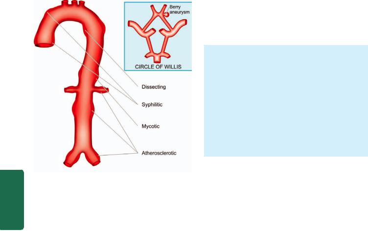
406
Pathology Systemic III SECTION
Figure 15.16 
 Sites of major forms of aneurysms.
5. Racemose or circoid having mass of intercommunicating small arteries and veins.
C. Based on pathogenetic mechanisms: This classification is followed most often (Fig. 15.16):
1.Atherosclerotic (arteriosclerotic) aneurysms are the most common type.
2.Syphilitic (luetic) aneurysms found in the tertiary stage of the syphilis.
3.Dissecting aneurysms (Dissecting haematoma) in which the blood enters the separated or dissected wall of the vessel.
4.Mycotic aneurysms which result from weakening of the arterial wall by microbial infection.
5.Berry aneurysms which are small dilatations especially affecting the circle of Willis in the base of the brain (Chapter 30).
The three common types of aortic aneurysms—athero- sclerotic, syphilitic and dissecting, are described below:
Atherosclerotic Aneurysms
Atherosclerotic aneurysms are the most common form of aortic aneurysms. They are seen more commonly in males and the frequency increases after the age of 50 years when the incidence of complicated lesions of advanced atherosclerosis is higher. They are most common in the abdominal aorta, so much so that all forms of aneurysms of abdominal aorta (fusiform, cylindrical and saccular) should be considered atherosclerotic until proved otherwise. Other locations include thoracic aorta (essentially the ascending part and arch of aorta), iliac arteries and other large systemic arteries.
PATHOGENESIS. Obviously, severe atherosclerotic lesions are the basic problem which cause thinning and destruction of the medial elastic tissue resulting in atrophy and
weakening of the wall. Since atherosclerotic lesions are most common and severe in the abdominal aorta, atherosclerotic aneurysms occur most frequently here. In the thoracic aorta, besides atherosclerotic lesions, medial degeneration is another additional factor implicated in pathogenesis.
MORPHOLOGIC FEATURES. Atherosclerotic aneurysms of the abdominal aorta are most frequently infrarenal, above the bifurcation of the aorta but may extend into common iliac arteries. They may be of variable size but are often larger than 5-6 cm in diameter. Atherosclerotic aneurysm is most frequently fusiform in shape and the lumen of aneurysm often contains mural thrombus.
Histologically, the wall of atherosclerotic aneurysm loses its normal arterial structure. Instead, there is predominance of fibrous tissue in the media and adventitia with mild chronic inflammatory reaction. The intima and inner part of the media show remnants of atheromatous plaques and mural thrombus.
EFFECTS. The clinical effects of atherosclerotic aneurysms are due to complications. These are as under:
1.Rupture. Rupture of the atherosclerotic aneurysm is the most serious and fatal complication. The risk of rupture depends upon the size and duration of the aneurysm and the blood pressure. Rupture of abdominal aneurysm may occur either into the peritoneum or into the retroperitoneum resulting in sudden and massive bleeding. Occasionally, there may be slow progressive leak from the aneurysm. A ruptured aneurysm is more likely to get infected.
2.Compression. The atherosclerotic aneurysm may press upon some adjacent structures such as compression of ureter and erosion on the vertebral bodies.
3.Arterial occlusion. Atherosclerotic aneurysms of the abdominal aorta may occlude the inferior mesenteric artery, or there may be development of occlusive thrombosis. However, collateral circulation develops slowly and is nearly always sufficient so as not to produce effects of ischaemia. Thromboembolism is rather common in abdominal aneurysms.
Syphilitic (Luetic) Aneurysms
Cardiovascular syphilis occurs in about 10% cases of syphilis. It causes arteritis—syphilitic aortitis and cerebral arteritis, both of which are already described in this chapter. One of the major complications of syphilitic aortitis is syphilitic or luetic aneurysm that develops in the tertiary stage of syphilis. It usually manifests after the age of 50 years and is more common in men. The predominant site of involvement is the thoracic aorta, especially in the ascending part and arch of aorta. It may extend proximally into the aortic valve causing aortic incompetence and may lead to syphilitic heart disease. Less often, it may extend distally to involve abdominal aorta.
PATHOGENESIS. About 40% cases of syphilitic aortitis develop syphilitic aneurysms. The process begins from inflammatory infiltrate around the vasa vasorum of the

adventitia, followed by endarteritis obliterans. This results in ischaemic injury to the media causing destruction of the smooth muscle and elastic tissue of the media and scarring. Since syphilitic aortitis involves the proximal aorta maximally, aortic aneurysm is found most frequently in the ascending aorta and in the aortic arch.
MORPHOLOGIC FEATURES. Syphilitic aneurysms occurring most often in the ascending part and the arch of aorta are saccular in shape and usually 3-5 cm in diameter. Less often, they are fusiform or cylindrical. The intimal surface is wrinkled and shows tree-bark appearance. When the aortic valve is involved, there is stretching and rolling of the valve-leaflets producing valvular incompetence and left ventricular hypertrophy due to volume overload. This results in massively enlarged heart called ‘cor bovinum’.
Histologically, the features of healed syphilitic aortitis are seen (page 401). The adventitia shows fibrous thickening with endarteritis obliterans of vasa vasorum. The fibrous scar tissue may extend into the media and the intima. Rarely, spirochaetes may be demonstrable in syphilitic aneurysm. Often, mural thrombus is found in the aneurysm.
EFFECTS. The clinical manifestations are found much more frequently in syphilitic aneurysms than in atherosclerotic aneurysms. The effects include the following:
1.Rupture. Syphilitic aneurysm is likely to rupture causing massive and fatal haemorrhage into the pleural cavity, pericardial sac, trachea and oesophagus.
2.Compression. The aneurysm may press on the adjacent tissues and cause symptoms such as on trachea causing dyspnoea, on oesophagus causing dysphagia, on recurrent laryngeal nerve leading to hoarseness; and erosion of vertebrae, sternum and ribs due to persistent pressure.
3.Cardiac dysfunction. When the aortic root and valve are involved, syphilitic aneurysm produces aortic incompetence and cardiac failure. Narrowing of the coronary ostia may further aggravate cardiac disease.
Dissecting Aneurysms and Cystic Medial Necrosis
The term dissecting aneurysm is applied for a dissecting haematoma in which the blood enters the separated (dissected) wall of the vessel and spreads for varying distance longitudinally. The most common site is the aorta and is an acute catastrophic aortic disease. The condition occurs most commonly in men in the age range of 50 to 70 years. In women, dissecting aneurysms may occur during pregnancy.
PATHOGENESIS. The pathogenesis of dissecting aneurysm is explained on the basis of weakened aortic media. Various conditions causing weakening in the aortic wall resulting in dissection are as under:
i) Hypertensive state. About 90% cases of dissecting aneurysm have hypertension which predisposes such patients to degeneration of the media in some questionable way.
ii) Non-hypertensive cases. These are cases in whom there is some local or systemic connective tissue disorder e.g.
a)Marfan’s syndrome, an autosomal dominant disease with genetic defect in fibrillin which is a connective tissue protein required for elastic tissue formation.
b)Development of cystic medial necrosis of Erdheim, especially in old age.
c)Iatrogenic trauma during cardiac catheterisation or coronary bypass surgery.
d)Pregnancy, for some unknown reasons.
Once medial necrosis has occurred, haemodynamic
factors, chiefly hypertension, cause tear in the intima and initiate the dissecting aneurysms. The media is split at its weakest point by the inflowing blood. An alternative suggestion is that the medial haemorrhage from the vasa vasorum occurs first and the intimal tear follows it. Further extension of aneurysm occurs due to entry of blood into the media through the intimal tear.
MORPHOLOGIC FEATURES. Dissecting aneurysm differs from atherosclerotic and syphilitic aneurysms in having no significant dilatation. Therefore, it is currently referred to as ‘dissecting haematoma’. Dissecting aneurysm classically begins in the arch of aorta. In 95% of cases, there is a sharply-incised, transverse or oblique intimal tear, 3- 4 cm long, most often located in the ascending part of the aorta. The dissection is seen most characteristically between the outer and middle third of the aortic media so that the column of blood in the dissection separates the intima and inner two-third of the media on one side from the outer one-third of the media and the adventitia on the other. The dissection extends proximally into the aortic valve ring as well as distally into the abdominal aorta
(Fig. 15.17).
Occasionally, the dissection may extend into the branches of aorta like into the arteries of the neck, coronaries, renal, mesenteric and iliac arteries. The dissection may affect the entire circumference of the aortic media or a segment of it. In about 10% of dissecting aneurysms, a second intimal tear is seen in the distal part of the dissection so that the blood enters the false lumen through the proximal tear and re-enters the true lumen through the distal tear. If the patient survives, the false lumen may develop endothelial lining and ‘double-barrel aorta’ is formed.
Two classification schemes for dissections of thoracic aorta and intramural haematoma have been described
(Fig. 15.18):
I. DeBakey classification. Depending upon the extent of aortic dissection, three types are described:
Type I: Comprises 75% of cases; the intimal tear begins in the ascending aorta but dissection extends distally for some distance.
Type II: Comprises 5% of cases and dissection is limited to the ascending aorta.
Type III: Constitutes the remaining 20% cases. In these cases, intimal tear begins in the descending thoracic aorta near the origin of subclavian artery and dissection extends distally.
407
Lymphatics and Vessels Blood The 15 CHAPTER
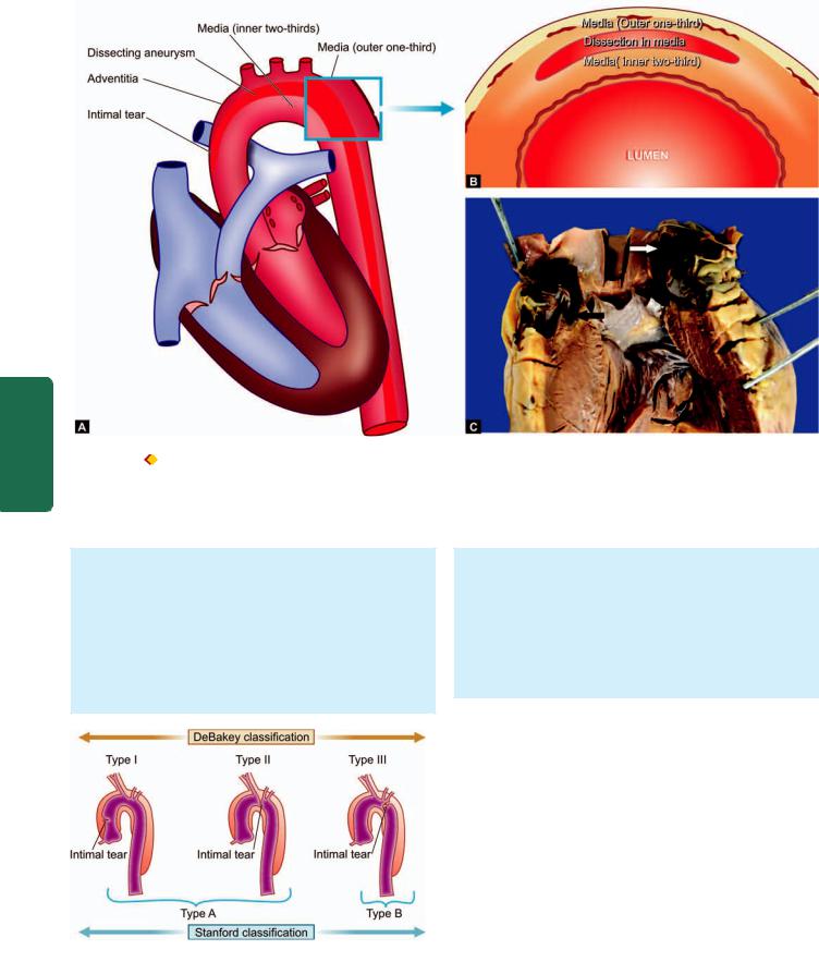
408
III SECTION
Figure 15.17 
 A, Dissecting aneurysm, (Type 1) beginning in the aortic arch and extending distally into the descending thoracic aorta as well as proximally into the ascending aorta. An intimal tear is seen in the arch. B, The cross section shows dissection typically separating the intima and inner two-thirds of the media on luminal side, from the outer one-third of the media and the adventitia. C, The ascending aorta is seen with the heart. There is an intimal tear in the aortic wall (black arrow) extending proximally upto aortic valve dissecting the media which contains clotted blood (white arrow)
Pathology Systemic
II. Stanford classification. Depending upon clinical management, these are divided into 2 types:
Type A (Proximal dissection): Involves the ascending aorta and includes type I and II of the above scheme because clinical management of DeBakey type I and II is not different.
Type B (Distal dissection): Limited to descending aorta and sparing the ascending aorta; it corresponds to DeBakey type III.
Figure 15.18 
 Two classification schemes of thoracic aortic dissection: Stanford and DeBakey. Stanford type A involving ascending aorta only includes DeBakey’s type I (involving ascending aorta and extending into descending aorta as well) and II (limited to ascending aorta only), while Stanford type B is limited to descending aorta corresponds to DeBakey type III.
Histologically, the characteristic features of cystic medial necrosis are found. These are as under:
 Focal separation of the fibromuscular and elastic tissue of the media.
 Numerous cystic spaces in the media containing basophilic ground substance.
 Fragmentation of the elastic tissue.
  Increased fibrosis of the media.
EFFECTS. The classical clinical manifestation of a dissecting aneurysm is excruciating tearing pain in the chest moving downwards. The complications arising from dissecting aneurysms are as under:
1.Rupture. Haemorrhage from rupture of a dissecting aneurysm in the ascending aorta results in mortality in 90% of cases. Most often, haemorrhage occurs into the pericardium; less frequently it may rupture into thoracic cavity, abdominal cavity or retroperitoneum.
2.Cardiac disease. Involvement of the aortic valve results in aortic incompetence. Obstruction of coronaries results in ischaemia causing fatal myocardial infarction. Rarely, dissecting aneurysm may extend into the cardiac chamber.
3.Ischaemia. Obstruction of the branches of aorta by dissection results in ischaemia of the tissue supplied. Thus, there may be renal infarction, cerebral ischaemia and infarction of the spinal cord.

FIBROMUSCULAR DYSPLASIA
Fibromuscular dysplasia first described in 1976, is a nonatherosclerotic and non-inflammatory disease affecting arterial wall, most often renal artery. Though the process may involve intima, media or adventitia, medial fibroplasia is the most common.
MORPHOLOGIC FEATURES. Grossly, the involvement is characteristically segmental—affecting vessel in a beadlike pattern with intervening uninvolved areas.
Microscopically, the beaded areas show collections of smooth muscle cells and connective tissue. There is often rupture and retraction of internal elastic lamina.
The main effects of renal fibromuscular dysplasia, depending upon the region of involvement, are renovascular hypertension and changes of renal atrophy.
VEINS
NORMAL STRUCTURE
The structure of normal veins is basically similar to that of arteries. The walls of the veins are thinner, the three tunicae (intima, media and adventitia) are less clearly demarcated, elastic tissue is scanty and not clearly organised into internal and external elastic laminae. The media contains very small amount of smooth muscle cells with abundant collagen. All veins, except vena cavae and common iliac veins, have valves best developed in veins of the lower limbs. The valves are delicate folds of intima, located every 1-6 cm, often next to the point of entry of a tributary vein. They prevent any significant retrograde venous blood flow.
VARICOSITIES
Varicosities are abnormally dilated and tortuous veins. The veins of lower extremities are involved most frequently, called varicose veins. The veins of other parts of the body which are affected are the lower oesophagus (oesophageal varices, Chapter 19), the anal region (haemorrhoids, Chapter 20) and the spermatic cord (varicocele, Chapter 23).
VARICOSE VEINS
Varicose veins are permanently dilated and tortuous superficial veins of the lower extremities, especially the long saphenous vein and its tributaries. About 10-12% of the general population develops varicose veins of lower legs, with the peak incidence in 4th and 5th decades of life. Adult females are affected more commonly than the males, especially during pregnancy. This is attributed to venous stasis in the lower legs because of compression on the iliac veins by pregnant uterus.
ETIOPATHOGENESIS. A number of etiologic and pathogenetic factors are involved in causing varicose veins. These are as follows:
i) Familial weakness of vein walls and valves is the most common cause.
ii) Increased intraluminal pressure due to prolonged upright 409 posture e.g. in nurses, policemen, surgeons etc.
iii)Compression of iliac veins e.g. during pregnancy, intravascular thrombosis, growing tumour etc.
iv)Hormonal effects on smooth muscle.
v)Obesity.
vi)Chronic constipation.
MORPHOLOGIC FEATURES. The affected veins, especially of the lower extremities, are dilated, tortuous, elongated and nodular. Intraluminal thrombosis and valvular deformities are often found.
Histologically, there is variable fibromuscular thickening of the wall of the veins due to alternate dilatation and hypertrophy. Degeneration of the medial elastic tissue may occur which may be followed by calcific foci. Mural thrombosis is commonly present which may get organised and hyalinised leading to irregular intimal thickening.
EFFECTS. Varicose veins of the legs result in venous stasis |
|
||
which is followed by congestion, oedema, thrombosis, stasis, |
|
||
dermatitis, cellulitis and ulceration. Secondary infection |
CHAPTER |
||
results in chronic varicose ulcers. |
|||
|
|||
PHLEBOTHROMBOSIS AND THROMBOPHLEBITIS |
|
||
The terms ‘phlebothrombosis’ or thrombus formation in veins, |
|
||
and ‘thrombophlebitis’ or inflammatory changes within the |
15 |
||
vein wall, are currently used synonymously. |
|||
|
|||
ETIOPATHOGENESIS. Venous thrombosis that precedes |
The |
||
thrombophlebitis is initiated by triad of changes: endothelial |
|||
|
|||
damage, alteration in the composition of blood and venous |
Blood |
||
stasis. The factors that predispose to these changes are cardiac |
|||
|
|||
failure, malignancy, use of oestrogen-containing compounds, |
|
||
postoperative state and immobility due to various reasons. |
Vessels |
||
|
|
||
MORPHOLOGIC FEATURES. The most common |
|
||
|
|
||
locations for phlebothrombosis and thrombophlebitis are |
|
|
|
the deep veins of legs accounting for 90% of cases; it is |
|
and |
|
commonly termed as deep vein thrombosis (DVT). Other |
|
||
|
|
||
locations are periprostatic venous plexus in males, pelvic |
|
Lymphatics |
|
distended and firm. Often, a mural or occlusive thrombus |
|
||
veins in the females, and near the foci of infection in the |
|
|
|
abdominal cavity such as acute appendicitis, peritonitis, |
|
|
|
acute salpingitis and pelvic abscesses. |
|
|
|
Grossly, the affected veins may appear normal or may be |
|
|
|
is present. |
|
|
|
Histologically, the thrombus that is attached to the vein |
|
|
|
wall induces inflammatory-reparative response beginning |
|
|
|
from the intima and infiltrating into the thrombi. The |
|
|
|
response consists of mononuclear inflammatory cells and |
|
|
|
fibroblastic proliferation. In late stage, thrombus is either |
|
|
|
organised or resolved leading to a thick-walled fibrous |
|
|
|
vein. |
|
|
|
|
|
|
|
EFFECTS. The clinical effects due to phlebothrombosis and |
|
||
thrombophlebitis may be local or systemic. |
|
||
Local effects are oedema distal to occlusion, heat, swelling, |
|
||
tenderness, redness and pain. |
|
||

410 Systemic effects are more severe and occur due to embolic phenomena, pulmonary thromboembolism being the most common and most important. Other systemic manifestations include bacteraemia and septic embolisation to brain, meninges, liver etc.
|
Special Types of Phlebothrombosis |
|
|
A few special variants of phlebothrombosis are considered |
|
|
below: |
|
|
1. THROMBOPHLEBITIS MIGRANS. Thrombophlebitis |
|
|
migrans or migratory thrombophlebitis or Trousseau’s |
|
|
syndrome is the term used for multiple venous thrombi that |
|
|
disappear from one site so as to appear at another site. The |
|
|
condition is not a morphologic entity but a clinical one, seen |
|
|
most often in disseminated visceral cancers (e.g. cancer of |
|
|
lungs, prostate, female reproductive tract, breast, pancreas |
|
|
and gastrointestinal tract) as part of paraneoplastic syndrome |
|
|
and is also found in nonbacterial thrombotic endocarditis. |
|
|
2. PHLEGMASIA ALBA DOLENS. This term meaning |
|
|
‘painful white leg’ refers to extensive swelling of the leg, |
|
SECTION |
occurring most frequently due to iliofemoral venous |
|
occur due to involvement of inferior vena cava. |
||
|
thrombosis. It occurs most often in women during late |
|
|
pregnancy or following delivery when the pregnant uterus |
|
|
causes pressure on the iliofemoral veins, or after extensive |
|
|
pelvic surgery. Development of pulmonary embolism may |
|
III |
3. PHLEGMASIA CERULEA DOLENS. This term |
|
|
||
|
meaning ‘painful blue leg’ refers to markedly swollen bluish |
|
Systemic |
skin with superficial gangrene. It is a serious complication |
|
of massive iliofemoral venous thrombosis and decreased |
||
|
||
|
arterial blood flow. |
|
|
4. SUPERIOR VENA CAVAL SYNDROME. Superior |
|
Pathology |
vena caval syndrome refers to obstruction of the superior |
|
vena cava. The obstruction results most often from external |
||
|
||
|
compression or from thrombosis. Some of the common |
|
|
causes of superior vena caval syndrome are malignancy |
|
|
(especially lung cancer and lymphoma), syphilitic aortic |
|
|
aneurysm and tuberculous mediastinitis. Clinical features |
|
|
include dilated veins of neck and thorax, oedema of the face, |
|
|
neck and upper chest, visual disturbances and disturbed |
|
|
sensorium. |
|
|
5. INFERIOR VENA CAVAL SYNDROME. Inferior vena |
|
|
caval syndrome is the obstruction of the inferior vena cava. |
|
|
Most often, obstruction results from thrombosis by extension |
|
|
from iliofemoral veins. Other causes of obstruction are |
|
|
external compression and neoplastic invasion. Clinical |
|
|
features are oedema of lower extremities, dilated leg veins |
|
|
and collateral venous channels in the lower abdomen |
|
|
|
|
|
LYMPHATICS |
|
|
||
|
NORMAL STRUCTURE |
|
|
Lymphatic capillaries, lymphatic vessels and lymph nodes |
|
|
comprise the lymphatic system. Lymphatic capillaries |
|
|
resemble blood capillaries, and larger lymphatics are |
|
|
identical to veins. However, lymphatics lined by a single |
layer of endothelium have thin muscle in their walls than in veins of the same size and the valves are more numerous. Lymphatic capillaries and lymphatics form plexuses around tissues and organs. The walls of lymphatic capillaries are permeable to tissue fluid, proteins and particulate matter.
LYMPHANGITIS
Inflammation of the lymphatics or lymphangitis may be acute or chronic.
Acute lymphangitis occurs in the course of many bacterial infections. The most common organisms are (β-haemolytic streptococci and staphylococci). Acute lymphangitis is often associated with lymphadenitis.
Grossly, the affected lymphatics are dilated and appear as cutaneous streaks.
Microscopically, the dilated lumen contains acute inflammatory exudate, cell debris and clotted lymph. There is inflammatory infiltration into the perilymphatic tissues alongwith hyperaemia and oedema. Acute lymphangitis generally heals completely.
Chronic lymphangitis occurs due to persistent and recurrent acute lymphangitis or from chronic infections like tuberculosis, syphilis and actinomycosis.
Histologically, there is permanent obstruction due to fibrosis of affected lymphatics called chronic lymphoedema.
LYMPHOEDEMA
Lymphoedema is swelling of soft tissues due to localised increase in the quantity of lymph (page 97). It may be primary (idiopathic) or secondary (obstructive).
I. PRIMARY (IDIOPATHIC) LYMPHOEDEMA. Lymphoedema occurring without underlying secondary cause is called primary or idiopathic lymphoedema. Its various types are as under:
1. Congenital lymphoedema. Congenital lymphoedema has further 2 subtypes—familial hereditary form (Milroy’s disease) and non-familial (simple) form.
i)Milroy’s disease is a form of congenital and familial oedema generally affecting one limb but at times may be more extensive and involve the eyelids and lips. The disease is inherited as an autosomal dominant trait and is often associated with other congenital anomalies. The condition results from developmental defect of lymphatic channels so that the affected tissue shows abnormally dilated lymphatics and the area shows honey-combed appearance. Recurrent infection of the tissue causes cellulitis and fibrosis of lymphatic vessels.
ii)Simple congenital lymphoedema is non-familial form with unknown etiology. It is often associated with Turner’s syndrome and affects one member of the family. The pathologic changes are similar to those of Milroy’s disease.
2. Lymphoedema praecox. This is a rare form of lymphoedema affecting chiefly young females. The oedema usually

begins in the foot and progresses slowly upwards to involve the whole extremity. With passage of time, the affected area becomes rough and the oedema is non-pitting. The etiology is unknown but probably the condition is related to female reproductive system because of preponderance in females and aggravation during menses.
II. SECONDARY (OBSTRUCTIVE) LYMPHOEDEMA.
This is more common form of lymphoedema. Various causes of lymphatic obstruction causing lymphoedema are as under:
i)Lymphatic invasion by malignant tumour.
ii)Surgical removal of lymphatics e.g. in radical mastectomy.
iii)Post-irradiation fibrosis.
iv)Parasitic infestations e.g. in filariasis of lymphatics producing elephantiasis.
v)Lymphangitis causing scarring and obstruction.
Obstructive lymphoedema occurs only when the obstruction is widespread as otherwise collaterals develop. The affected area consists of dilatation of lymphatics distal to obstruction with increased interstitial fluid. With passage of time, there is inflammatory scarring and the lymphatics become fibrosed with enlargement of the affected part. Rupture of dilated large lymphatics may result in escape of milky chyle into the peritoneum (chyloperitoneum), into the pleural cavity (chylothorax), into pericardial cavity (chylopericardium) and into the urinary tract (chyluria).
TUMOURS AND TUMOUR-LIKE LESIONS
Majority of benign vascular tumours are malformations or hamartomas. A hamartoma is a tumour-like lesion made up of tissues indigenous to the part but lacks the true growth potential of true neoplasms. However, there is no clear-cut distinction between vascular hamartomas and true benign tumours and are often described together. On the other hand, there are true vascular tumours which are of intermediate grade and there are frank malignant tumours.
A classification of vascular tumours and tumour-like conditions is given in Table 15.5.
TABLE 15.5: Tumours and Tumour-like Lesions of Blood |
|
411 |
|||
|
Vessels and Lymphatics. |
|
|
||
A. Benign Tumours and Hamartomas |
|
||||
|
1. |
Haemangioma |
|
||
|
2. |
Lymphangioma |
|
||
|
3. |
Glomus tumour (glomangioma) |
|
||
|
4. |
Arteriovenous malformations |
|
||
|
5. |
Bacillary angiomatosis |
|
||
B. |
Intermediate Grade Tumours |
|
|||
|
|
Haemangioendothelioma |
|
||
C. |
Malignant Tumours |
|
|||
|
1. |
Haemangiopericytoma |
|
||
|
2. |
Angiosarcoma |
|
||
|
3. |
Kaposi’s sarcoma |
|
||
|
|
|
|||
A. BENIGN TUMOURS AND HAMARTOMAS |
|
||||
Haemangioma |
|
||||
Haemangiomas are quite common lesions, especially in |
|
||||
infancy and childhood. The most common site is the skin of |
|
||||
the face. Amongst the various clinical and histologic types, |
|
||||
three important forms are described below. |
CHAPTER |
||||
CAPILLARY HAEMANGIOMA. These are the most |
|||||
|
|||||
common type. Clinically, they appear as small or large, flat |
|
||||
or slightly elevated, red to purple, soft and lobulated lesions, |
|
||||
varying in size from a few millimeters to a few centimeters |
|
||||
in diameter. They may be present at birth or appear in early |
15 |
||||
childhood. Strawberry birthmarks and ‘port-wine mark’ are |
|||||
|
|||||
some good examples. The common sites are the skin, |
|
||||
subcutaneous tissue and mucous membranes of oral cavity |
The |
||||
and lips. Less common sites are internal visceral organs like |
|||||
|
|||||
liver, spleen and kidneys. |
Blood |
||||
|
|
||||
Histologically, capillary haemangiomas are well-defined |
|
||||
but unencapsulated lobules. These lobules are composed |
|
||||
of capillary-sized, thin-walled, blood-filled vessels. These |
|
Vessels |
|||
vessels are lined by single layer of plump endothelial cells |
|
||||
|
|
||||
surrounded by a layer of pericytes. The vessels are |
|
|
|||
separated by some connective tissue stroma (Fig. 15.19). |
|
Lymphatics and |
|||
|
|
|
|
||
|
|
|
|
||
Figure 15.19 
 Capillary haemangioma of the skin. There are capillaries lined by plump endothelial cells and containing blood. The intervening stroma consists of scant connective tissue.
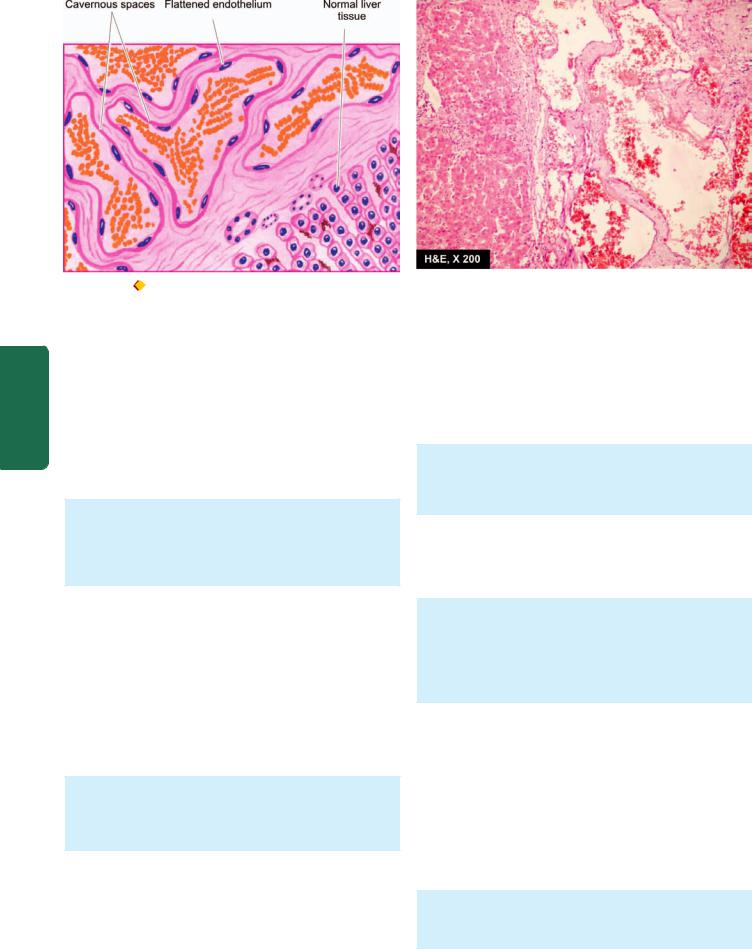
412
Pathology Systemic III SECTION
Figure 15.20 |
Cavernous haemangioma of the liver. The vascular spaces are large, dilated, many containing blood, and are lined by flattened |
endothelial cells. Scanty connective tissue stroma is seen between the cavernous spaces.
Many of the capillary haemangiomas regress spontaneously within a few years.
CAVERNOUS HAEMANGIOMA. Cavernous haemangiomas are single or multiple, discrete or diffuse, red to blue, soft and spongy masses. They are often 1 to 2 cm in diameter. They are most common in the skin (especially of the face and neck); other sites are mucosa of the oral cavity, stomach and small intestine, and internal visceral organs like the liver and spleen.
Histologically, cavernous haemangiomas are composed of thin-walled cavernous vascular spaces, filled partly or completely with blood. The vascular spaces are lined by flattened endothelial cells. They are separated by scanty connective tissue stroma (Fig. 15.20).
Cavernous haemangiomas rarely involute spontaneously.
GRANULOMA PYOGENICUM. Granuloma pyogenicum is also referred to as haemangioma of granulation tissue type.
True to its name, it appears as exophytic, red granulation tissue just like a nodule, commonly on the skin and mucosa of gingiva or oral cavity. Pregnancy tumour or granuloma gravidarum is a variant occurring on the gingiva during pregnancy and regresses after delivery. Granuloma pyogenicum often develops following trauma and is usually 1 to 2 cm in diameter.
Histologically, it shows proliferating capillaries similar to capillary haemangioma but the capillaries are separated by abundant oedema and inflammatory infiltrate, thus resembling inflammatory granulation tissue.
Lymphangioma
Lymphangiomas are lymphatic counterparts of vascular angiomas. Lymphangiomas are congenital lesions which are classified as capillary, cavernous and cystic hygroma. Combinations are also often seen.
CAPILLARY LYMPHANGIOMA. It is also called as lymphangioma simplex. It is a small, circumscribed, slightly elevated lesion measuring 1 to 2 cm in diameter. The common locations are the skin of head and neck, axilla and mucous membranes. Rarely, these may be found in the internal organs.
Histologically, capillary lymphangioma is composed of a network of endothelium-lined, capillary-sized spaces containing lymph and often separated by lymphoid aggregates.
CAVERNOUS LYMPHANGIOMA. It is more common than the capillary type. The common sites are in the region of head and neck or axilla. A large cystic variety called cystic hygroma occurs in the neck producing gross deformity in the neck.
Histologically, cavernous lymphangioma consists of large dilated lymphatic spaces lined by flattened endothelial cells and containing lymph. Scanty intervening stromal connective tissue is present (Fig. 15.21). These lesions, though benign, are often difficult to remove due to infiltration into adjacent tissues.
Glomus Tumour (Glomangioma)
Glomus tumour is an uncommon true benign tumour arising from contractile glomus cells that are present in the arteriovenous shunts (Sucquet-Hoyer anastomosis). These tumours are found most often in the dermis of the fingers or toes under a nail; other sites are mucosa of the stomach and nasal cavity. These lesions are characterised by extreme pain. They may be single or multiple, small, often less than 1 cm in diameter, flat or slightly elevated, red-blue, painful nodules.
Histologically, the tumours are composed of small blood vessels lined by endothelium and surrounded by aggregates, nests and masses of glomus cells. The glomus
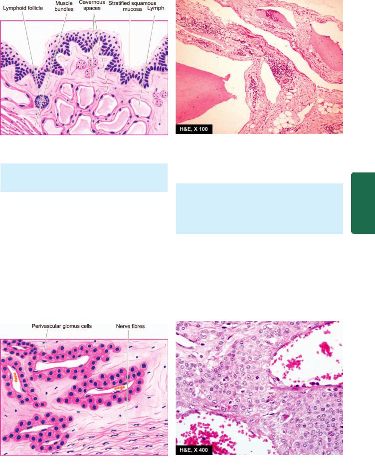
413
Figure 15.21 
 Cavernous lymphangioma of the tongue. Large cystic spaces lined by the flattened endothelial cells and containing lymph are present. Stroma shows scattered collection of lymphocytes.
cells are round to cuboidal cells with scanty cytoplasm (Fig. 15.22). The intervening connective tissue stroma contains some non-myelinated nerve fibres.
Arteriovenous Malformations
An arteriovenous (AV) malformation is a communication between an artery and vein without an intervening capillary bed. It may be congenital or acquired type. Congenital AV malformations have thick-walled vessels with hyalinisation and calcification. Acquired AV malformations reveal changes mainly in the veins which are dilated and thick-walled.
Bacillary Angiomatosis and Peliosis Hepatis
Bacillary angiomatosis is a tumour-like lesion reported in association with HIV-AIDS with CD4+ T cell counts below 100/μl. In fact, it is an opportunistic infection with gramnegative bacilli of Bartonella genus. Most common site of
involvement is the skin and bones while a closely related condition peliosis hepatis is seen in the liver (Chapter 21).
Grossly, the lesions on the skin are in the form of variablesized red papules.
Histologically, lobules of proliferating blood vessels are seen lined by epithelioid endothelial cells having mild atypia. Mixed inflammatory cell infiltrate with nuclear debris of neutrophils is present in these areas.
The condition is treated with antibiotics.
B. INTERMEDIATE GRADE TUMOURS
Haemangioendothelioma
Haemangioendothelioma is a true tumour of endothelial cells, the behaviour of which is intermediate between a haemangioma and haemangiosarcoma. It is found most often in the skin and subcutaneous tissue in relation to medium-
Lymphatics and Vessels Blood The 15 CHAPTER
Figure 15.22 
 Glomus tumour. There are blood-filled vascular channels lined by endothelial cells and surrounded by nests and masses of glomus cells.
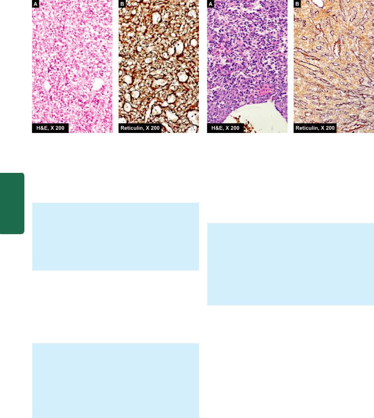
414
Pathology Systemic III SECTION
Figure 15.23 
 Haemangioendothelioma nose. A, The vascular channels are lined by multiple layers of plump endothelial cells having minimal mitotic activity obliterating the lumina. B, Reticulin stain shows condensation of reticulin around the vessel wall but not between the proliferating cells.
sized and large veins. Haemangioblastoma is the term used for similar tumour occurring in the cerebellum (Chapter 30).
Grossly, the tumour is usually well-defined, grey-red, polypoid mass.
Microscopically, there is active proliferation of endothelial cells forming several layers around the blood vessels so that vascular lumina are difficult to identify. These cells may have variable mitotic activity. Reticulin stain delineates the pattern of cell proliferation inner to the basement membrane (Fig. 15.23).
C. MALIGNANT TUMOURS
Haemangiopericytoma
Haemangiopericytoma is an uncommon tumour arising from pericytes. Pericytes are cells present external to the endothelial cells of capillaries and venules. This is a rare tumour that can occur at any site and at any age and may vary in size from 1 to 8 cm.
Microscopically, the tumour is composed of capillaries surrounded by spindle-shaped pericytes outside the vascular basement membrane forming whorled arrangement. These tumour cells may have high mitotic rate and areas of necrosis. Silver impregnation stain (i.e. reticulin stain) is employed to confirm the presence of pericytes outside the basement membrane of capillaries and to distinguish it from haemangioendothelioma
(Fig. 15.24).
Local recurrences are common and distant spread occurs in about 20% of cases.
Angiosarcoma
Also known as haemangiosarcoma and malignant haemangioendothelioma, it is a malignant vascular tumour
Figure 15.24 
 Haemangiopericytoma liver. Spindled cells surround the vascular lumina in a whorled fashion, highlighted by reticulin stain.
These tumour cells have bland nuclei and few mitoses.
occurring most frequently in the skin, subcutaneous tissue, liver, spleen, bone, lung and retroperitoneal tissues. It can occur in both sexes and at any age. Hepatic angiosarcomas are of special interest in view of their association with carcinogens like polyvinyl chloride, arsenical pesticides and radioactive contrast medium, thorotrast, used in the past.
Grossly, the tumours are usually bulky, pale grey-white, firm masses with poorly-defined margins. Areas of haemorrhage, necrosis and central softening are frequently present.
Microscopically, the tumours may be well-differentiated masses of proliferating endothelial cells around wellformed vascular channels, to poorly-differentiated lesions composed of plump, anaplastic and pleomorphic cells in solid clusters with poorly identifiable vascular channels
(Fig. 15.25).
These tumours invade locally and frequently have distant metastases in the lungs and other organs. Lymphangiosarcoma is a histologically similar tumour occurring in obstructive lymphoedema of long duration.
Kaposi’s Sarcoma
Kaposi’s sarcoma is a malignant angiomatous tumour, first described by Kaposi, Hungarian dermatologist, in 1872. However, the tumour has attracted greater attention in the last two decades due to its frequent occurrence in patients with HIV/AIDS.
CLASSIFICATION. Presently, four forms of Kaposi’s sarcoma are described:
1. Classic (European) Kaposi’s sarcoma. This is the form which was first described by Kaposi. It is more common in men over 60 years of age of Eastern European descent. The disease is slow growing and appears as multiple, small, purple, dome-shaped nodules or plaques in the skin,
415
Figure 15.25 
 Angiosarcoma spleen. A, Gross appearance of lobulated masses of grey white necrotic and haemorrhagic parenchyma. B, The tumour cells show proliferation of moderately pleomorphic anaplastic cells. C, These tumour cells show positive staining for endothelial marker,
CD34.
especially on the legs. Involvement of visceral organs occurs in about 10% cases after many years.
2.African (Endemic) Kaposi’s sarcoma. This form is common in equatorial Africa. It is so common in Uganda that it comprises 9% of all malignant tumours in men. It is found in younger age, especially in boys and in young men and has a more aggressive course than the classic form. The disease begins in the skin but grows rapidly to involve other tissues, especially lymph nodes and the gut.
3.Epidemic (AIDS-associated) Kaposi’s sarcoma. This form is seen in about 30% cases of AIDS, especially in young male homosexuals than the other high-risk groups. The cutaneous lesions are not localised to lower legs but are more extensively distributed involving mucous membranes, lymph nodes and internal organs early in the course of disease.
4.Kaposi’s sarcoma in renal transplant cases. This form is associated with recipients of renal transplants who have been administered immunosuppressive therapy for a long time. The lesions may be localised to the skin or may have widespread systemic involvement.
PATHOGENESIS. Pathogenesis of Kaposi’s sarcoma is complex. It is an opportunistic neoplasm in immunosuppressed patients which has excessive proliferation of spindle cells of vascular origin having features of both endothelium and smooth muscle cells:

 Epidemiological studies have suggested a viral association implicating HIV and human herpesvirus 8 (HSV 8, also called Kaposi’s sarcoma-associated herpesvirus or KSHV).
 Occurrence of Kaposi’s sarcoma involves interplay of HIV-1 infection, HHV-8 infection, activation of the immune system and secretion of cytokines (IL-6, TNF-α, GM-CSF, basic fibroblast factor, and oncostain M). Higher incidence of Kaposi’s sarcoma in male homosexuals is explained by increased secretion of cytokines by their activated immune system.
 Defective immunoregulation plays a role in its pathogenesis is further substantiated by observation of second malignancy (e.g. leukaemia, lymphoma and myeloma) in about one-third of patients with Kaposi’s sarcoma.
MORPHOLOGIC FEATURES. Pathologically, all forms of Kaposi’s sarcoma are similar
Lymphatics and Vessels Blood The 15 CHAPTER
Figure 15.26 |
Kaposi’s sarcoma in late nodular stage. There are slit-like blood-filled vascular spaces. Between them are present bands of |
plump spindle-shaped tumour cells.

416Grossly, the lesions in the skin, gut and other organs form prominent, irregular, purple, dome-shaped plaques or nodules.
Histologically, the changes are nonspecific in the early patch stage and more characteristic in the late nodular stage.
 Early patch stage: There are irregular vascular spaces separated by interstitial inflammatory cells and extravasated blood and haemosiderin.
 Late nodular stage: There are slit-like vascular spaces containing red blood cells and separated by spindle-
shaped, plump tumour cells. These spindle-shaped tumour cells are probably of endothelial origin (Fig. 15.26).
CLINICAL COURSE. The clinical course and biologic behaviour of Kaposi’s sarcoma is quite variable. The classic form of Kaposi’s sarcoma is largely confined to skin and the course is generally slow and insidious with long survival. The endemic (African) and epidemic (AIDS-associated) Kaposi’s sarcoma, on the other hand, has a rapidly progressive course, often with widespread cutaneous as well as visceral involvement, and high mortality.
Pathology Systemic III SECTION