

- •Foreword
- •Preface
- •Contents
- •1. Introduction to Pathology
- •2. Techniques for the Study of Pathology
- •6. Inflammation and Healing
- •8. Neoplasia
- •16. The Heart
- •17. The Respiratory System
- •18. The Eye, ENT and Neck
- •20. The Gastrointestinal Tract
- •24. The Female Genital Tract
- •25. The Breast
- •26. The Skin
- •27. The Endocrine System
- •28. The Musculoskeletal System
- •29. Soft Tissue Tumours
- •30. The Nervous System
- •Appendix
- •Further Readings
- •Index

538
Pathology Systemic III SECTION
Chapter 20 |
The Gastrointestinal Tract |
OESOPHAGUS
NORMAL STRUCTURE
The oesophagus is a muscular tube extending from the pharynx to the stomach. In an adult, this distance measures 25 cm. However, from the clinical point of view, the distance from the incisor teeth to the gastro-oesophageal (GE) junction is about 40 cm. The region of proximal oesophagus at the level of cricopharyngeus muscle is called the upper oesophageal sphincter, while the portion adjacent to the anatomic gastrooesophageal junction is referred to as lower oesophageal
sphincter.
Histologically, the wall of the oesophagus consists of mucosa, submucosa, muscularis propria and adventitia/ serosa.

 The mucosa is composed of non-keratinising stratified squamous epithelium overlying lamina propria except at the lower end for a distance of 0.5 to 1.5 cm. The basal layer of the epithelium may contain some melanocytes, argyrophil cells and Langerhans’ cells. At the lower end of the oesophagus, there is sudden change from stratified squamous epithelium to mucin-secreting columnar epithelium; this is called the junctional mucosa.
 The submucosa consists of loose connective tissue with sprinkling of lymphocytes, plasma cells, and occasional eosinophil and mast cell. Mucus-producing glands are scattered throughout the submucosa.

 The muscularis propria is composed of 2 layers of smooth muscle—an inner circular coat and an outer longitudinal coat. The proximal portion of oesophagus contains skeletal muscle fibres from cricopharyngeus muscle. The parasympathetic nerve supply by the vagus nerve is in the form of extrinsic and intrinsic plexuses.
 The adventitia/serosa is the outer covering of oesophagus. Serosa is present in intra-abdominal part of oesophagus only, while elsewhere the perioesophageal adventitia covers it.
The major functions of oesophagus are swallowing by peristaltic activity and to prevent the reflux of gastric contents into the oesophagus.
CONGENITALANOMALIES
Congenital anomalies of the oesophagus are uncommon and are detected soon after birth. These include a few rare anomalies such as agenesis (congenital absence of oesophagus) which is incompatible with life, duplication of oesophagus (double oesophagus) and congenital stenosis (i.e. fibrous thickening of the oesophageal wall and atrophy of
the muscularis propria). However, oesophageal atresia and tracheooesophageal fistula are relatively more common.
OESOPHAGEAL ATRESIA AND TRACHEO-OESO- PHAGEAL FISTULA. In about 85% of cases, congenital atresia of the oesophagus is associated with tracheooesophageal fistula, usually at the level of tracheal bifurcation. For survival, the condition must be recognised and corrected surgically within 48 hours of birth of the newborn. Clinically, the condition is characterised by regurgitation of every feed, hypersalivation, attacks of cough and cyanosis. Death usually results from asphyxia, aspiration pneumonia and fluid-electrolyte imbalance.
Morphologically, the condition is recognised by cord-like non-canalised segment of oesophagus having blind pouch at both ends.
MUSCULAR DYSFUNCTIONS
These are disorders in which there is motor dysfunction of the oesophagus, manifested clinically by dysphagia. These include achalasia, hiatus hernia, oesophageal diverticula, and webs and rings.
Achalasia (Cardiospasm)
Achalasia of the oesophagus is a neuromuscular dysfunction due to which the cardiac sphincter fails to relax during swallowing and results in progressive dysphagia and dilatation of the oesophagus (mega-oesophagus).
ETIOLOGY. There is loss of intramural neurons in the wall of the oesophagus. Most cases are of primary idiopathic achalasia which may be congenital. Secondary achalasia may occur from some other causes which includes: Chagas’ disease (an epidemic parasitosis with Trypansoma cruzi), infiltration into oesophagus by gastric carcinoma or lymphoma, certain viral infections, and neurodegenerative diseases.
MORPHOLOGIC FEATURES. There is dilatation above the short contracted terminal segment of the oesophagus. Muscularis propria of the wall may be of normal thickness, hypertrophied as a result of obstruction, or thinned out due to dilatation. Secondary oesophagitis may supervene and cause oesophageal ulceration and haematemesis.
Hiatus Hernia
Hiatus hernia is the herniation or protrusion of part of the stomach through the oesophageal hiatus of the diaphragm. Oesophageal hiatal hernia is the cause of diaphragmatic
hernia in 98% of cases. The condition is diagnosed radiologically in about 5% of apparently normal asymptomatic individuals. In symptomatic cases, especially the elderly women, the clinical features are heartburn (retrosternal burning sensation) and regurgitation of gastric juice into the mouth, both of which are worsened due to heavy work, lifting weights and excessive bending.
ETIOLOGY. The basic defect is the failure of the muscle fibres of the diaphragm that form the margin of the oesophageal hiatus. This occurs due to shortening of the oesophagus which may be congenital or acquired.
 Congenitally short oesophagus may be the cause of hiatus hernia in a small proportion of cases.

 More commonly, it is acquired due to secondary factors which cause fibrous scarring of the oesophagus as follows:
a)Degeneration of muscle due to aging.
b)Increased intra-abdominal pressure such as in pregnancy, abdominal tumours etc.
c)Recurrent oesophageal regurgitation and spasm causing inflammation and fibrosis.
d)Increase in fatty tissue in obese people causing decreased muscular elasticity of diaphragm.
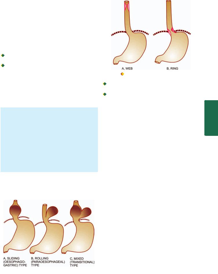
MORPHOLOGIC FEATURES. There are 3 patterns in hiatus hernia (Fig. 20.1):
i)Sliding or oesophago-gastric hernia is the most common, occurring in 85% of cases. The herniated part of the stomach appears as supradiaphragmatic bell due to sliding up on both sides of the oesophagus.
ii)Rolling or para-oesophageal hernia is seen in 10% of cases. This is a true hernia in which cardiac end of the stomach rolls up para-oesophageally, producing an intrathoracic sac.
iii)Mixed or transitional hernia constitutes the remaining 5% cases in which there is combination of sliding and rolling hiatus hernia.
Oesophageal Diverticula
Diverticula are the outpouchings of oesophageal wall at the point of weakness. They may be congenital or acquired.
Figure 20.1 
 Patterns of hiatus hernia.
539
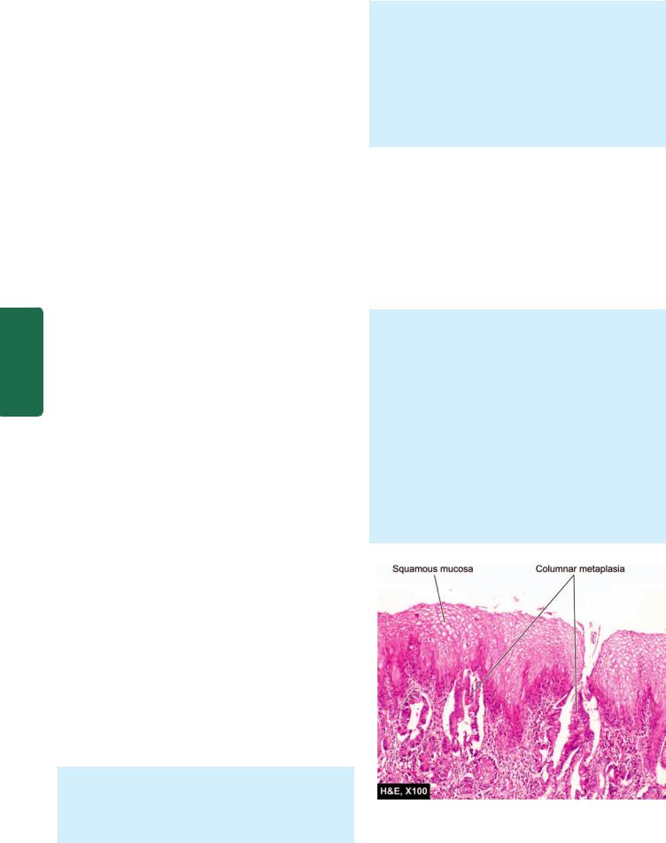
Figure 20.2 |
Oesophageal webs and rings. |
|
||
Congenital diverticula occur either at the upper end of |
|
|||
the oesophagus or at the bifurcation of trachea. |
|
|||
Acquired diverticula may be of 2 types: |
|
|||
a) Pulsion (Zenker’s) type—is seen in the region of hypo- |
|
|||
pharynx and occurs due to oesophageal obstruction such as |
CHAPTER |
|||
due to chronic oesophagitis, carcinoma etc. The mucosa and |
||||
|
||||
submucosa herniate through the weakened area or through |
|
|||
defect in the muscularis propria. |
|
|||
b) Traction type—occurs in the lower third of oesophagus |
|
|||
from contraction of fibrous tissue such as from pleural |
20 |
|||
adhesions, scar tissue of healed tuberculous lesions in the |
||||
hilum, silicosis etc. |
|
|||
Complications of diverticula include obstruction, infec- |
The |
|||
tion, perforation, haemorrhage and carcinoma. |
||||
Gastrointestinal |
||||
Oesophageal Webs and Rings |
||||
|
||||
Radiological shadows in the oesophagus resembling ‘webs’ |
|
|||
and ‘rings’ are observed in some patients complaining of |
|
|||
dysphagia. |
|
|
|
|
WEBS. Those located in the upper oesophagus, seen more |
|
|||
commonly in adult women, and associated with dysphagia, |
|
|||
iron deficiency anaemia and chronic atrophic glossitis |
Tract |
|||
(Plummer-Vinson syndrome) are called ‘webs’. |
||||
|
||||
RINGS. Those located in the lower oesophagus, not |
|
|||
associated with iron-deficiency anaemia, nor occurring in |
|
|||
women alone, are referred to as ‘Schatzki’s rings’. |
|
|||
|
|
|
||
MORPHOLOGIC FEATURES. The rings and webs are |
|
|
||
transverse folds of mucosa and submucosa encircling the |
|
|
||
entire circumference, or are localised annular thickenings |
|
|
||
of the muscle (Fig. 20.2). These give characteristic |
|
|
||
radiological shadows. |
|
|
||
HAEMATEMESIS OF OESOPHAGEAL ORIGIN |
|
|||
Massive haematemesis (vomiting of blood) may occur due |
|
|||
to vascular lesions in the oesophagus. These lesions are as |
|
|||
under: |
|
|
|
|
1. OESOPHAGEAL VARICES. Oesophageal varices are |
|
|||
tortuous, dilated and engorged oesophageal veins, seen along |
|
|||
540 the longitudinal axis of oesophagus. They occur as a result of elevated pressure in the portal venous system, most commonly in cirrhosis of the liver (Chapter 22). Less common causes are: portal vein thrombosis, hepatic vein thrombosis (Budd-Chiari syndrome) and pylephlebitis. The lesions occur as a result of bypassing of portal venous blood from the liver to the oesophageal venous plexus. The increased venous pressure in the superficial veins of the oesophagus may result in ulceration and massive bleeding.
2. MALLORY-WEISS SYNDROME. In this condition, there is lacerations of mucosa at the gastro-oesophageal junction following minor trauma such as by vomiting, retching or vigorous coughing. Patients present with upper gastro-oesophageal bleeding.
3. RUPTURE OF THE OESOPHAGUS. Rupture of the oesophagus may occur following trauma, during oesophagoscopy, indirect injury (e.g. due to sudden acceleration and deceleration of the body) and spontaneous rupture (e.g. after overeating, extensive aerophagy etc).
|
4. OTHER CAUSES. Oesophageal haematemesis may also |
|
SECTION |
occur in the following conditions: |
|
i) Bursting of aortic aneurysm into the lumen of oesophagus |
||
|
||
|
ii) Vascular erosion by malignant growth in the vicinity |
|
|
iii) Hiatus hernia |
|
|
iv) Oesophageal cancer |
|
III |
v) Purpuras |
|
vi) Haemophilia. |
||
|
||
Systemic |
INFLAMMATORY LESIONS |
|
Inflammation of the oesophagus, oesophagitis, occurs most |
||
|
||
|
commonly from reflux, although a number of other clinical |
|
|
conditions and infections may also cause oesophagitis as |
|
Pathology |
under: |
|
Reflux (Peptic) Oesophagitis |
||
|
||
|
Reflux of the gastric juice is the commonest cause of |
|
|
oesophagitis. |
PATHOGENESIS. Gastro-oesophageal reflux, to an extent, may occur in normal healthy individuals after meals and in early pregnancy. However, in some clinical conditions, the gastro-oesophageal reflux is excessive, resulting in inflammation of the lower oesophagus. These conditions are as under:
i)Sliding hiatus hernia
ii)Chronic gastric and duodenal ulcers
iii)Nasogastric intubation
iv)Persistent vomiting
v)Surgical vagotomy
vi)Neuropathy in alcoholics, diabetics
vii)Oesophagogastrostomy.
MORPHOLOGIC FEATURES. Endoscopically, the demarcation between normal squamous and columnar epithelium at the junctional mucosa is lost. The affected distal oesophageal mucosa is red, erythematous, friable and bleeds on touch. In advanced cases, there are features
of chronic disease such as nodularity, strictures, ulcerations and erosions.
Microscopically, the reflux changes in the distal oesophagus include basal cell hyperplasia and deep elongation of the papillae touching close to the surface epithelium. Inflammatory changes vary according to the stage of the disease. In early stage, mucosa and submucosa are infiltrated by some polymorphs and eosinophils; in chronic stage, there is lymphocytic infiltration and fibrosis of all the layers of the oesophageal wall.
Barrett’s Oesophagus
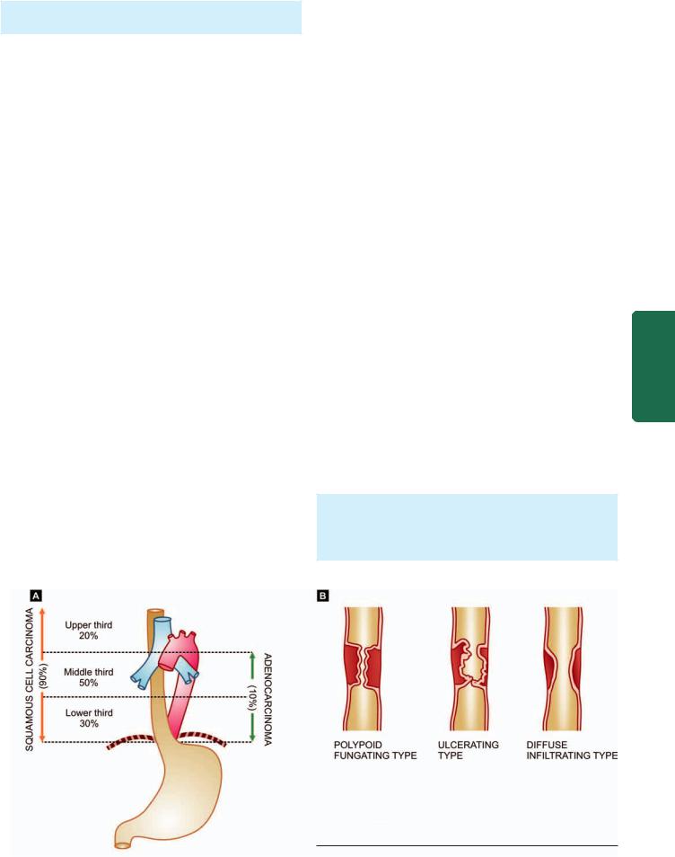
This is a condition in which, following reflux oesophagitis, stratified squamous epithelium of the lower oesophagus is replaced by columnar epithelium (columnar metaplasia). The condition is seen more commonly in later age and is caused by factors producing gastro-oesophageal reflux disease (described above). Barrett’s oesophagus is a premalignant condition evolving sequentially from Barrett’s epithelium (columnar metaplasia) → dysplasia → carcinoma in situ → oesophageal adenocarcinoma.
MORPHOLOGIC FEATURES. Endoscopically, the affected area is red and velvety. Hiatus hernia and peptic ulcer at squamocolumnar junction (Barrett’s ulcer) are frequently associated.
Microscopically, the most common finding is the replacement of squamous epithelium by metaplastic columnar cells. Barrett’s oesophagus may be composed of intestinal epithelium, fundic gastric glands, or cardiac mucous glands. Other cells present in the glands may be Paneth cells (Fig. 20.3), goblet cells, chief cells, parietal cells, mucus-secreting cells and endocrine cells.
Inflammatory changes, acute or chronic, are commonly accompanied. Dysplastic changes of the columnar epithelium or glands may be present.
Surveillance endoscopic biopsies are advised because Barrett’s intestinal metaplasaia may develop dysplasia.
Figure 20.3 
 Barrett’s oesophagus. Part of the oesophagus which is normally lined by squamous epithelium undergoes metaplastic change to columnar epithelium of intestinal type.
High-grade dysplasia may progress to invasive adenocarcinoma of the oesophagus in up to 20% cases.
Infectious Oesophagitis
A number of opportunistic infections in immunosuppressed individuals can cause oesophagitis. Some of these agents are as follows:
i)Candida (Monilial) oesophagitis
ii)Herpes simplex (Herpetic) oesophagitis
iii)Cytomegalovirus
iv)Tuberculosis.
Other Causes of Oesophagitis
i)Eosinophilic oesophagitis caused by radiation, corrosives
ii)Intake of certain drugs (anticholinergic drugs, doxycycline, tetracycline)
iii)Ingestion of hot, irritating fluids
iv)Crohn’s disease
v)Various vesiculobullous skin diseases.
TUMOURS OF OESOPHAGUS
Benign tumours of the oesophagus are uncommon and small in size (less than 3 cm). The epithelial benign tumours project as intraluminal masses arising from squamous epithelium (squamous cell papilloma), or from columnar epithelium (adenoma). The stromal or mesenchymal benign tumours are intramural masses such as leiomyoma and others like lipoma, fibroma, neurofibroma, rhabdomyoma, lymphangioma and haemangioma.
For all practical purposes, malignant tumours of the oesophagus are carcinomas because sarcomas such as leiomyosarcoma and fibrosarcoma occur with extreme rarity.
Carcinoma of Oesophagus
Carcinoma of the oesophagus is diagnosed late, after symptomatic oesophageal obstruction (dysphagia) has developed and the tumour has transgressed the anatomical limits of the organ. The tumour occurs more commonly in
men over 50 years of age. Prognosis is dismal: with standard methods of therapy (surgical resection and/or irradiation), 70% of the patients die within one year of diagnosis. Fiveyear survival rate is 5-10%.
ETIOLOGY. Although exact etiology of carcinoma of the oesophagus is not known, a number of conditions and factors have been implicated as under:
1. Diet and personal habits:
i)Heavy smoking
ii)Alcohol consumption
iii)Intake of foods contaminated with fungus
iv)Nutritional deficiency of vitamins and trace elements.
2. Oesophageal disorders:
i)Oesophagitis (especially Barrett’s oesophagus in adenocarcinoma)
ii)Achalasia
iii)Hiatus hernia
iv)Diverticula
v)Plummer-Vinson syndrome.
3. Other factors:
i)Race—more common in the Chinese and Japanese than in Western races; more frequent in blacks than whites.
ii)Family history—association with tylosis (keratosis palmaris et plantaris).
iii)Genetic factors—predisposition with coeliac disease, epidermolysis bullosa, tylosis.
iv)HPV infection—is the recent addition in etiologic factors.
At molecular level, abnormality of p53 tumour suppressor gene has been found associated with a number of above risk factors, notably with consumption of tobacco and alcohol, and in cases having proven Barrett’s oesophagus.
MORPHOLOGIC FEATURES. Carcinoma of the oesophagus is mainly of 2 types—squamous cell (epidermoid) and adenocarcinoma. The sites of predilection for each of these 2 forms is shown in Fig. 20.4,A.
541
Tract Gastrointestinal The 20 CHAPTER
Figure 20.4 
 A, Carcinoma oesophagus—sites of predilection for squamous cell carcinoma and adenocarcinoma. B, Gross patterns of squamous cell carcinoma of the oesophagus.
542
Pathology Systemic III SECTION
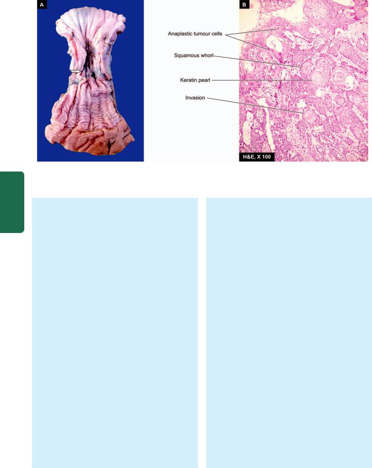
Figure 20.5 
 Squamous cell carcinoma oesophagus. A, Gross appearance. The tubular structure has thick muscle in its wall and has longitudinal mucosal folds. There is a concentric circumferential thickening in the middle (arrow) causing narrowing of the lumen (arrow). The mucosal surface is ulcerated. B, Photomicrograph shows whorls of anaplastic squamous cells invading the underlying soft tissues.
SQUAMOUS CELL (EPIDERMOID) CARCINOMA.
Squamous cell or epidermoid carcinoma comprises 90% of primary oesophageal cancers. It is exceeded in incidence by carcinoma colon, rectum and stomach amongst all the gastrointestinal cancers. The disease occurs in 6th to 7th decades of life and is more common in men than women. The sites of predilection are the three areas of oesophageal constrictions. Half of the squamous cell carcinomas of oesophagus occur in the middle third, followed by lower third, and the upper third of oesophagus in that order of frequency.
Grossly, 3 types of patterns are recognised (Fig. 20.4,B):
i)Polypoid fungating type—is the most common form. It appears as a cauliflower-like friable mass protruding into the lumen.
ii)Ulcerating type—is the next common form. It looks grossly like a necrotic ulcer with everted edges (Fig. 20.5, A).
iii)Diffuse infiltrating type—appears as an annular, stenosing narrowing of the lumen due to infiltration into the wall of oesophagus.
Microscopically, majority of the squamous cell carcinomas of the oesophagus are well-differentiated or moderatelydifferentiated (Fig. 20.5, B). Prickle cells, keratin formation and epithelial pearls are commonly seen. However, non-keratinising and anaplastic growth patterns can also occur. An exophytic, slow-growing, extremely welldifferentiated variant, verrucous squamous cell carcinoma, has also been reported in the oesophagus.
ADENOCARCINOMA. Adenocarcinoma of the oesophagus constitutes less than 10% of primary oesophageal cancer. It occurs predominantly in men in their 4th to 5th decades. The common locations are lower
and middle third of the oesophagus. These tumours have a strong and definite association with Barrett’s oesophagus in which there are foci of gastric or intestinal type of epithelium.
Grossly, oesophageal adenocarcinoma appears as nodular, elevated mass in the lower oesophagus.
Microscopically, adenocarcinoma of the oesophagus can have 3 patterns:
i)Intestinal type—is the adenocarcinoma with a pattern similar to that seen in adenocarcinoma of intestine or stomach.
ii)Adenosquamous type—is the pattern in which there is an irregular admixture of adenocarcinoma and squamous cell carcinoma.
iii)Adenoid cystic type—is an uncommon variety and is akin to similar growth in salivary gland i.e. a cribriform appearance in an epithelial tumour.
Adenocarcinoma of the oesophagus must be distinguished from adenocarcinoma of the gastric cardia. This is done by identifying normal oesophageal mucosa on distal as well as proximal margin of the tumour.
OTHER CARCINOMAS. Besides the two main histological types of oesophageal cancer, a few other varieties are occasionally encountered. These are as follow:
i)Mucoepidermoid carcinoma is a tumour having characteristics of squamous cell as well as mucus-secreting carcinomas.
ii)Malignant melanoma is derived from melanoblasts in the epithelium of the oesophagus.
iii)Oat cell carcinoma arises from argyrophil cells in the basal layer of the epithelium.
iv)Undifferentiated carcinoma is an anaplastic carcinoma which cannot be classified into any recognisable type of carcinoma.
v)Carcinosarcoma consists of malignant epithelial as well as sarcomatous components.
vi)Secondary tumours rarely occur in the oesophagus from carcinomas of the breast, kidney and adrenals.
SPREAD. The oesophageal cancer spreads locally as well as to distant sites.
i)Local spread. This is the most important mode of spread and is of great importance for surgical treatment. The local spread may occur in the transverse as well as longitudinal direction. The tumour may invade below into the stomach, above into the hypopharynx, into the trachea resulting in tracheo-oesophageal fistula, and may involve larynx causing hoarseness. The tumour may invade the muscular wall of the oesophagus and involve the mediastinum, lungs, bronchi, pleura and aorta.
ii)Lymphatic spread. Submucosal lymphatic permeation may lead to multiple satellite nodules away from the main tumour. Besides, the lymphatic spread may result in metastases to the cervical, para-oesophageal, tracheobronchial and subdiaphragmatic lymph nodes.
iii)Haematogenous spread. Blood-borne metastases from the oesophageal cancer are rare, probably because the death occurs early due to invasion of important structures by other modes of spread. However, metastatic deposits by haematogenous route can occur in the lungs, liver and adrenals.
STOMACH
NORMAL STRUCTURE
The stomach is ‘gland with cavity’, extending from its junction with lower end of the oesophagus (cardia) to its junction with the duodenum (pylorus). The lesser curvature
is inner concavity on the right, while the greater curvature is the outer convexity on the left side of the stomach.
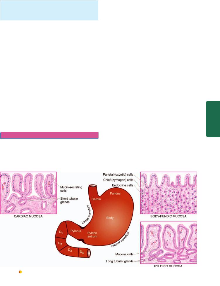
The stomach has 5 anatomical regions (Fig. 20.6):
1.Cardia is the oesophagogastric junction and lacks the sphincter.
2.Fundus is the portion above the horizontal line drawn across the oesophagogastric junction.
3.Body is the middle portion of the stomach between the fundus and the pyloric antrum.
4.Pyloric antrum is the distal third of the stomach.
5.Pylorus is the junction of distal end of the stomach with the duodenum. It has powerful sphincter muscle.
The mucosal folds in the region of the body and the fundus are loose (rugae), while the antral mucosa is somewhat flattened. Gastric canal is the relatively fixed portion of the pyloric antrum and the adjoining lesser curvature; it is the site for numerous pathological changes such as gastritis, peptic ulcer and gastric carcinoma.
The stomach receives its blood supply from the left gastric artery and the branches of the hepatic and splenic arteries with widespread anastomoses. Numerous gastric lymphatics which communicate freely with each other are also present. The innervation of the stomach is by the vagi and branches of the sympathetic which are connected with ganglia in the muscular and submucous layers.
Histologically, the wall of the stomach consists of 4 layers— serosa, muscularis, submucosa and mucosa.
1.Serosa is derived from the peritoneum which is deficient in the region of lesser and greater curvatures.
2.Muscularis consists of 3 layers of smooth muscle fibres— the outer longitudinal, the middle circular and the inner oblique. Nerve plexuses and ganglion cells are present between the longitudinal and circular layers of muscle. The pyloric sphincter is the thickened circular muscle layer at the gastroduodenal junction.
543
Tract Gastrointestinal The 20 CHAPTER
Figure 20.6 
 Anatomical subdivisions of the stomach correlated with histological appearance of gastric mucosa in different regions. D1, D2, D3 and D4 are the first to fourth parts of the duodenum.

5443. Submucosa is a layer of loose fibroconnective tissue binding the mucosa to the muscularis loosely and contains branches of blood vessels, lymphatics and nerve plexuses and ganglion cells.
4. Mucosa consists of 2 layers—superficial and deep. Between the two layers is the lamina propria composed of network of fibrocollagenic tissue with a few lymphocytes, plasma cells, macrophages and eosinophils. The mucosa is externally bounded by muscularis mucosae:
i) Superficial layer. It consists of a single layer of surface epithelium composed of regular, mucin-secreting, tall columnar cells with basal nuclei. There is a very rapid turnover of these cells. These dip down at places to form crypts (or pits or foveolae).
 Cardiac mucosa is the transition zone between the oesophageal squamous mucosa and the oxyntic mucosa of the fundus and body with which it gradually merges.

 Oxyntic mucosa lines both gastric fundus and body.

 Antral mucosa lines the pyloric antrum.
ii) Deep layer: It consists of glands that open into the bottom of the crypts. Depending upon the structure, these glands are of 3 types:
SECTION |
a) |
Glands of the cardia are simple tubular or compound |
||
b) |
Glands of the body-fundus are long, tubular and tightly |
|||
|
tubulo-racemose, lined by mucin secreting cells. A few |
|||
|
endocrine cells and occasional parietal and chief cells are also |
|||
|
present. |
|
|
|
III |
packed which may be coiled or dilated. There are 4 types of |
|||
cells present in the glands of body-fundic mucosa: |
|
|||
|
|
|||
Systemic |
|
Parietal (Oxyntic) cells—are the most numerous and line |
||
|
the superficial (upper) part of the glands. Parietal cells |
|||
|
are triangular in shape, have dark-staining nuclei and |
|||
|
|
|||
|
|
eosinophilic cytoplasm. These cells are responsible for |
||
|
|
production of hydrochloric acid of the gastric juice and |
||
Pathology |
|
the blood group substances. |
|
|
|
Chief (Peptic) cells—are the dominant cells in the deeper |
|||
|
|
|||
|
|
(lower) parts of the glands. Their basal nuclei are large |
||
|
|
with prominent nucleoli and the cytoplasm is coarsely |
||
|
|
granular and basophilic. These cells secrete pepsin of the |
||
|
|
gastric juice. |
|
|
|
|
Mucin-secreting neck cells—are small and fewer. These |
||
|
|
cells are present in the region of the narrow neck of the |
||
|
|
gastric glands i.e. at the junction of the glands with the |
||
|
|
pits. |
|
|
|
|
Endocrine (Kulchitsky or Enterochromaffin) cells—are |
||
|
|
widely distributed in the mucosa of all parts of the |
||
|
|
alimentary tract and are described later (page 561). |
||
|
c) |
Glands of the pylorus are much longer than the body-fundic |
||
|
glands. These are simple tubular glands which are often |
|||
|
coiled. They are lined mainly by small, granular, mucin- |
|||
|
secreting cells resembling neck cells and occasional parietal |
|||
|
cells but no chief cells. Gastrin-producing G-cells are present |
|||
|
predominantly in the region of antropyloric mucosa, with a |
|||
|
small number of these cells in the crypts and Brunner’s glands |
|||
|
of the proximal duodenum. |
|
|
|
|
|
The secretory products of the gastric mucosa are the |
||
|
gastric juice and the intrinsic factor, required for absorption of |
|||
|
vitamin B12. Gastric juice consists of hydrochloric acid, |
|||
|
pepsin, mucin and electrolytes like Na+, K+, HCO’ |
3 |
and Cl–. |
|
|
|
|
|
|
Hydrochloric acid is produced by the parietal (oxyntic) cells by the interaction of Cl’ ions of the arterial blood with water and carbon dioxide in the presence of the enzyme, carbonic anhydrase. The degree of gastric activity is correlated with the ‘total parietal cell mass’. Injection of histamine can stimulate the production of acid component of the gastric juice, while the pepsin-secreting chief cells do not respond to histamine. Physiologically, the gastric secretions are stimulated by the food itself.
The control of gastric secretions chiefly occurs in one of the following 3 ways:
1.Cephalic phase—is stimulated by the sight, smell, taste or even thought of food. A neural reflex is initiated via branches of the vagus nerve that promotes the release of hydrochloric acid, pepsinogen and mucus.
2.Gastric phase—is triggered by the mechanical and chemical stimuli.
i)Mechanical stimulation comes from stretching of the wall of the stomach and conveying neural messages to the medulla for gastric secretion.
ii)Chemical stimulation is by digested proteins, amino acids, bile salts and alcohol which act on gastrin-producing G cells. Gastrin then passes into the blood stream and on return to the stomach promotes the release of gastric juice.
3. Intestinal phase—is triggered by the entry of proteinrich food in the small intestine. An intestinal hormone capable of stimulating gastric secretion is probably released into the blood stream.
GASTRIC ANALYSIS
In various diseases of the stomach, the laboratory tests to measure gastric secretions (consisting of gastric acid, pepsin, mucus and intrinsic factor) and serum gastrin are of particular significance (Table 20.1).
A.TESTS FOR GASTRIC SECRETIONS
1.Tests for Gastric Acid Secretions
The conventional fractional test meal (FTM) has been totally superseded by newer tests. These tests are based on the
TABLE 20.1: Gastric Analysis.
A.TESTS FOR GASTRIC SECRETIONS
1.Tests for gastric acid secretions
i)Histamine stimulation
ii)Histalog stimulation
iii)Pentagastrin (peptavlon) stimulation
iv)Insulin meal (Hollander test)
v)Tubeless analysis
2.Tests for pepsin
Pepsin inhibitors
3.Tests for mucus
Protein content of mucus
4.Tests for intrinsic factor
B.TESTS FOR GASTRIN
1.Serum gastrin
2.Gastrin provocation tests
i)Secretin test
ii)Calcium infusion test
principle of measuring basal acid output (BAO) and maximal acid output (MAO) produced by the stomach under the influence of a variety of stimulants, and then comparing the readings of BAO and MAO with the normal values.
Quantitative analysis is performed after an overnight fast. The stomach is intubated and gastric secretion collected in 4 consecutive 15-minute intervals. This unstimulated, one-hour collection after titration for the acid concentration in it, is called BAO, expressed in mEq 1-hour. Subsequently, the stomach is stimulated to secrete maximal acid which is similarly collected for one hour and the acid content called as MAO, expressed in mEq-1-hour. Two highest 15-minute acid outputs are added and then multiplied by 2; this gives the peak acid output (PAO).
The tests for gastric acid secretion are named after the stimulants used for MAO. Some of the commonly used substances are as under:
i)HISTAMINE. Histamine was the first standard stimulant used for gastric acid secretion test. Subcutaneous injection of histamine phosphate (0.04 mg/kg body weight) is given with simultaneous administration of antihistaminic agent to prevent the untoward side-effects of histamine.
ii)HISTALOG (BETAZOLE). Subcutaneous injection of histalog (1-15 mg/kg body weight) is preferable over histamine due to fewer undesired side-effects and no need for administration of antihistaminic agent.
iii)PENTAGASTRIN (PEPTAVLON). Pentagastrin is currently the most preferred agent administered in the dose of 6 μg/kg body weight. Its activity is similar to gastrin.
iv)INSULIN MEAL (HOLLANDER TEST). This test is based on the fact that in a state of hypoglycaemia, direct vagal action on the parietal cell mass is responsible for acid secretion. Hypoglycaemia induced by intravenous insulin (15 IU soluble insulin) can be used as a test for evaluating the completeness of vagotomy. No increase in acid production should occur if the vagal resection is complete.
v)TUBELESS ANALYSIS. A resin-bound dye, diagnex blue, is given orally. The release of dye by the action of gastric acid and its appearance in the urine indicates the presence of gastric acid. The test can be repeated after giving stimulant of gastric secretion.
SIGNIFICANCE
Normal value for BAO is 1.5-2.0 mEq 1-hour and for MAO is 12-40 mEq 1-hour. In gastric ulcer, the values of BAO and MAO are usually normal or slightly below normal.
Higher values are found in:

 duodenal ulcer;

 Zollinger-Ellison syndrome (gastrinoma); and

 anastomotic ulcer.
Low value or achlorhydria are observed in:

 pernicious anaemia (atrophic gastritis); and
 achlorhydria in the presence of gastric ulcer is highly
suggestive of gastric malignancy.
2. Tests for Pepsin
Pepsin inhibitors are used for analysis of pepsin derived from pepsinogen for research purposes. The levels of pepsin are low in atrophic gastritis.
3. Tests for Mucus
Protein content of gastric mucus is measured, normal value being 1.8 mg/ml. The level is increased in chronic hypertrophic gastritis (Ménétrier’s disease).
4. Test for Intrinsic Factor
Intrinsic factor (IF) is essential for vitamin B12 absorption from the small intestine. In its absence, the absorption of vitamin B12 is impaired as occurs in chronic atrophic gastritis and gastric atrophy. Schilling test is used for evaluation of patients with suspected pernicious anaemia but can also be used as a diagnostic test for pancreatic insufficiency resulting in impaired absorption of vitamin B12 since gastric R-binder protein is not cleared from intrinsic factor due to reduced pancreatic proteolytic activity. Schilling test is discussed on page 308.
B. TESTS FOR GASTRIN
Circulating gastrin secreted by G-cells present in the antropyloric and proximal duodenal mucosa is normally 0-200 pg/ml. It can be tested by the following methods:
1. Serum Gastrin Levels
Radioimmunoassay (RIA) is the commonly used method of measurement of serum gastrin levels. Normal fasting values are 20-150 pg/ml. The levels are high in:

 atrophic gastritis (with low gastric acid secretion);
 Zollinger-Ellison syndrome or gastrinoma (with high gastric acid secretion); and

 following surgery on the stomach.
2. Gastrin Provocation Tests
These tests are used to differentiate between hypergastrinaemia and gastric acid hypersecretion as follows:
i)SECRETIN TEST. An intravenous injection of secretin (1 unit/kg body weight) is given. If the serum gastrin levels rise by more than 50% of basal value in 5-15 minutes, it is diagnostic of Zollinger-Ellison syndrome (gastrinoma). This rise does not occur in other conditions.
ii)CALCIUM INFUSION TEST. Intravenous infusion of calcium (5 mg/kg per hour) is given for 3 hour. Rise in serum gastrin levels by more than 50% of basal value is diagnostic of Zollinger-Ellison syndrome (gastrinoma).
CONGENITAL ANOMALIES
Pancreatic Heterotopia
Heterotopic pancreatic tissue may present clinically as a gastric mass or may be an incidental finding. Symptomatic cases may present in newborn or later in life.
Grossly, it is seen as a mass projecting into the gastric lumen, generally in the region of submucosa and less often in the muscular layer. In most cases, the mass is located in the region of antrum or pylorus.
Microscopically, both normal mature pancreatic acinar and ductal tissue are seen. Islets are seen in about a third of cases.
545
Tract Gastrointestinal The 20 CHAPTER
546
|
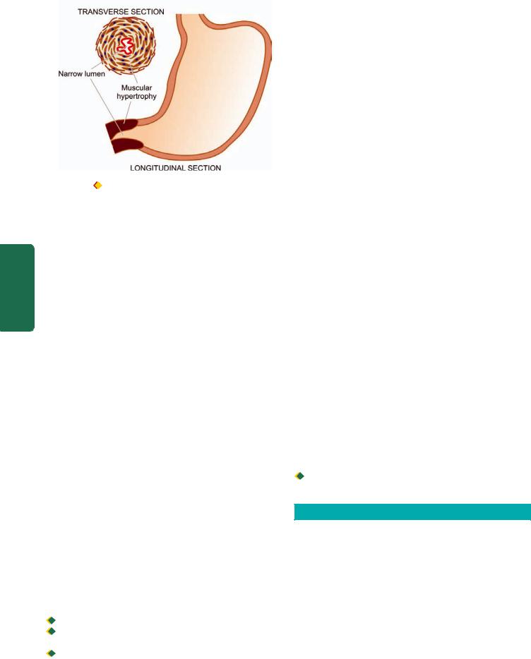
Figure 20.7 |
Pyloric stenosis, infantile type. Longitudinal and |
||
|
transverse section of the stomach showing hypertrophy of the circular |
|||
|
layer of the muscularis in the pyloric sphincter. |
|||
|
Pyloric Stenosis |
|||
|
Hypertrophy and narrowing of the pyloric lumen occurs |
|||
SECTION |
predominantly in male children as a congenital defect |
|||
(infantile pyloric stenosis). The adult form is rarely seen, either |
||||
|
||||
|
as a result of late manifestation of mild congenital anomaly |
|||
|
or may be acquired type due to inflammatory fibrosis or |
|||
|
invasion by tumours. |
|||
III |
ETIOLOGY. The exact cause of congenital (infantile) pyloric |
|||
stenosis is not known but it appears to have familial |
||||
|
||||
|
clustering and recessive genetic origin. The acquired (adult) |
|||
Systemic |
pyloric stenosis is related to antral gastritis, and tumours in |
|||
MORPHOLOGIC FEATURES. Grossly and micros- |
||||
|
the region (gastric carcinoma, lymphoma, pancreatic |
|||
|
carcinoma). |
|
||
Pathology |
copically, there is hypertrophy as well as hyperplasia of |
|||
the circular layer of muscularis in the pyloric sphincter |
||||
|
||||
|
accompanied by mild degree of fibrosis (Fig. 20.7). |
|||
|
CLINICAL FEATURES. The patient, usually a first born |
|||
|
male infant 3 to 6 weeks old, presents with the following |
|||
|
clinical features: |
|||
|
1. Vomiting, which may be projectile and occasionally |
|||
|
contains bile or blood. |
|||
|
2. Visible peristalsis, usually noticed from left to right side |
|||
|
of the upper abdomen. |
|||
|
3. Palpable lump, better felt after an episode of vomiting. |
|||
|
4. |
Constipation. |
||
|
5. |
Loss of weight. |
||
MISCELLANEOUS ACQUIRED CONDITIONS
Bezoars
Bezoars are foreign bodies in the stomach, usually in patients with mental illness who chew these substances. Some of the common bezoars are as follows:

 Trichobezoars composed of a ball of hair.

 Phytobezoars composed of vegetable fibres, seeds or fruit skin.
 Trichophytobezoars combining both hair and vegetable matter.
Acute Dilatation
Sudden and enormous dilatation of the stomach by gas or fluids due to paralysis of the gastric musculature may occur after abdominal operations, generalised peritonitis, and, in pyloric stenosis.
Gastric Rupture
The stomach may rupture rarely and prove fatal e.g. due to blunt trauma, external cardiac massage, ingestion of heavy meal or large quantity of liquid intake like beer.
INFLAMMATORY CONDITIONS
The two important inflammatory conditions of the stomach are gastritis and peptic ulcer. Rarely, stomach may be involved in tuberculosis, sarcoidosis and Crohn’s disease.
GASTRITIS
The term ‘gastritis’ is commonly employed for any clinical condition with upper abdominal discomfort like indigestion or dyspepsia in which the specific clinical signs and radiological abnormalities are absent. The condition is of great importance due to its relationship with peptic ulcer and gastric cancer. Broadly speaking, gastritis may be of 2 types—acute and chronic. Chronic gastritis can further be of various types.
A simple classification of various types of gastritis is presented in Table 20.2.
Acute Gastritis
Acute gastritis is a transient acute inflammatory involvement of the stomach, mainly mucosa.
ETIOPATHOGENESIS. A variety of etiologic agents have been implicated in the causation of acute gastritis. These are as follows:
1. Diet and personal habits:

 Highly spiced food

 Excessive alcohol consumption

 Malnutrition

 Heavy smoking.
2. Infections:

 Bacterial infections e.g. Helicobacter pylori, diphtheria, salmonellosis, pneumonia, staphylococcal food poisoning.
TABLE 20.2: Classification of Gastritis.
A.ACUTE GASTRITIS
1. Acute H. pylori gastritis
2. Other acute infective gastritis (bacteria, viruses, fungi, parasites)
3. Acute non-infective gastritis
B.CHRONIC GASTRITIS
1. |
Type A (autoimmune) |
: |
Body-fundic predominant |
2. |
Type B (H. pylori-related) |
: |
Antral-predominant |
|
gastritis |
|
|
3. |
Type AB (mixed environmental) |
: |
Antral-body gastritis |
4. |
Chemical (reflux) gastritis |
: |
Antral-body predominant |
5. |
Miscellaneous forms of gastritis |
|
|
|
|
|
|


 Viral infections e.g. viral hepatitis, influenza, infectious mononucleosis.
3. Drugs:

 Intake of drugs like non-steroidal anti-inflammatory drugs (NSAIDs), aspirin, cortisone, phenylbutazone, indomethacin, preparations of iron, chemotherapeutic agents.
4. Chemical and physical agents:
 Intake of corrosive chemicals such as caustic soda, phenol, lysol

 Gastric irradiation

 Freezing.
5. Severe stress:

 Emotional factors like shock, anger, resentment etc.

 Extensive burns

 Trauma

 Surgery.
The mucosal injury and subsequent acute inflammation in acute gastritis occurs by one of the following mechanisms:
1.Reduced blood flow, resulting in mucosal hypoperfusion due to ischaemia.
2.Increased acid secretion and its accumulation due to H. pylori infection resulting in damage to epithelial barrier.
3.Decreased production of bicarbonate buffer.
MORPHOLOGIC FEATURES. Grossly, the gastric mucosa is oedematous with abundant mucus and haemorrhagic spots.
Microscopically, depending upon the stage, there is variable amount of oedema and infiltration by neutrophils in the lamina propria. In acute haemorrhagic and erosive gastritis, the mucosa is sloughed off and there are haemorrhages on the surface.
Chronic Gastritis
Chronic gastritis is the commonest histological change observed in biopsies from the stomach. The microscopic change is usually poorly correlated to the symptomatology, as the change is observed in about 35% of endoscopically normal mucosal biopsies. The condition occurs more frequently with advancing age; average age for symptomatic chronic gastritis being 45 years which corresponds well with the age incidence of gastric ulcer.
ETIOPATHOGENESIS. In the absence of clear etiology of chronic gastritis, a number of etiologic factors have been implicated. All the causative factors of acute gastritis described above may result in chronic gastritis too. Recurrent attacks of acute gastritis may result in chronic gastritis. Some additional causes are as under:
1.Reflux of duodenal contents into the stomach, especially in cases which have undergone surgical intervention in the region of pylorus.
2.Infection with H. pylori is strongly implicated in the etiology of chronic gastritis and is more common.
3.Associated disease of the stomach and duodenum, such as gastric or duodenal ulcer, gastric carcinoma.
4.Chronic hypochromic anaemia, especially associated with atrophic gastritis.
5.Immunological factors such as autoantibodies to gastric parietal cells in atrophic gastritis and autoantibodies against intrinsic factor.
The mechanism of chronic gastric injury by any of the etiologic agents is by cytotoxic effect of the injurious agent on the gastric mucosal epithelium, thus breaking the barrier and then inciting the inflammatory response.
CLASSIFICATION. Based on the type of mucosa affected (i.e. cardiac, body, pyloric, antral or transitional), a clinicopathologic classification has been proposed
(Table 20.2).
1.Type A gastritis (Autoimmune gastritis). Type A gastritis involves mainly the body-fundic mucosa. It is also called autoimmune gastritis due to the presence of circulating antibodies and is sometimes associated with other autoimmune diseases such as Hashimoto’s thyroiditis and Addison’s disease. As a result of the antibodies against parietal cells and intrinsic factor, there is depletion of parietal cells and impaired secretion of intrinsic factor. These changes may lead to significant gastric atrophy where intestinal metaplasia may occur, and a small proportion of these patients may develop pernicious anaemia. Due to depletion of gastric acid-producing mucosal area, there is hypoor achlorhydria, and hyperplasia of gastrin-producing G cells in the antrum resulting in hypergastrinaemia.
2.Type B gastritis (H. pylori-related). Type B gastritis mainly involves the region of antral mucosa and is more common. It is also called hypersecretory gastritis due to excessive secretion of acid, commonly due to infection with H. pylori. These patients may have associated peptic ulcer. Unlike type A gastritis, this form of gastritis has no autoimmune basis nor has association with other autoimmune diseases.
3.Type AB gastritis (Mixed gastritis, Environmental gastritis, Chronic atrophic gastritis). Type AB gastritis affects the mucosal region of A as well as B types (body-fundic and antral mucosa). This is the most common type of gastritis in all age groups. It is also called environmental gastritis because a number of unidentified environmental factors have been implicated in its etiopathogenesis. Chronic atrophic gastritis is also used synonymously with type AB gastritis because in advanced stage, there is progression from chronic superficial gastritis to chronic atrophic gastritis, characterised by mucosal atrophy and metaplasia of intestinal or pseudopyloric type.
MORPHOLOGIC FEATURES. Grossly, the features of all forms of gastritis are inconclusive. The gastric mucosa may be normal, atrophied, or oedematous.
Histologically, criteria for categorisation are based on the following:
i) Extent of inflammatory changes in the mucosa (i.e. superficial or deep).
547
Tract Gastrointestinal The 20 CHAPTER
548
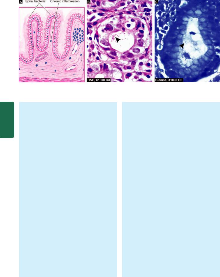
Figure 20.8 
 Histologic appearance of H. pylori chronic gastritis. A, Diagrammatic representation. B, H&E stained section. C, Demonstration of
H. pylori in Giemsa stain.
Pathology Systemic III SECTION
ii)Activity of inflammation (i.e. quiscent or active; acute or chronic).
iii)Presence of and type of metaplasia (i.e. intestinal or pseudopyloric).
Based on above, following simple morphologic classification has been proposed:
1.Chronic superficial gastritis
2.Chronic atrophic gastritis
3.Gastric atrophy
4.Chronic hypertrophic gastritis (Ménétrier’s disease)
5.Uncommon forms of chronic gastritis.
However, Sydney system of recording of histologic changes in gastritis is more acceptable since it takes into account following multiple parameters as well:
i)Etiology (H. pylori, autoimmune, NSAIDs, infections).
ii)Location (pangastritis, predominant antral, predominant body-fundic).
iii)Morphology (depth of inflammation—superficial or deep, severity of inflammation, type of inflammation, atrophy, metaplasia).
iv)Some special features (e.g. granulomas, eosinophilic gastritis, erosions, necrosis, haemorrhages).
1.CHRONIC SUPERFICIAL GASTRITIS. As the name suggests there is inflammatory infiltrate consisting of plasma cells and lymphocytes in the superficial layer of the gastric mucosa, but there are no histological changes in the deep layer of mucosa containing gastric glands. Chronic superficial gastritis may resolve completely or may progress to chronic gastric atrophy.
H. pylori, a spiral-shaped bacteria, was first reported by Warren and Marshall in Australia in 1984 as inhabitant of the acid environment of the stomach causing gastritis. After intial skepticism, numerous workers subsequently verified its association with gastritis and peptic ulcer (Warren and Marshall shared Nobel Prize in medicine in 2005 for their discovery). It is now known that H. pylori is
causative for almost all active cases of chronic superficial gastritis and about 65% of quiscent cases. The organism is identified on the epithelial layer on the luminal surface and does not invade the mucosa (Fig. 20.8). It is not seen on areas with intestinal metaplasia. H. pylori gastritis can be diagnosed by the following techniques:
i) Invasive tests (Endoscopic biopsy):
a)histologic examination combined with special stains for identification of microorganism: Giemsa, Steiner silver or Warthin-Starry stains;
b)biopsy urease test which is quick and simple but not fully sensitive; and
c)culture of the microorganism that helps in determining
specific antibiotic sensitivity.
ii)Non-invasive tests:
a)serologic tests (Immunoblot, ELISA) which are cheap and convenient but may not be helpful in early follow-up cases; and
b)14C urea breath test.
Although most patients of chronic superficial gastritis due to H. pylori remain asymptomatic, they may develop chronic atrophic gastritis, gastric atrophy, peptic ulcer disease. H. pylori infection is now considered an independent risk factor for gastric cancer: 3-6 fold increased risk for gastric adenocarcinoma and 6-50 times risk of MALT lymphoma (Fig. 20.9).
2. CHRONIC ATROPHIC GASTRITIS. In this stage, there is inflammatory cell infiltrate in the deeper layer of the
mucosa and atrophy of the epithelial elements including destruction of the glands. Two types of metaplasia are commonly associated with atrophic gastritis:
i) Intestinal metaplasia. Intestinal metaplasia is more common and involves antral mucosa more frequently. Characteristic histologic feature is the presence of intestinal type mucus-goblet cells; Paneth cells and endocrine cells may also be present. Parietal cells are very
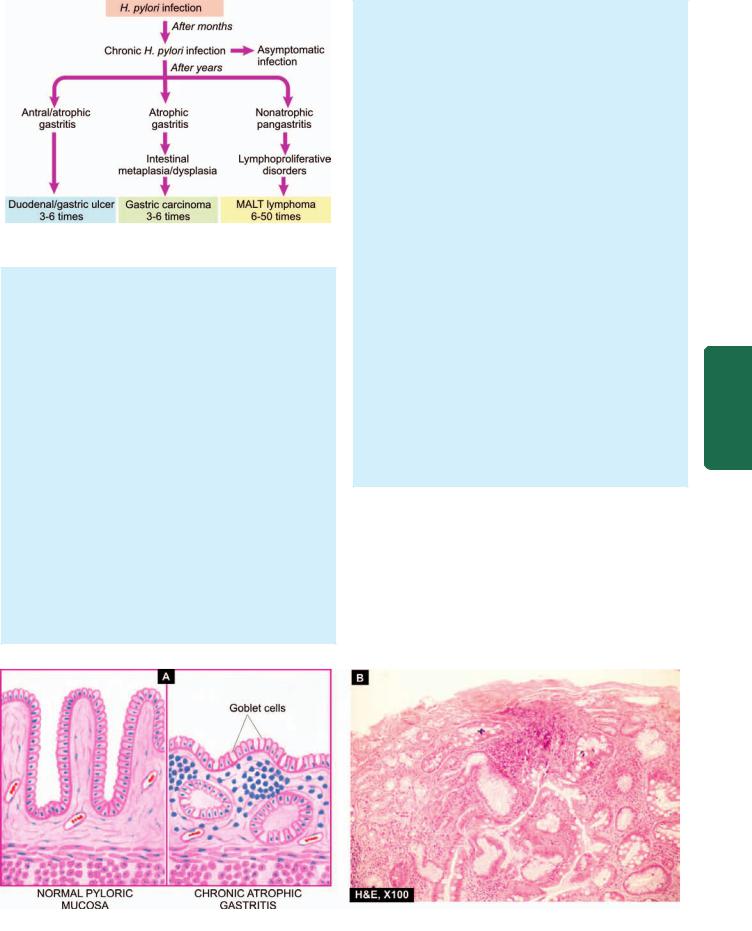
Figure 20.9 
 Consequences of long-term H. pylori gastritis.
few or absent (Fig. 20.10). Intestinal metaplasia, focal or extensive, in atrophic gastritis is significant because its incidence is high in populations having high prevalence rate of gastric cancer like in Japan. However, areas of intestinal metaplasia are not colonised by H. pylori.
ii) Pseudopyloric metaplasia. It involves the body glands which are replaced by proliferated mucus neck cells, conforming in appearance to normal pyloric glands. Its significance is not known.
3.GASTRIC ATROPHY. In this, there is thinning of the gastric mucosa with loss of glands but no inflammation though lymphoid aggregates may be present.
4.CHRONIC HYPERTROPHIC GASTRITIS (MÉNÉTRIER’S DISEASE). This is an uncommon condition characterised pathologically by enormous thickening of gastric rugal folds resembling cerebral convolutions, affecting mainly the region of fundic-body mucosa and characteristically sparing antral mucosa. The patients present with dyspepsia, haematemesis, melaena or protein-losing enteropathy.
Histologically, the gastric pits are elongated and are tortuous. The mucosa is markedly thickened and parts of
muscularis mucosae may extend into the thickened folds. Epithelium-lined cysts are commonly seen in the glandular layer. Inflammatory infiltrate is usually mild but lymphoid follicles may be present. The condition is considered significant in view of the risk of developing cancer.
5. MISCELLANEOUS FORMS OF CHRONIC GASTRITIS. A few other types of gastritis which do not fit into the description of the types of gastritis described above are as under:
i) Eosinophilic gastritis. This condition is characterised by diffuse thickening of the pyloric antrum due to oedema and extensive infiltration by eosinophils in all the layers of the wall of antrum. Eosinophilic gastritis probably has an allergic basis.
iii)Chronic follicular gastritis. This is a variant of chronic atrophic gastritis in which numerous lymphoid follicles are present in the mucosa and submucosa of the stomach.
iv)Haemorrhagic (Erosive) gastritis. In this condition, there are superficial erosions and mucosal haemorrhages, usually following severe haematemesis. The causes for such erosions and haemorrhages are duodenal-gastric reflux, administration of non-steroidal anti-inflammatory drugs (NSAIDs), portal hypertension.
v)Granulomatous gastritis. Rarely, granulomas may be present in the gastric mucosa such as in tuberculosis, sarcoidosis, Crohn’s disease, syphilis, various mycoses, and as a reaction to endogenous substance or foreign material.
PEPTIC ULCERS
Peptic ulcers are the areas of degeneration and necrosis of gastrointestinal mucosa exposed to acid-peptic secretions. Though they can occur at any level of the alimentary tract that is exposed to hydrochloric acid and pepsin, they occur most commonly (98-99%) in either the duodenum or the stomach in the ratio of 4:1. Each of the two main types may be acute or chronic.
549
Tract Gastrointestinal The 20 CHAPTER
Figure 20.10 
 of gastric glands metaplasia.
A, Chronic atrophic gastritis (right) contrasted with normal pyloric mucosa (left). There is marked gastric atrophy with disappearance and appearance of goblet cells (intestinal metaplasia). B, Photomicrograph showing chronic atrophic gastritis with intestinal

550Acute Peptic (Stress) Ulcers
Acute peptic ulcers or stress ulcers are multiple, small mucosal erosions, seen most commonly in the stomach but occasionally involving the duodenum.
ETIOLOGY. These ulcers occur following severe stress. The causes are as follows:
i)Psychological stress
ii)Physiological stress as in the following:

 Shock
Shock

 Severe trauma

 Septicaemia

 Extensive burns (Curling’s ulcers in the posterior aspect of the first part of the duodenum).

 Intracranial lesions (Cushing’s ulcers developing from hyperacidity following excessive vagal stimulation).

 Drug intake (e.g. aspirin, steroids, butazolidine, indomethacin).
|
Local irritants (e.g. alcohol, smoking, coffee etc). |
|
|
PATHOGENESIS. It is not clear how the mucosal erosions |
|
SECTION |
occur in stress ulcers because actual hypersecretion of gastric |
|
acid is demonstrable in only Cushing’s ulcers occurring from |
||
|
||
|
intracranial conditions such as due to brain trauma, |
|
|
intracranial surgery and brain tumours. In all other etiologic |
|
|
factors, gastric acid secretion is normal or below normal. In |
|
III |
these conditions, the possible hypotheses for genesis of stress |
|
ulcers are as under: |
||
|
1. Ischaemic hypoxic injury to the mucosal cells. |
|
Systemic |
2. Depletion of the gastric mucus ‘barrier’ rendering the |
|
ulcers are multiple (more than three ulcers in 75% of cases). |
||
|
mucosa susceptible to attack by acid-peptic secretions. |
|
|
MORPHOLOGIC FEATURES. Grossly, acute stress |
|
Pathology |
They are more common anywhere in the stomach, |
|
Microscopically, the stress ulcers are shallow and do not |
||
|
followed in decreasing frequency by occurrence in the first |
|
|
part of duodenum. They may be oval or circular in shape, |
|
|
usually less than 1 cm in diameter. |
|
|
invade the muscular layer. The margins and base may |
|
|
show some inflammatory reaction depending upon the |
|
|
duration of the ulcers. These ulcers commonly heal by |
|
|
complete re-epithelialisation without leaving any scars. |
|
|
Complications such as haemorrhage and perforation may |
|
|
occur. |
|
|
Chronic Peptic Ulcers (Gastric and Duodenal Ulcers) |
|
|
If not specified, chronic peptic ulcers would mean gastric |
|
|
and duodenal ulcers, the two major forms of ‘peptic ulcer |
|
|
disease’ of the upper GI tract in which the acid-pepsin |
|
|
secretions are implicated in their pathogenesis. Peptic ulcers |
|
|
are common in the present-day life of the industrialised and |
|
|
civilised world. |
|
|
Gastric and duodenal ulcers represent two distinct |
|
|
diseases as far as their etiology, pathogenesis and clinical |
|
|
features are concerned. However, morphological findings in |
|
|
both are similar and quite diagnostic. The features of gastric |
and duodenal peptic ulcers are described together below while their contrasting features are presented in Table 20.3.
INCIDENCE. Peptic ulcers are more frequent in middle-aged adults. The peak incidence for duodenal ulcer is 5th decade, while for gastric ulcer it is a decade later (6th decade). Duodenal as well as gastric ulcers are more common in males than in females. Duodenal ulcer is almost four times more common than gastric ulcer; the overall incidence of gastroduodenal ulcers being approximately 10% of the male population.
ETIOLOGY. The immediate cause of peptic ulcer disease is disturbance in normal protective mucosal ‘barrier’ by acidpepsin, resulting in digestion of the mucosa. However, in contrast to duodenal ulcers, the patients of gastric ulcer have
low-to-normal gastric acid secretions, though true achlorhydria in
response to stimulants never occurs in benign gastric ulcer.
Besides, 10-20% patients of gastric ulcer may have coexistent duodenal ulcer as well. Thus, the etiology of peptic ulcers possibly may not be explained on the basis of a single factor but is multifactorial. These factors are discussed below but the first two—H. pylori gastritis and NSAIDs-induced injury are considered most important.
1.Helicobacter pylori gastritis. About 15-20% cases infected with H. pylori in the antrum develop duodenal ulcer in their life time while gastric colonisation by H. pylori never develops ulceration and remain asymptomatic. H. pylori can be identified in mucosal samples by histologic examination, culture and serology as discussed on page 548.
2.NSAIDs-induced mucosal injury. Non-steroidal antiinflammatory drugs are most commonly used medications in the developed countries and are responsible for direct toxicity, endothelial damage and epithelial injury to both gastric as well as duodenal mucosa.
3.Acid-pepsin secretions. There is conclusive evidence that some level of acid-pepsin secretion is essential for the development of duodenal as well as gastric ulcer. Peptic ulcers never occur in association with pernicious anaemia in which there are no acid and pepsin-secreting parietal and chief cells respectively.
4.Gastritis. Some degree of gastritis is always present in the region of gastric ulcer, though it is not clear whether it is the cause or the effect of ulcer. Besides, the population distribution pattern of gastric ulcer is similar to that of chronic gastritis.
5.Other local irritants. Pyloric antrum and lesser curvature of the stomach are the sites most exposed for longer periods to local irritants and thus are the common sites for occurrence of gastric ulcers. Some of the local irritating substances implicated in the etiology of peptic ulcers are heavily spiced foods, alcohol, cigarette smoking, unbuffered aspirin.
6.Dietary factors. Nutritional deficiencies have been regarded as etiologic factors in peptic ulcers e.g. occurrence of gastric ulcer in poor socioeconomic strata, higher incidence of duodenal ulcer in parts of South India. However, malnutrition does not appear to have any causative role in peptic ulceration in European countries and the U.S.

TABLE 20.3: Distinguishing Features of Two Major Forms of Peptic Ulcers. |
|
551 |
||
|
Feature |
Duodenal Ulcer |
Gastric Ulcer |
|
1. |
Incidence |
i) Four times more common than gastric ulcers |
Less common than duodenal ulcers |
|
|
|
ii) Usual age 25-50 years |
Usually beyond 6th decade |
|
|
|
iii) More common in males than in females (4:1) |
More common in males than in females (3.5:1) |
|
2. |
Etiology |
Most commonly as a result of H. pylori infection |
Gastric colonisation with H. pylori asymptomatic |
|
|
|
Other factors—hypersecretion of acid-pepsin, |
but higher chances of development of duodenal ulcer. |
|
|
|
association with alcoholic cirrhosis, tobacco, |
Disruption of mucus barrier most important factor. |
|
|
|
hyperparathyroidism, chronic pancreatitis, |
Association with gastritis, bile reflux, drugs, |
|
|
|
blood group O, genetic factors |
alcohol, tobacco |
|
3. |
Pathogenesis |
i) Mucosal digestion from hyperacidity most |
Usually normal-to-low acid levels; hyperacidity |
|
|
|
significant factor |
if present is due to high serum gastrin |
|
|
|
ii) Protective gastric mucus barrier may be damaged |
Damage to mucus barrier significant factor |
|
4. |
Pathologic changes |
i) Most common in the first part of duodenum |
Most common along the lesser curvature |
|
|
|
|
and pyloric antrum |
|
ii)Often solitary, 1-2.5 cm in size, round to oval, punched out
iii)Histologically, composed of 4 layers—necrotic, superficial exudative, granulation tissue and cicatrisation
5.Complications Commonly haemorrhage, perforation,
sometimes obstruction; malignant transformation never occurs
6.Clinical features i) Pain-food-relief pattern
ii)Night pain common
iii)No vomiting
iv)Melaena more common than haematemesis
v)No loss of weight
vi)No particular choice of diet
vii)Deep tenderness in the right hypochondrium
viii)Marked seasonal variation
ix)Occurs more commonly in people at greater stress
Grossly similar to duodenal ulcer
Histologically, indistinguishable from duodenal ulcer
Perforation, haemorrhage and at times obstruction; malignant transformation in less than 1% cases
Food-pain pattern
No night pain
Vomiting common
Haematemesis more common
Significant loss of weight
Patients choose bland diet devoid of fried foods, curries etc.
Deep tenderness in the midline in epigastrium
No seasonal variation
More often in labouring groups
7.Psychological factors. Psychological stress, anxiety, fatigue and ulcer-type personality may exacerbate as well as predispose to peptic ulcer disease.
8.Genetic factors. People with blood group O appear to be more prone to develop peptic ulcers than those with other blood groups. Genetic influences appear to have greater role in duodenal ulcers as evidenced by their occurrence in families, monozygotic twins and association with HLA-B5 antigen.
9.Hormonal factors. Secretion of certain hormones by tumours is associated with peptic ulceration e.g. elaboration of gastrin by islet-cell tumour in Zollinger-Ellison syndrome, endocrine secretions in hyperplasia and adenomas of parathyroid glands, adrenal cortex and anterior pituitary.
10.Miscellaneous. Duodenal ulcers have been observed to occur in association with various other conditions such as
alcoholic cirrhosis, chronic renal failure, hyperparathyroidism, chronic obstructive pulmonary disease, and chronic pancreatitis.
PATHOGENESIS. Although the role of various etiologic factors just described is well known in ulcerogenesis, two most important factors in peptic ulcer are as under:

 Exposure of mucosa to gastric acid and pepsin secretion.

 Strong etiologic association with H. pylori infection.
There are distinct differences in the pathogenetic mechanisms involved in duodenal and gastric ulcers as under:
Duodenal ulcer. There is conclusive evidence to support the role of high acid-pepsin secretions in the causation of duodenal ulcers. Besides this, a few other noteworthy features in the pathogenesis of duodenal ulcers are as follows:
Tract Gastrointestinal The 20 CHAPTER
5521. There is generally hypersecretion of gastric acid into the fasting stomach at night which takes place under the influence of vagal stimulation. There is high basal as well as maximal acid output (BAO and MAO) in response to various stimuli.
2.Patients of duodenal ulcer have rapid emptying of the stomach so that the food which normally buffers and neutralises the gastric acid, passes down into the small intestine, leaving the duodenal mucosa exposed to the aggressive action of gastric acid.
3.Helicobacter gastritis caused by H. pylori is seen in 95-100% cases of duodenal ulcers. The underlying mechanisms are as under:
|
i) Gastric mucosal defense is broken by bacterial elaboration |
|
|
of urease, protease, catalase and phospholipase. |
|
|
ii) Host factors: H. pylori-infected mucosal epithelium releases |
|
|
proinflammatory cytokines such as IL-1, IL-6, IL-8 and tumour |
|
|
necrosis factor-α, all of which incite intense inflammatory |
|
|
reaction. |
|
|
iii) Bacterial factors: Epithelial injury is also induced by |
|
|
cytotoxin-associated gene protein (CagA), while vacuolating |
|
SECTION |
cytotoxin (VacA) induces elaboration of cytokines. |
|
Gastric ulcer. The pathogenesis of gastric ulcer is mainly |
||
explained on the basis of impaired gastric mucosal defenses |
||
against acid-pepsin secretions. Some other features in the |
||
pathogenesis of gastric ulcer are as follows: |
||
III |
1. Hyperacidity may occur in gastric ulcer due to increased |
|
|
serum gastrin levels in response to ingested food in an atonic |
|
Systemic |
stomach. |
|
such as gastritis, bile reflux, cigarette smoke etc. |
||
|
2 However, many patients of gastric ulcer have low-to- |
|
|
normal gastric acid levels. Ulcerogenesis in such patients is |
|
Pathology |
explained on the basis of damaging influence of other factors |
|
3. The normally protective gastric mucus ‘barrier’ against |
||
|
||
|
acid-pepsin is deranged in gastric ulcer. There is depletion |
|
|
in the quantity as well as quality of gastric mucus. One of |
|
|
the mechanisms for its depletion is colonisation of the gastric |
|
|
mucosa by H. pylori seen in 75-80% patients of gastric ulcer. |
|
|
|
|
|
MORPHOLOGIC FEATURES. Gross and microscopic |
|
|
changes in gastric and duodenal ulcers are similar and |
|
|
quite characteristic. Gastric ulcers are found predominantly |
|
|
along the lesser curvature in the region of pyloric antrum, |
|
|
more commonly on the posterior than the anterior wall. |
|
|
Most duodenal ulcers are found in the first part of the |
|
|
duodenum, usually immediate post-pyloric, more |
|
|
commonly on the anterior than the posterior wall. |
|
|
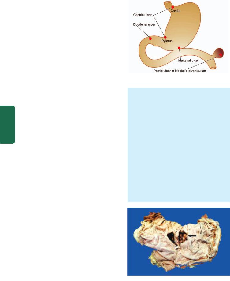
Uncommon locations include ulcer in the cardia, marginal |
|
|
ulcer and in the Meckel’s diverticulum (Fig. 20.11). |
|
|
Grossly, typical peptic ulcers are commonly solitary (80%), |
|
|
small (1-2.5 cm in diameter), round to oval and |
|
|
characteristically ‘punched out’. Benign ulcers usually |
|
|
have flat margins in level with the surrounding mucosa. |
|
|
The mucosal folds converge towards the ulcer. The ulcers |
|
|
may vary in depth from being superficial (confined to |
|
|
mucosa) to deep ulcers (penetrating into the muscular |
|
|
layer) (Fig. 20.12). In about 10-20% of cases, gastric and |
|
|
|
Figure 20.11 
 Distribution of peptic ulcers.
duodenal ulcers are coexistent. Vast majority of the peptic ulcers are benign. Chronic duodenal ulcer never turns malignant, while chronic gastric ulcer may develop carcinoma in less than 1% of cases. Malignant gastric ulcers are larger, bowl-shaped with elevated and indurated mucosa at the margin (Fig. 20.13).
Microscopically, chronic peptic ulcers have 4 histological zones. From within outside, these are as under (Fig. 20.14):
1.Necrotic zone—lies in the floor of the ulcer and is composed of fibrinous exudate containing necrotic debris and a few leucocytes.
2.Superficial exudative zone—lies underneath the necrotic zone. The tissue elements here show coagulative necrosis giving eosinophilic, smudgy appearance with nuclear debris.
3.Granulation tissue zone—is seen merging into the necrotic zone. It is composed of nonspecific inflammatory infiltrate and proliferating capillaries.
4.Zone of cicatrisation—is seen merging into thick layer of granulation tissue. It is composed of dense fibrocollagenic scar tissue over which granulation tissue rests. Thrombosed or sclerotic arteries may cross the ulcer which on erosion may result in haemorrhage.
Figure 20.12 
 Benign chronic peptic ulcer. Partial gastrectomy specimen showing a punched out round to oval ulcer on the mucosa, about 1 cm in diameter (arrow) and penetrating into muscularis layer.
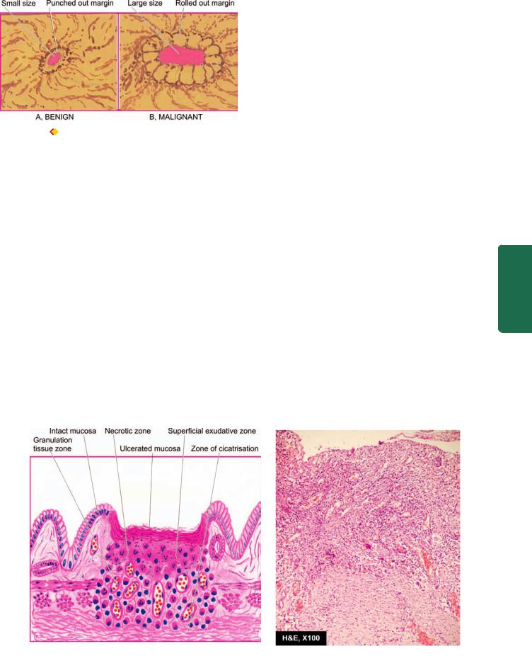
Figure 20.13 
 Chronic gastric ulcer (A) contrasted with malignant gastric ulcer (B).
COMPLICATIONS. Acute and subacute peptic ulcers usually heal without leaving any visible scar. However, healing of chronic, larger and deeper ulcers may result in complications. These are as follows:
1.Obstruction. Development of fibrous scar at or near the pylorus results in pyloric stenosis. In the case of healed duodenal ulcer, it causes duodenal stenosis. Healed ulcers along the lesser curvatures may produce ‘hourglass’ deformity due to fibrosis and contraction.
2.Haemorrhage. Minor bleeding by erosion of small blood vessels in the base of an ulcer occurs in all the ulcers and can be detected by testing the stool for occult blood. Chronic blood loss may result in iron deficiency anaemia. Severe bleeding may cause ‘coffee ground’ vomitus or melaena. A penetrating chronic ulcer may erode a major artery (e.g. left gastric, gastroduodenal or splenic artery) and cause a massive and severe hematemesis and sometimes death.
3.Perforation. A perforated peptic ulcer is an acute abdominal emergency. Perforation occurs more commonly in chronic duodenal ulcers than chronic gastric ulcers. Following sequelae may result:
i)On perforation the contents escape into the lesser sac or into the peritoneal cavity, causing acute peritonitis.
ii)Air escapes from the stomach and lies between the liver and the diaphragm giving the characteristic radiological appearance of air under the diaphragm.
iii)Subphrenic abscess between the liver and the diaphragm may develop due to infection.
iv)Perforation may extend to involve the adjacent organs e.g. the liver and pancreas.
4. Malignant transformation. The dictum ‘cancers ulcerate but ulcers rarely cancerate’ holds true for most peptic ulcers. A chronic duodenal ulcer never turns malignant, while less than 1% of chronic gastric ulcers may transform into carcinoma.
CLINICAL FEATURES. Peptic ulcers are remitting and relapsing lesions. Their chronic and recurrent behaviour is summed up the saying: ‘once a peptic ulcer patient, always a peptic ulcer patient.’ The two major forms of chronic peptic ulcers show variations in clinical features which are as follows:
1.Age. The peak incidence of duodenal ulcer is in 5th decade while that for gastric ulcer is a decade later.
2.People at risk. Duodenal ulcer occurs more commonly in people faced with more stress and strain of life (e.g. executives, leaders), while gastric ulcer is seen more often in labouring groups.
3.Periodicity. The attacks in gastric ulcers last from 2-6 weeks, with interval of freedom from 1-6 months. The attacks of duodenal ulcer, are classically worsened by ‘work, worry
and weather.’
4. Pain. In gastric ulcer, epigastric pain occurs immediately or within 2 hours after food and never occurs at night. In duodenal ulcer, pain is severe, occurs late at night (‘hunger pain’) and is usually relieved by food.
553
Tract Gastrointestinal The 20 CHAPTER
Figure 20.14 
 Chronic peptic ulcer. Histologic zones of the ulcer are illustrated in the diagram. The photomicrograph on right shows necrotic debris, ulceration and inflammation on the mucosal surface.

554 5. Vomiting. Vomiting which relieves the pain is a conspicuous feature in patients of gastric ulcer. Duodenal ulcer patients rarely have vomiting but instead get heart-burn (retrosternal pain) and ‘water brash’ (burning fluid into the mouth).
6. Haematemesis and melaena. Haematemesis and melaena occur in gastric ulcers in the ratio of 60:40, while in duodenal ulcers in the ratio of 40:60. Both may occur together more commonly in duodenal ulcer than in gastric ulcer patients.
7. Appetite. The gastric ulcer patients, though have good appetite but are afraid to eat, while duodenal ulcer patients have very good appetite.
8. Diet. Patients of gastric ulcer commonly get used to a bland diet consisting of milk, eggs etc and avoid taking fried foods, curries and heavily spiced foods. In contrast, duodenal ulcer patients usually take all kinds of diets.
9. Weight. Loss of weight is a common finding in gastric ulcer patients while patients of duodenal ulcer tend to gain weight due to frequent ingestion of milk to avoid pain.
|
10. Deep tenderness. Deep tenderness is demonstrable in |
|
SECTION |
both types of peptic ulcers. In the case of gastric ulcer it is in |
|
the midline of the epigastrium, while in the duodenal ulcer |
||
|
||
|
it is in the right hypochondrium. |
|
|
HAEMATEMESIS AND MELAENA OF GASTRIC ORIGIN |
|
III |
In continuity with the discussion on peptic ulcers which are |
|
the commonest cause of haematemesis and melaena, it is |
||
|
||
Systemic |
worthwhile listing various causes of haematemesis of gastric |
|
origin (causes of haematemesis of oesophageal origin are |
||
already given on page 539). |
||
|
||
|
i) Chronic peptic ulcers (gastric as well as duodenal) |
|
|
ii) Acute peptic ulcers (stress ulcers) |
|
Pathology |
iii) Multiple gastric and duodenal erosions |
|
iv) Carcinoma of the stomach |
||
|
||
|
v) Peptic ulcer in Meckel’s diverticulum |
|
|
vi) Mallory-Weiss syndrome |
|
|
vii) Anaemias |
|
|
viii) Purpuras |
|
|
ix) Haemophilia. |
|
|
TUMOURS AND TUMOUR-LIKE LESIONS |
|
|
The various types of tumour-like lesions (polyps) and benign |
|
|
and malignant tumours of the stomach are given in |
|
|
Table 20.4. |
|
|
A. TUMOUR-LIKE LESIONS (POLYPS) |
|
|
Hyperplastic (Inflammatory) Polyps |
|
|
Hyperplastic or inflammatory polyps are regenerative, non- |
|
|
neoplastic lesions which are the most common type (90%). |
|
|
They may be single or multiple and are more often located |
|
|
in the pyloric antrum. |
|
|
|
|
|
Grossly, the lesions may be sessile or pedunculated, 1 cm |
|
|
or larger in size, smooth and soft. The surface may be |
|
|
ulcerated or haemorrhagic. |
|
|
|
TABLE 20.4: Gastric Tumours and Tumour-like Lesions.
A.TUMOUR-LIKE LESIONS (POLYPS)
1.Hyperplastic (inflammatory) polyps
2.Hamartomatous polyps
B.BENIGN TUMOURS
1.Epithelial
Adenomas (adenomatous or neoplastic polyps)
2.Non-epithelial
Gastrointestinal spindle cell (stromal) tumours (GIST)
C.MALIGNANT TUMOURS
1.Epithelial (90%)
(i)Adenocarcinoma
(ii)Others
2.Non-epithelial (2%)
(i)Leiomyosarcoma
(ii)Leiomyoblastoma (epithelioid leiomyoma)
3.Carcinoid tumour (3%)
4.Lymphoma (4%)
Microscopically, they are composed of irregular hyperplastic glands, which may show cystic change. The lining epithelium is mostly superficial gastric type but antral glands, chief cells and parietal cells may be present. These lesions do not have cellular atypia and do not have malignant potential.
Hamartomatous Polyps
Hamartomatous polyps are not true neoplasms but are malformations. They are of various types such as gastric polyps of the Peutz-Jeghers syndrome (page 582), juvenile polyp, pancreatic heterotopia, heterotopia of Brunner’s glands and inflammatory fibroid polyps (eosinophilic granulomatous polyps).
B. BENIGN TUMOURS
Adenomas (Adenomatous or Neoplastic Polyps)
Adenomas, also, referred to as adenomatous or neoplastic polyps, are true benign epithelial neoplasms and are much rare in the stomach than in the large intestine. They are also found more often in the region of pyloric antrum. They are commonly associated with atrophic gastritis and pernicious anaemia. Morphologically, adenomatous polyps of the stomach resemble their counterparts in the large bowel and are described on page 583.
Stromal Tumours
Stomach may be the site for occurrence of various uncommon benign tumours of stromal cell origin e.g. leiomyomas (being the most common); others are neurofibromas, schwannomas and lipomas. They are usually firm, circumscribed nodules, less than 4 cm in size and appear as submucosal nodules. They resemble in gross and microscopic appearance with their counterparts in other parts of the body.
Currently, the term gastrointestinal stromal tumours (GISTs) is used for a group of uncommon benign tumours composed of spindle cells or stromal cells but lacking the
true phenotypic features of smooth muscle cells, neural cells or Schwann cells. They are uncommon but as compared to other sites in the GIT, are most common in the stomach. Their behaviour is generally benign but may be recurrent, aggressive or even metastasis may occur.
C. MALIGNANT TUMOURS
Gastric Carcinoma
INCIDENCE. Carcinoma of the stomach comprises more than 90% of all gastric malignancies and is the leading cause of cancer-related deaths in countries where its incidence is high. The highest incidence is between 4th to 6th decades of life and is twice more common in men than in women.
ETIOLOGY. A number of etiologic factors have been implicated in causation of gastric cancer. These are as under:
1.H. pylori infection. H. pylori infection of the stomach is an important risk factor for the development of gastric cancer. Epidemiologic studies throughout world have shown that a seropositivity with H. pylori is associated with 3 to 6 times higher risk of development of gastric cancer. It may be mentioned here that similar association of H. pylori infection exists with gastric lymphomas (MALT type) as well.
2.Dietary factors. Epidemiological studies suggest that dietary factors are most significant in the etiology of gastric cancer. The evidences in support of this are multifold:
i)Occurrence of gastric cancer in the region of gastric canal (i.e. along the lesser curvature and the pyloric antrum) where irritating foods exert their maximum effect.
ii)Populations consuming certain foodstuffs have high risk of developing gastric cancer e.g. ingestion of smoked foods, high intake of salt, pickled raw vegetables, high intake of carcinogens as nitrates in foods and drinking water, nitrites as preservatives for certain meats etc. However, intake of green leafy vegetables, citrus fruits and animal fats has been reported to have protective role in gastric cancer.
iii)Tobacco smoke, tobacco juice and consumption of alcohol have all been shown to have carcinogenic effect on gastric mucosa.
3.Geographical factors. There are geographic variations in the incidence of gastric cancer. Japan, Chile and Italy have the highest recorded death rate from gastric cancer, while the incidence is considerably low in the US, UK and Canada. The higher incidence in certain geographic regions is the result of environmental influences as observed from the finding of incidence of gastric cancer in the next generation of Japanese immigrants to the US which is comparable to that of native Americans.
4.Racial factors. Within the country, different ethnic groups may have variations in incidence of gastric cancer e.g. incidence is higher in Blacks, American Indians, Chinese in Indonesia, North Wales than other parts of Wales.
5.Genetic factors. Genetic influences have some role in the etiology of gastric cancer. Not more than 4% of patients of gastric cancer have a family history of this disease. Individuals with blood group A have higher tendency to develop gastric cancer (Recall that the peptic ulcer is more
555
Figure 20.15 |
Distribution of gastric carcinoma in the anatomical |
|
||
subdivisions of the stomach. The serial numbers in the figure indicate |
|
|||
the order of frequency of occurrence of gastric cancer. |
|
|||
common in individuals with blood group O). A germ line |
|
|||
mutation in E-cadherin gene inherited as a autosomal |
CHAPTER |
|||
dominant pattern has been linked to higher incidence of |
||||
|
||||
occult gastric cancer in younger individuals. |
|
|||
6. Pre-malignant changes in the gastric mucosa. There are |
|
|||
some conditions of gastric mucosa which have increased risk |
|
|||
to development of gastric cancer: |
20 |
|||
i) Hypoor achlorhydria in atrophic gastritis of gastric |
||||
|
||||
mucosa with intestinal metaplasia. |
|
|||
ii) Adenomatous (neoplastic) polyps of the stomach. |
The |
|||
iii) Chronic gastric ulcer (ulcer-cancer), and its association |
||||
|
||||
with achlorhydria. |
Gastrointestinal |
|||
iv) Stump carcinoma in patients who have undergone partial |
||||
|
||||
gastrectomy. |
|
|
|
|
|
|
|
||
MORPHOLOGIC FEATURES. Gastric carcinoma is most |
|
|||
commonly located in the region of gastric canal (prepyloric |
|
|||
region) formed by lesser curvature, pylorus and antrum. |
|
|||
Other less common locations are the body, cardia and |
|
|||
fundus (Fig. 20.15). |
Tract |
|||
Pathogenetically, a sequential evolution of all gastric |
||||
|
||||
carcinomas from an initial stage of in situ carcinoma |
|
|||
confined to mucosal layers called early gastric carcinoma |
|
|||
(EGC) has been found. EGC eventually penetrates the |
|
|||
muscularis or beyond, resulting in advanced gastric |
|
|||
carcinoma. Accordingly, gastric carcinomas are broadly |
|
|||
classified into 2 main groups: |
|
|||
I. Early gastric carcinoma (EGC). |
|
|||
II. Advanced gastric carcinoma, which has 5 further major |
|
|||
gross subtypes: |
|
|||
i)Ulcerative carcinoma
ii)Fungating (Polypoid) carcinoma
iii)Scirrhous carcinoma (Linitis plastica)
iv)Colloid (Mucoid) carcinoma
v)Ulcer-cancer
In addition to the above classification, gastric carcinomas have been classified, on the basis of extent of invasion, into 2 groups:
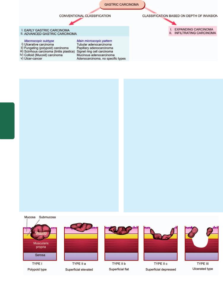
556
Pathology Systemic III SECTION
Figure 20.16 
 Classifications of gastric carcinomas. A, Conventional classification, showing correlation of the macroscopic subtypes with the main histological patterns. B, Classification based on the depth of invasion by the tumour.
I. Expanding (formerly intestinal type) carcinomas that grow laterally by an invasive margin. The tumour cells are in the form of cohesive clusters.
II. Infiltrating (formerly diffuse type) carcinomas have poorly-defined invasive border. The tumour cells are loose and invade singly or in small group.
These classifications are summarised in Fig. 20.16 and comparative morphology of various types is shown diagrammatically in Fig. 20.18.
I. EARLY GASTRIC CARCINOMA (EGC) (Fig. 20.18,A). EGC is the term used to describe cancer limited to the mucosa and submucosa. The diagnosis of this condition has been made possible by extensive work on histogenesis of gastric cancer by Japanese pathologists by the use of fibreoptic endoscope and gastrocamera. In Japan, EGC comprises 35% of newly-diagnosed cases of gastric cancer.
Grossly, the lesion of EGC may have 3 patterns—polypoid (protruded), superficial and ulcerated (Fig. 20.17):
Type I |
: Polypoid type |
Type IIa |
: Superficial elevated |
Type II b |
: Superficial flat |
Type II c |
: Superficial depressed |
Type III |
: Ulcerated type |
Histologically, EGC is a typical glandular adenocarcinoma, usually well-differentiated type.
Prognosis of EGC after surgical resection is quite good; 5-year survival rate being 93-99%.
Early gastric carcinoma must be distinguished from certain related terms as under:
 Epithelial dysplasia is cellular atypia seen in intestinal metaplasia such as in atrophic gastritis and pernicious anaemia.
 Carcinoma in situ in the stomach is a state of severe cellular atypia or dysplasia, without invasion across the basement membrane of the glands.
II. ADVANCED GASTRIC CARCINOMA. When the carcinoma crosses the basement membrane into the muscularis propria or beyond, it is referred to as advanced gastric carcinoma. Advanced gastric carcinoma has following 5 patterns:
i)Ulcerative carcinoma (Fig. 20.18,B). This is the most common pattern. The tumour appears as a flat, infiltrating and ulcerative growth with irregular necrotic base and raised margin. It is seen more commonly in the region of gastric canal (Fig. 20.19,A).
Histologically, ulcerative carcinomas are poorlydifferentiated adenocarcinomas, which invade deeply into the stomach wall. Tubular and acinar patterns are seen more commonly (Fig. 20.19, B).
ii)Fungating (polypoid) carcinoma (Fig. 20.18,C). The second common pattern is a cauliflower growth projecting into the lumen, similar to what is commonly seen in the
Figure 20.17 
 Diagrammatic representation of gross patterns of early gastric carcinoma.

large intestine. It is seen more often in the fundus. The tumour undergoes necrosis and infection commonly. Histologically, fungating or polypoid carcinomas are well-differentiated adenocarcinomas, commonly papillary type.
iii)Scirrhous carcinoma (Linitis plastica) (Fig. 20.18,D). In this pattern, the stomach wall is thickened due to extensive desmoplasia giving the appearance as ‘leatherbottle stomach’ or ‘linitis plastica’. The involvement may be localised to pyloric antrum, or diffuse affecting whole of the stomach from the cardia to pylorus. The lumen of the stomach is reduced. There are no ulcers but rugae are prominent (Fig. 20.19,C).
Histologically, it may be an adenocarcinoma or signetring cell carcinoma, extensively infiltrating the stomach wall, but due to marked desmoplasia cancer cells may be difficult to find (Fig. 20.19,D).
iv)Colloid (Mucoid) carcinoma (Fig. 20.18,E). This pattern is usually seen in the fundus. The tumour grows like masses having gelatinous appearance due to secretion of large quantities of mucus.
Histologically, mucoid carcinoma contains abundant pools of mucin in which are seen a small number of tumour cells, sometimes having signet-ring appearance.
v)Ulcer-cancer (Fig. 20.18,F). Development of cancer in chronic gastric ulcer is a rare occurrence (less than 1%). Majority of ulcer-cancers are malignant lesions from the beginning. For confirmation of cancer in a pre-existing gastric ulcer, the characteristic microscopic appearance of peptic ulcer should be demonstrable with one portion of the base or the margin of the ulcer showing carcinomatous changes.
Histologically, ulcer-cancers are adenocarcinomas without any specific features. The differences between a benign and malignant gastric ulcer are summarised in Table 20.5 (also see Fig. 20.13).
SPREAD. Carcinoma of the stomach may spread by the following routes:
1.Direct spread. Direct spread by local extension is the most common feature of gastric carcinoma. The spread occurs mainly from the loose submucosal layer but eventually muscularis and serosa are also invaded. After the peritoneal covering of the stomach has been invaded, transcoelomic dissemination may occur in any other part of the peritoneal cavity but ovarian masses (one sided or both-sided) occur more commonly, referred to as Krukenberg tumours (Chapter 24). Submucosal spread occurs more often upwards into the oesophagus due to continuity of the layers of stomach with those of oesophagus, while the spread downwards into the duodenum occurs less often due to the presence of pyloric sphincter and submucosal Brunner’s glands. The tumour may directly involve other neighbouring structures and organs like lesser and greater omentum, pancreas, liver, common bile duct, diaphragm, spleen and transverse colon.
2.Lymphatic spread. Metastases to regional lymph nodes occur early, especially in the scirrhous carcinoma. The groups of lymph nodes involved are along the lesser and greater curvature around the cardia and suprapancreatic lymph nodes. Involvement of left supraclavicular lymph node, Virchow or Troisier’s sign, is sometimes the presenting feature of gastric carcinoma.
3.Haematogenous spread. Blood spread of gastric carcinoma may occur to the liver, lungs, brain, bones, kidneys and adrenals. It occurs more commonly with the poorlydifferentiated carcinoma.
The American Joint Committee on Cancer has developed TNM staging system for gastric carcinoma based on tumour invasion (T), lymph node involvement (N) and distant
metastasis (M) into earliest stage Tis N0 M0 (intraepithelial tumour) to most advanced stage Tany Nany M1.
CLINICAL FEATURES. Gastric carcinoma may have diverse presentations. The usual clinical features are as under:
i) Persistent abdominal pain
TABLE 20.5: Differences between Benign and Malignant Gastric Ulcers.
|
Feature |
Benign Ulcer |
Malignant Ulcer |
|
|
|
|
|
|
1. |
Age |
|
Younger age |
Older age |
2. |
Sex |
|
Markedly common in males |
Slightly common in males |
3. |
Duration of symptoms |
Weeks to years |
Weeks to months |
|
4. |
Location |
Commonly lesser curvature of pylorus and antrum |
Commonly greater curvature of pylorus and antrum |
|
5. |
Gross features |
|
|
|
|
a) |
Size |
Small |
Large |
|
b) |
Shape |
Regular |
Irregular |
|
c) |
Mucosal folds |
Radiating |
Interrupted |
|
d) |
Ulcer bed |
Haemorrhagic |
Necrotic |
6. |
Barium studies |
Punched out ulcer |
Irregular filling defect |
|
7. |
Acidity |
Usually normal-to-low |
May be normal-to-even achlorhydria |
|
8. |
Therapy |
Responds well to medical therapy |
Usually does not respond to medical therapy |
|
|
|
|
|
|
557
Tract Gastrointestinal The 20 CHAPTER
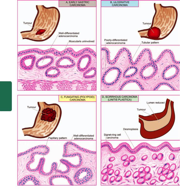
558
Pathology Systemic III SECTION
Figure 20.18 
 Gastric carcinoma, gross appearance of subtypes and their corresponding dominant histological patterns.
ii)Gastric distension and vomiting
iii)Loss of weight (cachexia)
iv)Loss of appetite (anorexia)
v)Anaemia, weakness, malaise.
The most common complication of gastric cancer is
haemorrhage (in the form of haematemesis and/or melaena); others are obstruction, perforation and jaundice.
Gastric carcinoma remains undiagnosed until late when the symptoms appear. Therefore, the prognosis is generally poor; 5-year survival rate being 5-15% from the time of diagnosis of advanced gastric carcinoma. However, 5-year survival rate for early gastric carcinoma is far higher (93-99%) and hence the need for early diagnosis of the condition.
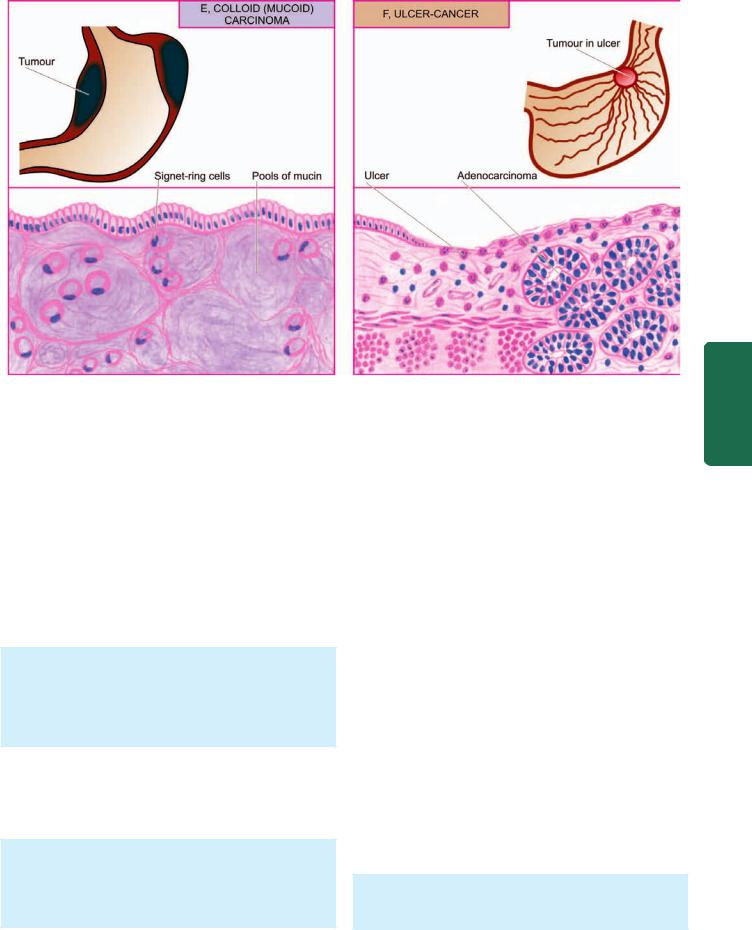
Figure 20.18 contd... 
 Gastric carcinoma, gross appearance of subtypes and their corresponding dominant histological patterns.
Other Carcinomas
Besides the various morphologic patterns of adenocarcinoma just described, other carcinomas that occur rarely in the stomach are: adenosquamous carcinoma, squamous cell carcinoma and undifferentiated carcinoma, all of which are morphologically similar to such tumours elsewhere.
Leiomyosarcoma
Leiomyosarcoma, though rare, is the commonest soft tissue sarcoma, the stomach being the more common site in the gastrointestinal tract.
Grossly, the tumour may be of variable size but is usually quite large, pedunculated and lobulated mass into the lumen.
Microscopically, leiomyosarcoma is characterised by high cellularity and presence of mitotic figures. Tumour is usually well-differentiated.
Leiomyoblastoma (Epithelioid Leiomyoma)
This is a rare tumour, the behaviour of which is intermediate between clearly benign and malignant tumour.
Grossly, the tumour is large, circumscribed and projects into the lumen.
Microscopically, it is characterised by round to polygonal cells with clear perinuclear halos. The number of mitoses determines the biological behaviour of the tumour.
Carcinoid Tumour
Carcinoid tumours are rare in the stomach and are usually non-argentaffin type but argentaffinomas also occur. Their behaviour is usually malignant; they are described on page 579.
Lymphomas of Gut
Primary gastrointestinal lymphomas are defined as lymphomas arising in the gut without any evidence of systemic involvement at the time of presentation.
Secondary gastrointestinal lymphomas, on the other hand, appear in the gut after dissemination from other primary site.
Gastric lymphomas constitute over 50% of all bowel lymphomas; other sites being small and large bowel in decreasing order of frequency. Prognosis of primary gastric lymphoma is better than for intestinal lymphomas. Primary lymphoma of stomach is the most common malignant gastric tumour (4%) next to carcinoma.
Clinical manifestations of gastric lymphomas may be similar to gastric carcinoma. Age incidence for lymphomas of the gastrointestinal tract is usually lower than that for carcinoma (30-40 years as compared to 40-60 years in gastric carcinoma) and may occur even in childhood. Relationship with long-standing chronic H. pylori gastritis with lymphoid hyperplasia has been strongly suggested.
Grossly, gastric lymphomas have 2 types of appearances:
1. Diffusely infiltrating type, producing thickening of the affected gut wall, obliteration of mucosal folds and
559
Tract Gastrointestinal The 20 CHAPTER
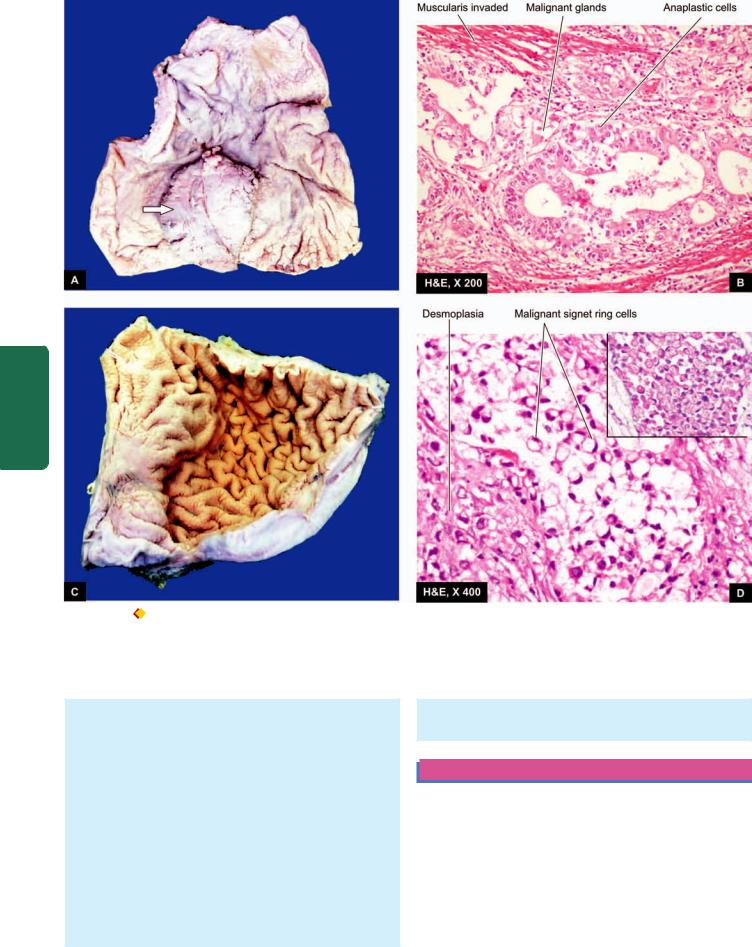
560
Pathology Systemic III SECTION
Figure 20.19 
 A, Ulcerative carcinoma stomach. The luminal surface of the stomach in the region of pyloric canal shows an elevated irregular growth with ulcerated surface and raised margins. B, Malignant cells forming irregular glands with stratification are seen invading the layers of the stomach wall. C, Linitis plastica. The wall of the stomach in the region of pyloric canal is markedly thickened and fibrotic while the mucosal folds are lost. D, Microscopy shows characteristic signet ring tumour cells having abundant mucinous cytoplasm positive for mucicarmine (inbox). The stroma is desmoplastic.
ulcerations. Cut section shows lesions in the mucosa and submucosa but in late stage whole thickness of the gut wall may be affected.
2. Polypoid type, which produces large protruding mass into the lumen with ulcerated surface.
Lymph node involvement may occur in either of the two patterns.
Microscopically, gastric lymphomas are most often nonHodgkin’s lymphomas of the following types:
 High-grade large cell immunoblastic lymphoma being the most common.
 Low-grade small lymphocytic well-differentiated B-cell lymphoma referred to as MALToma is the next in frequency (arising from Mucosa Associated Lymphoid
Tissue). The term pseudolymphoma is sometimes used for non-invasive stage of MALToma.
SMALL INTESTINE
NORMAL STRUCTURE
Anatomically, the small bowel having a length of 550-650 cm, includes the duodenum, jejunum and ileum and tends to become narrower throughout its course.
Histologically, the small bowel is identified by recognition of villi. The wall of the small intestine consists of 4 layers:
1. The serosa is the outer covering of the small bowel which is complete except over a part of the duodenum.

2.The muscularis propria is composed of 2 layers of smooth muscle tissue—outer thinner longitudinal and inner thicker circular layer. These muscles are functionally important for peristalsis. Between the two layers of muscle lie ganglionated plexus, myenteric plexus of Auerbach.
3.The submucosa is composed of loose fibrous tissue with blood vessels and lacteals in it. It contains a gangliated plexus, Meissner’s plexus, having fewer and smaller cells than the Auerbach’s plexus.
4.The mucosa consists of glandular epithelium overlying the lamina propria composed of loose connective tissue and contains phagocytic cells and abundance of lymphoid cells (Peyer’s patches in the ileum) and plasma cells. It is supported externally by thin layer of smooth muscle fibres, muscularis mucosae. The mucous membrane is thrown into folds or plicae which are more in the jejunum and less in the ileum, thus increasing the absorptive surface enormously. The absorptive surface is further increased by the intestinal villi. Villi are finger-like or leaf-like projections which contain
3types of cells:
i)Simple columnar cells. They perform absorptive function due to the presence of brush border consisting of large number of microvilli.
ii)Goblet cells. These are mucus-secreting cells and are interspersed between the columnar cells.
iii)Endocrine cells. These are scattered in the villi as well as are widely distributed throughout the gastrointestinal tract. These cells have various synonyms as under:

 Kulchitsky cells, after the name of its discoverer.

 Enterochromaffin cells, due to their resemblance to chromaffin cells of the adrenal medulla.

 Argentaffin cells, as the intracytoplasmic granules stain positively with silver salts by reduction reaction (argyrophil cells, on the other hand, require the addition of exogenous reducing substance for staining).

 Endocrine cells, as these specialised cells are considered to be part of APUD cell system (having common properties as Amine content, amine Precursor Uptake and Decarboxylation). APUD cells are considered to be endodermal in origin, while previously they were thought to be neural crest derivative. Other endocrine cells belonging to the APUD cell system are C-cells of the thyroid, chromaffin cells of the adrenal medulla, certain cells of the carotid body, bronchi, hypothalamus, pituitary and sympathetic ganglia.
Endocrine cells are heavily populated in the proximal small bowel as this is the most active site for absorption and secretory activities. They are sparse in the colon which is less active site for such functions.
The duodenum contains distinctively branched Brunner’s glands present in the submucosa and going up to muscularis mucosae. The deeper layer of the mucosa of the small intestine elsewhere contains intestinal glands or crypts of Lieberkuhn. They are lined by columnar cells, goblet cells, endocrine cells and Paneth cells. Paneth cells are normally exclusively found in the small intestine and occasionally in the caecum. These cells are characterised by the presence of supranuclear granules rich in lysozyme.
The blood supply of the whole of small intestine, except the first part of the duodenum, is by the superior mesenteric artery which supplies blood by mesenteric arterial arcades and the straight arteries.
The main functions of the small intestine are digestion and absorption so that ultimately nutrients passing into the bloodstream are utilised by the cells in metabolism. The mucosal layer of the small intestine has remarkable capacity for regeneration and new lining is laid every 3-4 days.
CONGENITALANOMALIES
Intestinal Atresia and Stenosis
 Intestinal atresia is congenital absence of lumen, most commonly affecting the ileum or duodenum. The proximal segment has a blind end which is separated from distal segment freely, or the two segments are joined by a fibrous cord. The condition must be recognised early and treated surgically, as otherwise it is incompatible with life.

 Intestinal stenosis is congenital narrowing of the lumen affecting a segment of the small intestine. Intestinal segment above the level of obstruction is dilated and that below it is collapsed.
Meckel’s Diverticulum
Meckel’s diverticulum is the most common congenital anomaly of the gastrointestinal tract, occurring in 2% of population. It is more common in males. The anomaly is commonly situated on the antimesenteric border of the ileum, about 1 meter above the ileocaecal valve. Like other true diverticula, Meckel’s diverticulum is an outpouching containing all the layers of the intestinal wall in their normal orientation (Fig. 20.20). It is almost always lined by small intestinal type of epithelium; rarely it may contain islands of gastric mucosa and ectopic pancreatic tissue. Embryologic origin of Meckel’s diverticulum is from incomplete obliteration of vitellointestinal duct. (Other anomalies resulting from the remnants of vitellointestinal duct are vitelline sinus and vitelline cyst).
The common complications of Meckel’s diverticulum are perforation, haemorrhage and diverticulitis.
In addition to congenital Meckel’s diverticulum, acquired diverticula also occur in the small intestine. These are commonly multiple (diverticulosis), frequently located on the mesenteric border, and are sometimes associated with malabsorption.
Intestinal Malrotation
Malrotation is a developmental abnormality of the midgut (i.e. the portion of intestine between the duodenojejunal flexure and the middle of transverse colon). Due to failure of normal rotation of midgut, the following consequences can occur:
i)Exomphalos i.e. intestinal eventration at the umbilicus.
ii)Misplacement of the caecum, appendix and ascending colon.
iii)Mobile caecum.
561
Tract Gastrointestinal The 20 CHAPTER
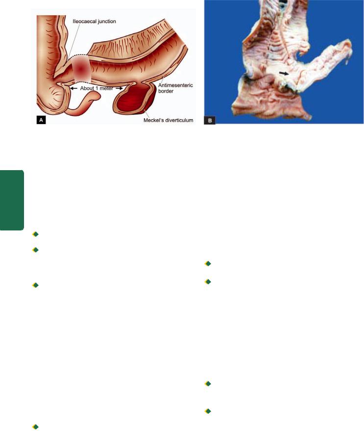
562
Pathology Systemic III SECTION
Figure 20.20 
 Meckel’s diverticulum. A, Common location and gross appearance. B, Resected segment of the small intestine shows an outpouching which on section is seen communicating with the intestinal lumen. The lumen contains necrotic debris.
INTESTINAL OBSTRUCTION
Conditions which interfere with the propulsion of contents in the intestine are considered under the heading of intestinal obstruction. The causes of intestinal obstruction can be classified under the following 3 broad groups:
1. Mechanical obstruction. It can occur as a result of the following causes:
i) Internal obstruction (intramural and intraluminal):

 Inflammatory strictures (e.g. Crohn’s disease) Congenital stenosis, atresia, imperforate anus

 Tumours

 Meconium in mucoviscidosis

 Roundworms

 Gallstones, faecoliths, foreign bodies

 Ulceration induced by potassium chloride tablets prescribed to counter hypokalaemia.
ii) External compression:

 Peritoneal adhesions and bands

 Strangulated hernias

 Intussusception

 Volvulus
 Intra-abdominal tumour.
2.Neurogenic obstruction. It occurs due to paralytic ileus i.e. paralysis of muscularis of the intestine as a result of shock after abdominal operation or by acute peritonitis.
3.Vascular obstruction. Obstruction of the superior mesenteric artery or its branches may result in infarction causing paralysis. The causes are as under:

 Thrombosis

 Embolism

 Accidental ligation.
Out of the various causes listed above, conditions producing external compression on the bowel wall are the most common causes of intestinal obstruction (80%). Some of these are described below.
Peritoneal Adhesions and Bands
Adhesions and bands in the peritoneum composed of fibrous tissue result following healing in peritonitis. Rarely, such fibrous adhesions and bands may be without any preceding peritoneal inflammation and are of congenital origin. In either case, peritoneal bands and adhesions result in partial or complete intestinal obstruction by outside pressure on the bowel wall.
Hernias
Hernia is protrusion of portion of a viscus through an abnormal opening in the wall of its natural cavity.

 External hernia is the protrusion of the bowel through a defect or weakness in the peritoneum.
 Internal hernia is the term applied for herniation that does not present on the external surface.
Two major factors involved in the formation of a hernia are as under:
i)Local weakness which may be congenital e.g. at the umbilicus, inguinal and femoral canals, and in surgical scars called ‘incisional hernia’.
ii)Increased intra-abdominal pressure that is produced by coughing, straining and exertion.
Inguinal hernias are more common, followed in decreasing frequency, by femoral and umbilical hernias. Inguinal hernias may be of 2 types:
 Direct when hernia passes medial to the inferior epigastric artery and it appears through the external abdominal ring.

 Indirect when it follows the inguinal canal lateral to the inferior epigastric artery.
When the contents of hernia such as loop of intestine can be returned to the abdominal cavity, it is called reducible. When it is not possible to reduce hernia due to large contents or due to adhesions in the hernial sac, it is referred to as
irreducible.
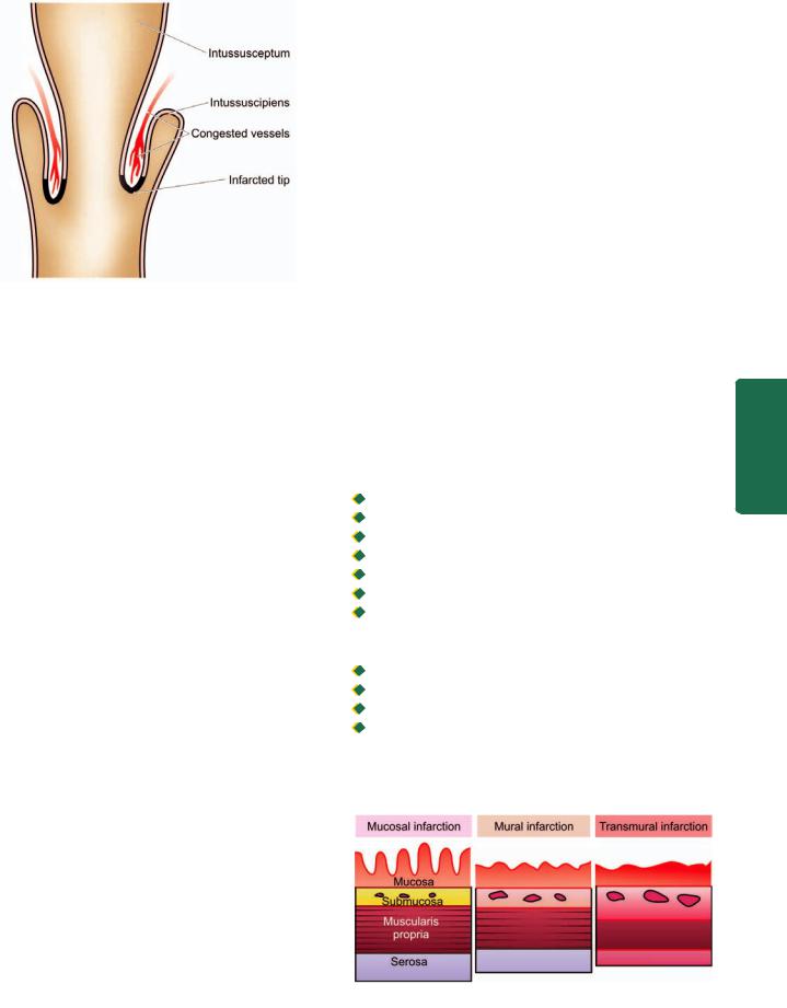
Figure 20.21 
 Ileocaecal intussusception.
When the blood flow in the hernial sac is obstructed, it results in strangulated hernia. Obstruction to the venous drainage and arterial supply may result in infarction or gangrene of the affected loop of intestine. The gross and microscopic appearance of strangulated intestine is the same as that of infarction of intestine.
Intussusception
Intussusception is the telescoping of a segment of intestine into the segment below due to peristalsis. The telescoped segment is called the intussusceptum and lower receiving segment is called the intussuscipiens. The condition occurs more commonly in infants and young children, more often in the ileocaecal region when the portion of ileum invaginates into the ascending colon without affecting the position of the ileocaecal valve (Fig. 20.21). Less common forms are ileoileal and colo-colic intussusception.
In children, the cause is usually not known though enlargement of the lymphoid tissue in the terminal ileum has been suggested by some. In the case of adults, the usual causes are foreign bodies and tumours.
The main complications of intussusception are intestinal obstruction, infarction, gangrene, perforation and peritonitis.
Volvulus
Volvulus is the twisting of loop of intestine upon itself through 180° or more. This leads to obstruction of the intestine as well as cutting off of the blood supply to the affected loop. The usual causes are bands and adhesions (congenital or acquired) and long mesenteric attachment. The condition is more common in the sigmoid colon than the small bowel.
ISCHAEMIC BOWEL DISEASE (ISCHAEMIC ENTEROCOLITIS)
Ischaemic lesions of the gastrointestinal tract may occur in the small intestine and/or colon; the latter is called ischaemic colitis or ischaemic enterocolitis and is commonly referred to
as ischaemic bowel disease. In either case, the cause of 563 ischaemia is compromised mesenteric circulation, while ischaemic effect is less likely to occur in the stomach, duodenum and rectum due to abundant collateral blood supply.
Depending upon the extent and severity of ischaemia, 3 patterns of pathologic lesions can occur (Fig. 20.22):
1. Transmural infarction, characterised by full thickness involvement i.e. transmural ischaemic necrosis and gangrene of the bowel.
2 Mural infarction, characterised by haemorrhagic gastroenteropathy (haemorrhage and necrosis). The ischaemic effect in mural infarction is limited to mucosa, submucosa and superficial muscularis, while mucosal infarction is confined to mucosal layers superficial to muscularis mucosae.
3. Ischaemic colitis, due to chronic colonic ischaemia causing fibrotic narrowing of the affected bowel.
These pathologic patterns are described below:
Transmural Infarction
Ischaemic necrosis of the full-thickness of the bowel wall is |
CHAPTER |
|
|
||
more common in the small intestine than the large intestine. |
|
|
ETIOPATHOGENESIS. The common causes of transmural |
|
|
infarction of small bowel are as under: |
|
|
i) |
Mesenteric arterial thrombosis such as due to the following: |
20 |
|
Atherosclerosis (most common) |
|
|
Aortic aneurysm |
|
|
Vasospasm |
The |
|
Fibromuscular hyperplasia |
|
|
Invasion by the tumour |
Gastrointestinal |
|
Use of oral contraceptives |
|
|
|
|
|
Arteritis of various types |
|
ii) |
Mesenteric arterial embolism arising from the following |
|
causes: |
|
|
|
Mural thrombi in the heart |
|
|
Endocarditis (infective and nonbacterial thrombotic) |
|
|
Atherosclerotic plaques |
Tract |
|
Atrial myxoma |
|
|
|
|
iii) Mesenteric venous occlusion is less common cause of fullthickness infarction of the bowel. The causes are as under:
Figure 20.22 
 Schematic diagram to show the three types of ischaemic bowel disease depending upon the extent of involvement.
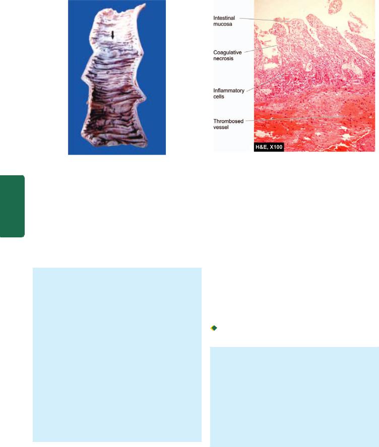
564
Pathology Systemic III SECTION
Figure 20.23 
 Haemorrhagic infarct of the small intestine. The infarcted area is swollen, dark in colour and coated with fibrinous exudate.
A sharp line of demarcation separates infarcted area from the normal bowel (arrow).

 Intestinal sepsis e.g. appendicitis

 Portal venous thrombosis in cirrhosis of the liver

 Tumour invasion

 Use of oral contraceptives
iv) Miscellaneous causes: 
 Strangulated hernia
 Torsion

 Fibrous bands and adhesions.
MORPHOLOGIC FEATURES. Grossly, irrespective of the underlying etiology, infarction of the bowel is haemorrhagic (red) type (page 126). A varying length of the small bowel may be affected. In the case of colonic infarction, the distribution area of superior and inferior mesenteric arteries (i.e. splenic flexure) is more commonly involved. The affected areas become dark purple and markedly congested and the peritoneal surface is coated with fibrinous exudate. The wall is thickened, oedematous and haemorrhagic. The lumen is dilated and contains blood and mucus. In arterial occlusion, there is sharp line of demarcation between the infarcted bowel and the normal intestine, whereas in venous occlusion the infarcted area merges imperceptibly into the normal bowel
(Fig. 20.23).
Microscopically, there is coagulative necrosis and ulceration of the mucosa and there are extensive submucosal haemorrhages. The muscularis is less severely affected by ischaemia. Subsequently, inflammatory cell infiltration and secondary infection occur, leading to gangrene of the bowel (Fig. 20.24).
The condition is clinically characterised by ‘abdominal angina’ in which the patient has acute abdominal pain, nausea, vomiting, and sometimes diarrhoea. The disease is rapidly fatal, with 50-70% mortality rate.
Figure 20.24 
 Infarct small intestine, microscopic appearance. The mucosa in the infarcted area shows coagulative necrosis and submucosal haemorrhages: muscularis is also partly affected. Inflammatory cell infiltration is marked at the line of demarcation between the infarcted and normal bowel.
Mural and Mucosal Infarction (Haemorrhagic
Gastroenteropathy, Membranous Colitis)
Mural and mucosal infarctions are limited to superficial layers of the bowel wall, sparing the deeper layer of the muscularis and the serosa. The condition is also referred to
as haemorrhagic gastroenteropathy, and in the case of colon as membranous colitis.
ETIOPATHOGENESIS. Haemorrhagic gastroenteropathy results from conditions causing non-occlusive hypoperfusion (compared from transmural infarction which occurs from occlusive causes). These are as under:

 Shock
Shock

 Cardiac failure

 Infections

 Intake of drugs causing vasoconstriction e.g. digitalis, norepinephrine.
MORPHOLOGIC FEATURES. Grossly, the lesions affect variable length of the bowel. The affected segment of the bowel is red or purple but without haemorrhage and exudation on the serosal surface. The mucosa is oedematous at places, sloughed and ulcerated at other places. The lumen contains haemorrhagic fluid.
Microscopically, there is patchy ischaemic necrosis of mucosa, vascular congestion, haemorrhages and inflammatory cell infiltrate. The changes may extend into superficial muscularis but deeper layer of muscularis and serosa are spared. Secondary bacterial infection may supervene resulting in pseudomembranous enterocolitis.
Clinically, as in transmural infarction, the features of abdominal pain, nausea, vomiting and diarrhoea are present, but the changes are reversible and curable. With adequate
therapy, normal morphology is completely restored in superficial lesions, while deeper lesions may heal by fibrosis leading to stricture formation.
Ischaemic Colitis
Although this condition affects primarily colon in the region of splenic flexure, it is described here due to its apparent pathogenetic relationship with ischaemic injury. Ischaemic colitis is characterised by chronic segmental colonic ischaemia followed by chronic inflammation and healing by fibrosis and scarring causing obstruction (ischaemic stricture).
Grossly, most frequently affected site is the splenic flexure; other site is rectum. Ischaemic colitis passes through 3 stages: infarct, transient ischaemia and ischaemic stricture. However, the surgical submitted specimens generally are of the ischaemic stricture. External surface of the affected area is fusiform or saccular. On cut section, there are patchy, segmental and longitudinal mucosal ulcers. Thus, the gross appearance can be confused with either of the two types of inflammatory bowel disease.
Microscopically, the ulcerated areas of the mucosa show granulation tissue. The submucosa is characteristically thickened due to inflammation and fibrosis. The muscularis may also show inflammatory changes and patchy replacement by fibrosis. The blood vessels may show atheromatous emboli, organising thrombi and endarteritis obliterans.
NECROTISING ENTEROCOLITIS
Necrotising enterocolitis is an acute inflammation of the terminal ileum and ascending colon, occurring primarily in premature and low-birth-weight infants within the first week of life and less commonly in full-term infants.
ETIOLOGY. The condition has been considered as a variant of the spectrum of ischaemic bowel disease. Important factors in the etiology of this disorder, thus, are as follows:
1.Ischaemia
2.Hypoxia/anoxia of the bowel due to bypassing of blood from the affected area
3.Bacterial infection and endotoxins
4.Establishment of feeding
5.Infants fed on commercial formulae than breast-fed, implying the role of immunoprotective factors.
MORPHOLOGIC FEATURES. Grossly, the affected segment of the bowel is dilated, necrotic, haemorrhagic and friable. Bowel wall may contain bubbles of air (pneumatosis intestinalis).
Microscopically, the changes are variable depending upon the stage. Initial changes are confined to mucosa and show oedema, haemorrhage and coagulative necrosis. A pseudomembrane composed of necrotic epithelium, fibrin and inflammatory cells may develop. As the ischaemic process extends to the subjacent layers, muscle layer is also involved and may lead to perforation and peritonitis.
In healed cases, stricture formation, malabsorption and short bowel syndrome are the usual complications.
INFLAMMATORY BOWEL DISEASE
(CROHN’S DISEASE AND ULCERATIVE COLITIS)
DEFINITION. The term ‘inflammatory bowel disease (IBD)’ is commonly used to include 2 idiopathic bowel diseases having many similarities but the conditions usually have distinctive morphological appearance. These 2 conditions are Crohn’s disease (regional enteritis) and ulcerative colitis:
1.Crohn’s disease or Regional enteritis is an idiopathic chronic ulcerative IBD, characterised by transmural, noncaseating granulomatous inflammation, affecting most commonly the segment of terminal ileum and/or colon, though any part of the gastrointestinal tract may be involved.
2.Ulcerative colitis is an idiopathic form of acute and chronic ulcero-inflammatory colitis affecting chiefly the mucosa and submucosa of the rectum and descending colon, though sometimes it may involve the entire length of the large bowel.
Both these disorders primarily affect the bowel but may have systemic involvement in the form of polyarthritis, uveitis, ankylosing spondylitis, skin lesions and hepatic involvement. Both diseases can occur at any age but are more frequent in 2nd and 3rd decades of life. Females are affected slightly more often.
ETIOPATHOGENESIS. The exact etiology of IBD remains unknown. However, multiple factors are implicated which can be considered under the following 3 groups:
1.Genetic factors. Genetic factors are implicated in the etiopathogenesis of IBD is supported by the following evidences:
i)There is about 3 to 20 times higher incidence of occurrence of IBD in first-degree relatives. This is due to genetic defect causing diminished epithelial barrier function.
ii)Overall, there is approximately 50% chance of development of IBD (Crohn’s disease about 60% concordance, ulcerative colitis about 6% concordance) in monozygotic twins.
iii)Although no specific and consistent gene association with IBD has been seen, genome wide search has revealed that disease-predisposing loci are present in chromosomes 16q, 12p, 6p, 14q and 5q. In particular, CARD15 (caspase- associated recruitment domain containing protein 15) on chromosome 16q is expressed by several cells in the intestinal mocosa which in mutated form results in loss of its function and renders an individual about 50-times higher risk to develop Crohn’s disease.
iv)HLA studies show that ulcerative colitis is more common in HLA-DRB1-alleles while Crohn’s disease is more common in HLA-DR7 and DQ4 alleles.
2.Immunologic factors. Defective immunologic regulation in IBD has been shown to play significant role in the pathogenesis of IBD:
i) Defective regulation of immune suppression. In a normal individual, there is lack of immune responsiveness to dietary
565
Tract Gastrointestinal The 20 CHAPTER

566antigens and commensal flora in the intestinal lumen. The mechanism responsible for this is by activation of CD4+ T cells secreting cytokines inhibitory to inflammation (IL-10, TGF-β) which suppress inflammation in the gut wall. In IBD, this immune mechanism of suppression of inflammation is defective and thus results in uncontrolled inflammation.
ii) Transgenic mouse experimental model studies. Gene ‘knock out’ studies on colitis in mice have revealed that multiple immune abnormalities may be responsible for IBD as under:
a)Deletion of inflammation inhibitory cytokines (e.g. IL-2, IL-10, TGF-β) or their receptors.
b)Deletion of molecules responsible for T cell recognition (e.g. T cell antigen receptors, MHC class II).
c)Interference with normal epithelial barrier function in the intestine (e.g. blocking N-cadherin, deletion of multi-drug resistance MDR gene).
iii) Type of inflammatory cells. In both types of IBD, activated CD4+ T cells are present in the lamina propria and in the
peripheral blood. These cells either activate other inflammatory cells (e.g. macrophages and B cells), or recruit more inflammatory cells by stimulation of homing receptor on leucocytes and vascular endothelium. There are two main types of CD4+ T cells in IBD:

 TH1 cells secrete proinflammatory cytokines IFN-γ and TNF which induce transmural granulomatous inflammation seen in Crohn’s disease. IL-12 initiates TH1 cytokine pathway.
 TH2 cells secrete IL-4, IL-5 and IL-13 which induce superficial mucosal inflammation characteristically seen in ulcerative colitis.
3. Exogenous factors. In addition to role of genetic factors and deranged T-cell mediated immunity, a role for several exogenous and environmental factors has been assigned: i) Microbial factors: At different times, role of a variety of microbes in initiation of inflammatory response by the body has been suspected. Accordingly, several microorganism species (bacteria, viruses, protozoa and fungi) have been
Pathology Systemic III SECTION
TABLE 20.6: Distinguishing Features of Crohn’s Disease and Ulcerative Colitis.
|
Feature |
Crohn’s Disease |
Ulcerative Colitis |
A. MACROSCOPIC FEATURES |
|
|
|
1. |
Distribution |
Segmental with skip areas |
Continuous without skip areas |
2. |
Location |
Commonly terminal ileum and/or |
Commonly rectum, sigmoid colon and |
|
|
ascending colon |
extending upwards |
3. |
Extent |
Usually involves the entire thickness |
Usually superficial, confined to mucosal |
|
|
of the affected segment of bowel wall |
layers |
4. |
Ulcers |
Serpiginous ulcers, may develop |
Superficial mucosal ulcers without fissures |
|
|
into deep fissures |
|
5. |
Pseudopolyps |
Rarely seen |
6. |
Fibrosis |
Common |
7. |
Shortening |
Due to fibrosis |
B. |
MICROSCOPIC FEATURES |
|
1. |
Depth of inflammation |
Typically transmural |
2. |
Type of inflammation |
Non-caseating granulomas and infiltrate |
|
|
of mononuclear cells (lymphocytes, |
|
|
plasma cells and macrophages) |
Commonly present
Rare
Due to contraction of muscularis
Mucosal and submucosal
Crypt abscess and non-specific acute and chronic inflammatory cells (lymphocytes, plasma cells, neutrophils, eosinophils, mast cells)
3. |
Mucosa |
Patchy ulceration |
Haemorrhagic mucosa with ulceration |
4. |
Submucosa |
Widened due to oedema and lymphoid |
Normal or reduced in width |
|
|
aggregates |
|
5. |
Muscularis |
Infiltrated by inflammatory cells |
Usually spared except in cases of toxic |
|
|
|
megacolon |
6. |
Fibrosis |
Present |
Usually absent |
C. |
IMMUNOLOGIC FEATURES |
|
|
1. |
Lymphocyte type |
CD4+ TH1 |
CD4+ TH2 |
2. |
Cytokines |
INF-γ, TNF, IL-12 |
TGF-β, IL-4, IL-5, IL-13 |
3. |
ANCA-P antibodies |
Positive in a few |
Positive in most |
D. |
COMPLICATIONS |
|
|
1. |
Fistula formation |
Internal and external fistulae in 10% cases |
Extremely rare |
2. |
Malignant changes |
Rare |
May occur infrequently in disease of more than |
|
|
|
10 years’ duration |
3. |
Type of malignancy |
Lymphoma more than carcinoma |
Carcinoma more than lymphoma |
4. |
Fibrous strictures |
Common |
Never |
|
|
|
|
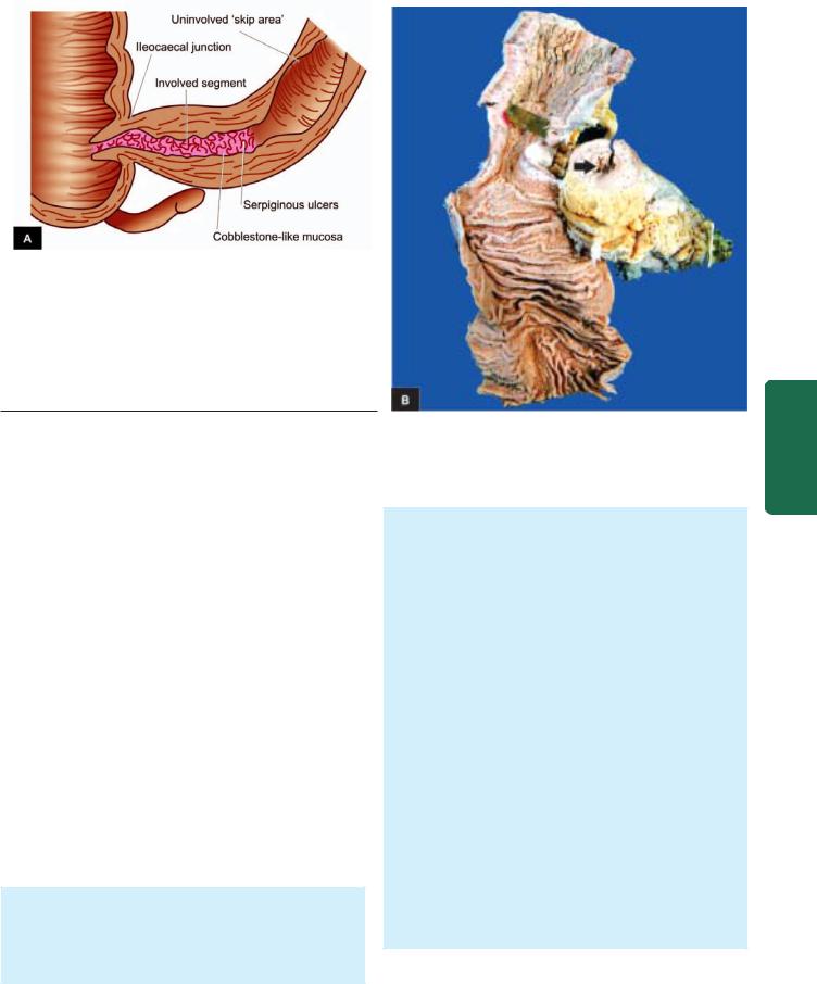
Figure 20.25 
 Crohn’s disease of the terminal ileum. A, The lesions are characteristically segmental with intervening uninvolved ‘skip areas’.
The bowel wall is thickened and the lumen narrowed, giving hose-pipe appearance. Serpiginous ulcers, some deep fissures and swollen intervening surviving mucosa giving ‘cobblestone appearance’, are present.
B, The specimen of small intestine is shown in longitudinal section along with a segment in cross section. External surface shows increased mesenteric fat, thickened wall and narrow lumen. Luminal surface of longitudinal cut section shows segment of thickened wall with narrow lumen which is better appreciated in cross section (arrow) while intervening areas of the bowel are uninvolved or skipped.
suspect but without definite evidence: Mycobacterium paratuberculosis, Salmonella, Shigella, Helicobacter, Clostridia, bacteroides, Escherichia, Measles virus etc.
ii)Psychosocial factors: It has been observed that individuals who are unduly sensitive, dependent on others and unable to express themselves, or some major life events such as illness or death in the family, divorce, interpersonal conflicts etc, suffer from irritable colon or have exacerbation of symptoms. Patients of IBD in the West have been found to suffer from greater functional impairment than the general population, as assessed by sickness impact profile which is a measure of overall psychological and physical functioning.
iii)Smoking: Role of smoking in causation of Crohn’s disease has been reported.
iv)Oral contraceptives: An increased risk to develop Crohn’s disease with long-term use of oral contraceptives has been found in some studies but there is no such increased risk for ulcerative colitis.
Consensus hypothesis in pathogenesis of IBD combines the role of above three major groups of etiologic factors: i.e.
in a genetically predisposed individual, the effects of exogenous and endogenous host factors result in dysregulation of mucosal immune function, which gets further modified by
certain environmental factors.
MORPHOLOGIC FEATURES. The morphologic features of Crohn’s disease and ulcerative colitis are sufficiently distinctive so as to be classified separately. These features are presented below; the distinguishing features of the two conditions are summarised in Table 20.6.
CROHN’S DISEASE. Crohn’s disease may involve any portion of the gastrointestinal tract but affects most
commonly 15-25 cm of the terminal ileum which may extend into the caecum and sometimes into the ascending colon:
Grossly, characteristic feature is the multiple, welldemarcated segmental bowel involvement with intervening uninvolved ‘skip areas’. The wall of the affected bowel segment is thick and hard, resembling a ‘hose pipe’. Serosa may be studded with minute granulomas. The lumen of the affected segment is markedly narrowed. The mucosa shows ‘serpiginous ulcers’, while intervening surviving mucosa is swollen giving ‘cobblestone appearance’. There may be deep fissuring into the bowel wall (Fig. 20.25).
Histologically, the characteristic features are as follows
(Fig. 20.26):
1.Transmural inflammatory cell infiltrate consisting of chronic inflammatory cells (lymphocytes, plasma cells and macrophages) is the classical microscopic feature.
2.Non-caseating, sarcoid-like granulomas are present in all the layers of the affected bowel wall in 60% of cases and may even be seen in the regional lymph nodes.
3.There is patchy ulceration of the mucosa which may take the form of deep fissures, accompanied by inflammatory infiltrate of lymphocytes and plasma cells.
4.There is widening of the submucosa due to oedema and foci of lymphoid aggregates.
5.In more chronic cases, fibrosis becomes increasingly prominent in all the layers disrupting muscular layer.
ULCERATIVE COLITIS. Classically, ulcerative colitis begins in the rectum, and in continuity extends upwards into the sigmoid colon, descending colon, transverse colon, and sometimes may involve the entire colon. The colonic contents
567
Tract Gastrointestinal The 20 CHAPTER
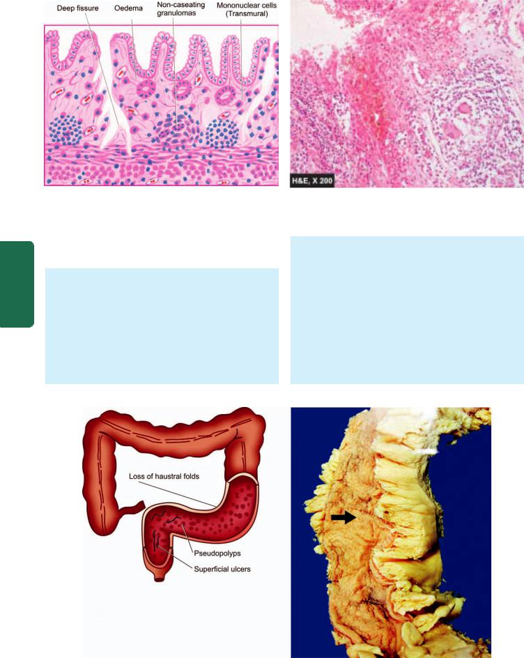
568
Pathology Systemic III SECTION
Figure 20.26 
 Crohn’s disease of the ileum. The histological features present are: transmural chronic inflammatory cell infiltration, deep fissures into the bowel wall, submucosal widening due to oedema, some prominent lymphoid follicles and a few non-caseating epithelioid cell granulomas in the bowel wall.
may rarely backflow into the terminal ileum in continuity, causing ‘back-wash ileitis’ in about 10% of cases.
Grossly, the characteristic feature is the continuous involvement of the rectum and colon without any uninvolved skip areas compared to Crohn’s disease. The appearance of colon may vary depending upon the stage and intensity of the disease because of remissions and exacerbations. Mucosa shows linear and superficial ulcers, usually not penetrating the muscular layer. The intervening intact mucosa may form inflammatory ‘pseudopolyps.’ The muscle layer is thickened due to contraction, producing shortening and narrowing of the
affected colon with loss of normal haustral folds giving ‘garden-hose appearance’ (Fig. 20.27).
Histologically, ulcerative colitis because of remission and exacerbations, is characterised by alternating ‘active disease process’ and ‘resolving colitis.’ The changes in the ‘active disease process’ are as under (Fig. 20.28):
1.Crypt distortion, cryptitis and focal accumulations of neutrophils forming crypt abscesses.
2.Marked congestion, dilatation and haemorrhages from mucosal capillaries.
3.Superficial mucosal ulcerations, usually not penetrating into the muscle coat, except in severe cases, and is accompanied by nonspecific inflammatory cell infiltrate
Figure 20.27 
 Ulcerative colitis. Continuous involvement of the rectum and colon without any uninvolved skip areas. The ulcers are superficial with intervening inflammatory pseudopolyps. The lumen is narrow and the haustral folds are lost giving ‘garden-hose appearance’.
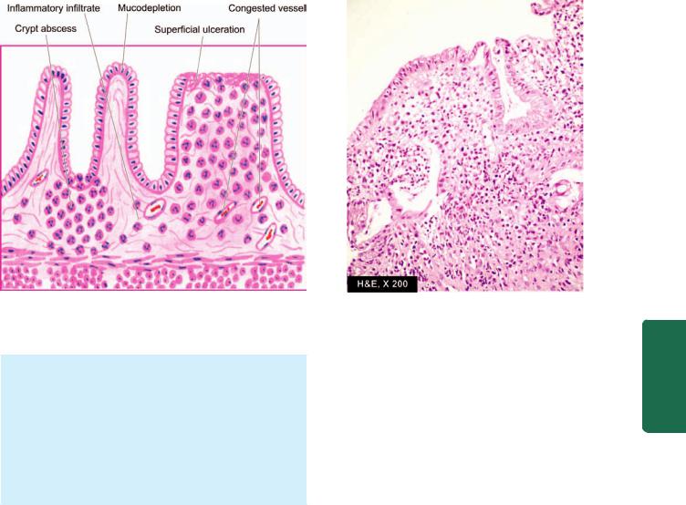
569
Figure 20.28 
 Ulcerative colitis in active phase. The microscopic features seen are superficial ulcerations, with mucosal infiltration by inflammatory cells and a ‘crypt abscess’.
of lymphocytes, plasma cells, neutrophils, some eosinophils and mast cells in the lamina propria.
4.Goblet cells are markedly diminished in cases of active disease.
5.Areas of mucosal regeneration and mucodepletion of lining cells.
6.In long-standing cases, epithelial cytologic atypia ranging from mild to marked dysplasia and sometimes developing into carcinoma in situ and frank adenocarcinoma.
COMPLICATIONS. Complications of Crohn’s disease and ulcerative colitis are as under:
Crohn’s disease:
1.Malabsorption due to impaired absorption of fat, vitamin B12, proteins and electrolytes from the diseased small bowel.
2.Fistula formation may occur in long-standing cases. These may be internal fistulae between the loops of the intestine, or external fistulae such as enterocutaneous, rectal and anal fistulae.
3.Stricture formation may occur in chronic cases due to extensive fibrosis in the affected bowel wall.
4.Development of malignancy in the small intestine as a late complication of Crohn’s disease is rarer than that in ulcerative colitis, but lymphoma may develop more often in Crohn’s disease than adenocarcinoma (seen in some long-standing cases of ulcerative colitis).
Ulcerative colitis:
1. Toxic megacolon (Fulminant colitis) is the acute fulminating colitis in which the affected colon is thin-walled and dilated and is prone to perforation and faecal peritonitis. There is deep penetration of the inflammatory cell infiltrate into muscle layer which is disrupted.
2.Perianal fistula formation may occur rarely.
3.Carcinoma may develop in long-standing cases of ulcerative colitis of more than 10 years duration.
4.Stricture formation almost never occurs in ulcerative colitis.
OTHER INFLAMMATORY LESIONS OF THE BOWEL
Besides IBD, a variety of other acute and chronic inflammatory conditions affect small bowel (enteritis), large bowel (colitis), or both (enterocolitis); the last named being more common. Hence, all these conditions involving small bowel and/or large bowel are described together here for better correlation of features.
The various forms of inflammations of the bowel after excluding IBD can be categorised broadly into ‘infective
enterocolitis’ and ‘pseudomembranous enterocolitis.’
INFECTIVE ENTEROCOLITIS
These are a group of acute and chronic inflammatory lesions of small intestine and/or colon caused by microorganisms (bacteria, viruses, fungi, protozoa and helminths). All these are characterised by diarrhoeal syndromes. Pathogenetically speaking, these microorganisms can cause enterocolitis by 2 mechanisms—by enteroinvasive bacteria producing ulcerative lesions, and by enterotoxin-producing bacteria resulting in nonulcerative lesions.
A list of common microorganisms producing enterocolitis is presented in Table 20.7. A few important forms are described below.
Intestinal Tuberculosis
Intestinal tuberculosis can occur in 3 forms—primary, secondary and hyperplastic caecal tuberculosis.
1. PRIMARY INTESTINAL TUBERCULOSIS. Though an uncommon disease in the developed countries of the world,
Tract Gastrointestinal The 20 CHAPTER
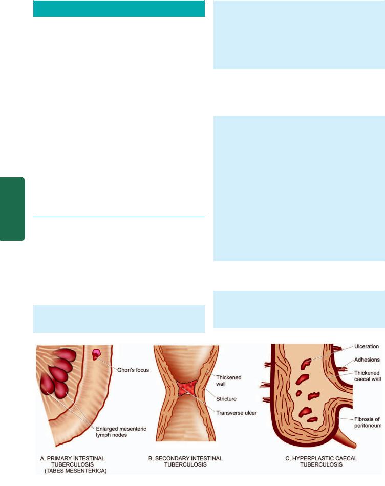
570
Pathology Systemic III SECTION
TABLE 20.7: Micro-organisms Causing Infective Enterocolitis.
A.BACTERIAL ENTEROCOLITIS
1.Entero-invasive bacteria
(i)Tuberculosis
(ii)Salmonella
(iii)Campylobacter jejuni
(iv)Shigella
(v)Escherichia coli
(vi)Yersinia enterocolitica
2.Enterotoxin-producing bacteria
(i)Vibrio cholerae
B.VIRAL ENTEROCOLITIS
C.FUNGAL ENTEROCOLITIS
(i)Candidiasis
(ii)Mucormycosis
D.PROTOZOAL AND METAZOAL INFESTATIONS
(i)Giardia lamblia
(ii)Entamoeba histolytica
(iii)Balantidium coli
(iv)Taenia solium
(v)Ascaris lumbricoides
(vi)Ancylostoma duodenale
(vii)Strongyloides stercoralis
primary tuberculosis of the ileocaecal region is quite common in developing countries including India. In the prepasteurisation era, it used to occur by ingestion of unpasteurised cow’s milk infected with Mycobacterium bovis. But now-a-days due to control of tuberculosis in cattle and pasteurisation of milk, virtually all cases of intestinal tuberculosis are caused by M. tuberculosis. The predominant changes are in the mesenteric lymph nodes without any significant intestinal lesion.
Grossly, the affected lymph nodes are enlarged, matted and caseous (tabes mesenterica). Eventually, there is healing by fibrosis and calcification (Fig. 20.29,A).
Microscopically, in the initial stage, there is primary complex or Ghon’s focus in the intestinal mucosa as occurs elsewhere in primary tuberculous infection (page 153). Subsequently, the mesenteric lymph nodes are affected which show typical tuberculous granulomatous inflammatory reaction with caseation necrosis. Tuberculous peritonitis may occur due to spread of the infection.
2. SECONDARY INTESTINAL TUBERCULOSIS. Selfswallowing of sputum in patients with active pulmonary tuberculosis may cause secondary intestinal tuberculosis, most commonly in the terminal ileum and rarely in the colon.
Grossly, the intestinal lesions are prominent than the lesions in regional lymph nodes as in secondary pulmonary tuberculosis (Fig. 20.29,B). The lesions begin in the Peyer’s patches or the lymphoid follicles with formation of small ulcers that spread through the lymphatics to form large ulcers which are transverse to the long axis of the bowel, (c.f. typhoid ulcers of small intestine, described below). These ulcers may be coated with caseous material. Serosa may be studded with visible tubercles. In advanced cases, transverse fibrous strictures and intestinal obstruction are seen (Fig. 20.30,A, B).
Histologically, the tuberculous lesions in the intestine are similar to those observed elsewhere i.e. presence of tubercles. Mucosa and submucosa show ulceration and the muscularis may be replaced by variable degree of fibrosis (Fig. 20.30,C). Tuberculous peritonitis may be observed.
3. HYPERPLASTIC CAECAL TUBERCULOSIS. This is a variant of occurring secondary to pulmonary tuberculosis.
Grossly, the caecum and/or ascending colon are thickwalled with mucosal ulceration. Clinically, the lesion is palpable and may be mistaken for carcinoma
(Fig. 20.29,C).
Figure 20.29 
 Intestinal tuberculosis, three patterns.
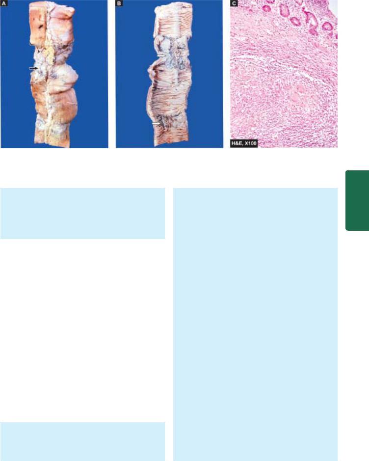
571
Figure 20.30 
 Intestinal tuberculosis. A, The external surface of small intestine shows stricture and a lymph node in section having caseation necrosis (arrows). B, The lumen shows characteristic transverse ulcers and two strictures (arrow). The wall of intestine in the area of narrowed lumen is thickened. C, Microscopy of intestine shows caseating epithelioid cell granulomas in the intestinal wall.
Microscopically, the presence of caseating tubercles distinguishes the condition from Crohn’s disease in which granulomas are non-caseating. Besides, bacteriological evidence by culture or animal inoculation and Mantoux test are helpful in differential diagnosis of the two conditions.
Enteric Fever
The term enteric fever is used to describe acute infection caused by Salmonella typhi (typhoid fever) or Salmonella paratyphi (paratyphoid fever). Besides these 2 salmonellae,
Salmonella typhimurium causes food poisoning.
PATHOGENESIS. The typhoid bacilli are ingested through contaminated food or water. During the initial asymptomatic incubation period of about 2 weeks, the bacilli invade the lymphoid follicles and Peyer’s patches of the small intestine and proliferate. Following this, the bacilli invade the bloodstream causing bacteraemia, and the characteristic clinical features of the disease like continuous rise in temperature and ‘rose spots’ on the skin are observed. Immunological reactions (Widal’s test) begin after about 10 days and peak titres are seen by the end of the third week. Eventually, the bacilli are localised in the intestinal lymphoid tissue (producing typhoid intestinal lesions), in the mesenteric lymph nodes (leading to haemorrhagic lymphadenitis), in the liver (causing foci of parenchymal necrosis), in the gall bladder (producing typhoid cholecystitis), and in the spleen (resulting in splenic reactive hyperplasia).
MORPHOLOGIC FEATURES. The lesions are observed in the intestines as well as in other organs.
1. INTESTINAL LESIONS. Grossly, terminal ileum is affected most often, but lesions may be seen in the jejunum
and colon. Peyer’s patches show oval typhoid ulcers with their long axis along the length of the bowel, (c.f. tuberculous ulcers of small intestine, described above). The base of the ulcers is black due to sloughed mucosa. The margins of the ulcers are slightly raised due to inflammatory oedema and cellular proliferation. There is never significant fibrosis and hence fibrous stenosis seldom occurs in healed typhoid lesions. The regional lymph nodes are invariably enlarged (Fig. 20.31,A).
Microscopically, there is hyperaemia, oedema and cellular proliferation consisting of phagocytic histiocytes (showing characteristic erythrophagocytosis), lymphocytes and plasma cells. Though enteric fever is an example of acute inflammation, neutrophils are invariably absent from the cellular infiltrate and this is reflected in the leucopenia with neutropenia and relative lymphocytosis in the peripheral blood (Fig. 20.31,B).
The main complications of the intestinal lesions of typhoid are perforation of the ulcers and haemorrhage.
2. OTHER LESIONS. Besides the intestinal involvement, various other organs and tissues showing pathological changes in enteric fever are as under:
i)Mesenteric lymph nodes—haemorrhagic lymphadenitis.
ii)Liver—foci of parenchymal necrosis.
iii)Gallbladder—typhoid cholecystitis.
iv)Spleen—splenomegaly with reactive hyperplasia.
v)Kidneys—nephritis.
vi)Abdominal muscles—Zenker’s degeneration.
vii)Joints—arthritis.
viii)Bones—osteitis.
ix)Meninges—Meningitis.
x)Testis—Orchitis.
Tract Gastrointestinal The 20 CHAPTER
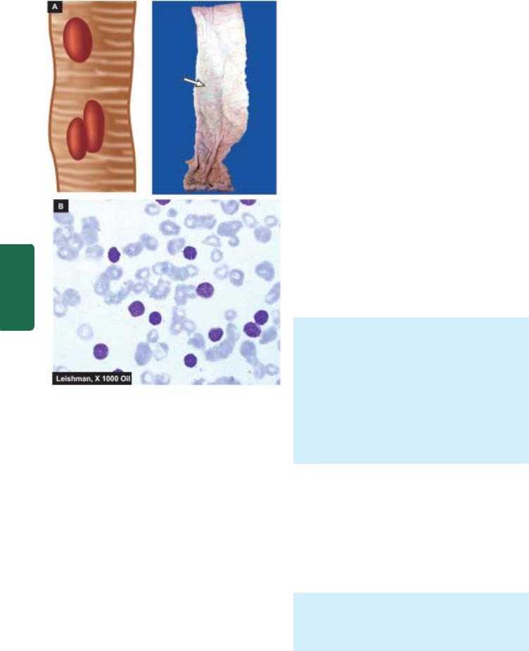
572
Pathology Systemic III SECTION
Figure 20.31 
 A, Typhoid ulcers in the small intestine appear characteristically oval with their long axis parallel to the long axis of the bowel. B, Blood picture in typhoid fever showing neutropenia and relative lymphocytosis.
Persistence of organism in the gallbladder or urinary tract may result in passage of organisms in the faeces or urine creating a ‘carrier state’ which is a source of infection to others.
Bacterial Food Poisoning
This is a form of acute bacterial illness that occurs following ingestion of food or water contaminated with bacteria other than those that cause specific acute intestinal infections like typhoid, paratyphoid, cholera or dysentery bacilli. The illness results from either bacterial invasion or bacterial toxigenic effect on the bowel.
The commonest causes of bacterial food poisoning resulting in enteritis or enterocolitis are as under:
1. Staphylococcal food poisoning. Staphylococcus aureus infection acquired from contaminated food produces either mild food poisoning by enterotoxins, or may cause more severe form of the illness called pseudomembranous enterocolitis
described below. Staphylococcal food poisoning occurs due to liberation of enterotoxins by the bacteria.
2.Clostridial food poisoning. Infection with anaerobic organisms Clostridium welchii, following consumption of contaminated meat results in acute food poisoning (page 181). The illness occurs both by bacterial invasion as well as by toxins.
3.Botulism. This is a severe form of paralysing illness caused by ingestion of organism, Clostridium botulinum, which produces neurotoxin.
4.Salmonella food poisoning (Salmonellosis). This is an infection (and not caused by toxins) occurring due to food contaminated by S. typhimurium or S. enteritidis. The condition manifests with fever, vomiting, and diarrhoea. Death may result from depletion of water and electrolytes.
Dysenteries
The term ‘dysentery’ is used to mean diarrhoea with abdominal cramps, tenesmus and passage of mucus in the stools, from any cause. There are 2 main forms of dysen- teries—bacillary and amoebic.
1. BACILLARY DYSENTERY. Bacillary dysentery is the term used for infection by shigella species: S. dysenteriae, S. flexneri, S. boydii and S. sonnei. Infection occurs by foeco-oral route and is seen with poor personal hygiene, in densely populated areas, and with contaminated food and water. The common housefly plays a role in spread of infection.
Grossly, the lesions are mainly found in the colon and occasionally in the ileum. Superficial transverse ulcerations of mucosa of the bowel wall occur in the region of lymphoid follicles but perforation is seldom seen. The intervening intact mucosa is hyperaemic and oedematous. Following recovery from the acute attack, complete healing usually takes place.
Microscopically, the mucosa overlying the lymphoid follicles is necrosed. The surrounding mucosa shows congestion, oedema and infiltration by neutrophils and lymphocytes. The mucosa may be covered by greyishyellow ‘pseudomembrane’ composed of fibrinosuppurative exudate.
The complications of bacillary dysentery are haemorrhage, perforation, stenosis, polyarthritis and iridocyclitis.
2. AMOEBIC DYSENTERY. This is due to infection by Entamoeba histolytica. It is more prevalent in the tropical countries and primarily affects the large intestine. Infection occurs from ingestion of cyst form of the parasite. The cyst wall is dissolved in the small intestine from where the liberated amoebae pass into the large intestine. Here, they invade the epithelium of the mucosa, reach the submucosa and produce the characteristic flask-shaped ulcers.
Grossly, early intestinal lesions appear as small areas of elevation on the mucosal surface. In advanced cases, typical flask-shaped ulcers having narrow neck and broad base are seen. They are more conspicuous in the caecum, rectum and in the flexures (Fig. 20.32).
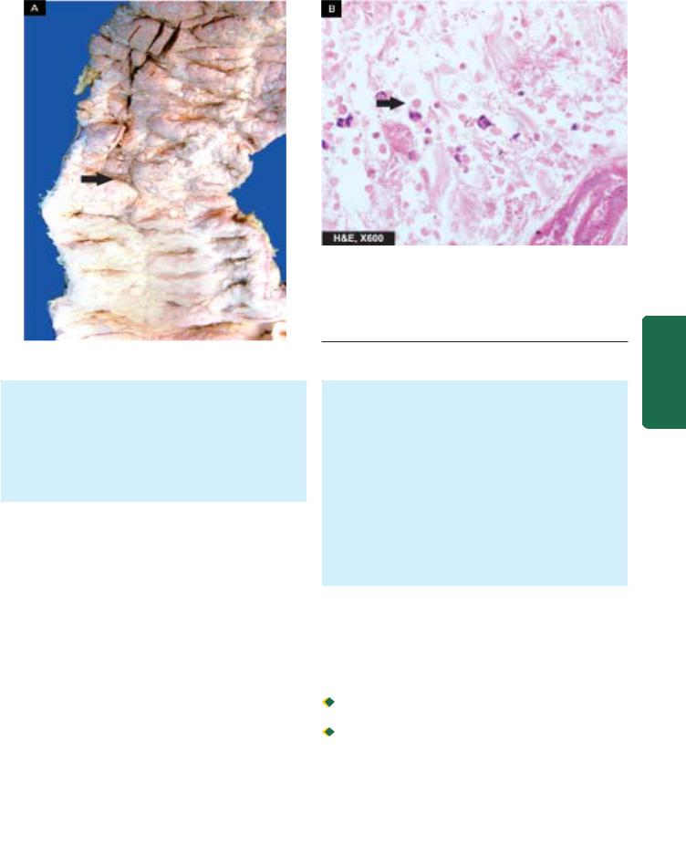
Microscopically, the ulcerated area shows chronic inflammatory reaction consisting of lymphocytes, plasma cells, macrophages and eosinophils. The trophozoites of Entamoeba are seen in the inflammatory exudate and are concentrated at the advancing margin of the lesion. Intestinal amoebae characteristically have ingested red cells in their cytoplasm. Oedema and vascular congestion are present in the area surrounding the ulcers.
Complications of intestinal amoebic ulcers are: amoebic liver abscess or amoebic hepatitis, perforation, haemorrhage and formation of amoeboma which is a tumour-like mass.
PSEUDOMEMBRANOUS ENTEROCOLITIS (ANTIBIOTIC-ASSOCIATED DIARRHOEA)
Pseudomembranous enterocolitis is a form of acute inflammation of colon and/or small intestine characterised by formation of ‘pseudomembrane’ over the site of mucosal injury.
ETIOLOGY. Numerous studies have established the overgrowth of Clostridium difficile with production of its toxin in the etiology of antibiotic-associated diarrhoea culminating in pseudomembranous colitis. Oral antibiotics such as clindamycin, ampicillin and the cephalosporins are more often (20%) associated with antibiotic-associated diarrhoea, while development of pseudomembranous colitis may occur in 1-10% cases.
Pseudomembrane formation may also occur in various other conditions as under:

 Staphylococcal enterocolitis

 Bacillary (Shigella) dysentery
 Candida enterocolitis
Figure 20.32 
 Amoebic ulcers large intestine. A, The luminal surface shows multiple ulcers some of which are deep and are flask-shaped with narrow neck and broad base (arrow) containing necrotic tissue and undermined margins. B, Trophozoites of Entamoeba histolytica are seen at the margin of ulcer (arrow).
MORPHOLOGIC FEATURES. Grossly, the lesions may be confined, to the large intestine or small intestine, or both may be involved. The mucosa of the bowel is covered by patchy, raised yellow-white plaques. Elsewhere, the mucosa is congested and may show small mucosal ulcerations.
Microscopically, the ‘pseudomembrane’ is composed of network of fibrin and mucus, in which are entangled inflammatory cells and mucosal epithelial cells. There is focal necrosis of surface epithelial cells. The lamina propria contains inflammatory cell infiltrate, mainly neutrophils. The submucosa has congested capillaries and may show microthrombi. The inflammation spreads laterally rather than deeply.
MALABSORPTION SYNDROME
DEFINITION AND CLASSIFICATION
The malabsorption syndrome (MAS) is characterised by impaired intestinal absorption of nutrients especially of fat; some other substances are proteins, carbohydrates, vitamins and minerals. MAS is subdivided into 2 broad groups:

 Primary MAS, which is due to primary deficiency of the absorptive mucosal surface and of the associated enzymes.
  Secondary MAS, in which mucosal changes result secondary to other factors such as diseases, surgery, trauma and drugs.
Each of the two main groups has a number of causes listed in Table 20.8.
CLINICAL FEATURES
The clinical manifestations of MAS vary according to the underlying cause. However, some common symptoms are as follows:
573
Tract Gastrointestinal The 20 CHAPTER
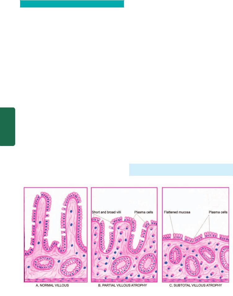
574 TABLE 20.8: Classification of Malabsorption Syndrome.
I.PRIMARY MALABSORPTION
1.Coeliac sprue
2.Collagenous sprue
3.Tropical sprue
4.Whipple’s disease
5.Disaccharidase deficiency
6.Allergic and eosinophilic gastroenteritis II. SECONDARY MALABSORPTION
1.Impaired digestion
(i)Mucosal damage e.g. in tuberculosis, Crohn’s disease, lymphoma, amyloidosis, radiation injury, systemic sclerosis
(ii)Hepatic and pancreatic insufficiency
(iii)Resection of bowel
(iv)Drugs e.g. methotrexate, neomycin, phenindione etc.
2.Impaired absorption
(i)Short or stagnant bowel (blind loop syndrome) from surgery or disease resulting in abnormal proliferation of microbial flora
(ii)Acute infectious enteritis
(iii)Parasitoses e.g. Giardia, Strongyloides, hookworms
|
3. |
Impaired transport |
|
|
(i) Lymphatic obstruction e.g. in lymphoma tuberculosis, lymph- |
||
SECTION |
|
angiectasia |
|
(ii) |
Abetalipoproteinaemia |
||
|
|||
|
|
|
|
|
1. |
Steatorrhoea (pale, bulky, foul-smelling stools) |
|
|
2. |
Chronic diarrhoea |
|
III |
3. |
Abdominal distension |
|
4. |
Barborygmi and flatulence |
||
|
|||
|
5. |
Anorexia |
|
Systemic |
6. |
Weight loss |
|
7. |
Muscle wasting |
||
|
|||
|
8. |
Dehydration |
|
|
9. |
Hypotension |
|
Pathology |
10. |
Specific malnutrition and vitamin deficiencies depending |
|
upon the cause. |
|||
|
|||
INVESTIGATIONS
When MAS is suspected on clinical grounds, the following investigations (laboratory tests) and endoscopic biopsy) may be carried out to confirm it:
I. LABORATORY TESTS:
1. Tests for fat malabsorption:
i)Faecal analysis for fat content
ii)Microscopic analysis for faecal fat
iii)Blood lipid levels after a fatty meal
iv)Tests based on absorption of radioactive-labelled fat.
2. Tests for protein malabsorption:
i)Bile acid malabsorption
ii)Radioactive-labelled glycine breath test.
iii)Prothrombin time (vitamin K deficiency)
iv)Secretin and other pancreatic tests.
3. Tests for carbohydrate malabsorption:
i)D-xylose tolerance test
ii)Lactose tolerance test
iii)Hydrogen breath test
iv)Bile acid breath test
4. Vitamin B12, malabsorption: i) Schilling test (page 308).
II. INTESTINAL MUCOSAL BIOPSY:
Mucosal biopsy of small intestine is essential for making the diagnosis of MAS and also evaluation of a patient on followup. The availability of endoscopes has enabled easy viewing of affected mucosa directly and taking mucosal biopsy under vision; this has largely replaced the earlier per-oral CrosbyKugler capsule biopsy of small intestine. The biopsy should first be examined under dissecting microscope before histologic sectioning.
Normal villous (Fig. 20.33,A). Under the dissecting microscope, the normal jejunal mucosa has tall, slender,
Figure 20.33 
 Jejunal biopsy diagrammatic appearance in malabsorption syndrome.
finger-shaped or leaf-shaped villi. It is lined by tall columnar absorptive epithelium and has scattered lymphocytes in the lamina propria.
Villous atrophy. Variable degree of flattening of intestinal mucosa in MAS is the commonest pathological change in mucosal pattern and is referred to as villous atrophy. It may be of 2 types—partial and subtotal/total type.
 Partial villous atrophy is the mild form of the lesion in which villi fuse with each other and thus become short and broad, commonly called as convolutions and irregular ridges (Fig. 20.33,B). The epithelial cells show compensatory hyperplasia suggesting a turnover of these cells (Fig. 20.34,A). Lamina propria shows increased cellular infiltrate, predominantly of plasma cells.
Partial villous atrophy is commonly found in children and adults with diarrhoea, parasitic infestations, Crohn’s disease, ulcerative colitis and malabsorption due to drugs and radiation injury.
 Subtotal/Total villous atrophy is the severe form of the lesion in which there is flattening of mucosa due to more advanced villous fusion (Fig. 20.33,C). The surface epithelium is cuboidal and there is increased plasma cell infiltrate in the lamina propria (Fig. 20.34,B).
Subtotal and total villous atrophy is exhibited by a number of conditions such as nontropical sprue, tropical sprue, intestinal lymphomas, carcinoma, protein-calorie malnutrition etc.
IMPORTANT TYPES OF MAS
Coeliac Sprue (Non-tropical Sprue, Gluten-Sensitive
Enteropathy, Idiopathic Steatorrhoea)
This is the most important cause of primary malabsorption occurring in temperate climates. The condition is characterised by significant loss of villi in the small intestine and thence diminished absorptive surface area. The condition occurs in 2 forms:
Childhood form, seen in infants and children and is commonly referred to as coeliac disease.
Adult form, seen in adolescents and early adult life and used
to be called idiopathic steatorrhoea.
In either case, there is genetic abnormality resulting in sensitivity to gluten (a protein) and its derivative, gliadin, present in diets such as grains of wheat, barley and rye. Serum antibodies—IgA antigliadin and IgA antiendomysial, have been found but is not known whether these antibodies are primary or appear secondary to tissue damage.
The symptoms are usually relieved on elimination of gluten from the diet. The role of heredity is further supported by the observation of familial incidence and HLA association of the disease. Exact pathogenesis of the condition is not clear. However, following hypotheses are significant in causing mucosal cell damage:
1.Hypersensitivity reaction as seen by gluten-stimulated antibodies.
2.Toxic effect of gluten due to inherited enzyme deficiency in the mucosal cells.
575
Figure 20.34 |
A, Partial villus atrophy. There is shortening and |
blunting of the villi with reduction in their height. There is crypt hyperplasia. |
|
B, Subtotal villous atrophy. There is near total flattening of the villi and |
|
crypt hyperplasia. |
|
||
|
|
||
Histologically, there are no differences in the pathological |
|
CHAPTER |
|
findings in children and adults. There is variable degree |
|
||
|
|
||
of flattening of the mucosa, particularly of the upper |
|
|
|
jejunum, and to some extent of the duodenum and ileum. |
|
|
|
The surface epithelial cells are cuboidal or low columnar |
|
|
|
type. There may be partial villous atrophy which is |
|
20 |
|
replacement of normal villous pattern by convolutions, |
|
||
|
|
||
or subtotal villous atrophy characterised by flat mucosal |
|
|
|
surface. Lamina propria shows increased number of |
|
The |
|
plasma cells and lymphocytes (Fig. 20.34). |
|
||
|
|
||
|
|
|
|
The major sequela of long-term coeliac sprue is increased |
Gastrointestinal |
||
incidence of intestinal carcinoma in these cases. |
|||
|
|||
Collagenous Sprue |
|
||
This entity is regarded as the end-result of coeliac sprue in |
|
||
which the villi are totally absent (total villous atrophy) and |
|
||
there are unique and diagnostic broad bands of collagen |
|
||
under the basal lamina of surface epithelium. The condition |
Tract |
||
is refractory to any treatment and the course is generally fatal. |
|||
Some workers consider collagenous sprue as a variant of |
|||
coeliac sprue without classifying it separately. |
|
||
Tropical Sprue |
|
||
This disease, as the name suggests, occurs in individuals |
|
||
living in or visiting tropical areas such as Caribbean |
|
||
countries, South India, Sri Lanka and Hong Kong. |
|
||
Pathogenesis of the condition is not clear but there is evidence |
|
||
to support enterotoxin production by some strains of E. coli |
|
||
which causes the intestinal injury. Severe cases are |
|
||
characterised by additional features such as macrocytic |
|
||
anaemia, glossitis and emaciation due to intestinal |
|
||
malabsorption of vitamin B12 and folate. |
|
||
|
|
|
|
Histologically, there is usually partial villous atrophy and |
|
|
|
sometimes subtotal atrophy. |
|
|
|

576 The lesions are relieved by removal of the patient from the tropical area and by oral administration of antibiotics but gluten-free diet has no role in improvement.
Whipple’s Disease (Intestinal Lipodystrophy)
This is an uncommon bacterial disease involving not only the intestines but also various other systems such as central nervous system, heart, blood vessels, skin, joints, lungs, liver, spleen and kidneys. The disease is more common in males in 4th to 5th decades of life. Patients may present with features of malabsorption or may have atypical presentation in the form of migratory polyarthritis, neurological disturbances and focal hyperpigmentation of the skin.
Histologically, the affected tissues show presence of characteristic macrophages containing PAS-positive granules and rod-shaped micro-organisms (Whipple’s bacilli). These macrophages are predominantly present in the lamina propria of the small intestine and mesenteric lymph nodes.
SECTION |
|
Patients respond very well to oral antibiotic therapy. |
|
Protein-Losing Enteropathies |
|||
A number of disorders of the gastrointestinal tract are |
|||
accompanied by excessive protein loss without concomitant |
|||
increase in protein synthesis, thus resulting in hypo- |
|||
III |
proteinaemia. These diseases are listed below: |
||
i) |
Whipple’s disease |
||
|
|||
Systemic |
ii) |
Crohn’s disease |
|
iii) Ulcerative colitis |
|||
iv) Sprue |
|||
|
|||
|
v) |
Intestinal lymphangiectasia |
|
Pathology |
vi) Ménétrier’s disease (Hypertrophic gastritis). |
||
are surprisingly rare. Most common benign tumours, in |
|||
|
SMALL INTESTINAL TUMOURS |
||
Although small intestine is about 6 meter long, but for obscure reasons, benign as well as malignant tumours in it
descending order of frequency, are: leiomyomas, adenomas and vascular tumours (haemangioma, lymphangioma). Amongst the malignant tumours, the most frequently encountered, in descending frequency, are: carcinoid tumours, lymphomas (Fig. 20.35) and adenocarcinoma. All these tumours are identical in morphology to those seen elsewhere in the alimentary tract. Carcinoid tumour, a peculiar neoplasm most common in the midgut, is described below.
Carcinoid Tumour (Argentaffinoma)
Carcinoid tumour or argentaffinoma is a generic term applied to tumours originating from endocrine cells (synonyms: argentaffin cells, Kulchitsky cells, enterochromaffin cells) belonging to APUD cell system and are therefore also called as apudomas (page 561). The endocrine cells are distributed throughout the mucosa of GI tract. These cells have secretory granules which stain positively with silver salts (argentaffin granules) or many stain after addition of exogenous reducing
Figure 20.35 
 Lymphoma small intestine. The polypoid growth is seen projecting into lumen while the covering mucosa is ulcerated.
agent (non-argentaffin or argyrophil granules). Accordingly, carcinoid tumour may be argentaffin or argyrophil type. Depending upon the embryologic derivation of the tissues where the tumour is located, these are classified as foregut, midgut, and hindgut carcinoids.

 Midgut carcinoids, seen in terminal ileum and appendix are the most common (60-80%) and are more often argentaffin positive.
 Hindgut carcinoids, occurring in rectum and colon are more commonly argyrophil type, and comprise about 1020% of carcinoids.

 Foregut carcinoids, located in the stomach, duodenum and oesophagus are also argyrophil type and are encountered as frequently as in the hindgut (10-20%).
Other uncommon locations are the bronchus, trachea, gallbladder, and Meckel’s diverticulum.
Appendix and terminal ileum, the two most common sites for carcinoids, depict variation in their age and sex incidence and biologic behaviour:
 Appendiceal carcinoids, occur more frequently in 3rd and 4th decades of life without any sex predilection, are often solitary and behave as locally malignant tumours.

 Ileal carcinoids, on the other hand, are seen more often in later age (7th decade) with female preponderance, are more commonly multiple and behave like metastasising carcinomas.
MORPHOLOGIC FEATURES. Grossly, all carcinoids are small, button-like submucosal elevations with intact or ulcerated overlying mucosa. They are usually small; those larger than 2 cm are more often metastasising. Ileal and gastric carcinoids are commonly multiple, whereas
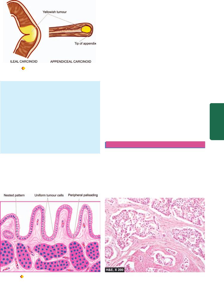
Figure 20.36 |
Carcinoid tumour. Gross appearance at common |
locations in longitudinal section.
appendiceal carcinoids commonly involve the tip of the organ and are solitary (Fig. 20.36). Cut section of all the carcinoids is bright yellow.
Histologically, the tumour cells may be arranged in a variety of patterns—solid nests, sheets, cords, trabeculae and clusters, all of which show characteristic palisading of the peripheral cells. Acinar arrangement and rosettes are rarely seen. The tumour cells are classically small, monotonous, having uniform nuclei and poorly-defined cell boundaries (Fig. 20.37). The argentaffin carcinoids show eosinophilic granules in the cytoplasm which stain positively by the argentaffin reaction. Mitotic figures are rare. However, the cytologic features are a poor guide for distinguishing clinically benign from malignant behaviour of the tumour, but all carcinoids infiltrate the bowel wall.
CARCINOID SYNDROME. Carcinoid tumours that metastasise, especially to the liver, are sometimes associated with the carcinoid syndrome. The syndrome consists of the following features:
1.Intermittent attacks of flushing of the skin of face
2.Episodes of watery diarrhoea
3.Abdominal pain
4.Attacks of dyspnoea due to bronchospasm
5.Right-sided heart failure due to involvement of tricuspid and pulmonary valves and endocardium (page 451).
A number of secretory products in a functioning carcinoid tumour have been demonstrated:
i)5-Hydroxytryptamine (5-HT, serotonin)
ii)5-Hydroxytryptophan
iii)5-Hydroxy-indole acetic acid (5-HIAA)
iv)Histamine
v)Kallikrein
vi)Bradykinin
However, 5-HT and its degradation product, 5-HIAA, are particularly significant in the production of carcinoid syndrome. 5-HT, a potent vasodilator and smooth muscle stimulant, is normally synthesised in the endocrine cells of the gut from dietary tryptophan. Tryptophan is first hydroxylated to 5-hydroxytryptophan, then decarboxylated to 5-HT and further oxidised to 5-HIAA by the monoamine oxidase in the liver cells. It is then excreted in the urine. This capacity to synthesise 5-HT and 5-HIAA is markedly elevated in primary and hepatic metastatic carcinoids. Midgut carcinoids have rich decarboxylating enzymes and are thus able to produce large quantities of 5-HT and 5-HIAA, accounting for high frequency of carcinoid syndrome in them. Foregut and hindgut carcinoids, on the other hand, lack decarboxylating enzymes and, therefore, are less often associated with carcinoid syndrome.
APPENDIX
NORMAL STRUCTURE
Appendix is a vestigial organ which serves no useful purpose in human beings but instead becomes the site of trouble at times. It is like a diverticulum of the caecum, usually lying behind the caecum and varies in length from 4 to 20 cm (average 7 cm).
577
Tract Gastrointestinal The 20 CHAPTER
Figure 20.37 
 Microscopic appearance showing solid masses and trabeculae of uniform, monotonous, small cells with palisading of the peripheral cells.
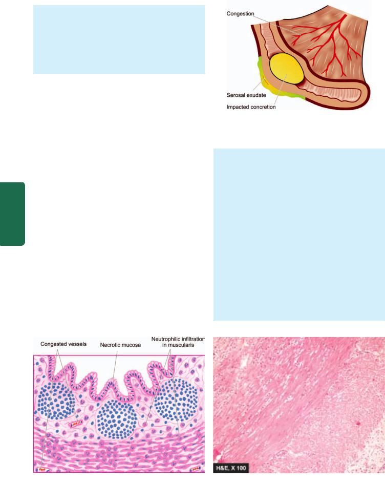
578Histologically, appendix has four layers in its wall— mucosa, submucosa, muscularis and serosa. The mucosa has patchy distribution of crypts and the submucosa has abundant lymphoid tissue. Argentaffin and nonargentaffin endocrine cells are present in the base of mucosal glands just as in the small intestine. The muscularis of the appendix has two layers (inner circular and outer longitudinal) as elsewhere in the alimentary tract.
Two important diseases involving the appendix are
|
appendicitis and appendiceal carcinoids. |
||
|
APPENDICITIS |
||
|
Acute inflammation of the appendix, acute appendicitis, is |
||
|
the most common acute abdominal condition confronting the |
||
|
surgeon. The condition is seen more commonly in older |
||
|
children and young adults, and is uncommon at the extremes |
||
|
of age. The disease is seen more frequently in the West and |
||
|
in affluent societies which may be due to variation in diet— |
||
|
a diet with low bulk or cellulose and high protein intake more |
||
|
often causes appendicitis. |
||
|
ETIOPATHOGENESIS. The most common mechanism is |
||
SECTION |
obstruction of the lumen from various etiologic factors that |
||
leads to increased intraluminal pressure. This presses upon |
|||
|
|||
|
the blood vessels to produce ischaemic injury which in turn |
||
|
favours the bacterial proliferation and hence acute |
||
|
appendicitis. The common causes of appendicitis are as |
||
III |
under: |
||
A. |
Obstructive: |
||
|
|||
|
1. |
Faecolith |
|
Systemic |
2. |
Calculi |
|
3. |
Foreign body |
||
4. |
Tumour |
||
5. |
Worms (especially Enterobius vermicularis) |
||
6. |
Diffuse lymphoid hyperplasia, especially in children. |
||
Pathology |
B. Non-obstructive: |
||
|
|||
|
1. |
Haematogenous spread of generalised infection |
|
|
2. |
Vascular occlusion |
|
|
3. |
Inappropriate diet lacking roughage. |
|
Figure 20.38 
 Acute appendicitis. Gross appearance of longitudinally opened appendix showing impacted faecolith in the lumen and exudate on the serosa.
MORPHOLOGIC FEATURES. Grossly, the appearance depends upon the stage at which the acutely-inflamed appendix is examined. In early acute appendicitis, the organ is swollen and serosa shows hyperaemia. In welldeveloped acute inflammation called acute suppurative appendicitis, the serosa is coated with fibrinopurulent exudate and engorged vessels on the surface. In further advanced cases called acute gangrenous appendicitis, there is necrosis and ulcerations of mucosa which extend through the wall so that the appendix becomes soft and friable and the surface is coated with greenish-black gangrenous necrosis (Fig. 20.38).
Microscopically, the most important diagnostic histological criterion is the neutrophilic infiltration of the muscularis. In early stage, the other changes besides acute inflammatory changes, are congestion and oedema of the appendiceal wall. In later stages, the mucosa is sloughed off, the wall becomes necrotic, the blood vessels may get thrombosed and there may be neutrophilic abscesses in the wall. In either case, an impacted foreign body, faecolith, or concretion may be seen in the lumen (Fig. 20.39).
Figure 20.39 
 Acute appendicitis. Microscopic appearance showing diagnostic neutrophilic infiltration into the muscularis. Other changes present are necrosis of mucosa and periappendicitis.

Thus, there is good correlation between macroscopic and microscopic findings in acute appendicitis.
CLINICAL COURSE. The patient presents with features of acute abdomen as under:
1.Colicky pain, initially around umbilicus but later localised to right iliac fossa
2.Nausea and vomiting
3.Pyrexia of mild grade
4.Abdominal tenderness
5.Increased pulse rate
6.Neutrophilic leucocytosis.
An attack of acute appendicitis predisposes the appendix
to repeated attacks (recurrent acute appendicitis) and thus surgery has to be carried out. If appendicectomy is done at a later stage following acute attack (interval appendicectomy), pathological changes of healing by fibrosis of the wall and chronic inflammation are observed.
COMPLICATIONS. If the condition is not adequately managed, the following complications may occur:
1.Peritonitis. A perforated appendix as occurs in gangrenous appendicitis may cause localised or generalised peritonitis.
2.Appendix abscess. This is due to rupture of an appendix giving rise to localised abscess in the right iliac fossa. This abscess may spread to other sites such as between the liver and diaphragm (subphrenic abscess), into the pelvis between the urinary bladder and rectum, and in the females may involve uterus and fallopian tubes.
3.Adhesions. Late complications of acute appendicitis are fibrous adhesions to the greater omentum, small intestine and other abdominal structures.
4.Portal pylephlebitis. Spread of infection into mesenteric veins may produce septic phlebitis and liver abscess.
5.Mucocele. Distension of distal appendix by mucus following recovery from an attack of acute appendicitis is referred to as mucocele. It occurs generally due to proximal obstruction but sometimes may be due to a benign or malignant neoplasm in the appendix. An infected mucocele may result in formation of empyema of the appendix.
TUMOURS OF APPENDIX
Tumours of the appendix are quite rare. These include: carcinoid tumour (the most common), pseudomyxoma peritonei and adenocarcinoma.
CARCINOID TUMOUR. It is already described on page 576. Both argentaffin and argyrophil types are encountered, the former being more common.
Grossly, carcinoid tumour of the appendix is mostly situated near the tip of the organ and appears as a circumscribed nodule, usually less than 1 cm in diameter, involving the wall but metastases are rare.
Histologically, carcinoid tumour of the appendix resembles other carcinoids of the midgut.
PSEUDOMYXOMA PERITONEI. Pseudomyxoma peritonei 579 is appearance of gelatinous mucinous material around the appendix admixed with epithelial tumour cells. It is generally
due to mucinous collection from benign mucinous cystadenoma of the ovary or mucin-secreting carcinoma of the appendix.
ADENOCARCINOMA. It is an uncommon tumour in the appendix and is morphologically similar to adenocarcinoma elsewhere in the alimentary tract.
LARGE INTESTINE
NORMAL STRUCTURE |
|
|
The large bowel consists of 6 parts—the caecum, ascending |
|
|
colon, transverse colon, descending colon, sigmoid colon and |
|
|
rectum, and in all measures about 1.5 meters in length. The |
|
|
serosal surface of the large intestine except the rectum is |
|
|
studded with appendices epiploicae which are small, rounded |
|
|
collections of fatty tissue covered by peritoneum. |
|
|
Histologically, the wall of large bowel consists of 4 layers as |
CHAPTER |
|
elsewhere in the alimentary tract—serosa, muscularis, |
||
|
||
submucosa and mucosa. |
|
|
The mucosa lacks villi and there is preponderance of |
|
|
goblet cells over columnar epithelial cells. The lymphoid |
|
|
tissue is less abundant than in the small bowel but lymphoid |
20 |
|
follicles are seen in the caecum and rectum. |
||
|
||
The muscularis propria of the large intestine is quite |
|
|
peculiar—the inner circular muscle layer ensheaths whole |
The |
|
length of the intestine, while the outer longitudinal muscle |
||
|
||
layer is concentrated into 3 muscle bands called taenia coli. |
Gastrointestinal |
|
The length of outer muscle layer is shorter than the length of |
||
|
||
the intestine and therefore, it forms the sacculations or haustra |
|
|
of the large intestine. At the rectosigmoid junction, the three |
|
|
muscle bands fuse to form a complete covering. |
|
|
The blood supply to the right colon is from the superior |
|
|
mesenteric artery which also supplies blood to the small |
|
|
bowel. The remaining portion of large bowel except the lower |
|
|
part of rectum receives blood supply from inferior mesenteric |
Tract |
|
branches. |
||
artery. The lower rectum is supplied by haemorrhoidal |
|
|
The innervation of the large bowel consists of 3 plexuses |
|
|
of ganglion cells—Auerbach’s or myenteric plexus lying |
|
|
between the two layers of muscularis, Henle’s plexus lying in |
|
|
the deep submucosa inner to circular muscle layer, and |
|
|
Meissner plexus that lies in the superficial mucosa just beneath |
|
|
the muscularis mucosae. These are interconnected by non- |
|
|
myelinated nerve fibres. |
|
|
Anal canal, 3-4 cm long tubular structure, begins at the |
|
|
lower end of the rectum, though is not a part of large bowel, |
|
|
but is included here to cover simultaneously lesions |
|
|
pertaining to this region. It is lined by keratinised or |
|
|
nonkeratinised stratified squamous epithelium. Anal verge |
|
|
is the junction between the anal canal and perineal skin, while |
|
|
pectinate line is the squamo-columnar junction between the |
|
|
anal canal and the rectum. |
|
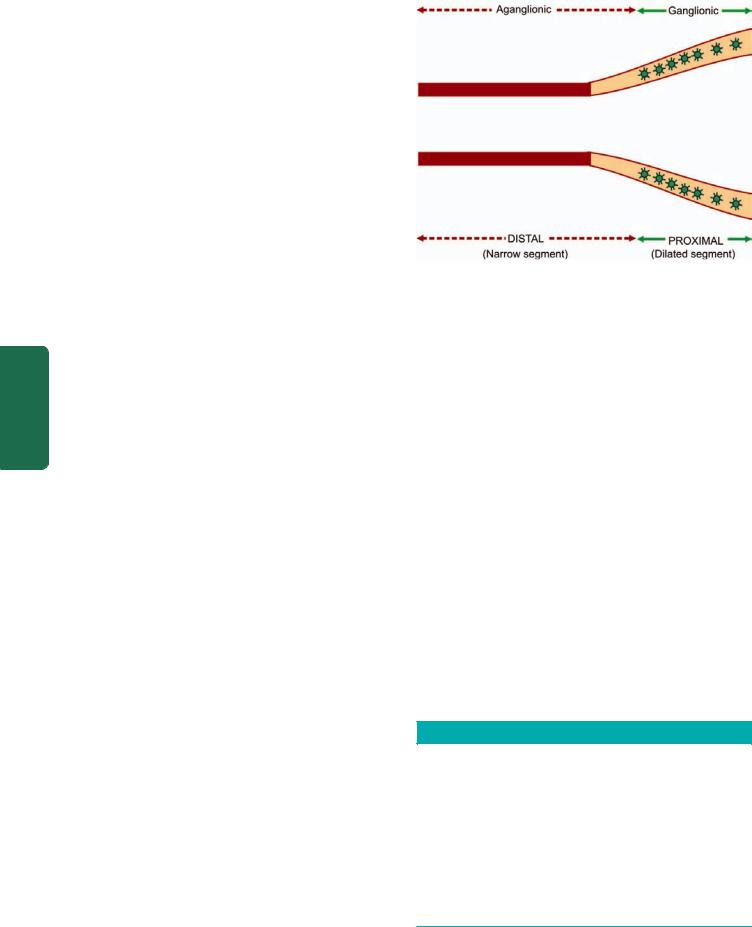
580CONGENITAL MALFORMATIONS Hirschsprung’s Disease (Congenital Megacolon)
The term ‘megacolon’ is used for any form of marked dilatation of the entire colon or its segment and may occur as a congenital or acquired disorder. Congenital form characterised by congenital absence of ganglion cells in the bowel wall (enteric neurons) is called Hirschsprung’s disease. As a result, the aganglionic segment remains contracted. Genetically, Hirschsprung’s disease is a heterogeneous disorder as under:
1.Autosomal dominant inheritance with mutation in RET proto-oncogene in some cases.
2.Autosomal recessive form with mutation in endothelin-B receptor gene in many other cases.
|
Clinically, the condition manifests shortly after birth with |
|
|
constipation, gaseous distension and sometimes with acute |
|
|
intestinal obstruction. Its frequency is 1 in 5,000 live-births, |
|
|
has familial tendency in about 4% of cases and has |
|
|
predilection for development in Down’s syndrome. |
|
|
Pathogenesis lies in the failure of neuroblasts to migrate to |
|
SECTION |
the rectum which normally occurs at about 12 weeks of |
|
gestation. |
||
|
||
|
|
|
|
MORPHOLOGIC FEATURES. Two types of biopsies may |
|
|
be done on infants suspected of having Hirschsprung’s |
|
III |
disease—full-thickness rectal biopsy, and suction biopsy |
|
that includes mucosa and submucosa. |
||
|
||
|
Grossly, typical case of Hirschsprung’s disease shows 2 |
|
Systemic |
segments—a distal narrow segment that is aganglionic and |
|
a dilated proximal segment that contains normal number of |
||
|
||
|
ganglion cells (Fig. 20.40). |
|
|
Microscopically, the distal narrow segment shows total |
|
Pathology |
absence of ganglion cells of all the three plexuses |
|
(Auerbach’s or myenteric plexus present between the two |
||
|
||
|
layers of muscularis, deep submucosal or Henle’s plexus, |
|
|
and superficial mucosal or Meissner’s plexus) and |
|
|
prominence of non-myelinated nerve fibres. Histo- |
|
|
chemical staining for acetylcholine esterase activity |
|
|
provides confirmation for identifying ganglion cells and |
|
|
nerve trunks. |
|
|
|
Depending upon the length of the segment affected by aganglionosis in Hirschsprung’s disease, following patterns are recognised:
1.Classic form: Anganglionosis from distal colorectal region to proximal dilated colon.
2.Short segment (rectal and recto-sigmoid) form: Aganglionosis involving a few centimeters of the rectum and rectosigmoid only.
3.Ultra-short form: Aganglionosis is in a very small segment which can be missed in a biopsy.
4.Long segment (subtotal colonic) form: Aganglionosis involves most of the colon from rectosigmoid to the ileocaecal valve, and sometimes may even extend into smll bowel.
Figure 20.40 
 Hirschsprung’s disease, diagrammatic representation of the pathologic changes.
5. Zonal colonic aganglionosis: A short segment is nvolved in agnaglionosis in which the ganglia cells are absent both above and below the aganglionic segment.
In addition to congenital megacolon discussed above, megacolon may occur from certain acquired causes as under:
i.Obstructive e.g. due to tumour, post-inflammatory strictures.
ii.Endocrine e.g. in myxoedema, cretinism.
iii.CNS disorders e.g. spina bifida, paraplegia, parkinsonism.
iv.Psychogenic e.g. emotional disturbances, psychiatric disorders.
v.Chagas’ disease due to infection with Trypanosoma cruzi is the only example resulting in acquired loss of ganglion cells. In all other acquired causes listed above, the bowel innervation is normal.
COLITIS
Colitis may occur in isolation but more commonly involvement of small intestine is also present (enterocolitis). In view of the considerable overlapping of enteritis and colitis, these lesions have already been described under small intestine (page 578). Table 20.9 presents a classification of the various types of colitis/enterocolitis.
TABLE 20.9: Classification of Colitis/Enterocolitis.
I.ISCHAEMIC BOWEL DISEASE Ischaemic colitis (‘Membranous’ colitis)
II.INFLAMMATORY BOWEL DISEASE
1.Ulcerative colitis
2.Crohn’s disease
III.OTHER INFLAMMATORY LESIONS
1.Infective enterocolitis (Dysenteries—bacillary, amoebic, other parasitic)
2.‘Pseudomembranous’ enterocolitis (Antibiotic-associated diarrhoea)
3.Necrotising enterocolitis

MISCELLANEOUS LESIONS
Diverticulosis Coli
Diverticula are the outpouchings or herniations of the mucosa and submucosa of the colon through the muscle wall. Diverticular disease, as it is commonly known, is rare under 30 years of age and is seen more commonly as the age advances. Multiple diverticula of the colon are very common in the Western societies, probably due to ingestion of lowfibre diet but is seen much less frequently in tropical countries and in Japan. Diverticulosis is often asymptomatic and may be detected as an incidental finding at autopsy. However, a proportion of patients develop clinical symptoms such as low abdominal pain, distension, constipation and sometimes intermittent bleeding.
Based on the etiologic role of low fibre diet, pathogenesis of diverticular disease of the colon can be explained as under:
1.Increased intraluminal pressure such as due to low fibre content of the diet causing hyperactive peristalsis and thereby sequestration, of mucosa and submucosa.
2.Muscular weakness of the colonic wall at the junction of the muscularis with submucosa.
MORPHOLOGIC FEATURES. Grossly, diverticulosis is seen most commonly in the sigmoid colon (95%) but any other part of the entire colon may be involved. They may vary in number from a few to several hundred. They appear as small, spherical or flask-shaped outpouchings, usually less than 1 cm in diameter, commonly extend into appendices epiploicae and may contain inspissated faeces. They are connected to the intestinal lumen by a narrow neck.
Histologically, the flask-shaped structures extend from the intestinal lumen through the muscle layer. The colonic wall in the affected area is thin and is composed of atrophic mucosa, compressed submucosa and thin or deficient muscularis. However, muscularis propria in between the diverticular protrusions is hypertrophied. While diverticular disease may remain asymptomatic, inflammatory changes in the diverticula (diverticulitis) produce clinical symptoms.
The complications of diverticulosis and diverticulitis are perforation, haemorrhage, intestinal obstruction and fistula formation.
Melanosis Coli
Melanosis coli is a peculiar condition in which mucosa of the large intestine acquires brown-black colouration. The condition is said to occur in individuals who are habitual users of cathartics of anthracene type.
Grossly, the mucosal surface is intact and is pigmented brown-black.
Microscopically, large number of pigment-laden macrophages are seen in the lamina propria. The nature of this pigment is found to be both melanin and lipofuscin.
Haemorrhoids (Piles)
Haemorrhoids or piles are the varicosities of the haemorrhoidal veins. They are called ‘internal piles’ if dilatation is of superior haemorrhoidal plexus covered over by mucous membrane, and ‘external piles’ if they involve inferior haemorrhoidal plexus covered over by the skin. They are common lesions in elderly and pregnant women. They commonly result from increased venous pressure. The possible causes include the following:
1.Portal hypertension
2.Chronic constipation and straining at stool
3.Cardiac failure
4.Venous stasis of pregnancy
5.Hereditary predisposition
6.Tumours of the rectum.
Microscopically, thin-walled and dilated tortuous veins are seen under the rectal mucosa (internal piles) or anal skin (external piles). Secondary changes and complications that may occur include: thrombosis, haemorrhage, inflammation, scarring and strangulation (prolapsed piles).
Angiodysplasia
Angiodysplasia is a submucosal telangiectasia affecting caecum and right colon that causes recurrent acute and chronic haemorrhage. The condition is more common in the elderly past 6th decade. The pathogenesis is obscure but is possibly due to mechanical obstruction of the veins.
MISCELLANEOUS INFLAMMATORY CONDITIONS

 ‘Fistula-in-ano’ is a well known and common condition in which one or more fistulous tracts pass from the internal opening at the pectinate line through the internal sphincter on to the skin surface. The condition probably results from infection of the anal glands.
Histologically, nonspecific inflammatory changes are seen.

 ‘Anal fissure’ is an ulcer in the anal canal below the level of the pectinate line, mostly in midline and posteriorly. The common cause is trauma due to passage of hard stools, followed by chronic infection.
 ‘Solitary rectal ulcer syndrome’ is a condition characterised usually by solitary, at times multiple, rectal ulcers with prolapse of rectal mucosa and development of proctitis. The histological appearance is quite characteristic. Besides ulceration and inflammation of the rectal mucosa, lamina propria is occupied by spindle-shaped fibroblasts and smooth muscle cells. The condition is also called as ‘localised form of colitis cystica profunda’ and must be differentiated from ‘diffuse form of colitis cystica profunda’ seen in cases of ulcerative colitis. Submucosal cysts lined by foreign body giant cells and containing gas are also seen in ‘pneumatosis cystoides
intestinalis’.
LARGE INTESTINAL POLYPS AND TUMOURS
Large bowel is the most common site for a variety of benign and malignant tumours, majority of which are of epithelial
581
Tract Gastrointestinal The 20 CHAPTER

582 TABLE 20.10: Polyps and Tumours of the Large Intestine.
I.COLORECTAL POLYPS
A.Non-neoplastic polyps
1.Hyperplastic (metaplastic) polyps
2.Hamartomatous polyps
(i)Peutz-Jeghers polyps and polyposis
(ii)Juvenile (Retention) polyps and polyposis
3.Inflammatory polyps (Pseudopolyps)
4.Lymphoid polyps
B.Neoplastic polyps (Adenomas)
1.Tubular adenoma (Adenomatous polyp)
2.Villous adenoma (Villous papilloma)
3.Tubulovillous adenoma (Papillary adenoma, villoglandular adenoma)
C.Familial polyposis syndromes
1.Familial polyposis coli (Adenomatosis)
2.Gardner’s syndrome
3.Turcot’s syndrome
4.Juvenile polyposis syndrome
II.OTHER BENIGN COLORECTAL TUMOURS
(Leiomyomas, leiomyoblastoma, neurilemmoma, lipoma and vascular tumours)
SECTION |
III. MALIGNANT COLORECTAL TUMOURS |
|||
A. |
Carcinoma |
|||
|
||||
|
|
1. |
Adenocarcinoma |
|
|
|
2. |
Other carcinomas |
|
|
|
|
(Mucinous adenocarcinoma, signet-ring cell carcinoma, |
|
III |
|
|
adenosquamous carcinoma, undifferentiated carcinoma) |
|
B. |
Other malignant tumours |
|||
|
||||
Systemic |
|
(Leiomyosarcoma, malignant lymphoma, carcinoid tumours) |
||
IV. TUMOURS OF THE ANAL CANAL |
||||
|
||||
|
A. Benign (viral warts or condyloma acuminata) |
|||
|
B. Malignant (squamous cell carcinoma, basaloid carcinoma, |
|||
|
|
mucoepidermoid carcinoma, adenocarcinoma, undifferentiated |
||
Pathology |
|
carcinoma, malignant melanoma) |
||
|
||||
and malignant tumours, is presented in Table 20.10. |
||||
|
origin. Most of the benign tumours present clinically as |
|||
polyps. A classification of polyps, alongwith benign tumours
COLORECTAL POLYPS
A polyp is defined as any growth or mass protruding from the mucous membrane into the lumen. Polyps are much more common in the large intestine than in the small intestine and are more common in the rectosigmoid colon than the proximal colon. Polyps are broadly classified into 2 groups— non-neoplastic and neoplastic. Non-neoplastic polyps have further subtypes indicating their mode of origin. Neoplastic polyps, on the other hand, include epithelial tumours, both benign and malignant (Table 20.10).
A. NON-NEOPLASTIC POLYPS
Non-neoplastic polyps are more common and include the following 4 subtypes:
Hyperplastic (Metaplastic) Polyps
The hyperplastic or metaplastic polyps are the most common amongst all epithelial polyps, particularly in the
rectosigmoid. They are called ‘hyperplastic’ because there is epithelial hyperplasia at the base of the crypts, and ‘metaplastic’ as there are areas of cystic metaplasia. They may be seen at any age but are more common in the elderly (6th-7th decade).
Grossly, hyperplastic polyps are generally multiple, sessile, smooth-surfaced and small (less than 0.5 cm). Microscopically, they are composed of long and cystically dilated glands and crypts lined by normal epithelial cells. Their lining is partly flat and partly papillary. The luminal border of the lining epithelium is often serrated or sawtoothed.
Hyperplastic polyps are usually symptomless and have no malignant potential unless there is a coexistent adenoma.
Hamartomatous Polyps
These are tumour-like lesions composed of abnormal mixture of tissues indigenous to the part. They are further of 2 types:
PEUTZ-JEGHERS POLYPS AND POLYPOSIS. PeutzJeghers syndrome is autosomal dominant defect, characterised by hamartomatous intestinal polyposis and melanotic pigmentation of lips, mouth and genitalia. The polyps may be located in the stomach, small intestine or colon but are most common in the jejunum and ileum. The most common age is adolescence and early childhood.
Grossly, these polyps are of variable size but are often large, multiple and pedunculated and more commonly situated in the small intestine.
Microscopically, the most characteristic feature is the treelike branching of muscularis mucosae. The lining epithelium is by normal-appearing epithelial cells. The glands may show hyperplasia and cystic change
(Fig. 20.41).
Peutz-Jeghers polyps do not undergo malignant transformation unless a coexistent adenoma is present. However, patients with Peutz-Jeghers syndrome are more prone to certain other cancers such as of pancreas, lung, breast, ovary and uterus.
JUVENILE (RETENTION) POLYPS. Juvenile or retention polyps, another form of hamartomatous polyps, occur more commonly in children below 5 years of age. Solitary juvenile polyps occur more often in the rectum, while juvenile polyposis may be present anywhere in the large bowel.
Grossly, juvenile polyps are spherical, smooth-surfaced, about 2 cm in diameter and are often pedunculated. Microscopically, the classical appearance is of cystically dilated glands containing mucus and lined by normal mucus-secreting epithelium. The stroma may show inflammatory cell infiltrate if there is chronic ulceration of the surface (Fig. 20.42).
Most cases, on becoming symptomatic in the form of rectal bleeding, are removed. In common with other nonneoplastic polyps, they are also not precancerous.
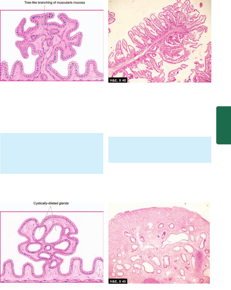
583
Figure 20.41 
 Hamartomatous polyps: Peutz-Jeghers polyp.
Inflammatory Polyps (Pseudopolyps)
Inflammatory polyps or pseudopolyps appear due to reepithelialisation of the undermined ulcers and overhanging margins in inflammatory bowel disease, most frequently in ulcerative colitis (colitis polyposa) and sometimes in Crohn’s disease.
Grossly, they are usually multiple, cylindrical to rounded overgrowths of mucosa and may vary from minute nodules to several centimeters in size.
Microscopically, the centre of inflammatory polyp consists of connective tissue core that shows some inflammatory cell infiltrate and is covered superficially by regenerating epithelial cells and some cystically-dilated glands.
These lesions have no malignant potential; carcinomas seen in long-standing cases of ulcerative colitis arise in the region of epithelial dysplasia and not from the polyps.
Lymphoid Polyps
Reactive hyperplasia of lymphoid tissue that is normally also more prominent in the rectum and terminal ileum, gives rise to localised or diffuse lymphoid polyps, also called rectal tonsils. Localised form occurs more often in the rectum in elderly, while diffuse form is seen at younger age and in children.
Grossly, they are solitary or multiple, tiny elevated lesions. Microscopically, they are composed of prominent lymphoid follicles with germinal centres located in the submucosa and mucosa, and are covered by epithelium that may be inflamed.
They are benign lesions and have to be distinguished from malignant lymphoma.
B. NEOPLASTIC POLYPS (ADENOMAS)
Neoplastic polyps are colorectal adenomas which have potential for malignant change while polypoid carcinoma is
Tract Gastrointestinal The 20 CHAPTER
Figure 20.42 
 Juvenile polyp.
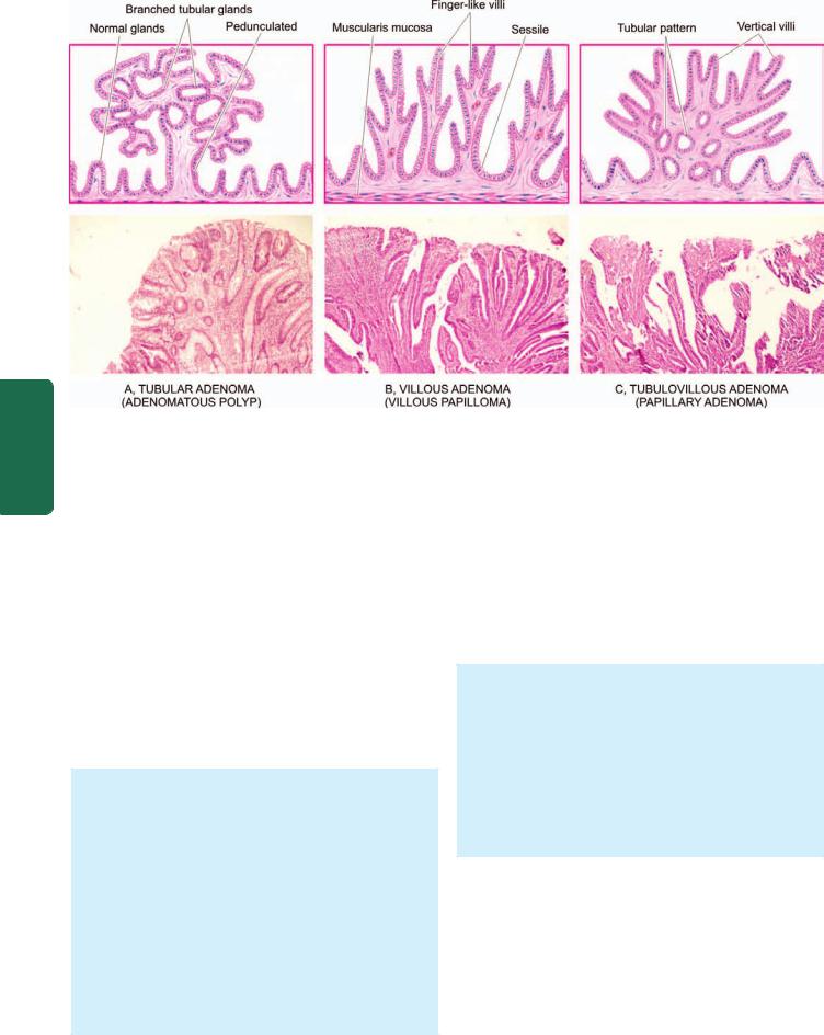
584
Pathology Systemic III SECTION
Figure 20.43 
 Adenomas (neoplastic polyps)—three main varieties.
the term used for invasive epithelial tumours. Adenomas have 3 main varieties (tubular, villous and tubulovillous), each of which represents a difference in the growth pattern of the same neoplastic process and variable biological behaviour.
Tubular Adenoma (Adenomatous Polyp)
Tubular adenomas or adenomatous polyps are the most common neoplastic polyps (75%). They are common beyond 3rd decade of life and have slight male preponderance. They occur most often in the distal colon and rectum. They may be found singly as sporadic cases, or multiple tubular adenomas as part of familial polyposis syndrome with autosomal dominant inheritance pattern. Tubular adenomas may remain asymptomatic or may manifest by rectal bleeding.
Grossly, adenomatous polyps may be single or multiple, sessile or pedunculated, vary in size from less than 1 cm to large, spherical masses with an irregular surface. Usually, the larger lesions have recognisable stalks.
Microscopically, the usual appearance is of benign tumour overlying muscularis mucosa and is composed of branching tubules which are embedded in the lamina propria. The lining epithelial cells are of large intestinal type with diminished mucus secreting capacity, large nuclei and increased mitotic activity (Fig. 20.43,A). However, tubular adenomas may show variable degree of cytologic atypia ranging from atypical epithelium restricted within the glandular basement membrane called as ‘carcinoma in situ’ to invasion into the fibrovascular stromal core termed as frank adenocarcinoma.
Malignant transformation is present in about 5% of tubular adenomas; the incidence being higher in larger adenomas.
Villous Adenoma (Villous Papilloma)
Villous adenomas or villous papillomas of the colon are much less common than tubular adenomas. The mean age at which they appear is 6th decade of life with approximatey equal sex incidence. They are seen most often in the distal colon and rectum, followed in decreasing frequency, by rest of the colon.
Grossly, villous adenomas are round to oval exophytic masses, usually sessile, varying in size from 1 to 10 cm or more in diameter. Their surface may be haemorrhagic or ulcerated.
Microscopically, the characteristic histologic feature is the presence of many slender, finger-like villi, which appear to arise directly from the area of muscularis mucosae. Each of the papillae has fibrovascular stromal core that is covered by epithelial cells varying from apparently benign to anaplastic cells. Excess mucus secretion is sometimes seen (Fig. 20.43,B).
Villous adenomas are invariably symptomatic; rectal bleeding, diarrhoea and mucus being the common features. The presence of severe atypia, carcinoma in situ and invasive carcinoma are seen more frequently. Invasive carcinoma has been reported in 30% of villous adenomas.
Tubulovillous Adenoma
(Papillary Adenoma, Villoglandular Adenoma)
Tubulovillous adenoma is an intermediate form of pattern between tubular adenoma and villous adenoma. It is also
TABLE 20.11: Contrasting Features of Non-neoplastic and Neoplastic Colorectal Polyps. |
585 |
|||
|
Feature |
Non-neoplastic polyps |
Neoplastic polyps (Adenomas) |
|
|
|
|
|
|
1. |
Frequency |
More common |
Less common |
|
2. |
Number |
Often sporadic |
Sporadic as well as multiple |
|
3. |
Familial predisposition |
No |
Yes, in sporadic cases |
|
4. |
Types |
Hyperplastic (90%) |
Tubular, villous and tubulovillous |
|
|
|
Others: hamartomatous (Peutz-Jeghers, juvenile) |
adenomas |
|
|
|
inflammatory, lymphoid |
|
|
5. |
Familial syndromes |
Juvenile polyposis syndrome |
Familial polyposis coli, Gardner’s, |
|
|
|
|
Turcot’s |
|
6. |
Biologic behaviour |
Always benign |
Variable malignant potential: |
|
|
|
|
Tubular adenoma 5%, villous 30%, |
|
|
|
|
tubulovillous intermediate |
|
|
|
|
|
|
known by other names like papillary adenoma and villoglandular adenoma. The distribution of these adenomas is the same as for tubular adenomas.
Grossly, tubulovillous adenomas may be sessile or pedunculated and range in size from 0.5-5 cm. Microscopically, they show intermediate or mixed pattern, characteristic vertical villi and deeper part showing tubular pattern (Fig. 20.43,C).
The behaviour of tubulovillous adenoma is intermediate between tubular and villous adenomas.
The contrasting features of non-neoplastic and neoplastic colorectal polyps are given in Table 20.11.
C. FAMILIAL POLYPOSIS SYNDROMES
Familial polyposis syndromes are a group of disorders with multiple polyposis of the colon with autosomal dominant inheritance pattern. Important conditions included in familial polyposis are:
1.Familial polyposis coli (adenomatosis)
2.Gardner’s syndrome
3.Turcot’s syndrome
4.Juvenile polyposis syndrome
Some other conditions in which multiple polyposis of colon occur but do not have familial basis are Peutz-Jeghers syndrome (hamartomatous), Cronkhite-Canada syndrome (inflammatory), and nodular lymphoid hyperplasia. The familial polyposis syndromes are as follows.
Familial Polyposis Coli (Adenomatosis)
This hereditary disease is defined as the presence of more than 100 neoplastic polyps (adenomas) on the mucosal surface of the colon; the average number is about 1000 (Fig. 20.44). Adenomatosis can be distinguished from multiple adenomas in which the number of adenomas is fewer, not exceeding 100. The condition has autosomal dominant transmission and is due to germline mutations in APC gene which results in occurrence of hundreds of adenomas which progress to invasive cancer. The average age at diagnosis is 2nd and 3rd decades of life with equal incidence in both the sexes.
Grossly and microscopically, the commonest pattern is that of adenomatous polyps (tubular adenomas) discussed above.
The malignant potential of familial polyposis coli is very high. Colorectal cancer develops virtually in 100% of cases by age of 50 years if not treated with colectomy. This subject of ‘adenoma-carcinoma sequence’ has been discussed again on page 586.
Gardner’s Syndrome
Gardner’s syndrome is combination of familial polyposis coli and certain extra-colonic lesions such as multiple osteomas (particularly of the mandible and maxilla), sebaceous cysts
Figure 20.44 |
Familial polyposis coli. The mucosal surface is |
straddled with multiple polyoid structures (arrow) of varying sizes many of which are pedunculated.
Tract Gastrointestinal The 20 CHAPTER

586 and connective tissue tumours. The number of polyps in Gardner’s syndrome is generally fewer than in the familial polyposis coli but their clinical behaviour is identical.
Turcot’s Syndrome
Turcot’s syndrome is combination of familial polyposis coli and malignant neoplasms of the central nervous system.
Juvenile Polyposis Syndrome
Juvenile polyposis is appearance of multiple juvenile polyps in the colon, stomach and small intestine but their number is not as high as in familial polyposis coli. Family history in some cases may show autosomal dominant inheritance pattern, while it may be negative in others. They resemble the typical juvenile polyps as regards their age (under 5 years), sex distribution and morphology. They lack the malignant potential.
|
OTHER BENIGN TUMOURS |
|
|
Some non-epithelial benign tumours that may rarely occur |
|
SECTION |
in large intestine are leiomyomas, leiomyoblastoma, neuri- |
|
lemmoma, lipoma and vascular tumours (haemangioma, |
||
|
||
|
lymphangioma). |
|
MALIGNANT COLORECTAL TUMOURS |
|
III |
A. Colorectal Carcinoma |
|
Colorectal cancer comprises 98% of all malignant tumours |
||
|
||
Systemic |
of the large intestine. It is the commonest form of visceral |
|
60 years. Cancer in the rectum is more common in males |
||
|
cancer accounting for deaths from cancer in the United States, |
|
|
next only to lung cancer. The incidence of carcinoma of the |
|
|
large intestine rises with age; average age of patients is about |
|
Pathology |
than females in the ratio of 2:1, while at other locations in |
|
the large bowel the overall incidence is equal for both sexes. |
||
|
||
|
ETIOLOGY. As with most other cancers, etiology of |
|
|
colorectal carcinoma is not clear but a few etiological factors |
|
|
have been implicated: |
|
|
1. Geographic variations. The incidence of large bowel |
|
|
carcinoma shows wide variation throughout the world. It is |
|
|
much more common in North America, Northern Europe |
|
|
than in South America, Africa and Asia. Colorectal cancer is |
|
|
generally thought to be a disease of affluent societies because |
|
|
its incidence is directly correlated with the socioeconomic |
|
|
status of the countries. In Japan, however, colon cancer is |
|
|
much less common than in the US but the incidence of rectal |
|
|
cancer is similar. |
|
|
2. Dietary factors. Diet plays a significant part in the |
|
|
causation of colorectal cancer: |
|
|
i) A low intake of vegetable fibre-diet leading to low stool |
|
|
bulk is associated with higher risk of colorectal cancer. |
|
|
ii) Consumption of large amounts of fatty foods by |
|
|
populations results in excessive cholesterol and their |
|
|
metabolites which may be carcinogenic. |
|
|
iii) Excessive consumption of refined carbohydrates that |
|
|
remain in contact with the colonic mucosa for prolonged |
duration changes the bacterial flora of the bowel, thus resulting in production of carcinogenic substances.
3. Adenoma-carcinoma sequence. There is strong evidence to suggest that colonic adenocarcinoma evolves from preexisting adenomas, referred to as adenoma-carcinoma sequence (Fig. 20.45). The following evidences are cited to support this hypothesis:
i)In a case with early invasive cancer, the surrounding tissue often shows preceding changes of evolution from adenoma → hyperplasia → dysplasia → carcinoma in situ → invasive carcinoma.
ii)Incidence of adenomas in a population is directly proportionate to the prevalence of colorectal cancer.
iii)The risk of adenocarcinoma colon declines with endoscopic removal of all identified adenomas.
iv)Peak incidence of adenomas generally precedes by some years to a few decades the peak incidence for colorectal cancer.
v)The risk of malignancy increases with the following
adenoma-related factors:
a)Number of adenomas: familial polyposis coli syndrome almost certainly evolves into malignancy.
b)Size of adenomas: large size increases the risk.
c)Type of adenomas: greater villous component associated with higher prevalence.
4.Hereditary non-polyposis colonic cancer (HNPCC or Lynch syndrome). HNPCC is an autosomal dominant condition in which colorectal cancer is seen in at least two generations of first-degree relatives before the age of 50 years, without evidence of familial polyposis coli. There are germline mutations in mismatch repair genes, human mutL homolog abbreviated as hMLH2 located on chromosome 2 and hMLH1 on chromosome 3 resulting in DNA instability. In HNPCC, colon cancer appears at a relatively younger age (<50 years), association with multiple primary cancers at different sites (e.g. endometrium, ovary), preferred location in proximal colon and better prognosis than other sporadic colon cancer cases.
5.Other factors. Presence of certain pre-existing diseases such as inflammatory bowel disease (especially ulcerative colitis) and diverticular disease for long duration increase the risk of developing colorectal cancer subsequently. It may be recalled here that low fibre diet is implicated in the pathogenesis of diverticular disease as well. Besides, there is an etiologic role of tobacco smoking in development of colorectal cancer in youger patients.
GENETIC BASIS OF COLORECTAL CARCINOGENESIS.
Studies by molecular genetics have revealed that there are sequential multistep mutations in evolution of colorectal cancer from adenomas by one of the following two mechanisms:
1. APC mutation/ β-catenin mechanism. This pathway of multiple mutations is generally associated with morphologically identifiable changes as described above in adenoma-carcinoma sequence. These changes are as under: i) Loss of tumour suppressor APC (adenomatous polyposis coli) gene located on the long arm of chromosome 5 (5q) is present
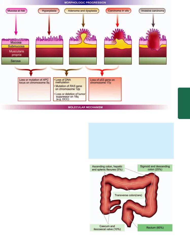
Figure 20.45 
 Adenoma-carcinoma sequence—schematic diagram of molecular and morphologic evolution.
in 80% cases of sporadic colon cancer. Since the function of APC gene is linked to β-catenin, loss of APC gene results in translocation of β-catenin to the nucleus where it activates transcription of other genes, mainly MYC and cyclin D1, both of which stimulate cell proliferation.
ii)Point mutation in K-RAS gene follows loss of APC gene and is seen in 10 to 50% cases of adenoma-carcinoma.
iii)Deletion of DCC gene located on long arm of chromosome
18i.e. 18q (DCC for deleted in colorectal cancer) in 60-70% cases of colon cancer.
iv)Loss of p53 tumour suppressor gene seen in 70-80% cases of colon cancer.
2.Microsatellite instability mechanism. In this pathway also, there are multiple mutations but of different genes, and unlike APC mutation/β-catenin mechanism there are no morphologically identifiable changes. This pathway accounts for 10-15% cases of colon cancer. Basic mutation is loss of DNA repair gene. This results in a situation in which repetitive DNA sequences (i.e. microsatellites) become unstable during replication cycle, termed microsatellite instability, which is the hallmark of this pathway. The significant DNA repair genes which are mutated in colon cancer are as under:
i)TGF-β receptor gene which normally inhibits cell proliferation but in mutated form allows the uncontrolled proliferation of colonic epithelium in adenoma.
ii)BAX gene which normally causes apoptosis but a defect in it results in loss of apoptosis and dysregulated growth.
MORPHOLOGIC FEATURES. Distribution of the primary colorectal cancer reveals that about 60% of the cases occur in the rectum, followed in descending order, by sigmoid and descending colon (25%), caecum and ileocaecal valve (10%); ascending colon, hepatic and splenic flexures (5%); and quite uncommonly in the transverse colon (Fig. 20.46).
Grossly, there are distinct differences between the growth on the right and left half of the colon (Fig. 20.47).
Figure 20.46 
 Distribution of the primary colorectal cancer.
587
Tract Gastrointestinal The 20 CHAPTER

588
Pathology Systemic III SECTION
Figure 20.47 |
Gross appearance of colorectal carcinoma. A, Right-sided growth—fungating polypoid carcinoma showing cauliflower-like growth |
projecting into the lumen. B, Left-sided growth—napkin-ring configuration with spread of growth into the bowel wall.
 Right-sided colonic growths tend to be large, cauli- flower-like, soft and friable masses projecting into the
lumen (fungating polypoid carcinoma).
 Left-sided colonic growths, on the other hand, have napkin-ring configuration i.e. they encircle the bowel wall circumferentially with increased fibrous tissue forming annular ring, and have central ulceration on the surface with slightly elevated margins (carcinomatous ulcers).
These differences in right and left colonic growths are probably due to the liquid nature of the contents in the ascending colon leaving space for luminal growth on right side, while the contents in left colon are more solid permitting the spread of growth into the bowel wall. However, early lesion in left as well as right colon are small, button-like areas of elevation.
Microscopically, the appearance of right and left-sided growths is similar. About 95% of colorectal carcinomas are adenocarcinomas of varying grades of differentiation, out of which approximately 10% are mucin-secreting colloid carcinomas (Fig. 20.48). The remaining 5% tumours include uncommon microscopic patterns like undifferentiated carcinoma, signet-ring cell carcinoma, and adenosquamous carcinomas seen in more distal colon near the anus. The histologic grades indicating the degree of differentiation are: well-differentiated, moderatelydifferentiated and poorly-differentiated.
SPREAD. Carcinoma of the large intestine may spread by the following routes:
1. Direct spread. The tumour spreads most commonly by direct extension in both ways— circumferentially into the
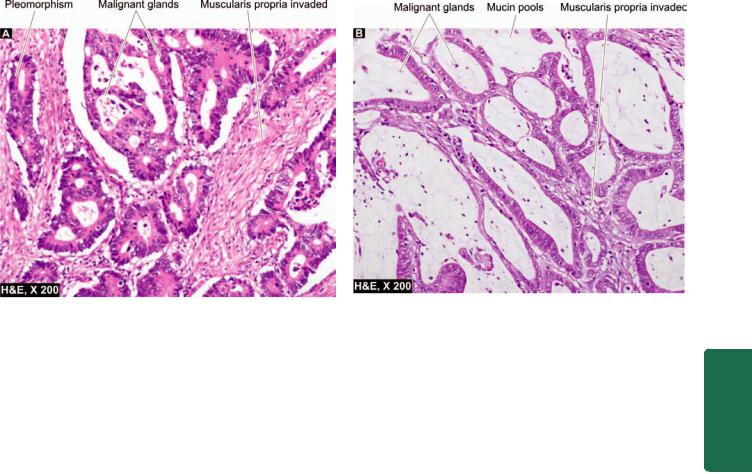
589
Figure 20.48 
 Colonic adenocarcinoma. A, Moderately differentiated. B, Mucin-secreting adenocarcinoma.
bowel wall as well as directly into the depth of the bowel wall to the serosa, pericolic fat, and sometimes into peritoneal cavity.
2.Lymphatic spread. Spread via lymphatics occurs rather commonly and involves, firstly the regional lymph nodes in the vicinity of the tumour, and then into other groups of lymph nodes like preaortic, internal iliac and the sacral lymph nodes.
3.Haematogenous spread. Blood spread of large bowel cancer occurs relatively late and involves the liver, lungs, brain, bones and ovary.
CLINICAL FEATURES. Clinical symptoms in colorectal cancer appear after considerable time. These are as follows:
i)Occult bleeding (melaena)
ii)Change in bowel habits, more often in left-sided growth
iii)Loss of weight (cachexia)
iv)Loss of appetite (anorexia)
v)Anaemia, weakness, malaise.
The most common complications are obstruction and haemorrhage; less often perforation and secondary infection may occur. Aside from the diagnostic methods like stool test for occult blood, PR examination, proctoscopy, radiographic contrast studies and CT scan, recently the role of tumourmarkers has been emphasised. Of particular importance is the estimation of carcinoembryonic antigen (CEA) level which is elevated in 100% cases of metastatic colorectal cancers, while it is positive in 20-40% of early lesions, and 60-70% of advanced primary lesions. However, the test may have prognostic significance only and is not diagnostic of colorectal cancer because it is positive in other cancers too such as of the lungs, breast, ovary, urinary bladder and prostate. CEA levels are elevated in some non-neoplastic conditions also like in ulcerative colitis, pancreatitis and alcoholic cirrhosis.
STAGING AND PROGNOSIS. The prognosis of colorectal cancer depends upon a few variables:
i)Extent of the bowel involvement
ii)Presence or absence of metastases
iii)Histologic grade of the tumour
iv)Location of the tumour
The most important prognostic factor in colorectal cancer is, however, the stage of the disease at the time of diagnosis. Three staging systems are in use:
1.Dukes’ ABC staging (modified Duke’s includes stage D as well).
2.Astler-Coller staging which is a further modification of Duke’s staging and is most widely used.
3.TNM staging described by American Joint Committee is also used.
Table 20.12 and Fig. 20.49 sum up the features of staging classification and the overall 5-year survival rate in disease stage.
B. Other Colorectal Malignant Tumours
Aside from colorectal carcinoma, other malignant tumours which are encountered sometimes in the large bowel are leiomyosarcoma (page 737) and malignant lymphoma (page 559). Hindgut carcinoids may occur in the rectum and colon (page 576).
TUMOURS OF THE ANAL CANAL
Epithelial tumours of the anal canal are uncommon and may be combination of several histological types. Amongst the benign tumours of the anal canal, multiple viral warts called as condyloma acuminata are the only tumours of note. Malignant tumours of the anal canal include the following:
1.Squamous cell carcinoma (Fig. 20.50)
2.Basaloid carcinoma
Tract Gastrointestinal The 20 CHAPTER
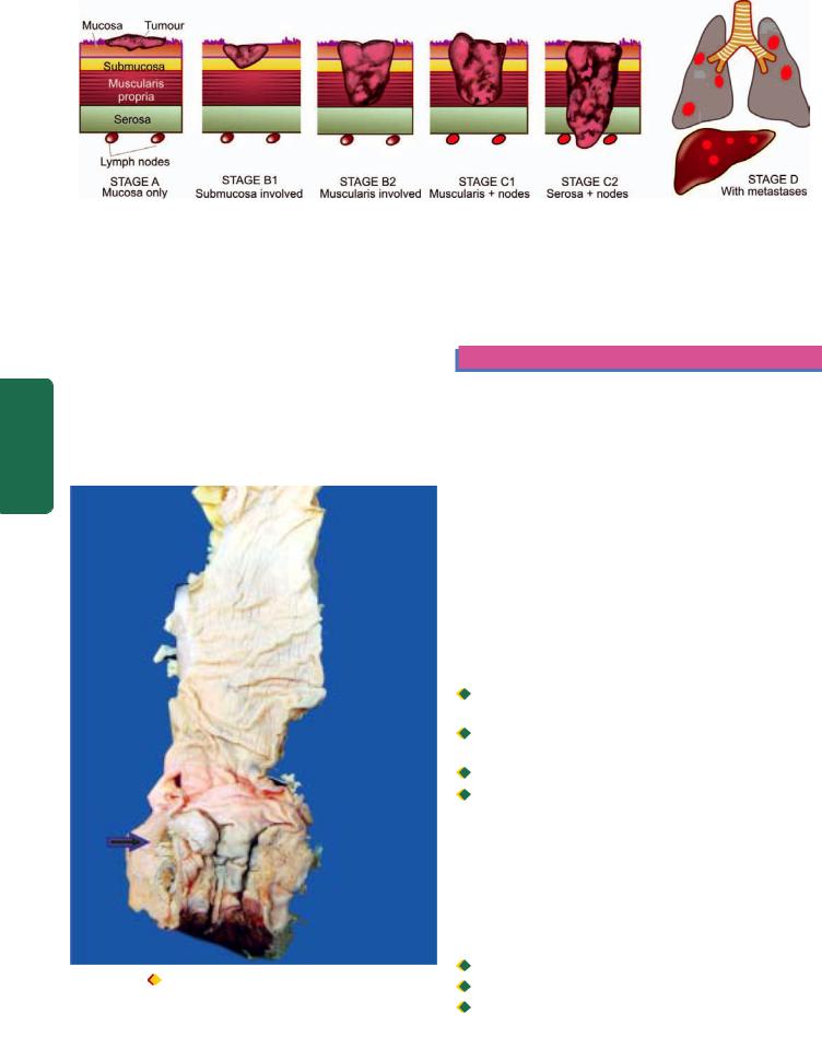
590
Figure 20.49 
 Pathologic staging according to Astler-Coller system. (Also see facing Table 20.12).
Pathology Systemic III SECTION
3.Mucoepidermoid carcinoma
4.Adenocarcinoma (rectal, of anal glands, within anorectal fistulas)
5.Undifferentiated carcinoma
6.Malignant melanoma.
These tumours resemble in morphology with similar
lesion elsewhere in the body.
CAUSES OF GASTROINTESTINAL BLEEDING
Gastrointestinal bleeding from upper (haematemesis), middle (small intestinal) and lower (melaena) is a major
Figure 20.50 |
Anorectal carcinoma. The anorectal margin shows |
an ulcerated mucosa with thickened wall (arrow). The sectioned surface of rectal wall is grey-white and fleshy due to infiltration by the tumour.
presenting clinical feature of a variety of gastrointestinal diseases. Table 20.13 summarises the main causes of gastrointestinal bleeding.
PERITONEUM
NORMAL STRUCTURE
The peritoneal cavity is lined by a layer of surface mesothelium derived from mesoderm. The lining rests on vascularised subserosal fibrous tissue. Other structures topographically related to peritoneum are retroperitoneum, omentum, mesentery and umbilicus. These structures are involved in a variety of pathologic states but a few important conditions included below are inflammation (peritonitis), tumour-like lesions (idiopathic retroperitoneal fibrosis and mesenteric cysts) and tumours (primary and metastatic).
PERITONITIS
Inflammatory involvement of the peritoneum may result from chemical agents or bacteria.
1. Chemical peritonitis can be caused by the following:
 Bile extravasated due to trauma or diseases of the gallbladder.

 Pancreatic secretions released from pancreas in acute haemorrhagic pancreatitis.

 Gastric juice leaked from perforation of stomach.

 Barium sulfate from perforation of bowel during radiographic studies.
Chemical peritonitis is localised or generalised sterile inflammation of the peritoneum.
2. Bacterial peritonitis may be primary or secondary; the latter being more common.
Primary form is caused by streptococcal infection, especially in children. Secondary bacterial peritonitis may occur from the following disorders:

 Appendicitis

 Cholecystitis

 Salpingitis

TABLE 20.12: Staging and Prognosis of Colorectal Cancer (Duke's System as Modified by Astler-Coller). |
591 |
||||
Stage |
|
TNM |
5-year Survival |
Pathologic Features |
|
A |
I |
T1 No Mo |
>90% |
Cancer confined to mucosa only |
|
B1 |
II |
T2 No Mo |
85% |
Cancer extends into submucosa |
|
B2 |
II |
T3 No Mo |
70-80% |
Cancer extends into muscularis+serosa |
|
C1 |
III |
Tx N1 Mo |
35-65% |
Cancer involves muscularis+regional lymph nodes |
|
C2 |
III |
Tx N1 Mo |
20-35% |
Cancer extends into serosa+regional lymph nodes |
|
D |
IV |
Tx Nx M1 |
5% |
Cancer with distant metastases |
|
Rupture of peptic ulcer |
|
|
MESENTERIC CYSTS |
|
||
Gangrene of bowel |
|
|
Mesenteric cysts of unknown etiology and varying sizes may |
|||
Tuberculosis (specific inflammation). |
||||||
be found in the peritoneal cavity. On the basis of their |
||||||
|
|
|
|
possible origin, they are of various types: |
||
MORPHOLOGIC FEATURES Depending upon duration, |
|
|||||
|
Chylous cyst is a thin-walled cyst arising from lymph |
|||||
the features in bacterial peritonitis vary. It may be |
|
vessels and lined by endothelium. |
||||
generalised or may get localised by omentum such as in |
|
Pseudocysts are those which are formed following walled- |
||||
appendiceal abscess following acute appendicitis. Depen- |
|
off infection or pancreatitis. |
||||
ding upon duration, the fluid accumulation varies from |
|
Neoplastic cysts occur due to cystic change in tumours. |
||||
serous, turbid, creamy to frankly suppurative. The fluid |
|
TUMOURS |
|
|||
may eventually resolve or may heal by organisation with |
|
|
||||
formation of fibrous adhesions. |
|
|
Peritoneum may be involved in malignant tumours— |
|||
|
|
|
|
primary and metastatic. |
|
|
IDIOPATHIC RETROPERITONEAL FIBROSIS |
Mesothelioma is an example of primary peritoneal |
|||||
|
|
|
|
tumour (benign and malignant) and is similar in morphology |
||
Also known as Ormond’s disease or sclerosing retro- |
as in pleural cavity (page 505). |
|||||
peritonitis, this rare entity of unknown etiology is characteri- |
Intra-abdominal desmoplastic small cell tumour is a |
|||||
sed by diffuse fibrous overgrowth and chronic inflammation. |
recently described highly malignant tumour belonging to the |
|||||
The condition is, therefore, more like inflammatory rather |
group of other round cell or blue cell tumours such as small |
|||||
than neoplastic in origin. It may be associated with similar |
cell carcinoma lung, Ewing’s sarcoma, rhabdomyosarcoma, |
|||||
process in the mediastinum, sclerosing cholangitis and |
neuroblastoma and others. |
|||||
Riedel’s thyroiditis and termed multifocal fibrosclerosis. |
Metastatic peritoneal tumours are quite common and |
|||||
Though idiopathic, the etiologic role of ergot derivative drugs |
may occur from dissemination from any intra-abdominal |
|||||
and autoimmune reaction has been suggested. |
malignancy. |
|
||||
|
|
|
||||
TABLE 20.13: Causes of Gastrointestinal (G.I.) Bleeding. |
|
|
||||
|
Upper G.I. Bleeding |
Small Intestinal Bleeding |
|
Lower G.I. Bleeding |
||
|
|
|
|
|
||
1. |
Oesophageal varices |
Vascular ectasias |
|
Inflammatory bowel disease (IBD) |
||
2. |
Mallory-Weiss tear |
Tumours (adenocarcinoma, lymphoma, leiomyoma) |
Carcinoma colon |
|||
3. |
Haemorrhagic/erosive gastritis |
NSAIDs |
|
Carcinoma rectosigmoid |
||
4. |
Duodenal ulcer |
Meckel's diverticulum |
|
Haemorrhoids |
||
5. |
Gastric ulcer |
Intussusception |
|
Anal fissure |
||
6. |
Cancer stomach |
Crohn's disease |
|
Diverticulosis |
||
|
|
|
|
|
|
|
Tract Gastrointestinal The 20 CHAPTER
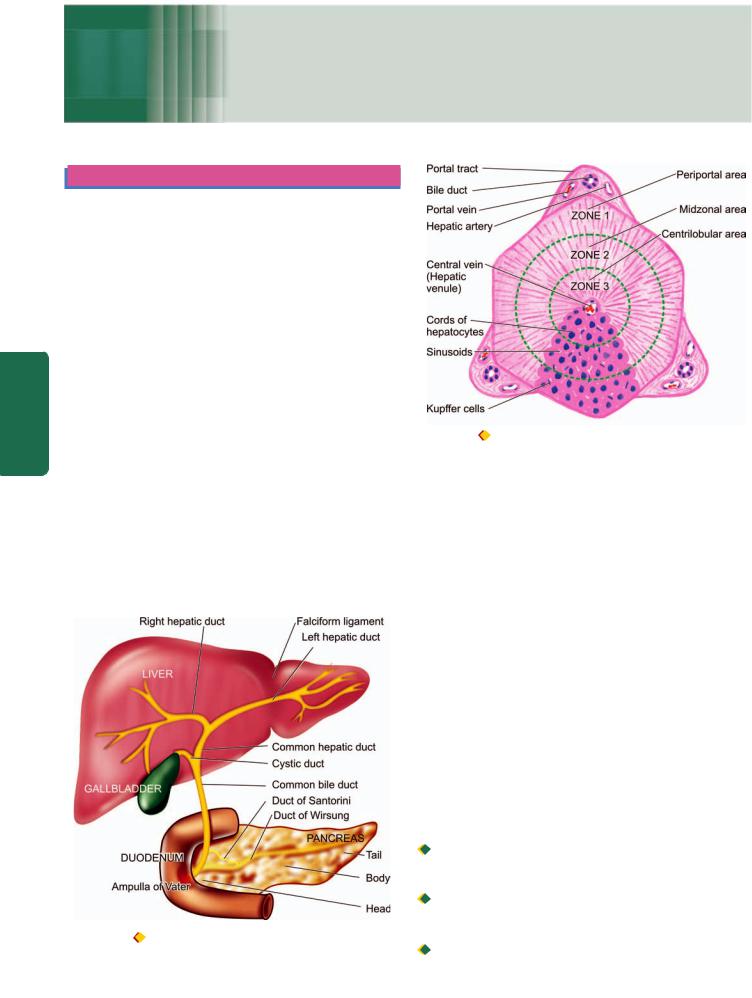
592
Chapter 21
The Liver, Biliary Tract and
Exocrine Pancreas
Pathology Systemic III SECTION
LIVER
NORMAL STRUCTURE
ANATOMY. The liver is the largest organ in the body weighing 1400-1600 gm in the males and 1200-1400 gm in the females. There are 2 main anatomical lobes—right and left, the right being about six times the size of the left lobe. The right lobe has quadrate lobe on its inferior surface and a caudate lobe on the posterior surface. The right and left lobes are separated anteriorly by a fold of peritoneum called the falciform ligament, inferiorly by the fissure for the ligamentum teres, and posteriorly by the fissure for the ligamentum venosum (Fig. 21.1).
The porta hepatis is the region on the inferior surface of the right lobe where blood vessels, lymphatics and common hepatic duct form the hilum of the liver. A firm smooth layer of connective tissue called Glisson’s capsule encloses the liver and is continuous with the connective tissue of the porta hepatis forming a sheath around the structures in the porta hepatis. The liver has a double blood supply—the portal vein brings the venous blood from the intestines and spleen, and the hepatic artery coming from the coeliac axis supplies arterial blood to the liver. This dual blood supply provides sufficient protection against infarction in the liver. The portal vein and hepatic artery divide into branches to the right and left lobes in the porta. The right and left hepatic ducts also
Figure 21.1 |
Anatomy of the liver and its relationship to the |
gallbladder, pancreas and duodenum.
Figure 21.2 |
Histology of hepatic lobule. The hexagonal or pyramidal |
structure with central vein and peripheral 4 to 5 portal triads is termed the classical lobule. The functional divisions of the lobule into 3 zones are shown by circles.
join in the porta to form the common hepatic duct. The venous drainage from the liver is into the right and left hepatic veins which enter the inferior vena cava. Lymphatics and the nerve fibres accompany the hepatic artery into their branchings and terminate around the porta hepatis.
HISTOLOGY. The hepatic parenchyma is composed of numerous hexagonal or pyramidal classical lobules, each with a diameter of 0.5 to 2 mm. Each classical lobule has a central tributary from the hepatic vein and at the periphery are 4 to 5 portal tracts or triads containing branches of bile duct, portal vein and hepatic artery. Cords of hepatocytes and blood-containing sinusoids radiate from the central vein to the peripheral portal triads. The functioning lobule or liver acinus as described by Rappaport has a portal triad in the centre and is surrounded at the periphery by portions of several classical lobules. However, in most descriptions on pathology of the liver, the term lobule is used in its classical form.
The blood supply to the liver parenchyma flows from the portal triads to the central veins. Accordingly, the hepatic parenchyma of liver lobule is divided into 3 zones (Fig. 21.2):

 Zone 1 or the periportal (peripheral) area is closest to the arterial and portal blood supply and hence bears the brunt of all forms of toxic injury.
 Zone 3 or the centrilobular area surrounds the central vein and is most remote from the blood supply and thus suffers from the effects of hypoxic injury.

 Zone 2 is the intermediate midzonal area.

The hepatocytes are polygonal cells with a round single nucleus and a prominent nucleolus. The liver cells have a remarkable capability to undergo mitosis and regeneration. Thus it is not uncommon to find liver cells containing more than one nuclei and having polyploidy up to octoploidy. A hepatocyte has 3 surfaces: one facing the sinusoid and the space of Disse, the second facing the canaliculus, and the third facing neighbouring hepatocytes.
The blood-containing sinusoids between cords of hepatocytes are lined by discontinuous endothelial cells and scattered flat Kupffer cells belonging to the reticuloendothelial system.
The space of Disse is the space between hepatocytes and sinusoidal lining endothelial cells. A few scattered fat storing Ito cells lie within the space of Disse.
The portal triad or tract besides containing portal vein radicle, the hepatic arteriole and bile duct, has a few mononuclear cells and a scanty connective tissue considered to be extension of Glisson’s capsule. The portal triads are surrounded by a limiting plate of hepatocytes.
The intrahepatic biliary system begins with the bile canaliculi interposed between the adjacent hepatocytes. The bile canaliculi are simply grooves between the contact surfaces of the liver cells and are covered by microvilli. These canaliculi join at the periphery of the lobule to drain eventually into terminal bile ducts or ductules (canal of Hering) which are lined by cuboidal epithelium.
FUNCTIONS. The liver performs multifold functions. These are briefly listed below:
1.Manufacture and excretion of bile.
2.Manufacture of several major plasma proteins such as albumin, fibrinogen and prothrombin.
3.Metabolism of proteins, carbohydrates and lipids.
4.Storage of vitamins (A, D and B12) and iron.
5.Detoxification of toxic substances such as alcohol and drugs.
LIVER FUNCTION TESTS
In view of multiplicity and complexity of the liver functions, it is obvious that no single test can establish the disturbance in liver function. Thus a battery of liver function tests are employed for accurate diagnosis, to assess the severity of damage, to judge prognosis and to evaluate therapy. These tests are described below in relation to major liver functions. A summary of various liver function tests is given in
Table 21.1.
I.TESTS FOR MANUFACTURE AND EXCRETION OF BILE
Bile is produced by the liver, stored in the gallbladder and secreted via biliary ducts into the duodenum. Bile consists of biliary phospholipids and primary and secondary bile acids. To understand the mechanisms underlying biliary pathology, it is important to understand normal bilirubin metabolism (page 596). In brief, jaundice will develop if bilirubin is excessively produced, or there is impaired hepatic uptake and conjugation of bilirubin, or it is insufficiently excreted into the duodenum. Tests employed to assess the
synthesis and elimination of bilirubin pigment, urobilinogen and bile acids are as follows:
1. BILIRUBIN. Bilirubin pigment can be detected in serum, faeces and urine.
i)Serum bilirubin estimation is based on van den Bergh diazo reaction by spectrophotometric method. Diazo reagent consists of diazotised sulfanilic acid. Water-soluble conjugated bilirubin gives direct van den Bergh reaction with diazo reagent within one minute, whereas alcohol-soluble unconjugated bilirubin is determined by indirect van den Bergh reaction. Addition of alcohol to the reaction mixture gives positive test for both conjugated and unconjugated bilirubin pigment. The unconjugated bilirubin level is then estimated by subtracting direct bilirubin value from this total value. The serum of normal adults contains less than 1 mg/ dl of total bilirubin, out of which less than 0.25 mg/dl is conjugated bilirubin. Bilirubin level rises in diseases of hepatocytes, obstruction to biliary excretion into the duodenum, in haemolysis, and defects of hepatic uptake and conjugation of bilirubin pigment such as in Gilbert’s disease.
ii)In faeces, excretion of bilirubin is assessed by inspection of stools. Clay-coloured stool due to absence of faecal excretion of the pigment indicates obstructive jaundice.
iii)In urine, conjugated bilirubin can be detected by commercially available ‘dipsticks’, Fouchet’s test, foam test or ictotest tablet method. Bilirubinuria does not occur in normal subjects nor is unconjugated bilirubin excreted in the urine. Bilirubinuria occurs only when there is raised level of conjugated bilirubin (filterable). Its excretion depends upon the level of conjugated bilirubin in plasma that is not proteinbound and is therefore available for glomerular filtration. Bilirubinuria appears in patients of hepatitis before the patient becomes jaundiced.
2.UROBILINOGEN. Urobilinogen is normally excreted in the urine. Its semiquantitative estimation in the urine can be done by preparing dilutions with Ehrlich’s aldehyde reagent or by ‘dipstick’ method. An increase in urobilinogen in the urine is found in hepatocellular dysfunctions such as in alcoholic liver disease, cirrhosis and malignancy of the liver. It is also raised in haemolytic disease and in pyrexia. In cholestatic jaundice due to complete biliary obstruction, urobilinogen disappears from the urine.
3.BROMSULPHALEIN EXCRETION. Bromsulphalein (BSP) is a dye which is removed from circulation by the same mechanisms of binding, conjugation and excretion as bilirubin. BSP is injected intravenously and a sample of venous blood 45 minutes later is tested for percentage of injected dye remaining in the blood. The test is rarely performed nowadays because of the availability of enzyme estimations which are better indicators of hepatic dysfunction. Presently, the only value of BSP excretion test is in the diagnosis of Dubin-Johnson’s syndrome (page 601).
4.BILE ACIDS (BILE SALTS). The primary bile acids (cholic acid and cheno-deoxycholic acid) are formed from cholesterol in the hepatocytes. These bile acids on secretion into the gut come in contact with colonic bacteria and undergo deconjugation with the production of secondary bile
593
Pancreas Exocrine and Tract Biliary Liver, The 21 CHAPTER

594
Pathology Systemic III SECTION
TABLE 21.1: Liver Function Tests.
Tests |
Significance |
|
|
I.TESTS FOR MANUFACTURE AND EXCRETION OF BILE
1.Bilirubin:
i) |
Serum bilirubin |
Increased in hepatocellular, obstructive and haemolytic disease, |
|
|
Gilbert’s disease |
ii) |
In faeces |
Absent in biliary obstruction |
iii) |
In urine |
Conjugated bilirubinuria in patients of hepatitis |
2. Urobilinogen: |
Increased in hepatocellular and haemolytic diseases, absent in |
|
|
|
biliary obstruction |
3.Bile acid (Bile salts):
II. SERUM ENZYME ASSAYS
1.Alkaline phosphatase:
2.γ-Glutamyl transpeptidase (γ-GT):
3.Transaminases:
i)SGOT (AST)
ii)SGPT (ALT)
4.Other enzymes:
i)5'-Nucleotidase
ii)Lactic dehydrogenase
iii)Cholinesterase
III. TESTS FOR METABOLIC FUNCTIONS
1.Amino acid and protein metabolism:
i)Serum proteins (total, A/G ratio, protein electrophoresis)
ii)Immunoglobulins
iii)Clotting factors
iv)Serum ammonia
v)Aminoaciduria
2.Lipid and lipoprotein metabolism:
Blood lipids (total serum cholesterol, triglycerides and lipoprotein fractions)
3.Carbohydrate metabolism:
Blood glucose and GTT
IV. IMMUNOLOGIC TESTS
1.Nonspecific immunologic reactions:
i)Smooth muscle antibody
ii)Mitochondrial antibody
iii)Antinuclear antibody and LE cell test
2.Antibodies to specific etiologic agents:
i)Antibodies to hepatitis B (HBsAg, HBc, HBeAg)
ii)Amoeba antibodies
V. ANCILLARY DIAGNOSTIC TESTS
1.Ultrasound examination
2.FNAC and/ or percutaneous liver biopsy
Increased in serum and detectable in urine in cholestasis
Increased in hepatobiliary disease (highest in biliary obstruction), bone diseases, pregnancy
Rise parallels alkaline phosphatase but is specific for hepatobiliary diseases
Increased in tissue injury to liver as well as to other tissues like in myocardial infarction
Increase is fairly specific for liver cell injury
Rise parallels alkaline phosphatase but more specific for diseases of hepatic origin
Increased in tumours involving the liver Decreased in hepatocellular disease, malnutrition
Hypoalbuminaemia in hepatocellular diseases; hyperglobulinaemia in cirrhosis and chronic active hepatitis
Nonspecific alterations in IgA, IgG and IgM
Prothrombin time and partial thromboplastin time prolonged in patients with hepatocellular disease
Increased in acute fulminant hepatitis, cirrhosis, hepatic encephalopathy In fulminant hepatitis
Increased in cholestasis, decreased in acute and chronic diffuse liver disease and in malnutrition
Decreased in hepatic necrosis
In hepatic necrosis
In primary biliary cirrhosis
In chronic active hepatitis
In hepatitis B
Amoebic liver abscess
Cholestasis of various etiologies; SOLs, US-guided-FNAC/liver biopsy
Unknown cause of hepatocellular disease, hepatomegaly and splenomegaly; long-standing hepatitis; PUO and SOLs of the liver
acids (deoxycholic acid and lithocholic acid). Most of these bile acids are reabsorbed through enterohepatic circulation and reach the liver. Only about 10% of the total bile acids are excreted in the faeces normally as unabsorbable toxic lithocholic acid.
Hepatobiliary diseases with cholestasis are associated with raised levels of serum bile acids which are responsible for producing itching (pruritus). These acids are excreted in the urine by active transport and passive diffusion and can be detected by simple methods as Hay’s test and ‘dipsticks’.
II. SERUM ENZYME ASSAYS
Determination of certain serum enzymes is considered useful in various types of liver injury, whether hepatocellular or cholestatic, as well as in quantifying liver damage. A combination of serum transaminases and alkaline phosphatase estimation is adequate to diagnose liver injury.
1.ALKALINE PHOSPHATASE. Serum alkaline phosphatase is produced by many tissues, especially bone, liver, intestine and placenta and is excreted in the bile. Most of the normal serum alkaline phosphatase (range 33-96 U/L) is derived from bone. Elevation in activity of the enzyme can thus be found in diseases of bone, liver and in pregnancy. In the absence of bone disease and pregnancy, an elevated serum alkaline phosphatase levels generally reflect hepatobiliary disease. The greatest elevation (3 to 10 times normal) occurs in biliary tract obstruction. Slight to moderate increase is seen in parenchymal liver diseases such as in hepatitis and cirrhosis and in metastatic liver disease. It is possible to distinguish serum hepatic alkaline phosphatase from bony alkaline phosphatase by fractionation into isoenzymes but this is not routinely done.
2.γ -GLUTAMYL TRANSPEPTIDASE (γ -GT). The primary source of the enzyme, γ-GT, in serum is the liver. Its serum level parallels serum alkaline phosphatase and is used to confirm that the elevated serum alkaline phosphatase is of hepatobiliary origin. Besides its elevation in cholestasis and hepatocellular disease, the levels are high in patients with alcohol abuse even without liver disease.
3.TRANSAMINASES (AMINOTRANSFERASES).
Assessment of liver cell necrosis is most frequently done by estimation of the following 2 serum enzymes:
i)Serum aspartate transaminase or AST (formerly glutamic oxaloacetic transaminase or SGOT): AST or SGOT is a mitochondrial enzyme released from heart, liver, skeletal muscle and kidney. Its normal serum level is 0.20-0.65 μkat/L (12-38 U/L).
ii)Serum alanine transaminase or ALT (formerly glutamic pyruvic transaminase or SGPT): ALT or SGPT is a cytosolic enzyme primarily present in the liver. Its normal serum level is 0.12-0.70 μkat/L (7-41 U/L).
Serum levels of SGOT and SGPT are increased on damage to the tissues producing them. Thus serum estimation of SGPT (ALT) which is fairly specific for liver tissue is of greater value in liver cell injury, whereas SGOT (AST) level may rise in acute necrosis or ischaemia of other organs such as the myocardium, besides liver cell injury.
Transaminase estimations are useful in the early diagnosis of viral hepatitis. Very high levels are seen in extensive acute hepatic necrosis such as in severe viral hepatitis and acute cholestasis. Alcoholic liver disease and cirrhosis are associated with mild to moderate elevation of transaminases.
4.OTHER SERUM ENZYMES. The determination of a few other serum enzymes is done sometimes but without any extra diagnostic advantage over the above mentioned enzyme assays. These are as under:
i) 5'-Nucleotidase is another phosphatase derived from the 595 liver. Its determination is useful to distinguish alkaline phosphatase of hepatic origin from that of bony tissue.
ii)Lactic dehydrogenase (LDH) is found to be elevated in serum of patients with metastatic liver involvement.
iii)Choline esterase synthesised by the liver is diminished in hepatocellular disease and malnutrition due to impaired synthesis.
III. TESTS FOR METABOLIC FUNCTIONS |
|
|
The liver is the principal site of metabolism and synthesis of |
|
|
plasma proteins and amino acids, lipids and lipoproteins, |
|
|
carbohydrates and vitamins, besides detoxification of drugs |
|
|
and alcohol. |
|
|
1. AMINO ACID AND PLASMA PROTEIN META- |
|
|
BOLISM. Amino acids derived from the diet and from |
|
|
tissue breakdown are metabolised in the liver to ammonia |
|
|
and urea. A number of plasma proteins and immuno- |
|
|
globulins are synthesised on polyribosomes bound to the |
|
|
rough endoplasmic reticulum within the hepatocytes and |
|
|
discharged into plasma. Based on these metabolic functions |
CHAPTER |
|
of the liver, serum estimation of proteins, immunoglobulins |
||
and ammonia and aminoaciduria are employed to assess |
||
the liver cell damage. |
||
i) Serum proteins. Liver cells synthesise albumin, |
||
fibrinogen, prothrombin, alpha-1-antitrypsin, haptoglobin, |
||
ceruloplasmin, transferrin, alpha fetoproteins and acute |
21 |
|
phase reactant proteins. The blood levels of these plasma |
||
|
||
proteins are decreased in extensive liver damage. Routinely |
The |
|
estimated are total concentration of serum proteins (normal |
||
6.7 to 8.6 gm/dl), serum albumin (normal 3.5 to 5.5 gm/dl), |
||
serum globulin (normal 2 to 3.5 gm/dl) and albumin/ |
Liver, |
|
globulin (A/G) ratio (normal 1.5-3:1). Electrophoresis is used |
||
|
||
to determine the proportions of α1, α2, β and γ globulins. |
Biliary |
|
Due to the availability of protein electrophoresis, thymol |
||
|
||
turbidity and flocculation tests based on altered plasma |
|
|
protein components have been discontinued. |
Tract |
|
Hypoalbuminaemia may occur in liver diseases having |
||
|
||
significant destruction of hepatocytes. Hyperglobulinaemia |
|
|
may be present in chronic inflammatory disorders such as |
and |
|
in cirrhosis and chronic hepatitis. |
||
|
||
ii) Immunoglobulins. The levels of serum immuno- |
Exocrine |
|
globulins produced by lymphocytes and plasma cells (IgG, |
||
|
||
IgM and IgA) show nonspecific abnormalities in liver |
|
|
diseases and represent inflammatory or immune response |
|
|
rather than liver cell dysfunction. IgA is the predominant |
Pancreas |
|
immunoglobulin in bile and its level is raised in cirrhosis, |
||
|
||
IgG is markedly raised in chronic active hepatitis and IgM |
|
|
is markedly increased in primary biliary cirrhosis. |
|
|
iii) Clotting factors. Hepatic synthetic function of several |
|
|
clotting factors can be assessed by a few simple coagulation |
|
|
tests. Prothrombin time and partial thromboplastin time, |
|
|
both of which reflect the activities of various clotting factors, |
|
|
are prolonged in patients with hepatocellular disease. |
|
|
Prothrombin time is dependent upon both hepatic synthesis |
|
|
of clotting factors and intestinal uptake of vitamin K, a fat- |
|
|
soluble vitamin. Thus, obstruction of the bile duct and intra- |
|

596hepatic cholestasis which result in vitamin K deficiency due to impaired lipid absorption, are associated with prolonged prothrombin time. However, parenteral injection of vitamin K will normalise prothrombin time if the prolongation was due to obstruction, but there will be no improvement in prothrombin time if there is extensive hepatocellular disease.
iv) Serum ammonia. High blood levels of ammonia are found in acute fulminant hepatitis, cirrhosis and hepatic encephalopathy. The rise in serum ammonia is due to inability of severely damaged liver to convert ammonia to urea. Thus, urea synthesis is reduced in chronic liver disease.
|
2. LIPID AND LIPOPROTEIN METABOLISM. Lipids |
|
|
synthesised in the liver include cholesterol and cholesterol |
|
|
esters, phospholipids and triglycerides. These lipids are |
|
|
insoluble in water and are carried in circulation with three |
|
|
major types of lipoproteins which contain apoproteins. These |
|
|
are: high density lipoproteins (HDL), low density |
|
|
lipoproteins (LDL) and very low density lipoproteins |
|
|
(VLDL). |
|
|
Blood lipids. Estimations of total serum cholesterol, |
|
|
triglycerides and lipoprotein fractions are frequently done |
|
SECTION |
in patients with liver disease. |
|
There is rise in total serum cholesterol in cholestasis, |
||
probably due to retention of cholesterol which is normally |
||
excreted in the bile (normal < 200 mg/dl). Serum triglyceride |
||
is also elevated in cholestasis. |
||
III |
Values are lowered in acute and chronic diffuse liver |
|
diseases and in malnutrition. |
||
|
||
Systemic |
3. CARBOHYDRATE METABOLISM. The liver plays a |
|
insulin resistance. |
||
|
central role in carbohydrate metabolism. Blood glucose level |
|
|
is lowered in fulminant acute hepatic necrosis. In chronic |
|
Pathology |
liver disease, there is impaired glucose tolerance and relative |
|
IV. IMMUNOLOGIC TESTS |
||
|
||
|
Liver diseases are associated with various immunologic |
|
|
abnormalities which may be nonspecific immunologic reac- |
|
|
tions or may be antibodies against specific etiologic agents. |
1. NONSPECIFIC IMMUNOLOGIC REACTIONS. These include the following:
i)Smooth muscle antibody to actin component of muscle is formed in certain hepatic disorders with hepatic necrosis. It appears that hepatocytes have a protein which is immunologically similar to actin.
ii)Mitochondrial antibody develops in patients with primary biliary cirrhosis.
iii)Antinuclear antibody is present in some patients of chronic hepatitis. The LE cell test may be positive in these cases.
2. ANTIBODIES TO SPECIFIC ETIOLOGIC AGENTS.
These vary according to the etiologic agent causing the liver cell injury.
i)Hepatitis B surface antigen (HBsAg) can be demonstrated in cases of serum hepatitis. A confirmed positive test for HBsAg is definite proof of hepatitis B infection.
ii)Hepatitis B core antibody (HBc) can be detected in all patients with hepatitis B.
iii)Hepatitis Be antigen (HBeAg) can be found in chronic varieties of hepatitis B.
iv)Amoeba antibodies to Entamoeba histolytica develop in patients with amoebic liver abscess.
V. ANCILLARY DIAGNOSTIC TESTS
In addition to laboratory tests described above, two ancillary tests which are invariably done by the physician are ultrasonography and percutaneous liver biopsy and/or FNAC.
1. ULTRASONOGRAPHY. Ultrasound (US) examination of the liver is indicated in the following situations:
i)Cholestasis of various etiologies to see the dilated intraand extrahepatic canalicular tree.
ii)Space-occupying lesions (SOLs) within the liver to determine whether they are neoplasms or non-neoplastic cysts.
iii)To provide US-guidance for FNAC or liver biopsy.
2. FNAC AND/OR PERCUTANEOUS LIVER BIOPSY.
Lastly, FNAC and percutaneous liver biopsy are employed to examine the microscopic changes of hepatic morphology in various diseases. Both these tests are done after evaluation of signs of obstruction since these tests are contraindicated in cholestasis. FNAC and liver biopsy are otherwise easily performed bedside tests of value. Their main indications are as follows:
i)hepatocellular disease of unknown cause;
ii)suspected cases of chronic hepatitis;
iii)hepatomegaly of various etiologies;
iv)splenomegaly of unknown cause;
v)fever of unknown cause; and
vi)SOLs visualised in radiologic examination
JAUNDICE—GENERAL
Jaundice or icterus refers to the yellow pigmentation of the skin or sclerae by bilirubin (page 42). Bilirubin pigment has high affinity for elastic tissue and hence jaundice is particularly noticeable in tissues rich in elastin content. Jaundice is the result of elevated levels of bilirubin in the blood termed hyperbilirubinaemia. Normal serum bilirubin concentration ranges from 0.3-1.3 mg/dl, about 80% of which is unconjugated. Jaundice becomes clinically evident when the total serum bilirubin exceeds 2 mg/dl. A rise of serum bilirubin between the normal and 2 mg/dl is generally not accompanied by visible jaundice and is called latent jaundice.
Before considering the features and types of jaundice, it is essential to review the normal bilirubin metabolism.
NORMAL BILIRUBIN METABOLISM
Normal metabolism of bilirubin can be conveniently described under 4 main headings—source, transport, hepatic phase and intestinal phase as illustrated schematically earlier in Chapter 12 (see Fig. 12.9, page 291).
1. SOURCE OF BILIRUBIN. About 80-85% of the bilirubin is derived from the catabolism of haemoglobin present in senescent red blood cells. The destruction of effete
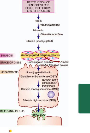
erythrocytes at the end of their normal lifespan of 120 days takes place in the reticuloendothelial system in the bone marrow, spleen and liver. The remaining 15-20% of the bilirubin comes partly from non-haemoglobin haemcontaining pigments such as myoglobin, catalase and cytochromes, and partly from ineffective erythropoiesis. In either case, haem moiety is formed which is converted to biliverdin by microsomal haem oxygenase for which oxygen and NADPH are essential requirements. Bilirubin is formed from biliverdin by biliverdin reductase.
2.TRANSPORT OF BILIRUBIN. Bilirubin on release from macrophages circulates as unconjugated bilirubin in plasma tightly bound to albumin. Certain drugs such as sulfonamides and salicylates compete with bilirubin for albumin binding and displace bilirubin from albumin, thus facilitating bilirubin to enter into the brain in neonates and increase the risk of kernicterus. Bilirubin is found in body fluids in proportion to their albumin content such as in CSF, joint effusions, cysts etc.
3.HEPATIC PHASE. On coming in contact with the hepatocyte surface, unconjugated bilirubin is preferentially metabolised which involves 3 steps: hepatic uptake, conjugation and secretion in bile.
i)Hepatic uptake: Albumin-bound unconjugated bilirubin upon entry into the hepatocyte, is dissociated into bilirubin and albumin. The bilirubin gets bound to cytoplasmic protein glutathione-S-transferase (GST) (earlier called ligandin).
ii)Conjugation: Unconjugated bilirubin is not water-soluble but is alcohol-soluble and is converted into water-soluble compound by conjugation. Conjugation occurs in endoplasmic reticulum and involves conversion to bilirubin monoand diglucuronide by the action of microsomal enzyme, bilirubin- UDP-glucuronosyl transferase (Fig. 21.3). The process of conjugation can be induced by drugs like phenobarbital.
Conjugated bilirubin is bound to albumin in two forms: reversible and irreversible. Reversible binding is similar to that of unconjugated bilirubin. However, when present in serum for a long time (e.g. in cholestasis, long-standing biliary obstruction, chronic active hepatitis), conjugated bilirubin is bound to albumin irreversibly and is termed delta bilirubin or biliprotein. This irreversible conjugated delta bilirubin is not excreted by the kidney, and remains detectable in serum for sufficient time after recovery from the diseases listed above.
iii)Secretion into bile: Conjugated (water-soluble) bilirubin is rapidly transported directly into bile canaliculi by energydependent process and then excreted into the bile.
4.INTESTINAL PHASE. Appearance of conjugated bilirubin in the intestinal lumen is followed by either direct excretion in the stool as stercobilinogen which imparts the normal yellow colour to stool, or may be metabolised to urobilinogen by the action of intestinal bacteria. Conjugated bilirubin is normally not reabsorbable whereas its metabolic product, urobilinogen, is reabsorbed from the small intestine and reaches enterohepatic circulation. Some of the absorbed urobilinogen in resecreted by the liver into the bile while the rest is excreted in the urine as urobilinogen.
Figure 21.3 |
Schematic representation of hepatic phase of bilirubin |
transport. |
|
The major differences between unconjugated and conjugated bilirubin are summarised in Table 21.2.
CLASSIFICATION AND FEATURES OF JAUNDICE
Based on pathophysiology, jaundice may result from one or more of the following mechanisms:
1.Increased bilirubin production
2.Decreased hepatic uptake
3.Decreased hepatic conjugation
4.Decreased excretion of bilirubin into bile
Accordingly, a simple age-old classification of jaundice was to divide it into 3 predominant types: pre-hepatic
(haemolytic), hepatic, and post-hepatic cholestatic. However, hyperbilirubinaemia due to first three mechanisms is mainly unconjugated while the last variety yields mainly conjugated hyperbilirubinaemia. Hence, currently pathophysiologic classification of jaundice is based on predominance of the type of hyperbilirubinaemia. A simple test to determine whether hyperbilirubinaemia is of unconjugated or conjugated variety is to determine whether bilirubin is present in urine or not; its absence in urine suggests unconjugated hyperbilirubinaemia since unconjugated
597
Pancreas Exocrine and Tract Biliary Liver, The 21 CHAPTER

598 TABLE 21.2: Major Differences between Unconjugated and Conjugated Bilirubin.
|
Feature |
Unconjugated Bilirubin |
Conjugated Bilirubin |
|
|
|
|
1. |
Normal serum level |
More |
Less (less than 0.25 mg/dl) |
2. |
Water solubility |
Absent |
Present |
3. |
Affinity to lipids |
Present |
Absent |
|
(alcohol solubility) |
|
|
4. |
Serum albumin binding |
High |
Low |
5. |
van den Bergh reaction |
Indirect |
Direct |
|
|
(Total minus direct) |
|
6. |
Renal excretion |
Absent |
Present |
7. |
Bilirubin albumin covalent |
Absent |
Present |
|
complex formation |
|
|
8. |
Affinity to brain tissue |
Present (Kernicterus) |
Absent |
|
|
|
|
Pathology Systemic III SECTION
bilirubin is not filtered by the glomerulus. The presence of bilirubin in the urine is evidence of conjugated hyperbilirubinaemia.
Based on these mechanisms, the pathogenesis and main features of the two predominant forms of hyperbilirubinaemia are discussed below (Table 21.3).
I.Predominantly Unconjugated Hyperbilirubinaemia
This form of jaundice can result from the following three sets of conditions:
TABLE 21.3: Pathophysiologic Classification of Jaundice.
I. PREDOMINANTLY UNCONJUGATED HYPERBILIRUBINAEMIA
1.Increased bilirubin production (Haemolytic, acholuric or prehepatic jaundice)
•Intraand extravascular haemolysis
•Ineffective erythropoiesis
2. Decreased hepatic uptake
•Drugs
•Prolonged starvation
•Sepsis
3. Decreased bilirubin conjugation
•Hereditary disorders (e.g. Gilbert’s syndrome, Crigler-Najjar syndrome)
•Acquired defects (e.g. drugs, hepatitis, cirrhosis)
•Neonatal jaundice
II.PREDOMINANTLY CONJUGATED HYPERBILIRUBINAEMIA (CHOLESTASIS)
1. Intrahepatic cholestasis (Impaired hepatic excretion)
•Hereditary disorders or ‘pure cholestasis’ (e.g. Dubin-Johnson syndrome, Rotor’s syndrome, fibrocystic disease of pancreas, benign familial recurrent cholestasis, intrahepatic atresia, cholestatic jaundice of pregnancy)
•Acquired disorders or ‘hepatocellular cholestasis’ (e.g. viral hepatitis, drugs, alcohol-induced injury, sepsis, cirrhosis)
2. Extrahepatic cholestasis (Extrahepatic biliary obstruction)
•Mechanical obstruction (e.g. gallstones, inflammatory strictures, carcinoma head of pancreas, tumours of bile ducts, sclerosing cholangitis, congenital atresia of extrahepatic ducts)
1.INCREASED BILIRUBIN PRODUCTION (HAEMOLYTIC, ACHOLURIC OR PREHEPATIC JAUNDICE). This results from excessive red cell destruction as occurs in intraand extravascular haemolysis or due to ineffective erythropoiesis. There is increased release of haemoglobin from excessive breakdown of red cells that leads to overproduction of bilirubin. Hyperbilirubinaemia develops when the capacity of the liver to conjugate large amount of bilirubin is exceeded. In premature infants, the liver is deficient in enzyme necessary for conjugation while the rate of red cell destruction is high. This results in icterus neonatorum which is particularly severe in haemolytic disease of the newborn due to maternal isoantibodies (Chapter 13). Since there is predominantly unconjugated hyperbilirubinaemia in such cases, there is danger of permanent brain damage in these infants from kernicterus when the serum level of unconjugated bilirubin exceeds 20 mg/dl.
Laboratory data in haemolytic jaundice, in addition to predominant unconjugated hyperbilirubinaemia, reveal normal serum levels of transaminases, alkaline phosphatase and proteins. Bile pigment being unconjugated type is absent from urine (acholuric jaundice). However, there is dark brown colour of stools due to excessive faecal excretion of bile pigment and increased urinary excretion of urobilinogen.
2.DECREASED HEPATIC UPTAKE. The uptake of bilirubin by the hepatocyte that involves dissociation of the pigment from albumin and its binding to cytoplasmic protein, GST or ligandin, may be deranged in certain conditions e.g. due to drugs, prolonged starvation and sepsis.
3.DECREASED BILIRUBIN CONJUGATION. This mechanism involves deranged hepatic conjugation due to defect or deficiency of the enzyme, glucuronosyl transferase. This can occur in certain inherited disorders of the enzyme (e.g. Gilbert’s syndrome and Crigler-Najjar syndrome), or acquired defects in its activity (e.g. due to drugs, hepatitis, cirrhosis). However, hepatocellular damage causes deranged excretory capacity of the liver more than its conjugating capacity (see below). The physiologic neonatal jaundice is also partly due to relative deficiency of UDP-glucuronosyl transferase in the neonatal liver and is partly as a result of increased rate of red cell destruction in neonates.
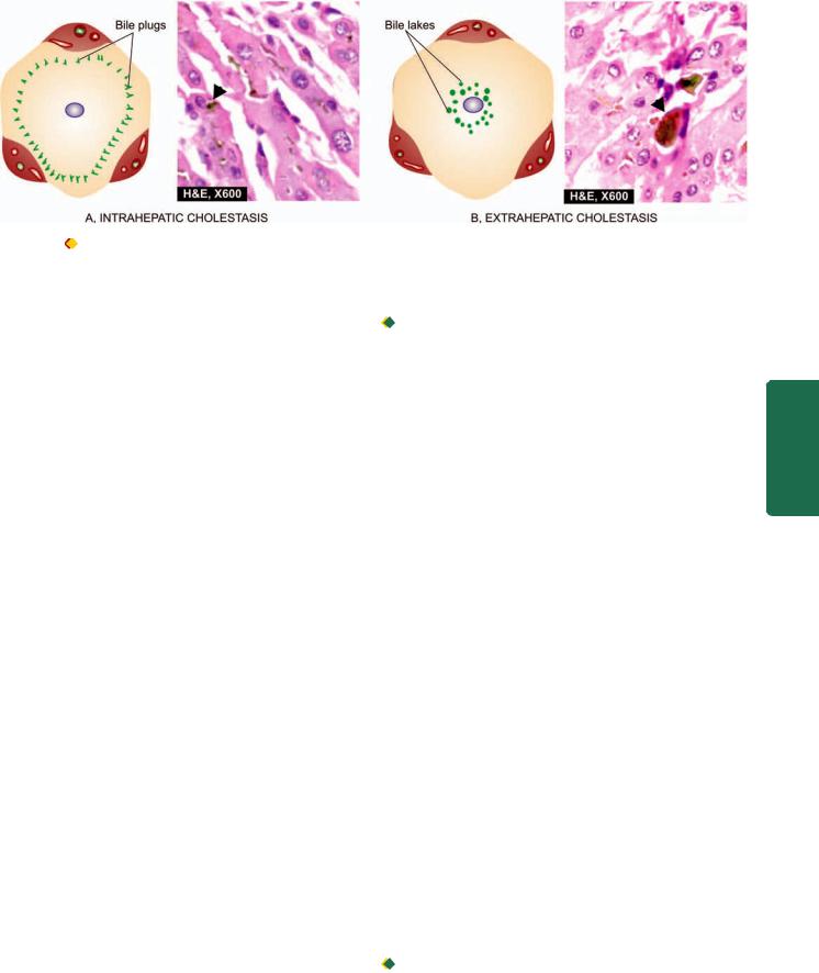
599
Figure 21.4 |
Salient features in morphology of liver in intraand extrahepatic cholestasis. A, Intrahepatic cholestasis is characterised by |
elongated bile plugs in the canaliculi of hepatocytes at the periphery of the lobule. B, Extrahepatic cholestasis shows characteristic bile lakes due to rupture of canaliculi in the hepatocytes in the centrilobular area.
II.Predominantly Conjugated Hyperbilirubinaemia (Cholestasis)
This form of hyperbilirubinaemia is defined as failure of normal amounts of bile to reach the duodenum. Morphologically, cholestasis means accumulation of bile in liver cells and biliary passages. The defect in excretion may be within the biliary canaliculi of the hepatocyte and in the microscopic
bile ducts (intrahepatic cholestasis or medical jaundice), or there may be mechanical obstruction to the extrahepatic biliary
excretory apparatus (extrahepatic cholestasis or obstructive jaundice). It is important to distinguish these two forms of cholestasis since extrahepatic cholestasis or obstructive jaundice is often treatable with surgery, whereas the intrahepatic cholestasis or medical jaundice cannot be benefitted by surgery but may in fact worsen by the operation. Prolonged cholestasis of either of the two types may progress to biliary cirrhosis (page 625).
1. INTRAHEPATIC CHOLESTASIS. Intrahepatic cholestasis is due to impaired hepatic excretion of bile and may occur from hereditary or acquired disorders.
i)Hereditary disorders producing intrahepatic obstruction to biliary excretion are characterised by ‘pure cholestasis’ e.g. in Dubin-Johnson syndrome, Rotor syndrome, fibrocystic disease of pancreas, benign familial recurrent cholestasis, intrahepatic atresia and cholestatic jaundice of pregnancy.
ii)Acquired disorders with intrahepatic excretory defect of bilirubin are largely due to hepatocellular diseases and hence are termed ‘hepatocellular cholestasis’ e.g. in viral hepatitis, alcoholic hepatitis, and drug-induced cholestasis such as from administration of chlorpromazine and oral contraceptives.
The features of intrahepatic cholestasis include: predominant conjugated hyperbilirubinaemia due to regurgitation of conjugated bilirubin into blood, bilirubinuria, elevated levels of serum bile acids and consequent pruritus, elevated serum alkaline phosphatase, hyperlipidaemia and hypoprothrombinaemia. ‘Pure cholestasis’ can be distinguished from ‘hepatocellular cholestasis’ by elevated serum levels of transaminases in the latter due to liver cell injury.
 Liver biopsy in cases with intrahepatic cholestasis reveals milder degree of cholestasis than the extrahepatic disorders (Fig. 21.4,A). The biliary canaliculi of the hepatocytes are dilated and contain characteristic elongated green-brown bile plugs. The cytoplasm of the affected hepatocytes shows feathery degeneration. Canalicular bile stasis eventually causes proliferation of intralobular ductules followed by periportal fibrosis and produces a picture resembling biliary cirrhosis (page 625).
2. EXTRAHEPATIC CHOLESTASIS. Extrahepatic cholestasis results from mechanical obstruction to large bile ducts outside the liver or within the porta hepatis. The common causes are gallstones, inflammatory strictures, carcinoma head of pancreas, tumours of bile duct, sclerosing cholangitis and congenital atresia of extrahepatic ducts. The obstruction may be complete and sudden with eventual progressive obstructive jaundice, or the obstruction may be partial and incomplete resulting in intermittent jaundice.
The features of extrahepatic cholestasis (obstructive jaundice), like in intrahepatic cholestasis, are: predominant conjugated hyperbilirubinaemia, bilirubinuria, elevated serum bile acids causing intense pruritus, high serum alkaline phosphatase and hyperlipidaemia. However, there are certain features which help to distinguish extrahepatic from intrahepatic cholestasis. In obstructive jaundice, there is malabsorption of fat-soluble vitamins (A,D,E and K) and steatorrhoea resulting in vitamin K deficiency. Prolonged prothrombin time in such cases shows improvement following parenteral administration of vitamin K, whereas hypoprothrombinaemia due to hepatocellular disease shows no such improvement in prothrombin time with vitamin K administration. The stools of such patients are clay-coloured due to absence of bilirubin metabolite, stercobilin, in faeces and there is virtual disappearance of urobilinogen from the urine. These patients may have fever due to high incidence of ascending bacterial infections (ascending cholangitis).

 Liver biopsy in cases with extrahepatic cholestasis shows more marked changes of cholestasis (Fig. 21.4,B). Since the obstruction is in the extrahepatic bile ducts, there is progressive retrograde extension of bile stasis into intrahepatic duct system. This results in dilatation of bile
Pancreas Exocrine and Tract Biliary Liver, The 21 CHAPTER

600
Pathology Systemic III SECTION
TABLE 21.4: Causes of Neonatal Jaundice.
A. UNCONJUGATED HYPERBILIRUBINAEMIA
1.Physiologic and prematurity jaundice
2.Haemolytic disease of the newborn and kernicterus (page 340)
3.Congenital haemolytic disorders (page 314)
4.Perinatal complications (e.g. haemorrhage, sepsis)
5.Gilbert’s syndrome
6.Crigler-Najjar syndrome (type I and II)
B. CONJUGATED HYPERBILIRUBINAEMIA
1.Hereditary (Dubin-Johnson syndrome, Rotor’s syndrome)
2.Infections (e.g. hepatitis B, hepatitis C or non-A non-B hepatitis, rubella, coxsackievirus, cytomegalovirus, echovirus, herpes simplex, syphilis, toxoplasma, gram-negative sepsis)
3.Metabolic (e.g. galactosaemia, alpha-1-antitrypsin deficiency, cystic fibrosis, Niemann-Pick disease)
4.Idiopathic (neonatal hepatitis, congenital hepatic fibrosis)
5.Biliary atresia (intrahepatic and extrahepatic)
6.Reye’s syndrome
ducts and rupture of canaliculi with extravasation of bile producing bile lakes. Since bile is toxic, the regions of bile lakes are surrounded by focal necrosis of hepatocytes. Stasis of bile predisposes to ascending bacterial infections with accumulation of polymorphs around the dilated ducts (ascending cholangitis). Eventually, there is proliferation of bile ducts and the appearance may mimic biliary cirrhosis (page 625).
NEONATAL JAUNDICE
Jaundice appears in neonates when the total serum bilirubin is more than 3 mg/dl. It may be the result of unconjugated or conjugated hyperbilirubinaemia; the former being more common. Important causes of neonatal jaundice are listed in Table 21.4. Some of these conditions are considered below, while others are discussed elsewhere in the relevant sections.
HEREDITARY NON-HAEMOLYTIC
HYPERBILIRUBINAEMIAS
Hereditary non-haemolytic hyperbilirubinaemias are a small group of uncommon familial disorders of bilirubin metabolism when haemolytic causes have been excluded. The commonest is Gilbert’s syndrome; others are CriglerNajjar syndrome, Dubin-Johnson syndrome, Rotor’s syndrome and benign familial recurrent cholestasis. The features common to all these conditions are presence of icterus but almost normal liver function tests and no welldefined morphologic changes except in Dubin-Johnson syndrome. Gilbert’s syndrome and Crigler-Najjar syndrome
are examples of hereditary non-haemolytic unconjugated hyperbilirubinaemia, whereas Dubin-Johnson syndrome, Rotor’s syndrome and benign familial recurrent cholestasis
are conditions with hereditary conjugated hyperbilirubinaemia.
These conditions are briefly described below. Their distinguishing features are summarised in Table 21.5.
Gilbert’s Syndrome
This is the commonest of the familial, genetically-determined diseases of the liver affecting 2-5% of the population. Gilbert’s syndrome is characterised by mild, benign, unconjugated hyperbilirubinaemia (serum bilirubin 1-5 mg/dl) which is not due to haemolysis. The condition is inherited as an autosomal dominant character. The defect in bilirubin metabolism is complex and appears to be reduced activity of UDP-glucuronosyl transferase with decreased conjugation, or an impaired hepatic uptake of bilirubin. The jaundice is usually mild and intermittent.
MORPHOLOGIC FEATURES. There are no morphologic abnormalities in the liver except some increased lipofuscin pigment in centrilobular hepatocytes. The prognosis of patients with Gilbert’s syndrome is excellent, though chronic jaundice persists throughout life.
TABLE 21.5: Contrasting Features of Major Hereditary Non-haemolytic Hyperbilirubinaemias.
|
Feature |
Gilbert’s |
Type 1 |
Type 2 |
Dubin-Johnson |
Rotor |
|
|
Syndrome |
Crigler-Najjar |
Crigler-Najjar |
Syndrome |
Syndrome |
|
|
|
Syndrome |
Syndrome |
|
|
|
|
|
|
|
|
|
1. |
Inheritance |
Autosomal |
Autosomal |
Autosomal |
Autosomal |
Autosomal |
|
|
dominant |
recessive |
dominant |
recessive |
recessive |
2. |
Predominant |
Unconjugated |
Unconjugated |
Unconjugated |
Conjugated |
Conjugated |
|
hyperbilirubinaemia |
|
|
|
|
|
3. |
Intensity of |
Mild |
Marked |
Mild to moderate |
Mild |
Mild |
|
jaundice |
(< 5 mg/dl) |
(>20 mg/dl) |
(<20 mg/dl) |
(<5 mg/dl) |
(< 5 mg/dl) |
4. |
Basic defect |
↓ UDP- |
Absence of UDP- |
↓ UDP- |
Defect in canali- |
Deranged hepatic |
|
|
glucuronosyl |
glucuronosyl |
glucuronosyl |
cular excretion |
storage |
|
|
transferase |
transferase |
transferase |
(Prolonged BSP |
|
|
|
activity |
|
|
excretion test) |
|
5. |
Hepatic |
Normal (except |
Normal (except |
Normal |
Greenish-black |
Normal |
|
morphology |
slightly increased |
mild canalicular |
|
pigment |
|
|
|
lipofuscin) |
stasis) |
|
|
|
6. |
Prognosis |
Excellent |
Poor (due to |
Good |
Excellent |
Excellent |
|
|
|
kernicterus) |
|
|
|
|
|
|
|
|
|
|

Crigler-Najjar Syndrome |
NEONATAL HEPATITIS |
601 |
Crigler-Najjar syndrome is a rare form of familial nonhaemolytic jaundice with very high unconjugated hyperbilirubinaemia. There are 2 forms of this condition: type I and type II.
Type I Crigler-Najjar syndrome. This is inherited as an autosomal recessive disorder. There is complete absence of conjugating enzyme UDP-glucuronosyl transferase in the hepatocytes and hence no conjugated bilirubin is formed. There is extreme elevation of unconjugated bilirubin (usually more than 20 mg/dl) with high risk of developing permanent CNS damage from kernicterus. The prognosis is generally fatal, with death coming from kernicterus usually in the first year of life.
MORPHOLOGIC FEATURES. There are no significant morphologic changes except some canalicular stasis.
Type II Crigler-Najjar syndrome. This is inherited as an autosomal dominant disease. There is deficiency of enzyme UDP-glucuronosyl transferase but not complete absence. Thus, unconjugated hyperbilirubinaemia is generally mild to moderate (usually less than 20 mg/dl). Occurrence of kernicterus is exceptional and patients respond well to phenobarbital therapy.
MORPHOLOGIC FEATURES. There are no morphologic changes in the liver.
Dubin-Johnson Syndrome
Dubin-Johnson syndrome is autosomal recessive disorder characterised by predominant conjugated hyperbilirubinaemia
(usually less than 5 mg/dl) with genetic defect in canalicular excretion of conjugated bilirubin. A prolonged BSP dye excretion test is diagnostic of Dubin-Johnson syndrome (page 593).
MORPHOLOGIC FEATURES. Grossly, the condition differs from other forms of hereditary hyperbilirubinaemias in producing greenish-black pigmented liver. Microscopically, The hepatocytes show dark-brown, melanin-like pigment in the cytoplasm, the exact nature of which is obscure but it is neither iron nor bile. Unrelated viral hepatitis mobilises the hepatic pigment of DubinJohnson syndrome leading to its excretion in urine but the pigment reappears after recovery from viral hepatitis.
The disease runs a benign course and does not interfere with life.
Rotor’s Syndrome
This is another form of familial conjugated hyperbilirubinaemia with mild chronic jaundice but differs from Dubin-Johnson syndrome in having no brown pigment in the liver cells. The disease is inherited as an autosomal recessive character. The defect probably lies in intrahepatic storage of bilirubin but the exact protein abnormality is not known.
Rotor’s syndrome has an excellent prognosis.
Neonatal hepatitis, also termed giant cell hepatitis or neonatal hepatocellular cholestasis, is a general term used for the constant morphologic change seen in conjugated hyperbilirubinaemia as a result of known infectious and metabolic causes listed in Table 21.4, or may have an idiopathic etiology. ‘Idiopathic’ neonatal hepatitis is more common and accounts for 75% of cases. Though all the cases with either known etiologies or idiopathic type are grouped together under neonatal hepatitis, all of them are not necessarily inflammatory conditions, thus belying their nomenclature as ‘hepatitis’. The condition usually presents in the first week of birth with jaundice, bilirubinuria, pale stools and high serum alkaline phosphatase.
MORPHOLOGIC FEATURES. Irrespective of the etiology, there is morphologic similarity in all these cases. The histologic features are as under:
1.Loss of normal lobular architecture of the liver.
2.Presence of prominent multinucleate giant cells derived from hepatocytes.
3.Mononuclear inflammatory cell infiltrate in the portal tracts with some periportal fibrosis.
4.Haemosiderosis.
5.Cholestasis in small proliferated ductules in the portal tract and between necrotic liver cells.
BILIARY ATRESIAS
Biliary atresias, also called as infantile cholangiopathies, are a group of intrauterine developmental abnormalities of the biliary system. Though they are often classified as congenital, the abnormality of development in most instances is extraneous infection during the intrauterine development or shortly after birth that brings about inflammatory destruction of the bile ducts. The condition may, therefore, have various grades of destruction ranging from complete absence of bile ducts termed atresia, to reduction in their number called
paucity of bile ducts.
Depending upon the portion of biliary system involved, biliary atresias may be extrahepatic or intrahepatic.
Extrahepatic Biliary Atresia
The extrahepatic bile ducts fail to develop normally so that in some cases the bile ducts are absent at birth, while in others the ducts may have been formed but start undergoing sclerosis in the perinatal period. It is common to have multiple defects and other congenital lesions. Extrahepatic biliary atresia is found in 1 per 10,000 livebirths. Cholestatic jaundice appears by the first week after birth. The baby has severe pruritus, pale stools, dark urine and elevated serum transaminases. In some cases, the condition is correctable by surgery, while in vast majority the atresia is not correctable and in such cases hepatic portoenterostomy (Kasai procedure) or hepatic transplantation must be considered. Death is usually due to intercurrent infection, liver failure, and bleeding due to vitamin K deficiency or oesophageal varices. Cirrhosis and ascites are late complications appearing within 2 years of age.
Pancreas Exocrine and Tract Biliary Liver, The 21 CHAPTER

602
Pathology Systemic III SECTION
MORPHOLOGIC FEATURES. Grossly, the liver is enlarged and dark green. The atretic segments of biliary system are reduced to cord-like structures.
Histologically, the condition must be distinguished from idiopathic neonatal hepatitis as surgical treatment is possible in extrahepatic biliary atresia but not in the latter. Besides, α-1-antitrypsin deficiency also produces similar appearance in liver biopsy. The main histologic features are as under:
1.Inflammation and fibrous obliteration of the extrahepatic ducts with absence of bile in them.
2.Ductular proliferation and periductular inflammation.
3.Cholestasis and bile thrombi in the portal area.
4.Periportal fibrosis and later secondary biliary cirrhosis (page 625).
5.Transformation of hepatic parenchyma to neonatal (giant cell) hepatitis in 15% of cases.
Intrahepatic Biliary Atresia
Intrahepatic biliary atresia is characterised by biliary hypoplasia so that there is paucity of bile ducts rather than their complete absence. The condition probably has its origin in viral infection acquired during intrauterine period or in the neonatal period. Cholestatic jaundice usually appears within the first few days of birth and is characterised by high serum bile acids with associated pruritus, and hypercholesterolaemia with appearance of xanthomas by first year of life. Hepatic as well as urinary copper concentrations are elevated. In some cases, intrahepatic biliary atresia is related to α-1-antitrypsin deficiency.
MORPHOLOGIC FEATURES. The microscopic features are as follows:
1.Paucity of intrahepatic bile ducts.
2.Cholestasis.
3.Increased hepatic copper.
4.Inflammation and fibrosis in the portal area, eventually leading to cirrhosis.
REYE’S SYNDROME
Reye’s syndrome is defined as an acute postviral syndrome of encephalopathy and fatty change in the viscera. The syndrome may follow almost any known viral disease but is most common after influenza A or B and varicella. Viral infection may act singly, but more often its effect is modified by certain exogenous factors such as by administration of salicylates, aflatoxins and insecticides. These effects cause mitochondrial injury and decreased activity of mitochondrial enzymes in the liver. This eventually leads to rise in blood ammonia and accumulation of triglycerides within hepatocytes.
The patients are generally children between 6 months and 15 years of age. Within a week after a viral illness, the child develops intractable vomiting and progressive neurological deterioration due to encephalopathy, eventually leading to stupor, coma and death. Characteristic laboratory findings are elevated blood ammonia, serum transaminases, bilirubin and prolonged prothrombin time.
MORPHOLOGIC FEATURES. Grossly, the liver is enlarged and yellowish-orange.
Microscopically, hepatocytes show small droplets of neutral fat in their cytoplasm (microvesicular fat). Similar fatty change is seen in the renal tubular epithelium and in the cells of skeletal muscles and heart. The brain shows oedema and sometimes focal necrosis of neurons.
HEPATIC FAILURE
Though the liver has a marked regenerative capacity and a large functional reserve, hepatic failure may develop from severe acute and fulminant liver injury with massive necrosis of liver cells (acute hepatic failure), or from advanced chronic liver disease (chronic hepatic failure). Acute hepatic failure develops suddenly with severe impairment of liver functions whereas chronic liver failure comes insidiously. The prognosis is much worse in acute hepatic failure than that in chronic liver failure.
ETIOLOGY. Acute and chronic hepatic failure result from different causes:

 Acute (fulminant) hepatic failure occurs most frequently in acute viral hepatitis. Other causes are hepatotoxic drug reactions (e.g. anaesthetic agents, nonsteroidal antiinflammatory drugs, anti-depressants), carbon tetrachloride poisoning, acute alcoholic hepatitis, mushroom poisoning and pregnancy complicated with eclampsia.
 Chronic hepatic failure is most often due to cirrhosis. Other causes include chronic active hepatitis, chronic cholestasis (cholestatic jaundice) and Wilson’s disease.
MANIFESTATIONS. In view of the diverse functions performed by the liver, the syndrome of acute or chronic hepatic failure produces complex manifestations. The major manifestations are briefly discussed below and diagrammatically illustrated in Fig. 21.5.
1.Jaundice. Jaundice usually reflects the severity of liver cell damage since it occurs due to failure of liver cells to metabolise bilirubin. In acute failure such as in viral hepatitis, jaundice nearly parallels the extent of liver cell damage, while in chronic failure such as in cirrhosis jaundice appears late and is usually of mild degree.
2.Hepatic encephalopathy (Hepatic coma). Neuropsychiatric syndrome may complicate liver disease of both acute and chronic types. The features include disturbed consciousness, personality changes, intellectual deterioration, low slurred speech, flapping tremors, and finally, coma and death. The genesis of CNS manifestations in liver disease is by toxic products not metabolised by the diseased liver. The toxic products may be ammonia and other nitrogenous substances from intestinal bacteria which reach the systemic circulation without detoxification in the damaged liver and thus damage the brain. Advanced cases of hepatic coma have poor prognosis but may respond favourably to hepatic transplantation.
3.Hyperkinetic circulation. All forms of hepatic failure are associated with a hyperkinetic circulation characterised by peripheral vasodilatation, increased splanchnic blood flow
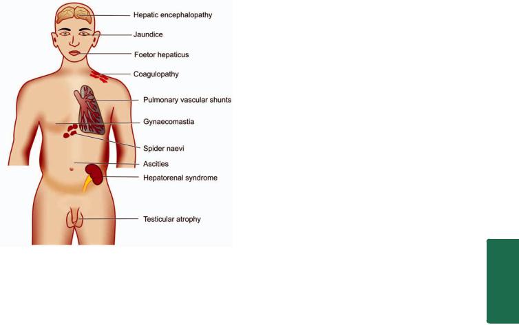
Figure 21.5 
 Complications of hepatic failure.
and increased cardiac output. There is increased splenic flow but reduced renal blood flow resulting in impaired renal cortical perfusion. These changes result in tachycardia, low blood pressure and reduced renal function.
4.Hepatorenal syndrome. The term hepatorenal syndrome is applied to patients of both acute and chronic hepatic failure who develop renal failure as well, in the absence of clinical, laboratory or morphologic evidence of other causes of renal dysfunction. Hepatorenal syndrome develops in about 10% cases of acute and chronic liver diseases. The acute renal failure is usually associated with oliguria and uraemia but with good tubular function. The histology of kidney is virtually normal, suggesting functional defect for the renal failure. The pathogenesis of the syndrome is poorly understood but appears to be initiated by effective reduction of the renal blood flow (effective hypovolaemia) as a consequence of systemic vasodilatation and pooling of blood in portal circulation. The renal failure in the hepatorenal syndrome is reversible with improvement in hepatic function.
Diagnosis of hepatorenal syndrome should be made only after excluding other causes producing concomitant damage to both the organs, circulatory failure leading to acute tubular necrosis and other forms of reversible tubular damage.
5.Hepatopulmonary syndrome. The pulmonary changes in chronic hepatic failure such as in cirrhosis consist of pulmonary vasodilatation with intra-pulmonary arteriovenous shunting. This results in ventilation-perfusion inequality that may lead to impaired pulmonary function, clubbing of fingers and sometimes cyanosis.
6.Coagulation defects. Impaired synthesis of a number of coagulation factors by the diseased liver may result in coagulation disorders. These include disseminated
intravascular coagulation (consumption coagulopathy), thrombocytopenia and presence of fibrin degradation products in the blood.
7.Ascites and oedema. Chronic liver failure due to cirrhosis may result in portal hypertension and ascites (page 630). Decreased synthesis of albumin by the liver resulting in hypoproteinaemia and consequent fall in plasma oncotic pressure, increased hydrostatic pressure due to portal hypertension and secondary hyperaldosteronism, contribute to the development of ascites and oedema in these patients.
8.Endocrine changes. Endocrine changes may be found in association with chronic hepatic failure. The changes are more common in alcoholic cirrhosis in active reproductive life. In the male, the changes are towards feminisation such as gynaecomastia and hypogonadism. In the female, the changes are less towards masculinisation but atrophy of gonads and breasts occurs. The underlying mechanism appears to be changed end-organ sensitiveness to sex hormones in cirrhosis.
9.Skin changes. In alcoholic cirrhosis ‘arterial spiders’ having radiating small vessels from a central arteriole are frequent in the vascular region drained by superior vena cava such as in the neck, face, forearms and dorsum of hands. Less frequently, palmar erythema, especially in the hypothenar and thenar eminences and on the pulps of the fingers, is observed in chronic liver disease.
10.Foetor hepaticus. A sweetish pungent smell of the breath is found in severe cases of acute and chronic hepatocellular diseases. It appears to be of intestinal origin, possibly due to failure of the liver to detoxify sulfur-containing substances absorbed from the gut.
CIRCULATORY DISTURBANCES
Vascular disorders of general nature involving the liver such as chronic passive congestion and infarction have already been discussed in Chapter 5. Hepatic and portal venous obstruction and hepatic arterial obstruction are considered here.
I.HEPATIC VENOUS OBSTRUCTION
The central veins of lobules of the liver are tributaries of the hepatic veins. In the normal liver, there are no anastomoses between hepatic vein and portal vein but in cirrhotic liver there are such anastomoses. Normal pressure in the free hepatic vein is about 6 mmHg.
Two uncommon diseases produced by obstruction of the hepatic veins are Budd-Chiari syndrome (hepatic vein thrombosis) and hepatic veno-occlusive disease.
Budd-Chiari Syndrome (Hepatic Vein Thrombosis)
Budd-Chiari syndrome in its pure form consists of slowly developing thrombosis of the hepatic veins and the adjacent inferior vena cava, while some workers include hepatic venoocclusive disease (described below) in this syndrome.
ETIOLOGY. The etiology of hepatic venous thrombosis in about a third of cases is unknown (idiopathic), while in the
603
Pancreas Exocrine and Tract Biliary Liver, The 21 CHAPTER

604remaining cases various causes associated with increased thrombotic tendencies are attributed to polycythaemia vera, paroxysmal nocturnal haemoglobinuria, oral contraceptives, pregnancy, postpartum state, intra-abdominal cancers (e.g. hepatocellular carcinoma), chemotherapy, radiation and myeloproliferative diseases. Formation of membranous webs, probably congenital or as a consequence of organised thrombosis, in the suprahepatic portion of inferior vena cava is another important cause.
MORPHOLOGIC FEATURES. Grossly, the liver is enlarged, swollen, red-purple and has a tense capsule. Histologically, the changes in sudden hepatic vein occlusion are those of centrilobular congestion, necrosis and rupture of sinusoids into the space of Disse. In slowly developing thrombosis, the changes are more chronic and include fibrosing reaction in the centrilobular zone that may progress to cardiac cirrhosis.
CLINICAL FEATURES. Budd-Chiari syndrome is clinically characterised by either an acute form or chronic form depending upon the speed of occlusion.
SECTION |
In the acute form, the features are abdominal pain, |
|
vomiting, enlarged liver, ascites and mild icterus. |
||
|
||
|
In the more usual chronic form, the patients present with |
|
|
pain over enlarged tender liver, ascites and other features of |
|
III |
portal hypertension. |
|
The acute form of illness leads to acute hepatic failure |
||
|
and death, whereas in chronic form the patient may live for |
|
Systemic |
months to a few years. |
|
stenosis and obliteration of the terminal central veins and |
||
|
Hepatic Veno-occlusive Disease |
|
|
Hepatic veno-occlusive disease consists of intimal thickening, |
|
Pathology |
medium-sized hepatic veins. The venous occlusion results |
|
veins. |
||
|
in pathologic changes similar to those of Budd-Chiari |
|
|
syndrome and can be distinguished from the latter by |
|
|
demonstration of absence of thrombosis in the major hepatic |
|
|
The cause and stimulus for hepatic veno-occlusive |
|
|
disease are obscure. The condition is more widespread in |
|
|
countries such as Africa, India and certain other tropical |
|
|
countries where ‘bush tea’ (medicinal tea) is consumed that |
|
|
contains hepatotoxic alkaloids. The disease has also been |
|
|
found in association with administration of antineoplastic |
|
|
drugs and immunosuppressive therapy. |
|
|
II. PORTAL VENOUS OBSTRUCTION |
|
|
Obstruction of the portal vein may occur within the |
|
|
intrahepatic course or in extrahepatic site. |
|
|
Intrahepatic cause of portal venous occlusion is hepatic |
|
|
cirrhosis as the commonest and most important, followed in |
|
|
decreasing frequency by tumour invasion, congenital hepatic |
|
|
fibrosis and schistosomiasis. |
|
|
Extrahepatic causes of portal vein obstruction are intra- |
|
|
abdominal cancers, intra-abdominal sepsis, direct invasion |
|
|
by tumour, myeloproliferative disorders and upper |
|
|
abdominal surgical procedure followed by thrombosis. |
The effects of portal venous obstruction depend upon the site of obstruction. The most important effect, irrespective of the site of occlusion or cause, is portal hypertension and its manifestations (page 630). If the obstruction is in the extrahepatic portal vein along with extension of occlusion into splenic vein, it may result in venous infarction of the bowel. Pylephlebitis may be followed by multiple pyaemic liver abscesses.
Peliosis Hepatis
Although sinusoidal dilatation can occur secondary to many liver diseases, peliosis hepatis is an uncommon condition of primary sinusoidal dilatation that results in blockage of blood outflow and may result in massive intraperitoneal haemorrhage. Although exact etiology is not known, peliosis hepatis and another related condition, bacillary angiomatosis (page 413), have been found to occur in HIV-infected patients whose CD4+ T cell counts fall below 100/μl. Opportunistic infection with Bartonella henselae in poor hygienic conditions in these cases results in blood-filled cysts in liver partly lined by endothelial cells and having mixed inflammatory cells in a fibromyxoid background.
Etiologic association of peliosis hepatis with consumption of anabolic steroids and oral contraceptives has also been suggested and is self-limiting with withdrawal of the offending agent.
III. HEPATIC ARTERIAL OBSTRUCTION
Diseases from obstruction of the hepatic artery are uncommon. Rarely, accidental ligation of the main hepatic artery or its branch to right lobe may be followed by fatal infarction. Obstruction of the small intrahepatic arterial branches usually does not produce any effects because of good collateral circulation.
LIVER CELL NECROSIS
All forms of injury to the liver such as microbiologic, toxic, circulatory or traumatic, result in necrosis of liver cells. The extent of involvement of hepatic lobule in necrosis varies. Accordingly, liver cell necrosis is divided into 3 types: diffuse (submassive to massive), zonal and focal.
1.DIFFUSE (SUBMASSIVE TO MASSIVE) NECROSIS.
When there is extensive and diffuse necrosis of the liver involving all the cells in groups of lobules, it is termed diffuse, or submassive to massive necrosis. It is most commonly caused by viral hepatitis or drug toxicity.
2.ZONAL NECROSIS. Zonal necrosis is necrosis of hepatocytes in 3 different zones of the hepatic lobule (page 592). Accordingly, it is of 3 types; each type affecting respective zone is caused by different etiologic factors:
i) Centrilobular necrosis is the commonest type involving hepatocytes in zone 3 (i.e. located around the central vein). Centrilobular necrosis is characteristic feature of ischaemic injury such as in shock and CHF since zone 3 is farthest from the blood supply. Besides, it also occurs in poisoning with chloroform, carbon tetrachloride and certain drugs.
ii)Midzonal necrosis is uncommon and involves zone 2 of the hepatic lobule. This pattern of necrosis is seen in yellow fever and viral hepatitis. In viral hepatitis, some of the necrosed hepatocytes of the mid-zone are transformed into acidophilic, rounded Councilman bodies.
iii)Periportal (peripheral) necrosis is seen in zone 1 involving the parenchyma closest to the arterial and portal blood supply. Since zone 1 is most well perfused, it is most vulnerable to the effects of circulating hepatotoxins e.g. in phosphorus poisoning and eclampsia.
3. FOCAL NECROSIS. This form of necrosis involves small groups of hepatocytes irregularly distributed in the hepatic lobule. Focal necrosis is most often caused by microbiologic infections. These include viral hepatitis, miliary tuberculosis, typhoid fever and various other forms of bacterial, viral and fungal infections. Focal necrosis may also occur in druginduced hepatitis.
VIRAL HEPATITIS
The term viral hepatitis is used to describe infection of the liver caused by hepatotropic viruses. Currently there are 5 main varieties of these viruses and a sixth poorlycharacterised virus, causing distinct types of viral hepatitis:

 Hepatitis A virus (HAV), causing a faecally-spread selflimiting disease.

 Hepatitis B virus (HBV), causing a parenterally transmitted disease that may become chronic.
 Hepatitis C virus (HCV), previously termed non-A, non-B (NANB) hepatitis virus involved chiefly in transfusionrelated hepatitis.

 Hepatitis delta virus (HDV) which is sometimes associated as superinfection with hepatitis B infection.

 Hepatitis E virus (HEV), causing water-borne infection.

 Hepatitis G virus (HGV), is a recently discovered transfusion-transmitted hepatotropic virus but is not known to cause hepatitis.
All these human hepatitis viruses are RNA viruses except HBV which is a DNA virus.
Though a number of other viral diseases such as infection with Epstein-Barr virus (in infectious mononucleosis), arbovirus (in yellow fever), cytomegalovirus, herpes simplex and several others affect the liver but the changes produced by them are nonspecific; the term ‘viral hepatitis’ is strictly applied to infection of the liver by the hepatitis viruses.
ETIOLOGIC CLASSIFICATION
Based on the etiologic agent, viral hepatitis is currently classified into 6 etiologic types—hepatitis A, hepatitis B, hepatitis C, hepatitis D, hepatitis E and hepatitis G. The contrasting features of major types are presented in
Table 21.6.
TABLE 21.6: Features of Various Types of Hepatitis Viruses.
|
Feature |
Hepatitis A |
Hepatitis B |
Hepatitis C |
Hepatitis D |
Hepatitis E |
1. |
Agent |
HAV |
HBV |
HCV |
HDV |
HEV |
2. |
Year identified |
1973 |
1965 |
1989 |
1977 |
1980 |
3. |
Viral particle |
27 nm |
42 nm |
30-60 nm |
35-37 nm |
32-34 nm |
4. |
Genome |
RNA, ss, linear |
DNA, ss/ds |
RNA, ss, linear |
RNA, ss, circular |
RNA, ss, linear |
|
|
|
|
circular |
|
|
5. |
Morphology |
Icosahedral |
Double-shelled, |
Enveloped |
Enveloped, replication |
Icosahedral, |
|
|
non-enveloped |
enveloped |
|
defective |
non-enveloped |
6. |
Spread |
Faeco-oral |
Parenteral, |
Parenteral, |
Parenteral, close |
Water-borne |
|
|
|
close contact |
close contact |
contact |
|
7. |
Incubation |
15-45 days |
30-180 days |
20-90 days |
30-50 days |
15-60 days |
|
period |
|
|
|
(In superinfection) |
|
8. |
Antigen(s) |
HAV |
HBsAg |
HCV RNA |
HBsAg |
HEV |
|
|
|
HBcAg |
C 100-3 |
HDV |
|
|
|
|
HBeAg |
C 33c |
|
|
|
|
|
HBxAg |
NS5 |
|
|
9. |
Antibodies |
anti-HAV |
anti-HBs |
anti-HCV |
anti-HBs |
anti-HEV |
|
|
|
anti-HBc |
|
anti-HDV |
|
|
|
|
anti-HBe |
|
|
|
10. |
Severity |
Mild |
Occasionally severe |
Moderate |
Occasionally severe |
Mild |
11. |
Chronic |
None |
Occasional |
Common |
Common |
None |
|
hepatitis |
|
|
|
|
|
12. |
Carrier state |
None |
<1% |
<1% |
1-10% |
Unknown |
13. |
Hepatocellular |
No |
+ |
+ |
± |
None |
|
carcinoma |
|
|
|
|
|
14. |
Prognosis |
Excellent |
Worse with age |
Moderate |
Acute good; chronic poor |
Good |
(ss= single-stranded; ss/ds= partially single-stranded partially double-stranded)
605
Pancreas Exocrine and Tract Biliary Liver, The 21 CHAPTER
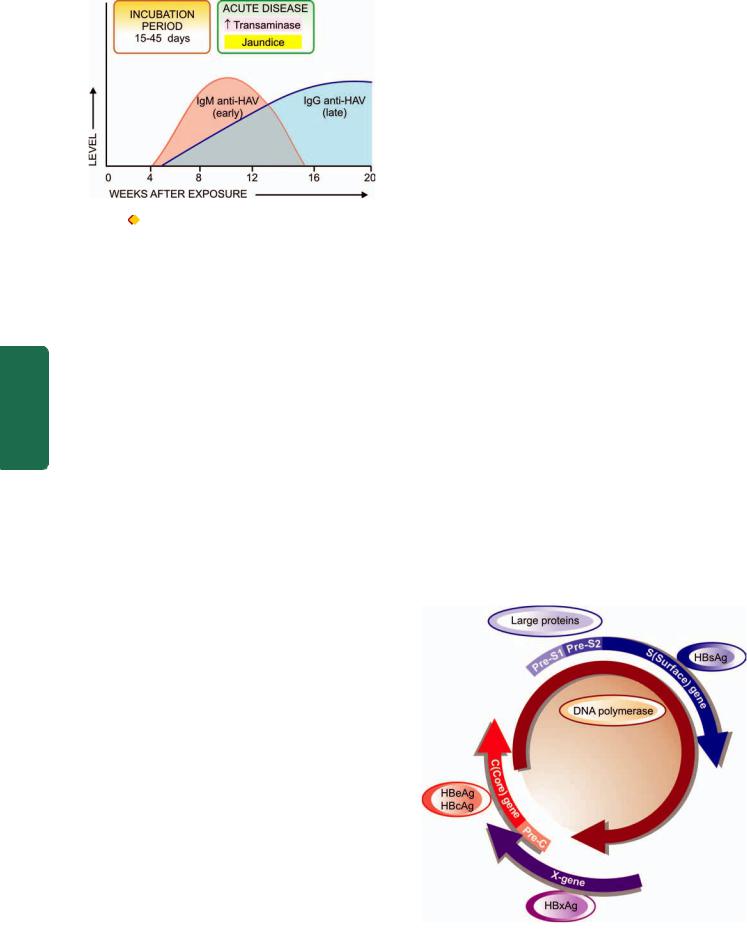
606
|
Figure 21.6 |
Sequence of appearance of antibodies to HAV. |
|
|
Hepatitis A |
|
|
|
Infection with HAV causes hepatitis A (infectious hepatitis). |
||
|
Hepatitis A is responsible for 20-25% of clinical hepatitis in |
||
|
the developing countries of the world but the incidence is |
||
|
much lower in the developed countries. Hepatitis A is |
||
SECTION |
usually a benign, self-limiting disease and has an incubation |
||
period of 15-45 days. The disease occurs in epidemic form |
|||
as well as sporadically. It is usually spread by faeco-oral |
|||
route. Parenteral transmission is extremely rare. The spread |
|||
is related to close personal contact such as in overcrowding, |
|||
poor hygiene and poor sanitation. Most frequently affected |
|||
III |
age is 5-14 years; adults are often infected by spread from |
||
children. |
|
||
|
|
||
Systemic |
HEPATITIS A VIRUS (HAV). The etiologic agent for |
||
hepatitis A, HAV, is a small, 27 nm diameter, icosahedral |
|||
|
|||
|
non-enveloped, single-stranded RNA virus. Viral genome |
||
|
has been characterised but only a single serotype has been |
||
Pathology |
identified. HAV infection can be transmitted to primates and |
||
the virus can be cultivated in vitro. Inactivation of viral |
|||
activity can be achieved by boiling for 1 minute, by ultraviolet radiation, or by contact with formaldehyde and chlorine. The virus is present in the liver cells, bile, stool and blood during the incubation period and in pre-icteric phase but viral shedding diminishes after the onset of jaundice. Chronic carriers have not been identified for HAV infection.
PATHOGENESIS. The mechanism by which HAV infection causes hepatitis A is poorly understood. An immunologic basis is suspected but the evidence in support is indirect in the form of immunologic markers but not direct demonstration of the etiologic agent in the affected hepatocytes. These markers are as follows (Fig. 21.6):
1.IgM anti-HAV antibody appears in the serum at the onset of symptoms of acute hepatitis A.
2.IgG anti-HAV antibody is detected in the serum after acute illness and remains detectable indefinitely. It gives life-long protective immunity against reinfection with HAV.
Hepatitis B
Hepatitis B (serum hepatitis) caused by HBV infection has a longer incubation period (30-180 days) and is transmitted parenterally such as in recipients of blood and blood
products, intravenous drug addicts, patients treated by renal dialysis and hospital workers exposed to blood, and by intimate physical contact such as from mother to child and by sexual contact. The disease may occur at any age. HBV infection causes more severe form of illness that includes: acute hepatitis B, chronic hepatitis, progression to cirrhosis, fulminant hepatitis and an asymptomatic carrier stage. HBV plays some role in the development of hepatocellular carcinoma as discussed later (page 634).
HEPATITIS B VIRUS (HBV). The etiologic agent for hepatitis B, HBV, is a DNA virus which has been extensively studied. Electron microscopic studies on serum of patients infected with HBV show 3 forms of viral particles of 2 sizes: small (spheres and tubules/filaments) and large (spheres) as under:
i)Small particles are most numerous and exist in two forms— as 22 nm spheres, and as tubules 22 nm in diameter and
100nm long. These are antigenically identical to envelope protein of HBV and represent excess of viral envelope protein referred as hepatitis B surface antigen (HBsAg).
ii)Large particles, 42 nm in diameter, are double-shelled spherical particles, also called as Dane particles. These are about 100 to 1000 times less in number in serum compared to small 22 nm particles and represent intact virion of HBV.
The genomic structure of HBV is quite compact and complex. The HBV DNA consists of 4 overlapping genes which encode for multiple proteins (Fig 21.7):
1.S gene codes for the surface envelope protein, hepatitis B surface antigen (HBsAg); this protein product is termed major protein. HBsAg is present on the outer surface of the large spherical particles as well as in small spherical and tubular structures. Pre-S1 and pre-S2 regions of genome are upstream of S gene and code for pre-S gene protein products that includes receptor on the HBV surface and for hepatocyte membrane proteins. The protein product of S-gene plus
Figure 21.7 
 Genomic structure of hepatitis B virus (Dane particle).
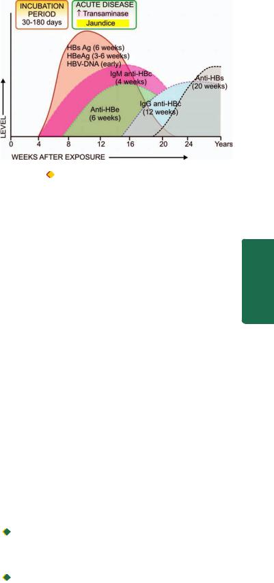
adjacent pre-S2 region is the middle protein, while the protein products of pre-S1 plus pre-S2 regions is the large protein. Large protein coming from both pre-S proteins is rich in complete virions.
2.P gene is the largest and codes for DNA polymerase.
3.C gene codes for two nucleocapsid proteins, HBeAg and a core protein termed HBcAg.
4.X gene codes for HBxAg which is a small non-particulate protein. HbxAg has a role in transactivation the transcription of both viral and cellular genes. The processes transactivated by X-genes include signal-transduction pathways, increased replication of HBV DNA, replication of other viruses including HIV, enhanced susceptibility of HBV-infected hepatocytes to cytolytic T cells, and pro-apoptotic pathway. Expression of HbxAg and its antibodies associated with enhanced HBV DNA replication has been implicated in hepatocelluar carcinoma in patients of chronic hepatitis.
PATHOGENESIS. The evidence linking immunopathogenetic mechanism with hepatocellular damage is much stronger in HBV infection than with HAV infection. In support of immune pathogenesis is the demonstration of several immunological markers (serologic as well as viral), and molecular and morphologic evidence that hepatocytic damage is initiated by virus-infected CD8+T cytotoxic cells.
Serologic and viral markers. Various immunological markers indicative of presence of HBV infection can be demonstrated in the sera as well as in the hepatocytes of infected individuals. These are as under (Fig. 21.8):
1.HBsAg. In 1965, Blumberg and colleagues in Philadelphia found a lipoprotein complex in the serum of a multipletransfused haemophiliac of Australian aborigine which was subsequently shown by them to be associated with serum hepatitis. This antigen was termed Australia antigen by them (In 1977, Blumberg was awarded the Nobel prize for his discovery). The term Australia antigen is now used synonymous with hepatitis B surface antigen (HBsAg). HBsAg appears early in the blood after about 6 weeks of infection and its detection is an indicator of active HBV infection. It usually disappears in 3-6 months. Its persistence for more than 6 months implies a carrier state. HBsAg may also be demonstrated in the cell membrane of hepatocytes of carriers and chronic hepatitis patients by Orcein staining (orange positivity) but not in the hepatocytes during acute stage of illness.
2.Anti-HBs. Specific antibody to HBsAg in serum called anti-HBs appears late, about 3 months after the onset. AntiHBs response may be both IgM and IgG type. The prevalence rate of anti-HBs ranges from 10-15%. In these individuals it persists for life providing protection against reinfection with HBV.
3.HBeAg. HBeAg derived from core protein is present transiently (3-6 weeks) during an acute attack. Its persistence beyond 10 weeks is indicative of development of chronic liver disease and carrier state.
4.Anti-HBe. Antibody to HBeAg called anti-HBe appears after disappearance of HBeAg. Seroconversion from HBeAg
607
Figure 21.8 |
Sequence of serologic and viral markers in acute |
|
|
hepatitis B. |
|
|
|
to anti-HBe during acute stage of illness is a prognostic sign |
|
||
for resolution of infection. |
|
||
5. HBcAg. HBcAg derived from core protein cannot be |
|
||
detected in the blood. But HBcAg can be demonstrated in |
CHAPTER |
||
the nuclei of hepatocytes in carrier state and in chronic |
|||
|
|||
hepatitis patients by Orcein staining but not in the liver cells |
|
||
during acute stage. |
|
||
6. Anti-HBc. Antibody to HBcAg called anti-HBc can, |
|
||
however, be detected in the serum of acute hepatitis B |
21 |
||
patients during pre-icteric stage. Anti-HBc may be IgM or |
|||
IgG class antibody. IgM anti-HBc persists for 4-6 months and |
|
||
is followed later by IgG anti-HBc. Thus detection of high titre |
The |
||
of IgM anti-HBc is indicative of recent acute HBV infection, |
|||
|
|||
while elevated level of IgG anti-HBc suggests HBV infection |
Liver, |
||
7. HBV-DNA. Detection of HBV-DNA by molecular |
|||
in the remote past. |
|
||
hybridisation using the Southern blot technique is the most |
Biliary |
||
symptomatic phase and transiently during early acute stage. |
|||
sensitive index of hepatitis B infection. It is present in pre- |
|
||
Hepatitis D |
|
Tract |
|
|
|
||
Infection with delta virus (HDV) in the hepatocyte nuclei of |
and |
||
HBsAg-positive patients is termed hepatitis D. HDV is a |
|||
|
|||
defective virus for which HBV is the helper. Thus, hepatitis |
Exocrine |
||
D develops when there is concomitant hepatitis B infection. |
|||
|
|||
HDV infection and hepatitis B may be simultaneous (co- |
|
||
infection), or HDV may infect a chronic HBsAg carrier |
|
||
(superinfection) (Fig. 21.9): |
Pancreas |
||
With coinfection, acute hepatitis D may range from mild |
|||
|
|||
to fulminant hepatitis but fulminant hepatitis is more likely |
|
||
in such simultaneous delta infection. Chronicity rarely |
|
||
develops in coinfection. |
|
||
With superinfection (incubation period 30-35 days), |
|
||
chronic HBV infection gets worsened indicated by |
|
||
appearance of severe and fulminant acute attacks, |
|
||
progression of carrier stage to chronic delta hepatitis or |
|
||
acceleration towards cirrhosis. Occurrence of hepatocellular |
|
||
carcinoma is, however, less common in HBsAg carriers with |
|
||
HDV infection. |
|
|
|
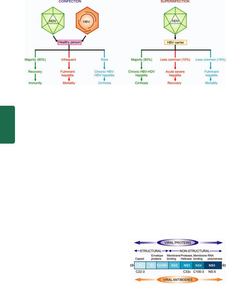
608
Figure 21.9 
 Consequences of coinfection versus superinfection in combined HDV-HBV infection.
Pathology Systemic III SECTION
HDV infection is worldwide in distribution though the incidence may vary in different countries. Endemic regions are Southern Europe, Middle-East, South India and parts of Africa. The high-risk individuals for HDV infection are the same as for HBV infection i.e. intravenous drug abusers, homosexuals, transfusion recipients, and health care workers.
HEPATITIS DELTA VIRUS (HDV). The etiologic agent, HDV, is a small single-stranded RNA particle with a diameter of 36 nm. It is double-shelled—the outer shell consists of HBsAg and the inner shell consists of delta antigen provided by a circular RNA strand. It is highly infectious and can induce hepatitis in any HBsAg-positive host. HDV replication and proliferation takes place within the nuclei of liver cells. Markers for HDV infection include the following:
1.HDV identification in the blood and in the liver cell nuclei.
2.HDAg detectable in the blood and on fixed liver tissue specimens.
3.Anti-HD antibody in acute hepatitis which is initially IgM type and later replaced by IgG type anti-HD antibody which persists for life to confer immunity against reinfection.
PATHOGENESIS. HDV, unlike HBV, is thought to cause direct cytopathic effect on hepatocytes. However, there are examples of transmission of HDV infection from individuals who themselves have not suffered from any attack of hepatitis, suggesting that it may not be always cytopathic. Thus, the exact mechanism remains unresolved.
Hepatitis C
The diagnosis of a third major category of hepatitis was earlier made after exclusion of infection with other known hepatitis viruses and was initially designated non-A, non-B (NANB) hepatitis. However, now this third type has been characterised and is called hepatitis C.
Hepatitis C infection is acquired by blood transfusions, blood products, haemodialysis, parenteral drug abuse and
accidental cuts and needle-pricks in health workers. About 90% of post-transfusion hepatitis is of hepatitis C type. About 1-2% of volunteer blood donors and up to 5% of professional blood donors are carriers of HCV. Hepatitis C has an incubation period of 20-90 days (mean 50 days). Clinically, acute HCV hepatitis is milder than HBV hepatitis but HCV has a higher rate of progression to chronic hepatitis than HBV. Persistence of infection and chronic hepatitis are the key features of HCV. Occurrence of cirrhosis after 5 to 10 years and progression to hepatocellular carcinoma are other late consequences of HCV infection. Currently, HCV is considered more important cause of chronic liver disease worldwide than HBV.
HEPATITIS C VIRUS (HCV). HCV is a single-stranded, enveloped RNA virus, having a diameter of 30-60 nm. HCV genome has about 3000 amino acids. The genomic organisation of HCV shows a 5’ terminal end, C (capsid) region and the envelope regions E1 and E2 in the exons
(Fig. 21.10).
The viral proteins result in corresponding serologic and virologic markers for HCV infection as under (Fig. 21.11):
1. Anti-HCV antibodies. Three generations of anti-HCV IgG assays are available:
i)First generation antibodies are against C100-3 region proteins and appear 1 to 3 months after infection.
Figure 21.10 
 Diagrammatic structure of hepatitis C virus.
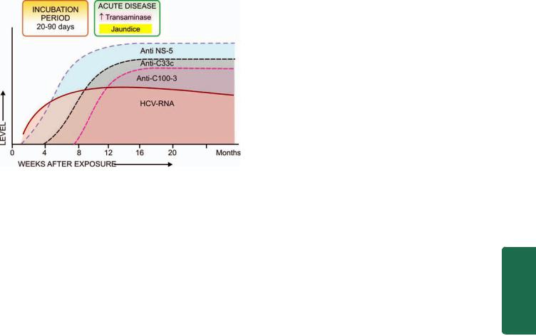
Figure 21.11 
 Sequence of serologic and viral markers of HCV infection.
ii)Second generation antibodies are against C200 and C33c proteins and appear about one month earlier than the first generation.
iii)Third generation antibodies are against C22-3 and NS-5 region proteins and are detected even earlier.
2. HCV-RNA. HCV infection is, however, confirmed by HCV-RNA employing PCR technique which can be detected within a few days after exposure to HCV infection, much before appearance of anti-HCV and persists for the duration of HCV infection.
PATHOGENESIS. Cell-mediated immune mechanism and production of antiviral cytokines by T-lymphocytes certainly play a role in hepatocytic injury due to HCV. HCV virions have not been identified in hepatocytes. Perhaps, HCV infection of lymphoid cells may induce immunologic injury to hepatocytes. In patients with chronic HCV hepatitis, HCVspecific CD4+ T cells and HLA-restricted CD8+ T cells have been identified. Crossreactivity between viral antigens and host autoantibodies to liver-kidney microsomal antigen (antiLKM) have been reported in a subset of patients that explains the association of autoimmune hepatitis and HCV hepatitis.
Hepatitis E
Hepatitis E is an enterically-transmitted virus, previously labelled as epidemic or enterically transmitted variant of non- A non-B hepatitis. The infection occurs in young or middleaged individuals, primarily seen in India, other Asian countries, Africa and central America.
The infection is generally acquired by contamination of water supplies such as after monsoon flooding. However, compared with HAV, secondary person-to-person infection does not occur with HEV. Thus HEV has some common epidemiologic features with HAV. HEV infection has a particularly high mortality in pregnant women but is otherwise a self-limited disease and has not been associated with chronic liver disease.
HEPATITIS E VIRUS (HEV). HEV is a single-stranded 3234 nm, icosahedral non-enveloped virus. The virus has been
isolated from stools, bile and liver of infected persons. 609 Serologic markers for HEV include the following:
1.Anti-HEV antibodies of both IgM and IgG class.
2.HEV-RNA.
However, testing for these markers for HEV is currently
not available.
Hepatitis G
A virus distinct from the foregoing hepatitis viruses has been designated separately as hepatitis G (HGV). HGV infection has been found in blood donors, patients on haemodialysis and as coinfection with HIV. However, unlike HCV, HGV is cleared from the plasma in majority of individuals while a small percentage of cases have chronic HGV infection who do not develop hepatitis, nor does infected blood require screening for HGV RNA.
HEPATITIS G VIRUS (HGV). HGV is a single-stranded RNA virus. The virus has been identified by PCR amplification technique.
CLINICOPATHOLOGIC SPECTRUM |
CHAPTER |
||
Among the various etiologic types of hepatitis, evidence |
|||
|
|||
linking HBV and HCV infection with the spectrum of clinico- |
|
||
pathologic changes is stronger than with other hepatotropic |
|
||
viruses. The typical pathologic changes of hepatitis by major |
|
||
hepatotropic viruses are virtually similar. HAV and HEV, |
21 |
||
however, do not have a carrier stage nor cause chronic |
|||
hepatitis. The various clinical patterns and pathologic |
|
||
consequences of different hepatotropic viruses can be |
The |
||
considered under the following headings: |
|||
|
|||
i) |
Carrier state |
Liver, |
|
iii) Acute hepatitis |
|||
ii) |
Asymptomatic infection |
|
|
iv) Chronic hepatitis |
Biliary |
||
v) Fulminant hepatitis (Submassive to massive necrosis) |
|||
|
|||
|
In addition, progression to cirrhosis (page 624) and |
|
|
association with hepatocellular carcinoma (page 634) are |
Tract |
||
known to occur in certain types of hepatitis which are |
|||
|
|||
discussed separately later. |
and |
||
I. |
Carrier State |
||
carriers: |
Exocrine |
||
An asymptomatic individual without manifest disease, |
|
||
harbouring infection with hepatotropic virus and capable of |
|
||
transmitting it is called carrier state. There can be 2 types of |
|
||
1. An ‘asymptomatic healthy carrier’ who does not suffer from |
Pancreas |
||
ill-effects of the virus infection but is capable of transmitting. |
|||
2. An ‘asymptomatic carrier with chronic disease’ capable of |
|||
transmitting the organisms. |
|||
As stated before, hepatitis A and E do not produce the carrier state. Hepatitis B is responsible for the largest number of carriers in the world, while concomitant infection with HDV more often causes progressive disease rather than an asymptomatic carrier state. There is geographic variation in incidence of HBV carrier state: while in normal population in US and western Europe it is less than 0.5%, its prevalence
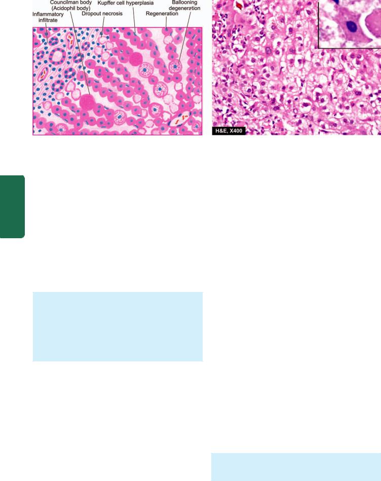
610
Figure 21.12 
 Acute viral hepatitis. The predominant histologic changes are: variable degree of necrosis of hepatocytes, most marked in zone
3 (centrilobular); and mononuclear cellular infiltrate in the lobule. Mild degree of liver cell necrosis is seen as ballooning degeneration while acidophilic Councilman bodies (inbox) are indicative of more severe liver cell injury.
Pathology Systemic III SECTION
is much higher in Asian and tropical countries (5-20%). An estimated 2-3% of the general population are asymptomatic carriers of HCV. Data on HBV carrier state reveal role of 2 important factors rendering the individual more vulnerable to harbour the organisms—early age at infection and impaired immunity. Whereas approximately 10% of adults contracting hepatitis B infection develop carrier state, 90% of infected neonates fail to clear HBsAg from the serum within 6 months and become HBV carriers.
Clinical recognition of carrier state of HBV is more frequently done by detection of HBsAg in the serum and less often by other markers such as HBeAg, HBcAg and antibodies. Concomitant infection of HDV with HBV depends upon the demonstration of anti-HD.
MORPHOLOGIC FEATURES. Carriers of HBV may or may not show changes on liver biopsy.
 Healthy HBV carriers may show no changes or minor hepatic change such as presence of finely granular, ground-glass, eosinophilic cytoplasm as evidence of HBsAg.
 Asymptomatic carriers with chronic disease may show changes of chronic hepatitis and even cirrhosis.
II. Asymptomatic Infection
These are cases who are detected incidentally to have infection with one of the hepatitis viruses as revealed by their raised serum transaminases or by detection of the presence of antibodies but are otherwise asymptomatic.
III. Acute Hepatitis
The most common consequence of all hepatotropic viruses is acute inflammatory involvement of the entire liver. In general, type A, B, C, D and E run similar clinical course and show identical pathologic findings.
Clinically, acute hepatitis is categorised into 4 phases: incubation period, pre-icteric phase, icteric phase and posticteric phase.
1.Incubation period: It varies among different hepatotropic viruses: for hepatitis A it is about 4 weeks (15-45 days); for hepatitis B the average is 10 weeks (30-180 days); for hepatitis D about 6 weeks (30-50 days); for hepatitis C the mean incubation period is about 7 weeks (20-90 days), and for hepatitis E it is 2-8 weeks (15-60 days). The patient remains asymptomatic during incubation period but the infectivity is highest during the last days of incubation period.
2.Pre-icteric phase: This phase is marked by prodromal constitutional symptoms that include anorexia, nausea, vomiting, fatigue, malaise, distaste for smoking, arthralgia and headache. There may be low-grade fever preceding the onset of jaundice, especially in hepatitis A. The earliest laboratory evidence of hepatocellular injury in pre-icteric phase is the elevation of transaminases.
3.Icteric phase: The prodromal period is heralded by the onset of clinical jaundice and the constitutional symptoms diminish. Other features include dark-coloured urine due to bilirubinuria, clay-coloured stools due to cholestasis, pruritus as a result of elevated serum bile acids, loss of weight and abdominal discomfort due to enlarged, tender liver. The diagnosis is based on deranged liver function tests (e.g. elevated levels of serum bilirubin, transaminases and alkaline phosphatase; prolonged prothrombin time and hyperglobulinaemia) and serologic detection of hepatitis antigens and antibodies.
4.Post-icteric phase: The icteric phase lasting for about 1 to 4 weeks is usually followed by clinical and biochemical recovery in 2 to 12 weeks. The recovery phase is more prolonged in hepatitis B and hepatitis C. Up to 1% cases of acute hepatitis may develop severe form of the disease (fulminant hepatitis); and 5-10% of cases progress on to chronic hepatitis. Evolution into the carrier state (except in HAV and HEV infection) has already been described above.
MORPHOLOGIC FEATURES. Grossly, the liver is slightly enlarged, soft and greenish.
Histologically, the changes are as follows (Fig. 21.12):

1. Hepatocellular injury: There may be variation in the degree of liver cell injury but it is most marked in zone 3 (centrilobular zone):
i) Mildly injured hepatocytes appear swollen with granular cytoplasm which tends to condense around the
nucleus (ballooning degeneration).
ii)Others show acidophilic degeneration in which the cytoplasm becomes intensely eosinophilic, the nucleus becomes small and pyknotic and is eventually extruded from the cell, leaving behind necrotic, acidophilic mass called Councilman body or acidophil body by the process known as apoptosis.
iii)Another type of hepatocellular necrosis is dropout necrosis in which isolated or small clusters of hepatocytes undergo lysis.
iv)Bridging necrosis is a more severe form of hepatocellular injury in acute viral hepatitis and may progress to fulminant hepatitis or chronic hepatitis (discussed below). Bridging necrosis is characterised by bands of necrosis linking portal tracts to central hepatic veins, one central hepatic vein to another, or a portal tract to another tract.
2.Inflammatory infiltrate: There is infiltration by mononuclear inflammatory cells, usually in the portal tracts, but may permeate into the lobules.
3.Kupffer cell hyperplasia: There is reactive hyperplasia of Kupffer cells many of which contain phagocytosed cellular debris, bile pigment and lipofuscin granules.
4.Cholestasis: Biliary stasis is usually not severe in viral hepatitis and may be present as intracytoplasmic bile pigment granules.
5.Regeneration: As a result of necrosis of hepatocytes, there is lobular disarray. Surviving adjacent hepatocytes undergo regeneration and hyperplasia. If the necrosis causes collapse of reticulin framework of the lobule, healing by fibrosis follows, distorting the lobular architecture.
The above histologic changes apply to viral hepatitis by various types of hepatotropic viruses in general, and by HBV in particular. It is usually not possible to distinguish histologically between viral hepatitis of various etiologies, but the following morphologic features may help in giving an etiologic clue:

 HAV hepatitis is a panlobular involvement by heavy inflammatory infiltrate compared to other types.

 HCV hepatitis causes milder necrosis, with fatty change in hepatocytes, presence of lymphoid aggregates in the portal triads and degeneration of bile duct epithelium.
IV. Chronic Hepatitis
Chronic hepatitis is defined as continuing or relapsing hepatic disease for more than 6 months with symptoms along with biochemical, serologic and histopathologic evidence of inflammation and necrosis. Majority of cases of chronic hepatitis are the result of infection with hepatotropic viruses—hepatitis B, hepatitis C and combined hepatitis B
and hepatitis D infection. However, some non-viral causes of chronic hepatitis include: Wilson’s disease, α-1-antitrypsin deficiency, chronic alcoholism, drug-induced injury and autoimmune diseases. The last named gives rise to autoimmune or lupoid hepatitis which is characterised by positive serum autoantibodies (e.g. antinuclear, anti-smooth muscle and anti-mitochondrial) and a positive LE cell test but negative for serologic markers of viral hepatitis.
Until recent years, prediction of prognosis of chronic hepatitis used to be made on the basis of morphology which divided it into 2 main types—chronic persistent and chronic
active (aggressive) hepatitis. A third form, chronic lobular hepatitis is distinguished separately by some as mild form of lobular inflammation without inflammation of portal tracts but these cases often recover completely. However, subsequent studies have revealed that morphologic subtypes do not necessarily correlate with prognosis since the disease is not essentially static but may vary from mild form to severe and vice versa. Besides, two other factors which determine the vulnerability of a patient of viral hepatitis to develop chronic hepatitis are: impaired immunity and extremes of age at which the infection is first contracted. Currently, therefore, chronic hepatitis is classified on the basis of etiology and hepatitis activity score (described below). The frequency and severity with which hepatotropic viruses cause chronic hepatitis varies with the organisms as under:
 HCV infection accounts for 40-60% cases of chronicity in adults. HCV infection is particularly associated with progressive form of chronic hepatitis that may evolve into cirrhosis.

 HBV causes chronic hepatitis in 90% of infected infants and in about 5% adult cases of hepatitis B.
 HDV superinfection on HBV carrier state may be responsible for chronic hepatitis in 10-40% cases.

 HAV and HEV do not produce chronic hepatitis.
MORPHOLOGIC FEATURES. The pathologic features are common to both HBV and HCV infection and include the following lesions (Fig. 21.13).
1. Piecemeal necrosis. Piecemeal necrosis is defined as periportal destruction of hepatocytes at the limiting plate (piecemeal = piece by piece). Its features in chronic hepatitis are as under:
i)Necrosed hepatocytes at the limiting plate in periportal zone.
ii)Interface hepatitis due to expanded portal tract by infiltration of lymphocytes, plasma cells and macrophages.
iii)Expanded portal tracts are often associated with proliferating bile ductules as a response to liver cell injury.
2. Portal tract lesions. All forms of chronic hepatitis are characterised by variable degree of changes in the portal tract.
i)Inflammatory cell infiltration by lymphocytes, plasma cells and macrophages (triaditis).
ii)Proliferated bile ductules in the expanded portal tracts.
iii)Additionally, chronic hepatitis C may show lymphoid aggregates or follicles with reactive germinal centre and
611
Pancreas Exocrine and Tract Biliary Liver, The 21 CHAPTER
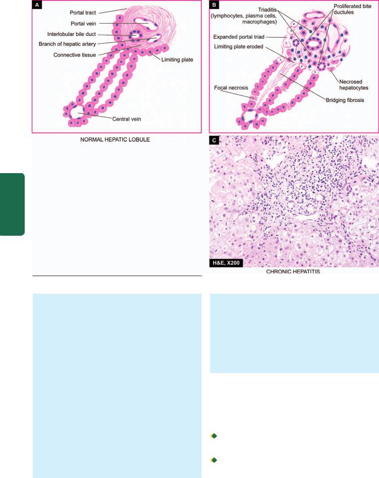
612
Pathology Systemic III SECTION
Figure 21.13 
 Chronic hepatitis. Diagrammatic representation of pathologic changes in chronic hepatitis (B) contrasted with normal morphology (A). Photomicrograph on right (C) shows stellate-shaped portal triad, with extension of fibrous spurs into lobules. The portal tract is expanded due to increased lymphomononuclear inflammatory cells which are seen to breach the limiting plate (i.e. hepatocytes at the interface of portal tract and lobule are destroyed).
infiltration of inflammatory cells in the damaged bile duct epithelial cells.
3. Intralobular lesions. Generally, the architecture of lobule is retained in mild to moderate chronic hepatitis.
i)There are focal areas of necrosis and inflammation within the hepatic parenchyma.
ii)Scattered acidophilic bodies in the lobule.
iii)Kupffer cell hyperplasia.
iv)More severe form of injury shows bridging necrosis (i.e. bands of necrosed hepatocytes that may bridge portal tract-to-central vein, central vein-to-central vein, and portal tract-to-portal tract).
v)Regenerative changes in hepatocytes in cases of persistent hepatocellular necrosis.
vi)Cases of chronic hepatitis C show moderate fatty change.
vii)Cases of chronic hepatitis B show scattered groundglass hepatocytes indicative of abundance of HBsAg in the cytoplasm.
4. Bridging fibrosis. The onset of fibrosis in chronic hepatitis from the area of interface hepatitis and bridging necrosis is a feature of irreversible damage.
i)At first, there is periportal fibrosis at the sites of interface hepatitis giving the portal tract stellate-shaped appearance.
ii)Progressive cases show bridging fibrosis connecting portal tract-to-portal tract or portal tract-to-central vein traversing the lobule.
iii)End-stage of chronic hepatitis is characterised by dense collagenous septa destroying lobular architecture and forming nodules resulting in postnecrotic cirrhosis.
As prognostic indicator of chronic hepatitis, criteria have been evolved to classify chronic hepatitis by giving hepatitis activity score (ranging from none to minimal/mild to moderate and severe) described by Knodell and Ishak based on the following features:
A. Necroinflammatory activity:
 Periportal necrosis i.e. piecemeal necrosis and/ or bridging necrosis (ranging from score 0 as ‘no necrosis’ to score 4 as ‘multilobular necrosis’).

 Intralobular necrosis, focal or confluent (ranging from score 0 as ‘none’ to score 4 for ‘>10 foci’ for focal necrosis, and score 6 as ‘panacinar/multiacinar’ for confluent necrosis).
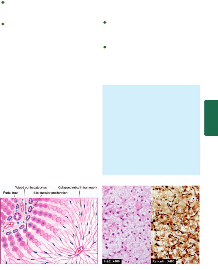

 Extent and depth of portal inflammation (ranging from grade 0 as ‘no inflammation’ to grade 4 having ‘marked portal inflammation’).
B. Stage of fibrosis:

 Extent and density of fibrosis (ranging from score 0 as ‘no fibrosis’ to score 6 as ‘cirrhosis’).
CLINICAL FEATURES. The clinical features of chronic hepatitis are quite variable ranging from mild disease to fullblown picture of cirrhosis.
i)Mild chronic hepatitis shows only slight but persistent elevation of transaminases (‘transaminitis’) with fatigue, malaise and loss of appetite.
ii)Other cases may show mild hepatomegaly, hepatic tenderness and mild splenomegaly.
iii)Laboratory findings may reveal prolonged prothrombin time, hyperbilirubinaemia, hyperglobulinaemia and markedly elevated alkaline phosphatase.
iv)Systemic features of circulating immune complexes due to HBV and HCV infection may produce features of immune complex vasculitis, glomerulonephritis and cryoglobulinaemia in a proportion of cases.
However, clinical features do not correlate with morphologic appearance of the liver biopsy. Some patients may have mild form of disease without progressing for several years while others may show rapid evolution into cirrhosis with its complications over a period of few years. Patients of longstanding HBV and HCV chronic infection are known to evolve into hepatocellular carcinoma.
V.Fulminant Hepatitis
(Submassive to Massive Necrosis)
Fulminant hepatitis is the most severe form of acute hepatitis in which there is rapidly progressive hepatocellular failure. Two patterns are recognised—submassive necrosis having a less rapid course extending up to 3 months; and massive
necrosis in which the liver failure is rapid and fulminant occurring in 2-3 weeks.
Fulminant hepatitis of either of the two varieties can occur from viral and non-viral etiologies:

 Acute viral hepatitis accounts for about half the cases, most often from HBV and HCV; less frequently from combined HBV-HDV and rarely from HAV. However, HEV infection is a serious complication in pregnant women. In addition, herpesvirus can also cause serious viral hepatitis.
 Non-viral causes include acute hepatitis due to drug toxicity (e.g. acetaminophen, non-steroidal antiinflammatory drugs, isoniazid, halothane and antidepressants), poisonings, hypoxic injury and massive infiltration of malignant tumours into the liver.
The patients present with features of hepatic failure with hepatic encephalopathy (page 602). The mortality rate is high if hepatic transplantation is not undertaken.
MORPHOLOGIC FEATURES. Grossly, the liver is small and shrunken, often weighing 500-700 gm. The capsule is loose and wrinkled. The sectioned surface shows diffuse or random involvement of hepatic lobes. There are extensive areas of muddy-red and yellow necrosis (previously called acute yellow atrophy) and patches of green bile staining.
Histologically, two forms of fulminant necrosis are distinguished—submassive and massive necrosis
(Fig. 21.14).
i)In submassive necrosis, large groups of hepatocytes in zone 3 (centrilobular area) and zone 2 (mid zone) are wiped out leading to a collapsed reticulin framework. Regeneration in submassive necrosis is more orderly and may result in restoration of normal architecture.
ii)In massive necrosis, the entire liver lobules are necrotic. As a result of loss of hepatic parenchyma, all that is left is the collapsed and condensed reticulin framework and portal tracts with proliferated bile ductules plugged
613
Pancreas Exocrine and Tract Biliary Liver, The 21 CHAPTER
Figure 21.14 
 Fulminant hepatitis. There is wiping out of liver lobules with only collapsed reticulin framework left out in their place, highlighted by reticulin stain (right photomicrograph). There is no significant inflammation or fibrosis.
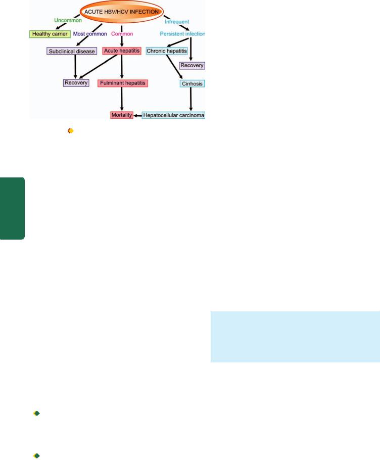
614
|
Figure 21.15 |
Clinicopathologic course of HBV and HCV infection. |
|
|
|
|
with bile. Inflammatory infiltrate is scanty. Regeneration, |
|
|
if it takes place, is disorderly forming irregular masses of |
|
|
hepatocytes. Fibrosis is generally not a feature of |
|
|
fulminant hepatitis. |
|
III SECTION |
The clinicopathologic course in two major forms of |
|
hepatitis, HBV and HCV, is summarised in Fig. 21.15. |
||
IMMUNOPROPHYLAXIS AND HEPATITIS VACCINES |
||
Best prophylaxis against the viral hepatitis remains |
||
|
prevention of its spread to the contacts after detection and |
|
Systemic |
identification of route by which infection is acquired such as |
|
are under development. The principle underlying either of |
||
|
from food or water contamination, sexual spread or |
|
|
parenteral spread. Of late, however, immunoprophylaxis and |
|
|
a few hepatitis vaccines have been developed and some more |
|
Pathology |
these two forms of prophylaxis is that the persons who |
|
persons may be carried out so as to determine their antibody |
||
|
develop good antibody response to the antigen of the |
|
|
hepatotropic virus following active infection are protected |
|
|
against the disease on reinfection. Thus, pre-testing of |
|
|
level. Immunoprophylaxis and hepatitis vaccination are |
|
|
unnecessary if the pre-testing for antibodies is positive. |
|
|
1. Hepatitis A. Passive immunisation with immune |
|
|
globulin as well as active immunisation with a killed vaccine |
|
|
are available. |
|
|
2. Hepatitis B. Earlier, only passive immunoprophylaxis |
|
|
with standard immune globulin was used. Later, active |
|
|
immunisation against HBsAg was introduced. Current |
|
|
recommendations include pre-exposure and post-exposure |
|
|
prophylaxis with recombinant hepatitis B vaccine: |
|
|
Pre-exposure prophylaxis is done for individuals at high- |
|
|
risk e.g. health care workers, haemodialysis patients and |
|
|
staff, haemophiliacs, intravenous drug users etc. Three |
|
|
intramuscular injections of hepatitis vaccine at 0, 1 and 6 |
|
|
months are recommended. |
|
|
Post-exposure prophylaxis is carried out for unvaccinated |
|
|
persons exposed to HBV infection and includes prophylaxis |
|
|
with combination of hepatitis B immune globulin and |
|
|
hepatitis B vaccine. |
|
3.Hepatitis D. Hepatitis D infection can also be prevented by hepatitis B vaccine.
4.Hepatitis C. Currently, hepatitis C vaccine has yet not been feasible though antibodies to HCV envelope have been developed.
5.Hepatitis E. It is not certain whether immune globulin (like for HAV) prevents hepatitis E infection or not but a vaccine against HEV is yet to be developed.
OTHER INFECTIONS AND INFESTATIONS
Apart from viral hepatitis, the liver is affected by infections with bacteria, spirochaetes and fungi and is involved in some parasitic infestations. Some common examples of such conditions are described below.
CHOLANGITIS
Cholangitis is the term used to describe inflammation of the extrahepatic or intrahepatic bile ducts, or both. There are two main types of cholangitis—pyogenic and primary sclerosing. While primary sclerosing cholangitis is discussed later with biliary cirrhosis (page 625), pyogenic cholangitis is described below.
Pyogenic Cholangitis
Cholangitis occurring secondary to obstruction of a major extrahepatic duct causes pyogenic cholangitis. Most commonly, the obstruction is from impacted gallstone; other causes are carcinoma arising in the extrahepatic ducts, carcinoma head of pancreas, acute pancreatitis and inflammatory strictures in the bile duct. Bacteria gain entry to the obstructed duct and proliferate in the bile. Infection spreads along the branches of obstructed duct and reaches the liver, termed ascending cholangitis. The common infecting bacteria are enteric organisms such as E.coli, Klebsiella and
Enterobacter.
MORPHOLOGIC FEATURES. The affected ducts show small beaded abscesses accompanied by bile stasis along their course and larger abscesses within the liver. The abscesses are composed of acute inflammatory cells which in time are replaced by chronic inflammatory cells and enclosed by fibrous capsule.
PYOGENIC LIVER ABSCESS
Most liver abscesses are of bacterial (pyogenic) origin; less often they are amoebic, hydatid and rarely actinomycotic. Pyogenic liver abscesses have become uncommon due to improved diagnostic facilities and the early use of antibiotics. However, their incidence is higher in old age and in immunosuppressed patients such as in AIDS, transplant recipients and those on intensive chemotherapy.
Pyogenic liver abscesses are classified on the basis of the mode of entry as under:
1. Ascending cholangitis through ascending infection in the biliary tract due to obstruction e.g. gallstones, cancer, sclerosing cholangitis and biliary strictures.
Figure 21.16 
 Gross appearance of pyogenic abscesses in the liver.
2.Portal pyaemia by means of spread of pelvic or gastrointestinal infection resulting in portal pylephlebitis or septic emboli e.g. from appendicitis, empyema of gallbladder, diverticulitis, regional enteritis, pancreatitis, infected haemorrhoids and neonatal umbilical vein sepsis.
3.Septicaemia through spread by hepatic artery.
4.Direct infection resulting in solitary liver abscess e.g. from adjacent perinephric abscess, secondary infection in amoebic liver abscess, metastasis and formation of haematoma following trauma.
5.Iatrogenic causes include liver biopsy, percutaneous biliary drainage and accidental surgical trauma.
6.Cryptogenic from unknown causes, especially in the elderly.
The commonest infecting organisms are gram-negative bacteria chiefly E. coli; others are Pseudomonas, Klebsiella, Enterobacter and a number of anaerobic organisms, bacteroides and actinomyces.
Liver abscesses are clinically characterised by pain in the right upper quadrant, fever, tender hepatomegaly and sometimes jaundice. Laboratory examination reveals leucocytosis, elevated serum alkaline phosphatase, hypoalbuminaemia and a positive blood culture.
MORPHOLOGIC FEATURES. Grossly depending upon the cause for pyogenic liver abscess, they occur as single or multiple yellow abscesses, 1 cm or more in diameter, in an enlarged liver. A single abscess generally has a thick fibrous capsule. The abscesses are particularly common in right lobe of the liver (Fig. 21.16).
Microscopically, typical features of abscess are seen. There are multiple small neutrophilic abscesses with areas of extensive necrosis of the affected liver parenchyma. The adjacent viable area shows pus and blood clots in the portal vein, inflammation, congestion and proliferating fibroblasts. Direct extension from the liver may lead to subphrenic or pleuro-pulmonary suppuration or peritonitis. There may be small pyaemic abscesses elsewhere such as in the lungs, kidneys, brain and spleen.
AMOEBIC LIVER ABSCESS
Amoebic liver abscesses are less common than pyogenic liver abscesses and have many similar features. They are caused
by the spread of Entamoeba histolytica from intestinal lesions. 615 The trophozoite form of amoebae in the colon invade the colonic mucosa forming flask-shaped ulcers from where they
are carried to the liver in the portal venous system (page 188) . Amoebae multiply and block small intrahepatic portal radicles resulting in infarction necrosis of the adjacent liver parenchyma.
|
The patients, generally from tropical and subtropical |
|
||
countries, may give history of amoebic dysentery in the past. |
|
|||
Cysts of E. histolytica in stools are present in only 15% of |
|
|||
patients of hepatic amoebiasis. Intermittent low-grade fever, |
|
|||
pain and tenderness in the liver area are common presenting |
|
|||
features. A positive haemagglutination test is quite sensitive |
|
|||
and useful for diagnosis of amoebic liver abscess. |
|
|
||
|
|
|
||
|
MORPHOLOGIC FEATURES. Grossly, amoebic liver |
|
||
|
abscesses are usually solitary and more often located in |
|
||
|
the right lobe in the posterosuperior portion. Amoebic |
|
||
|
liver abscess may vary greatly in size but is generally of |
|
||
|
the size of an orange. The centre of the abscess contains |
|
||
|
large necrotic area having reddish-brown, thick pus |
|
||
|
resembling anchovy or chocolate sauce. The abscess wall |
|
||
|
consists of irregular shreds of necrotic liver tissue |
(Fig. |
CHAPTER |
|
|
21.17). |
|
||
|
|
|
||
|
Histologically, the necrotic area consists of degenerated |
|
||
|
liver cells, leucocytes, red blood cells, strands of |
|
||
|
connective tissue and debris. Amoebae are most easily |
|
||
|
found in the liver tissue at the margin of abscess. PAS- |
21 |
||
|
staining is employed to confirm the trophozoites of E. |
|||
|
|
|||
|
histolytica. |
|
|
|
|
|
|
The |
|
HEPATIC TUBERCULOSIS |
|
|||
|
|
|||
Tuberculosis of the liver occurs as a result of miliary |
Liver, |
|||
dissemination from primary complex or from chronic adult |
||||
|
||||
pulmonary tuberculosis. The diagnosis is possible by liver |
Biliary |
|||
elevated serum alkaline phosphatase levels |
and |
|||
biopsy. The patients may have unexplained fever, jaundice, |
|
|||
hepatomegaly or hepatosplenomegaly. There may be |
|
|||
hyperglobulinaemia. |
|
Tract |
||
|
|
|
||
|
MORPHOLOGIC FEATURES. The basic lesion is the |
|||
|
Pancreas Exocrine and |
|||
|
epithelioid cell granuloma characterised by central |
|||
|
|
|
||
|
|
|
||
Figure 21.17 
 Amoebic liver abscess is commonly solitary and its wall is irregular and necrotic.
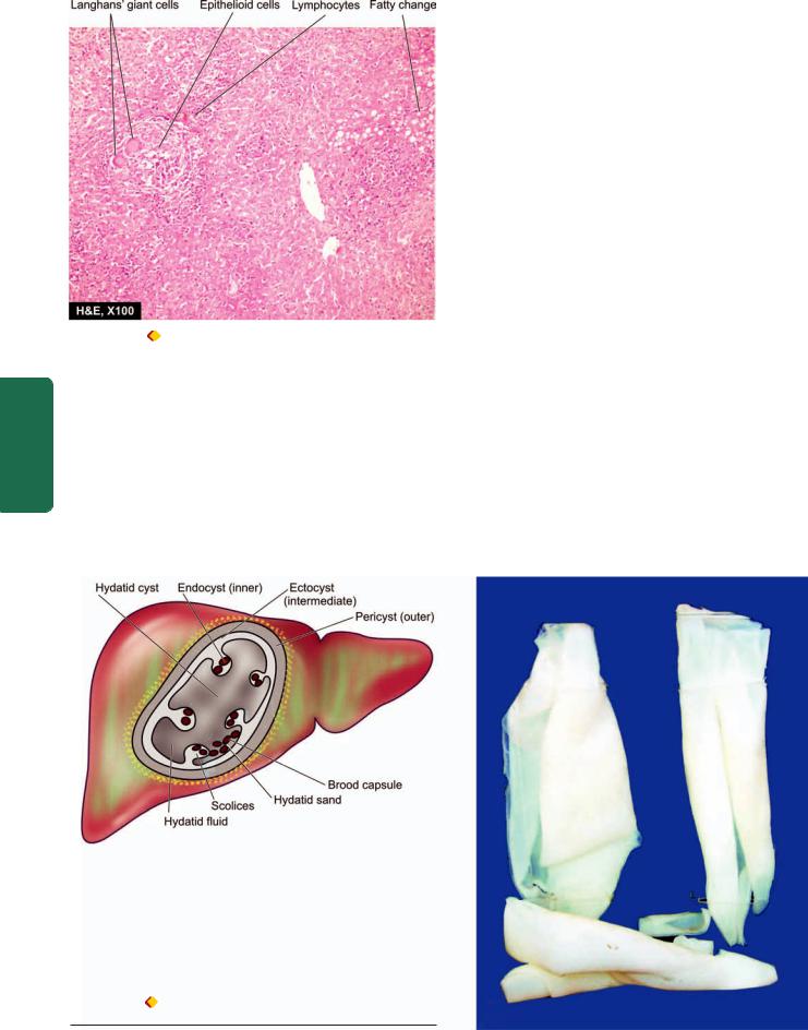
616
Figure 21.18 |
Miliary tuberculosis liver. The hepatic parenchyma |
shows epithelioid granulomas with small areas of central necrosis and |
|
surrounded peripherally by Langhans’ giant cells and lymphocytes. |
|
SECTION |
|
|
caseation necrosis with destruction of the reticulin |
||
|
||
|
framework and peripheral cuff of lymphocytes |
|
|
(Fig. 21.18). Ziehl-Neelsen staining for AFB or culture of |
|
III |
the organism from the biopsy tissue is confirmatory. Rare |
|
lesions consist of tuberculous cholangitis and tuberculous |
||
|
pylephlebitis. |
|
|
|
|
Pathology Systemic |
|
HYDATID DISEASE (ECHINOCOCCOSIS)
Hydatid disease occurs as a result of infection by the larval cyst stage of the tapeworm, Echinococcus granulosus. The dog is the common definite host, while man, sheep and cattle are the intermediate hosts. The dog is infected by eating the viscera of sheep containing hydatid cysts. The infected faeces of the dog contaminate grass and farmland from where the ova are ingested by sheep, pigs and man. Thus, man can acquire infection by handling dogs as well as by eating contaminated vegetables. The ova ingested by man are liberated from the chitinous wall by gastric juice and pass through the intestinal mucosa from where they are carried to the liver by portal venous system. These are trapped in the hepatic sinusoids where they eventually develop into hydatid cyst. About 70% of hydatid cysts develop in the liver which acts as the first filter for ova. However, ova which pass through the liver enter the right side of the heart and are caught in the pulmonary capillary bed and form pulmonary hydatid cysts. Some ova which enter the systemic circulation give rise to hydatid cysts in the brain, spleen, bone and muscles.
The disease is common in sheep-raising countries such as Australia, New Zealand and South America. The uncomplicated hydatid cyst of the liver may be silent or may produce dull ache in the liver area and some abdominal distension.
Complications of hydatid cyst include its rupture (e.g. into the peritoneal cavity, bile ducts and lungs), secondary infection and hydatid allergy due to sensitisation of the host with cyst fluid. The diagnosis is made by peripheral blood eosinophilia, radiologic examination and serologic tests such as indirect haemagglutination test and Casoni skin test.
Figure 21.19 |
Hydatid cyst in the liver. The cyst wall is composed of |
whitish membrane resembling the membrane of a hard boiled egg.
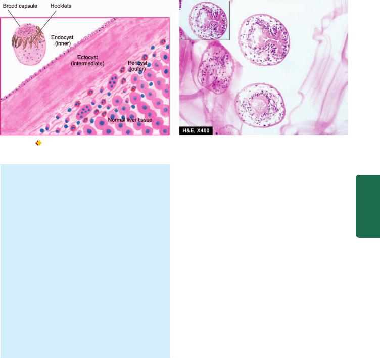
617
Figure 21.20 
 Microscopy shows three layers in the wall of hydatid cyst. Inbox in the right photomicrograph shows a scolex with a row of hooklets.
MORPHOLOGIC FEATURES. Hydatid cyst grows slowly and may eventually attain a size over 10 cm in diameter in about 5 years. E. granulosus generally causes unilocular hydatid cyst while E. multilocularis results in multilocular or alveolar hydatid disease in the liver.
The cyst wall is composed of 3 distinguishable zones— outer pericyst, intermediate characteristic ectocyst and inner endocyst (Fig. 21.19):
1.Pericyst is the outer host inflammatory reaction consisting of fibroblastic proliferation, mononuclear cells, eosinophils and giant cells, eventually developing into dense fibrous capsule which may even calcify.
2.Ectocyst is the intermediate layer composed of characteristic acellular, chitinous, laminated hyaline material (Fig. 21.20).
3.Endocyst is the inner germinal layer bearing daughter cysts (brood-capsules) and scolices projecting into the lumen.
Hydatid sand is the grain-like material composed of numerous scolices present in the hydatid fluid. Hydatid fluid, in addition, contains antigenic proteins so that its liberation into circulation gives rise to pronounced eosinophilia or may cause anaphylaxis.
CHEMICAL AND DRUG INJURY
HEPATIC DRUG METABOLISM. The liver plays a central role in the metabolism of a large number of organic and inorganic chemicals and drugs which gain access to the body by inhalation, injection, or most commonly, via the intestinal tract. The main drug metabolising system resides in the microsomal fraction of the smooth endoplasmic reticulum of the liver cells via P-450 cytochrome and cytochrome reductase enzyme systems. Other steps involved in the drug metabolism are its conjugation with an endogenous molecule, its active transport from the hepatocytes and ultimately its excretion in the bile or in urine depending upon the molecular weight of the substance. A number of risk factors
predispose an individual to hepatic drug injury such as preexisting liver disease, aging, female sex and genetic inability to perform a particular biotransformation.
HEPATOTOXICITY. Toxic liver injury produced by drugs and chemicals may virtually mimic any form of naturallyoccurring liver disease. In fact, any patient presenting with liver disease or unexplained jaundice is thoroughly questioned about history of drug intake or exposure to chemicals. Hepatotoxicity from drugs and chemicals is the commonest form of iatrogenic disease. Severity of hepatotoxicity is greatly increased if the drug is continued after symptoms develop.
Among the various inorganic compounds producing hepatotoxicity are arsenic, phosphorus, copper and iron. Organic agents include certain naturally-occurring plant toxins such as pyrrolizidine alkaloids, mycotoxins and bacterial toxins. The synthetic group of organic compounds are a large number of medicinal agents. In addition, exposure to hepatotoxic compounds may be occupational, environmental or domestic that could be accidental, homicidal or suicidal ingestion.
In general, drug reactions affecting the liver are divided into two main classes:
1.Direct or predictable, when the drug or one of its metabolites is either directly toxic to the liver or it lowers the host immune defense mechanism. The adverse effects occur in most individuals who consume them and their hepatotoxicity is dose-dependent e.g. carbon tetrachloride.
2.Indirect or unpredictable or idiosyncratic, when the drug or one of its metabolites acts as a hapten and induces hypersensitivity in the host. In many instances, drug hepatotoxicity is associated with appearance of autoantibodies to liver-kidney microsomes (i.e. anti-LKM2) directed against cytochrome P450 enzyme. The hepatotoxicity by this group does not occur regularly in all individuals and the effects are usually not dose-related e.g. acetaminophen.
Pancreas Exocrine and Tract Biliary Liver, The 21 CHAPTER

618 |
TABLE 21.7: Classification of Hepatic Drug Reactions. |
|||
|
|
Pathologic Changes |
Agents |
|
|
|
|
||
|
A. ACUTE LIVER DISEASE |
|
||
|
1. |
Zonal necrosis |
Carbon tetrachloride |
|
|
|
|
Acetaminophen |
|
|
|
|
Halothane |
|
|
2. |
Massive necrosis |
Halothane |
|
|
|
|
Acetaminophen |
|
|
|
|
Methyldopa |
|
|
3. |
Fatty change |
Tetracycline |
|
|
|
|
Salicylates |
|
|
|
|
Methotrexate |
|
|
|
|
Ethanol |
|
|
4. |
Hepatitis |
Methyldopa |
|
|
|
|
Isoniazid |
|
|
|
|
Halothane |
|
|
|
|
Ketoconazole |
|
|
5. |
Granuloma formation |
Sulfonamides |
|
|
|
|
Methyldopa |
|
|
|
|
Quinidine |
|
SECTION |
|
|
Allopurinol |
|
7. |
Veno-occlusive disease |
Cytotoxic drugs |
||
|
6. |
Cholestasis |
Sex hormones (including |
|
|
|
|
oral contraceptives) |
|
|
|
|
Chlorpromazine |
|
|
|
|
Nitrofurantoin |
|
III |
8. |
Hepatic/portal vein |
Oral contraceptives |
|
|
||||
|
|
thrombosis |
|
|
Systemic |
B. CHRONIC LIVER DISEASE |
|
||
1. |
hyperplasia |
Vitamin A |
||
|
Fibrosis-cirrhosis |
Methotrexate |
||
|
2. |
Focal nodular |
Vinyl chloride |
|
Pathology |
|
|
Sex hormones |
|
3. |
Adenoma |
Sex hormones |
||
|
||||
|
4. |
Hepatocellular carcinoma |
Sex hormones |
|
|
|
|
|
|
A simplified clinicopathologic classification of important hepatic drug reactions and the agents causing them is presented in Table 21.7. The changes produced by hepatotoxic agents may vary from mild, which are diagnosed only by elevated serum transaminases, to instances of massive necrosis and death. The pathologic changes by hepatotoxins include 2 large categories:
1.Acute liver disease characterised by cholestasis, hepatocellular necrosis, fatty change, granulomatous reaction or vascular disease.
2.Chronic liver disease characterised by variable degree of fibrosis, cirrhosis or neoplasia.
As such, the pathologic changes induced by hepatotoxins are indistinguishable from the respective disease states.
CIRRHOSIS
Cirrhosis of the liver is one of the ten leading causes of death in the Western world. It represents the irreversible end-stage
of several diffuse diseases causing hepatocellular injury and is characterised by the following 4 features:
1.It involves the entire liver.
2.The normal lobular architecture of hepatic parenchyma is disorganised.
3.There is formation of nodules separated from one another by irregular bands of fibrosis.
4.It occurs following hepatocellular necrosis of varying etiology so that there are alternate areas of necrosis and regenerative nodules. However, regenerative nodules are not essential for diagnosis of cirrhosis since biliary cirrhosis and cirrhosis in haemochromatosis have little regeneration.
PATHOGENESIS
Irrespective of the etiology, cirrhosis in general is initiated by hepatocellular necrosis. Continued destruction of hepatocytes causes collapse of normal lobular hepatic parenchyma followed by fibrosis around necrotic liver cells and proliferated ductules and there is formation of compensatory regenerative nodules.
FIBROGENESIS. Fibrosis in the liver lobules may be portalcentral, portal-portal, or both. The mechanism of fibrosis is by increased synthesis of all types of collagen and increase in the number of collagen-producing cells. In cirrhosis, there is proliferation of fat-storing Ito cells underlying the sinusoidal epithelium which become transformed into myofibroblasts and fibrocytes. Besides collagen, two glycoproteins, fibronectin and laminin, are deposited in excessive amounts in area of liver cell damage. The nature of factors acting as stimulants for fibrosis is not clearly known, but possible candidate mediators are lymphokines and monokines.
REGENERATIVE NODULE. The cause of compensatory proliferation of hepatocytes to form regenerative nodules is obscure. Possibly, growth factors, chalones and hormonal imbalance, play a role in regeneration.
CLASSIFICATION
Cirrhosis can be classified on the basis of morphology and etiology (Table 21.8).
A. MORPHOLOGIC CLASSIFICATION. There are 3 morphologic types of cirrhosis—micronodular, macronodular and mixed. Each of these forms may have an active and inactive form.

 An active form is characterised by continuing hepatocellular necrosis and inflammatory reaction, a process that closely resembles chronic hepatitis.
 An inactive form, on the other hand, has no evidence of continuing hepatocellular necrosis and has sharply-defined nodules of surviving hepatic parenchyma without any significant inflammation.
1. Micronodular cirrhosis. In micronodular cirrhosis, the nodules are usually regular and small, less than 3 mm in diameter. There is diffuse involvement of all the hepatic lobules forming nodules by thick fibrous septa which may
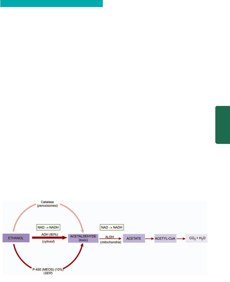
TABLE 21.8: Classification of Cirrhosis.
A. |
Morphologic |
B. |
Etiologic |
|
|
|
|
I. |
Micronodular |
1. |
Alcoholic cirrhosis |
|
(nodules less than 3 mm) |
|
(the most common, 60-70%) |
II. |
Macronodular |
2. |
Post-necrotic cirrhosis (10%) |
|
(nodules more than 3 mm) |
3. |
Biliary cirrhosis (5-10%) |
III. |
Mixed |
4. |
Pigment cirrhosis in |
|
|
|
haemochromatosis (5%) |
|
|
5. |
Cirrhosis in Wilson’s disease |
|
|
6. |
Cirrhosis in α-1-antitrypsin |
|
|
|
deficiency |
|
|
7. |
Cardiac cirrhosis |
|
|
8. |
Indian childhood cirrhosis (ICC) |
|
|
9. |
Cirrhosis in autoimmune hepatitis |
|
|
10. |
Cirrhosis in non-alcoholic |
|
|
|
steatohepatitis |
|
|
11. |
Miscellaneous forms of |
|
|
|
cirrhosis (metabolic, infectious, |
|
|
|
GI, infiltrative) diseases |
|
|
12. |
Cryptogenic cirrhosis |
|
|
|
|
be portal-portal, portal-central, or both. The micronodular cirrhosis includes etiologic type of alcoholic cirrhosis (or nutritional cirrhosis or Laennec’s cirrhosis) and represents impaired capacity for regrowth as seen in alcoholism, malnutrition, severe anaemia and old age.
2.Macronodular cirrhosis. In this type, the nodules are of variable size and are generally larger than 3 mm in diameter. The pattern of involvement is more irregular than in micronodular cirrhosis, sparing some portal tracts and central veins, and more marked evidence of regeneration. Macronodular cirrhosis corresponds to post-necrotic (or posthepatitis) cirrhosis of the etiologic classification.
3.Mixed cirrhosis. In mixed type, some parts of the liver show micronodular appearance while other parts show macronodular pattern. All the portal tracts and central veins are not involved by fibrosis but instead some of them are spared. Mixed pattern is a kind of incomplete expression of micronodular cirrhosis.
B. ETIOLOGIC CLASSIFICATION. Based on the etiologic agent for cirrhosis, various categories of cirrhosis are described as given in Table 21.8.
SPECIFIC TYPES OF CIRRHOSIS
Alcoholic Liver Disease and Cirrhosis
Alcoholic liver disease is the term used to describe the spectrum of liver injury associated with acute and chronic alcoholism. There are three sequential stages in alcoholic liver disease: alcoholic steatosis (fatty liver), alcoholic hepatitis and
alcoholic cirrhosis.
Before discussing the features of alcoholic liver disease and cirrhosis, a brief outline of ethanol metabolism is outlined below and is discussed earlier in Chapter 9 (page 239).
ETHANOL METABOLISM. One gram of alcohol gives 7 calories. But alcohol cannot be stored in the body and must undergo obligatory oxidation, chiefly in the liver. Thus, these empty calories make no contribution to nutrition other than to give energy.
Ethanol after ingestion and absorption from the small bowel circulates through the liver where about 90% of it is oxidised to acetate by a two-step enzymatic process involving two enzymes: alcohol dehydrogenase (ADH) present in the cytosol, and acetaldehyde dehydrogenase (ALDH) in the mitochondria of hepatocytes (Fig. 21.21). The remaining 10% of ethanol is oxidised elsewhere in the body.
First step: Ethanol is catabolised to acetaldehyde in the liver by the following three pathways, one major and two minor:
i)In the cytosol, by the major rate-limiting pathway of alcohol dehydrogenase (ADH).
ii)In the smooth endoplasmic reticulum, via microsomal P-450 oxidases (also called microsomal ethanol oxidising system, MEOS), where only part of ethanol is metabolised.
iii)In the peroxisomes, minor pathway via catalase such as H2O2.
619
Pancreas Exocrine and Tract Biliary Liver, The 21 CHAPTER
Figure 21.21 
 Metabolism of ethanol in the liver. Thickness and intensity of colour of arrows on left side of figure corresponds to extent of metabolic pathway followed (ADH = alcohol dehydrogenase; ALDH or ACDH = hepatic acetaldehyde dehydrogenase; NAD = nicotinamide adenine dinucleotide; NADH = reduced NAD).

620 Acetaldehyde is toxic and may cause membrane damage and cell necrosis. Simultaneously, the cofactor nicotinamideadenine dinucleotide (NAD) which is a hydrogen acceptor, is reduced to NADH.
Second step: The second step occurs in the mitochondria where acetaldehyde is converted to acetate with ALDH acting as a co-enzyme. Most of the acetate on leaving the liver is finally oxidised to carbon dioxide and water, or converted by the citric acid cycle to other compounds including fatty acids. Simultaneously, the same cofactor, NAD, is reduced to NADH resulting in increased NADH: NAD redox ratio which is the basic biochemical alteration occurring during ethanol metabolism. A close estimate of NADH:NAD ratio is measured by the ratio of its oxidised and reduced metabolites in the form of lactate-pyruvate ratio
and β-hydroxy butyrate-acetoacetate ratio.
|
RISK FACTORS FOR ALCOHOLIC LIVER DISEASE. All |
|
|
those who indulge in alcohol abuse do not develop liver |
|
|
damage. The incidence of cirrhosis among alcoholics at |
|
|
autopsy is about 10-15%. Why some individuals are |
|
|
predisposed to alcoholic cirrhosis is not clearly known, but |
|
SECTION |
a few risk factors have been implicated. These are as under: |
|
1. Drinking patterns. Most epidemiologic studies have |
||
|
||
|
attributed alcoholic cirrhosis to chronic alcoholism. Available |
|
|
evidence suggests that chronic and excessive consumption |
|
|
of alcohol invariably leads to fatty liver in >90% of chronic |
|
III |
alcoholics, progression to alcoholic hepatitis in 10-20% cases, |
|
and eventually to alcoholic cirrhosis in more than 10 years. |
||
|
||
|
It is generally agreed that continued daily imbibing of 60-80 |
|
Systemic |
gm of ethanol in any type of alcoholic beverage for at least |
|
10 years is likely to result in alcoholic cirrhosis. Liver injury |
||
|
||
|
is related to the quantity of ethanol contained in alcoholic |
|
|
beverage consumed and its duration, but not related to the |
|
Pathology |
type of alcoholic beverage consumed. Ethanol content in an |
|
alcoholic beverage is given on the label of the container, but |
||
|
||
|
in general, it s about 4-6% in beer, 10-12% in wine, and about |
|
|
40-50% in brandy, whisky and scotch. Intermittent drinking |
|
|
for long duration is less harmful since the liver is given |
|
|
chance to recover. |
|
|
2. Gender. Women have increased susceptibility to develop |
|
|
advanced alcoholic liver disease with much lesser alcohol |
|
|
intake (20-40 g/day). This gender difference in disease |
|
|
progression is unclear but is probably linked to effects of |
|
|
oestrogen. |
|
|
3. Malnutrition. Absolute or relative malnutrition of |
|
|
proteins and vitamins is regarded as a contributory factor in |
|
|
the evolution of cirrhosis. The combination of chronic alcohol |
|
|
ingestion and impaired nutrition leads to alcoholic liver |
|
|
disease and not malnutrition per se. It appears that calories |
|
|
derived from alcohol displace other nutrients leading to |
|
|
malnutrition and deficiency of vitamins in alcoholics. |
|
|
Additional factors contributing to malnutrition in alcoholics |
|
|
are chronic gastritis and pancreatitis. The evidence in favour |
|
|
of synergistic effect of malnutrition in chronic alcoholism |
|
|
comes from clinical and morphologic improvement in cases |
|
|
of alcoholic cirrhosis on treatment with protein-rich diets. |
|
|
4. Infections. Intercurrent bacterial infections are common |
|
|
in cirrhotic patients and may accelerate the course of the |
disease. Lesions similar to alcoholic cirrhosis may develop in non-alcoholic patients who have had viral infections in the past.
5.Genetic factors. The rate of ethanol metabolism is under genetic control. It is chiefly related to altered rates of elimination of ethanol due to genetic polymorphism for the two main enzyme systems, MEOS (microsomal P-450 oxidases) and alcohol dehydrogenase (ADH). Various HLA histocompatibility types have been associated with susceptibility of different populations to alcoholic liver damage but no single genotype has been identified yet.
6.Hepatitis C infection. Concurrent infection with HCV is an important risk factor for progression of alcoholic liver disease. HCV infection in chronic alcoholic leads to development of alcoholic liver disease with much less alcohol consumption (20-50 g/day), disease progression at a younger age, having greater severity, and increased risk to develop cirrhosis and hepatocellular carcinoma, and overall poorer survival.
PATHOGENESIS. Exact pathogenesis of alcoholic liver injury is yet unclear as to why only some chronic alcoholics develop the complete sequence of changes in the liver while others don’t. However, knowledge and understanding of the ethanol metabolism has resulted in discarding the old concept of liver injury due to malnutrition. Instead, now it is known that ethanol and its metabolites are responsible for ill-effects on the liver in a susceptible chronic alcoholic having above-mentioned risk factors. Briefly, the biomedical and cellular pathogenesis due to chronic alcohol consumption culminating in morphologic lesions of alcoholic steatosis (fatty liver), alcoholic hepatitis and alcoholic cirrhosis can be explained as under and is schematically illustrated in
Fig. 21.22:
1.Direct hepatotoxicity by ethanol. There is evidence to suggest that ethanol ingestion for a period of 8-10 days regularly may cause direct hepatotoxic effect on the liver and produce fatty change. Ethanol is directly toxic to microtubules, mitochondria and membrane of hepatocytes.
2.Hepatotoxicity by ethanol metabolites. The major hepatotoxic effects of ethanol are exerted by its metabolites, chiefly acetaldehyde. Acetaldehyde levels in blood are elevated in chronic alcoholics. Acetaldehyde produces hepatotoxicity by production of two adducts:
i) Production of protein-aldehyde adducts which are extremely toxic and can cause cytoskeletal and membrane damage and bring about hepatocellular necrosis.
ii) Formation of malon-di-aldehyde-acetaldehyde (MAA) adducts which produce autoantibodies and initiate autoimmune response. Theses adducts have also a role in hepatic fibrogenesis due to peroxisome proliferator-activated receptor (PPAR)-γ on hepatocytes.
3.Oxidative stress. Oxidation of ethanol by the cytochrome-
450oxidases (MEOS) leads to generation of free radicals which causes oxidative damage to the membranes and proteins.
4.Immunological mechanism. Cell-mediated immunity is impaired in alcoholic liver disease. Ethanol causes direct
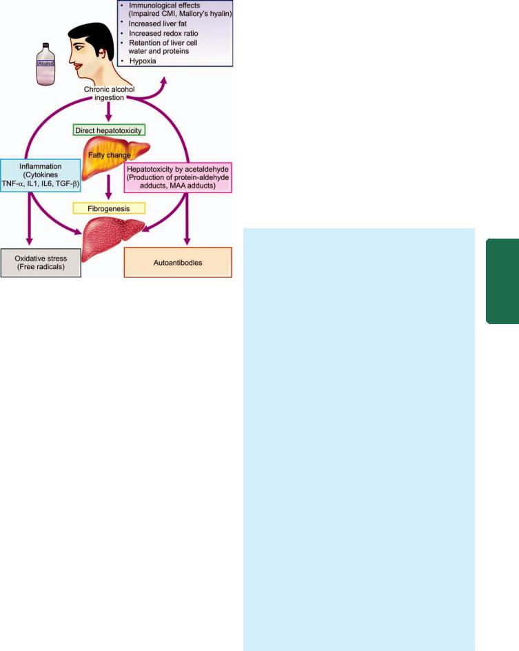
Figure 21.22 
 Pathogenesis of alcoholic liver disease.
immunologic attack on hepatocytes. In a proportion of cases, alcohol-related liver cell injury continues unabated despite cessation of alcohol consumption which is attributed to immunologic mechanisms. Immunological mechanism may also explain the genesis of Mallory’s alcoholic hyalin though more favoured hypothesis for its origin is the aggregation of intermediate filaments of prekeratin type due to alcoholinduced disorganisation of cytoskeleton.
5.Inflammation. Chronic ethanol ingestion is not only injurious to hepatocytes but also damages the intestinal cells. The injured intestinal cells elaborate endotoxins which release proinflammatory cytokines, chiefly tumour necrosis factor-α, IL-1, IL-6 and TGF-β . These cytokines and endotoxinaemia produce apoptosis and necrosis of hepatocytes and initiate inflammatory reaction in the alcohol damaged liver.
6.Fibrogenesis. Main event facilitating hepatic fibrogenesis is activation of stellate cells by various stimuli:
i)by damaged hepatocytes,
ii)by malon-di-aldehyde-acetaldehyde adducts,
iii)by activated Kupffer cells, and
iv)direct stimulation by acetaldehyde.
All forms of collagen are increased and there is increased transformation of fat-storing lto cells into myofibroblasts and fibrocytes.
7. Increased redox ratio. Marked increase in the NADH:NAD redox ratio in the hepatocytes results in increased redox ratio of lactate-pyruvate, leading to lactic acidosis. This altered redox potential has been implicated in a number of metabolic consequences such as in fatty liver, collagen formation,
occurrence of gout, impaired gluconeogenesis and altered steroid metabolism.
8.Retention of liver cell water and proteins. Alcohol is inhibitory to secretion of newly-synthesised proteins by the liver leading to their retention in the hepatocytes. Water is simultaneously retained in the cell in proportion to the protein and results in swelling of hepatocytes resulting in hepatomegaly in alcoholics.
9.Hypoxia. Chronic ingestion of alcohol results in increased oxygen demand by the liver resulting in a hypoxic state which causes hepatocellular necrosis in centrilobular zone (zone 3). Redox changes are also more marked in zone 3.
10.Increased liver fat. The origin of fat in the body was discussed in Chapter 3 (page 37). In chronic alcoholism, there is rise in the amount of fat available to the liver which could be from exogenous (dietary) sources, excess mobilisation from adipose tissue or increased lipid synthesis by the liver itself. This may account for lipid accumulation in the hepatocytes.
MORPHOLOGIC FEATURES. Three types of morphologic lesions are described in alcoholic liver disease— alcoholic steatosis (fatty liver), alcoholic hepatitis and alcoholic cirrhosis.
1.ALCOHOLIC STEATOSIS (FATTY LIVER). The morphologic changes in fatty change in liver have already been described on page 37 and are briefly considered here. Grossly, the liver is enlarged, yellow, greasy and firm with a smooth and glistening capsule.
Microscopically, the features consist of initial microvesicular droplets of fat in the hepatocyte cytoplasm followed by more common and pronounced feature of macrovesicular large droplets of fat displacing the nucleus to the periphery (Fig. 21.23). Fat cysts may develop due to coalescence and rupture of fat-containing hepatocytes. Less often, lipogranulomas consisting of collection of lymphocytes, macrophages and some multinucleate giant cells may be found.
2.ALCOHOLIC HEPATITIS. Alcoholic hepatitis develops acutely, usually following a bout of heavy drinking. Repeated episodes of alcoholic hepatitis superimposed on pre-existing fatty liver are almost certainly a forerunner of alcoholic cirrhosis.
Histologically, the features of alcoholic hepatitis are as follows (Fig. 21.24):
i)Hepatocellular necrosis: Single or small clusters of hepatocytes, especially in the centrilobular area (zone 3), undergo ballooning degeneration and necrosis.
ii)Mallory bodies or alcoholic hyalin: These are eosinophilic, intracytoplasmic inclusions seen in perinuclear location within swollen and ballooned hepatocytes. They represent aggregates of cytoskeletal intermediate filaments (prekeratin). They can be best visualised with connective tissue stains like Masson’s trichrome and chromophobe aniline blue, or by the use of immunoperoxidase methods. Mallory bodies are highly suggestive of, but not specific for, alcoholic hepatitis since
621
Pancreas Exocrine and Tract Biliary Liver, The 21 CHAPTER
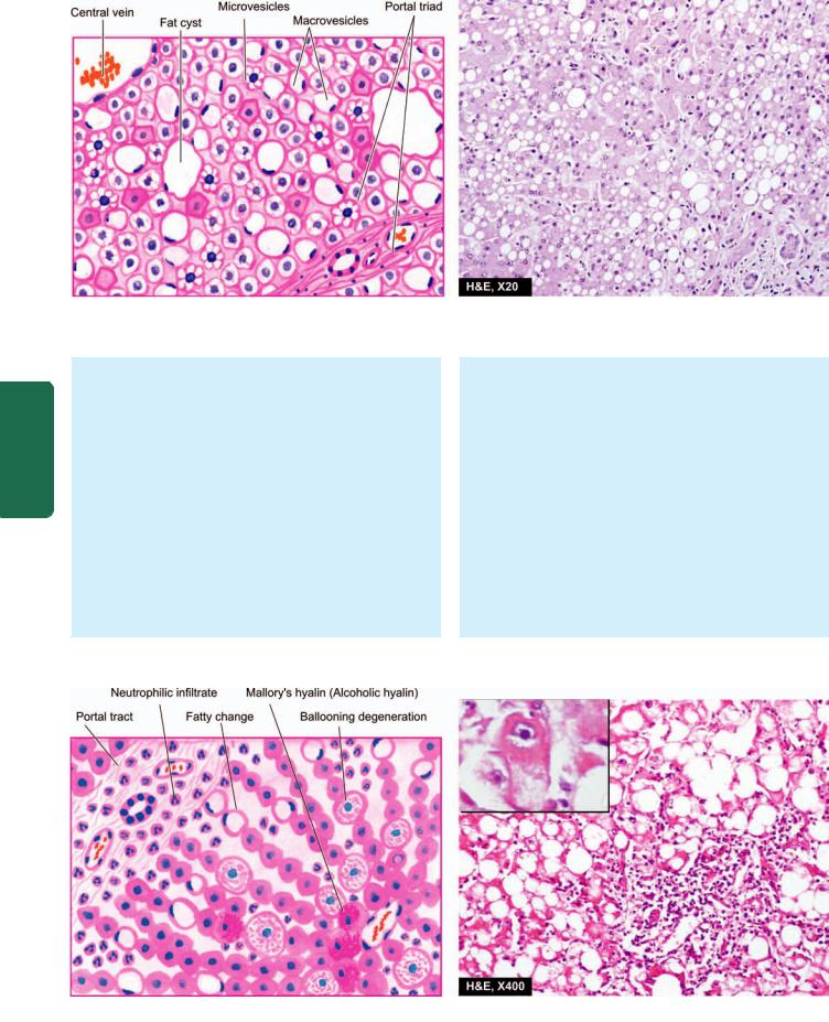
622
Figure 21.23 
 Fatty liver (alcoholic steatosis). Most of the hepatocytes are distended with large lipid vacuoles with peripherally displaced nuclei.
Pathology Systemic III SECTION
Mallory bodies are also found in certain other conditions such as: primary biliary cirrhosis, Indian childhood cirrhosis, cholestatic syndromes, Wilson’s disease, intestinal bypass surgery, focal nodular hyperplasia and hepatocellular carcinoma.
iii)Inflammatory response: The areas of hepatocellular necrosis and regions of Mallory bodies are associated with an inflammatory infiltrate, chiefly consisting of polymorphs and some scattered mononuclear cells. In more extensive necrosis, the inflammatory infiltrate is more widespread and may involve the entire lobule.
iv)Fibrosis: Most cases of alcoholic hepatitis are accompanied by pericellular and perivenular fibrosis, producing a web-like or chickenwire-like appearance. This
is also termed as creeping collagenosis.
3. ALCOHOLIC CIRRHOSIS. Alcoholic cirrhosis is the most common form of lesion, constituting 60-70% of all cases of cirrhosis. Several terms have been used for this type of cirrhosis such as Laennec’s cirrhosis, portal cirrhosis,
hobnail cirrhosis, nutritional cirrhosis, diffuse cirrhosis and micronodular cirrhosis.
Grossly, alcoholic cirrhosis classically begins as micronodular cirrhosis (nodules less than 3 mm diameter), the liver being large, fatty and weighing usually above 2 kg (Fig. 21.25). Eventually over a span of years, the liver shrinks to less than 1 kg in weight, becomes non-fatty, having macronodular cirrhosis (nodules larger than 3 mm in diameter), resembling post-necrotic cirrhosis. The nodules of the liver due to their fat content are tawnyyellow, on the basis of which Laennec in 1818 introduced
Figure 21.24 
 Alcoholic hepatitis. Liver cells show ballooning degeneration and necrosis with some containing Mallory’s hyalin (Inbox). Fatty change and clusters of neutrophils are also present.
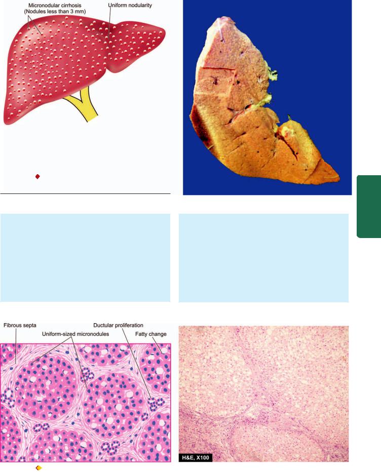
Figure 21.25 Alcoholic cirrhosis, showing the typical micronodular
pattern in gross specimen. There is diffuse nodularity (nodules less than
PP GLDPHWHU RQ VHFWLRQHG VXUIDFH RI WKH OLYHU
the term cirrhosis first of all (from Greek kirrhos = tawny). The surface of liver in alcoholic cirrhosis is studded with diffuse nodules which vary little in size, producing hobnail liver (because of the resemblance of the surface with the sole of an old-fashioned shoe having short nails with heavy heads). On cut section, spheroidal or angular nodules of fibrous septa are seen.
Microscopically, alcoholic cirrhosis is a progressive alcoholic liver disease. Its features include the following
(Fig. 21.26):
i)Nodular pattern: Normal lobular architecture is effaced in which central veins are hard to find and is replaced with nodule formation.
ii)Fibrous septa: The fibrous septa that divide the hepatic parenchyma into nodules are initially delicate and extend from central vein to portal regions, or portal tract to portal tract, or both. As the fibrous scarring increases with time, the fibrous septa become dense and more confluent.
iii)Hepatic parenchyma: The hepatocytes in the islands of surviving parenchyma undergo slow proliferation
623
Pancreas Exocrine and Tract Biliary Liver, The 21 CHAPTER
Figure 21.26 
 Alcoholic cirrhosis, microscopic appearance. It shows nearly uniform-sized micronodules, devoid of central veins and having thick fibrous septa dividing them. There is minimal inflammation and some reactive bile duct proliferation in the septa.
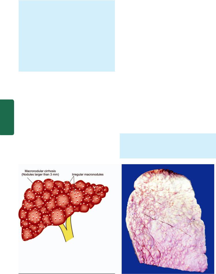
624
Pathology Systemic III SECTION
forming regenerative nodules having disorganised masses of hepatocytes. The hepatic parenchyma within the nodules shows extensive fatty change early in the disease. But as the fibrous septa become more thick, the amount of fat in hepatocytes is reduced. Thus, there is an inverse relationship between the amount of fat and the amount of fibrous scarring in the nodules.
iv) Necrosis, inflammation and bile duct proliferation:
The etiologic clue to diagnosis in the form of Mallory bodies is hard to find in a fully-developed alcoholic cirrhosis. The fibrous septa usually contain sparse infiltrate of mononuclear cells with some bile duct proliferation. Bile stasis and increased cytoplasmic haemosiderin deposits due to enhanced iron absorption in alcoholic cirrhosis are some other noticeable findings.
LABORATORY DIAGNOSIS. The clinical manifestations and complications of cirrhosis in general are described on page 630. The laboratory findings in the course of alcoholic liver disease may be quite variable and liver biopsy is necessary in doubtful cases. Progressive form of the disease, however, generally presents the following biochemical and haematological alterations:
1.Elevated transaminases: increase in SGOT (AST) is more than that of SGPT (ALT).
2.Rise in serum γ-glutamyl transpeptidase (γ-GT).
3.Elevation in serum alkaline phosphatase.
4.Hyperbilirubinaemia.
5.Hypoproteinaemia with reversal of albumin-globulin ratio.
6.Prolonged prothrombin time and partial thromboplastin time.
7.Anaemia.
8.Neutrophilic leucocytosis in alcoholic hepatitis and in secondary infections.
Post-necrotic Cirrhosis
Post-necrotic cirrhosis, also termed post-hepatitic cirrhosis,
macronodular cirrhosis and coarsely nodular cirrhosis, is characterised by large and irregular nodules with broad bands of connective tissue and occurring most commonly after previous viral hepatitis.
ETIOLOGY. Based on epidemiologic and serologic studies, the following factors have been implicated in the etiology of post-necrotic cirrhosis.
1.Viral hepatitis. About 25% of patients give history of recent or remote attacks of acute viral hepatitis followed by chronic viral hepatitis. Most common association is with hepatitis B and C; hepatitis A is not known to evolve into cirrhosis. It is estimated that about 20% cases of HBV chronic hepatitis and about 20-30% cases of HCV chronic hepatitis go to develop cirrhosis over 20-30 years.
2.Drugs and chemical hepatotoxins. A small percentage of cases may have origin from toxicity due to chemicals and drugs such as phosphorus, carbon tetrachloride, mushroom poisoning, acetaminophen and α-methyl dopa.
3.Others. Certain infections (e.g. brucellosis), parasitic infestations (e.g. clonorchiasis), metabolic diseases (e.g. Wilson’s disease or hepatolenticular degeneration) and advanced alcoholic liver disease may produce a picture of post-necrotic cirrhosis.
4.Idiopathic. After all these causes have been excluded, a group of cases remain in which the etiology is unknown.
MORPHOLOGIC FEATURES. Typically, post-necrotic cirrhosis is macronodular type.
Grossly, the liver is usually small, weighing less than 1 kg, having distorted shape with irregular and coarse scars and nodules of varying size (Fig. 21.27). Sectioned
Figure 21.27 
 Post-necrotic cirrhosis, showing the typical irregular macronodular pattern (nodules larger than 3 mm diameter). Externally the liver is small, distorted and irregularly scarred.
625
Figure 21.28 
 Post-necrotic cirrhosis. Fibrous septa dividing the hepatic parenchyma into nodules are thick and contain prominent mononuclear inflammatory cell infiltrate and bile ductular hyperplasia. A few intact hepatic lobules remain.
surface shows scars and nodules varying in diameter from 3 mm to a few centimeters.
Microscopically, the features are as follows (Fig. 21.28):
1.Nodular pattern: The normal lobular architecture of hepatic parenchyma is mostly lost and is replaced by nodules larger than those in alcoholic cirrhosis. However, uninvolved portal tracts and central veins in the hepatic lobules can still be seen in some parts of surviving parenchyma.
2.Fibrous septa: The fibrous septa dividing the variablesized nodules are generally thick.
3.Necrosis, inflammation and bile duct proliferation:
Active liver cell necrosis is usually inconspicuous. Fibrous septa contain prominent mononuclear inflammatory cell infiltrate which may even form follicles, especially in cases following HCV chronic hepatitis. Often there is extensive proliferation of bile ductules derived from collapsed liver lobules.
4.Hepatic parenchyma: Liver cells vary considerably in size and multiple large nuclei are common in regenerative nodules. Fatty change may or may not be present in the hepatocytes.
CLINICAL FEATURES. Besides the general clinical features described on page 630, post-necrotic cirrhosis is seen as frequent in women as in men, especially in the younger age group. Like in alcoholic cirrhosis, the patients may remain asymptomatic or may present with prominent signs and symptoms of chronic hepatitis (page 611). Splenomegaly and hypersplenism are other prominent features. The results of haematologic and liver function test are similar to those of alcoholic cirrhosis. Out of the various types of cirrhosis, postnecrotic cirrhosis, especially when related to hepatitis B and C virus infection in early life, is more frequently associated with hepatocellular carcinoma.
Biliary Cirrhosis
Biliary cirrhosis is defined as a chronic disorder characterised by clinical, biochemical and morphological features of longcontinued cholestasis of intrahepatic or extrahepatic origin. Biliary cirrhosis is of following types:
 Primary biliary cirrhosis in which the destructive process of unknown etiology affects intrahepatic bile ducts.

 Secondary biliary cirrhosis resulting from prolonged mechanical obstruction of the extrahepatic biliary passages.
 Primary sclerosing cholangitis and autoimmune cholangiopathy causing biliary cirrhosis.
ETIOLOGY. The etiology of these forms of biliary cirrhosis is distinctive:
A. Primary biliary cirrhosis. The etiology of this type remains unknown. However, a few factors have been implicated:
1.The condition is predominant in middle-aged women (male: female ratio = 1:9) and has led to the suggestion of a possible endocrine origin.
2.Familial incidence has been observed suggesting the role of some genetic influence and certain HLA types.
3.There is elevated cholesterol level with appearance of xanthoma and xanthelasma. Hepatomegaly and chronic liver disease are late features of the disease.
4.However, presently the most widely accepted hypothesis is autoimmune origin of the disease. In support are the following observations:

 increased incidence of associated autoimmune diseases (e.g. scleroderma, Sjögren’s syndrome, CREST syndrome, and autoimmune thyroiditis),

 circulating anti-mitochondrial antibody of IgG class detected in more than 90% cases;

 elevated levels of immunoglobulins, particularly of IgM;

 increased levels of circulating immune complexes;

 decreased number of circulating T-cells; and

 accumulation of T cells around bile ducts.
Pancreas Exocrine and Tract Biliary Liver, The 21 CHAPTER
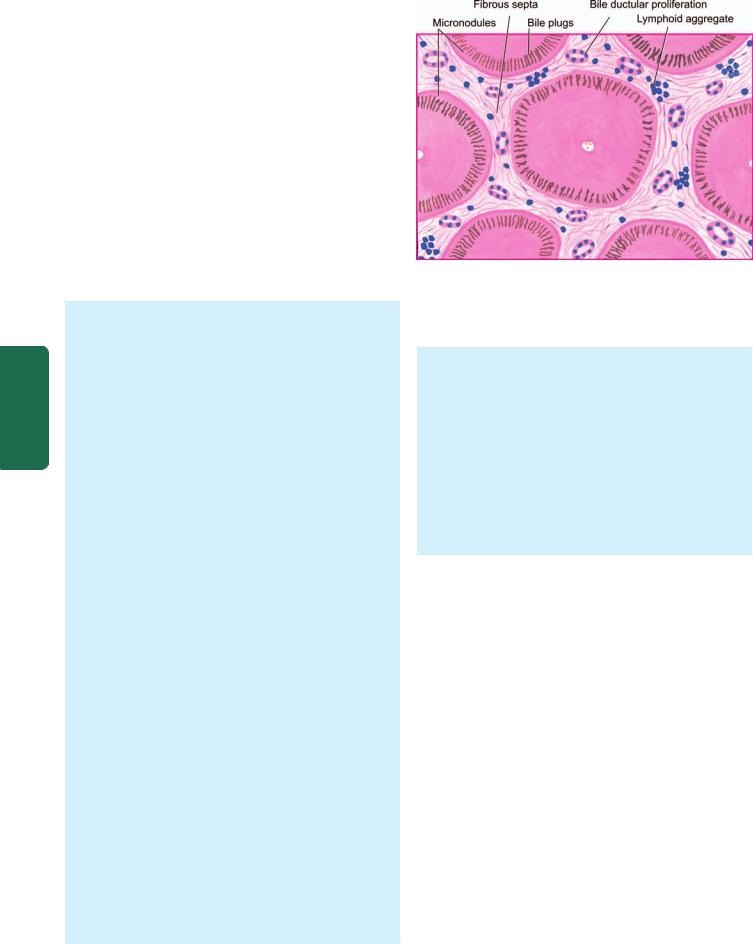
626B. Secondary biliary cirrhosis. Most cases of secondary biliary cirrhosis result from prolonged obstruction of extrahepatic biliary passages (page 599). These causes include the following:
1.Extrahepatic cholelithiasis, most common
2.Biliary atresia
3.Cancer of biliary tree and of head of pancreas
4.Postoperative strictures with superimposed ascending cholangitis.
C. Cirrhosis due to primary sclerosing cholangitis. Primary or idiopathic sclerosing cholangitis is a chronic cholestatic syndrome of unknown etiology. It is characterised by progressive, inflammatory, sclerosing and obliterative process affecting the entire biliary passages, both extrahepatic and intrahepatic ducts. Although etiology remains unknown, various mechanisms have been postulated which include viral and bacterial infections, immunologic injury, toxins, and genetic predisposition.
|
MORPHOLOGIC FEATURES. Grossly, in biliary |
|
|
cirrhosis of all types, the liver is initially enlarged and |
|
|
characteristically greenish in appearance, but later becomes |
|
SECTION |
smaller, firmer and coarsely micronodular. In cirrohosis |
|
due to primary sclerosing cholangitis, there is charac- |
||
|
||
|
teristic beading of intraand extrahepatic bile ducts due |
|
|
to irregular strictures and dilatation. |
|
|
Microscopically, the features of intraand extrahepatic |
|
III |
cholestasis correspond to primary and secondary biliary |
|
cirrhosis respectively discussed on page 599. The salient |
||
|
||
|
features of various forms of biliary cirrhosis are as under: |
|
Systemic |
A. Primary biliary cirrhosis: The diagnostic histologic |
|
feature is a chronic, non-suppurative, destructive |
||
|
||
|
cholangitis involving intrahepatic bile ducts. The disease |
|
|
evolves through the following 4 histologic states: |
|
Pathology |
Stage I: There are florid bile duct lesions confined to portal |
|
tracts. The changes in the affected area consist of |
||
|
||
|
destruction of bile ducts, presence of bile plugs, infiltration |
|
|
with acute and chronic inflammatory cells and sometimes |
|
|
formation of granulomas and lymphoid follicles. |
|
|
Stage II: There is ductular proliferation. The ductal involve- |
|
|
ment is quite widespread with very few normal bile ducts. |
|
|
The inflammatory infiltrate too extends beyond the portal |
|
|
tracts into surrounding hepatic parenchyma. Periportal |
|
|
Mallory bodies may be present. |
|
|
Stage III: This stage is characterised by fibrous scarring |
|
|
interconnecting the portal areas. There is diminished |
|
|
inflammatory infiltrate and reduced number of bile ducts. |
|
|
Stage IV: Well-formed micronodular pattern of cirrhosis |
|
|
develops in a period of a few years (Fig. 21.29). |
B. Secondary biliary cirrhosis: Prolonged obstruction of extrahepatic bile ducts may produce the following histologic changes:
1.Bile stasis, degeneration and focal areas of centrilobular necrosis of hepatocytes.
2.Proliferation, dilatation and rupture of bile ductules in the portal area with formation of bile lakes.
3.Cholangitis, sterile or pyogenic, with accumulation of polymorphs around the bile ducts.
Figure 21.29 
 Primary biliary cirrhosis, diagrammatic representation.
There are fibrous scars dividing the hepatic parenchyma into the micronodules. The fibrous septa contain prominent lymphoid infiltrate and proliferated bile ducts. Many of the hepatocytes contain elongated bile plugs.
4. Progressive expansion of the portal tract by fibrosis and evolution into micronodular cirrhosis.
C. Cirrhosis due to primary sclerosing cholangitis:
Following changes are seen:
1.Fibrosing cholangitis with lymphocytic infiltrate around bile ducts with segmental involvement.
2.Periductal fibrosis with eventual obliteration of lumen of affected bile ducts.
3.Intervening bile ducts are dilated, tortuous and inflamed.
4.Late cases show cholestasis and full-blown picture of biliary cirrhosis.
CLINICAL FEATURES. Clinical features of the three types of biliary cirrhosis are variable:

 Primary biliary cirrhosis may remain asymptomatic for months to years. Symptoms develop insidiously. Basically, it is a cholestatic disorder. The patients present with persistent pruritus, dark urine, pale stools, steatorrhoea, jaundice and skin pigmentation. The earliest laboratory finding is a markedly elevated serum alkaline phosphatase level. Elevation of serum lipids is accompanied by appearance of periorbital xanthelasma and xanthomas over joints. Death usually results from hepatic failure, variceal bleeding, intercurrent infections and concomitant development of cancers of liver and breast.

 The diagnosis of secondary biliary cirrhosis is considered in patients with previous history of gallstones, biliary tract surgery or clinical features of ascending cholangitis.

 The patients of primary sclerosing cholangitis may remain asymptomatic or may show features of cholestatic jaundice (raised alkaline phosphatase, pruritus, fatigue). Late cases show manifestations of chronic liver disease. The disease occurs in 3rd to 5th decade of life with two fold preponderance in males.

TABLE 21.9: Contrasting Features of Major Forms of Biliary Cirrhosis. |
|
627 |
|||
|
Feature |
Primary Biliary Cirrhosis |
Secondary Biliary Cirrhosis |
Primary Sclerosing Cholangitis |
|
|
|
|
|
|
|
1. |
Etiology |
Possibly autoimmune; association |
Extrahepatic biliary |
Possibly autoimmune; association |
|
|
|
with other autoimmune diseases |
obstruction; biliary atresia |
with inflammatory bowel disease |
|
2. |
Age and sex |
Middle-aged women |
Any age and either sex |
Middle age |
|
|
|
Male: Female = 1:9 |
|
Male: Female = 2:1 |
|
3. |
Laboratory tests |
↑ Alkaline phosphatase |
↑ Alkaline phosphatase |
↑ Alkaline phosphatase |
|
|
|
↑ Conjugated bilirubin |
↑ Conjugated bilirubin |
↑ Conjugated bilirubin |
|
|
|
Autoantibodies present |
|
Hypergammaglobulinaemia |
|
4. |
Pathologic changes |
Chronic destructive; |
Bile stasis in bile |
Fibrosing cholangitis |
|
|
|
cholangitis of intrahepatic |
ducts, and sterile or |
with periductal fibrosis |
|
|
|
bile ducts |
pyogenic cholangitis |
|
|
|
|
|
|
|
|
The contrasting features of three main types of intrahepatic disorders leading to biliary cirrhosis are summarised in Table 21.9.
Pigment Cirrhosis in Haemochromatosis
Haemochromatosis is an iron-storage disorder in which there is excessive accumulation of iron in parenchymal cells with eventual tissue damage and functional insufficiency of organs such as the liver, pancreas, heart and pituitary gland. The condition is characterised by a triad of features—
micronodular pigment cirrhosis, diabetes mellitus and skin pigmentation. On the basis of the last two features, the disease has also come to be termed as ‘bronze diabetes’. Males predominate and manifest earlier since women have physiologic iron loss delaying the effects of excessive accumulation of iron. Haemochromatosis exists in 2 main forms:
1.Idiopathic (primary, genetic) haemochromatosis is an autosomal recessive disorder of excessive accumulation of iron. It is associated with overexpression of HFE gene located on chromosome 6 close to the HLA gene locus, and normally regulates intestinal absorption of iron. Mutated (overexpressed) HFE gene complexes with transferrin receptor on intestinal crypt epithelial cells and results in excessive absoption of dietary iron throughout life.
2.Secondary (acquired) haemochromatosis is gross iron overload with tissue injury arising secondary to other diseases such as thalassaemia, sideroblastic anaemias, alcoholic cirrhosis or multiple transfusions.
ETIOPATHOGENESIS. A general discussion of iron metabolism and iron excess states is given on page 41.
Normally, the body iron content is 3-4 gm which is maintained in such a way that intestinal mucosal absorption of iron is equal to its loss. This amount is approximately 1 mg/day in men and 1.5 mg/day in menstruating women. In haemochromatosis, however, this amount goes up to 4 mg/day or more, as evidenced by elevated serum iron (normal about 125 μg/dl) and increased serum transferrin saturation (normal 30%).

 In idiopathic or hereditary haemochromatosis, the primary mechanism of disease appears to be the genetic basis in which the defect may either lie at the intestinal mucosal level causing excessive iron absorption, or at the post-
absorption excretion level leading to excessive accumulation of iron. The excess iron in primary haemochromatosis is deposited mainly in the cytoplasm of parenchymal cells of organs such as the liver, pancreas, spleen, heart and endocrine glands. Tissue injury results from iron-laden lysosomes of parenchymal cells and lipid peroxidation of cell organelles by excess iron.

 In secondary or acquired haemochromatosis, there is excessive accumulation of iron due to acquired causes like ineffective erythropoiesis, defective haemoglobin synthesis, multiple blood transfusions and enhanced absorption of iron due to alcohol consumption. The last-named phenomenon is observed in Bantu siderosis affecting South African Bantu tribals who consume large quantities of home-brew prepared in iron vessels. Cases of secondary haemochromatosis have increased iron storage within the reticuloendothelial system and liver. However, the magnitude of the iron excess in secondary haemochromatosis is generally insufficient to cause tissue damage.
MORPHOLOGIC FEATURES. Excessive deposition of iron in organs and tissues is ferritin and haemosiderin, both of which appear as golden-yellow pigment granules in the cytoplasm of affected parenchymal cells and haemosiderin stains positively with Prussian blue reaction. The organs most frequently affected are the liver and pancreas, and to a lesser extent, the heart, endocrine glands, skin, synovium and testis.
 In the liver, excess of pigment accumulates in the hepatocytes, and less often Kupffer cells and in bile duct epithelium. The deposits in the initial stage may be prominent in the periportal liver cells along with increased fibrosis in the portal zone. Eventually, micronodular cirrhosis develops. The deposits may produce grossly chocolate-brown colour of the liver and nodular surface.
 In the pancreas, pigmentation is less intense and is found in the acinar and islet cells. The deposits in pancreas produce diffuse interstitial fibrosis and atrophy of parenchymal cells leading to occurrence of diabetes mellitus.
CLINICAL FEATURES. The major clinical manifestations of haemochromatosis include skin pigmentation, diabetes
Pancreas Exocrine and Tract Biliary Liver, The 21 CHAPTER

628 mellitus, hepatic and cardiac dysfunction, arthropathy and hypogonadism. Characteristic bronze pigmentation is the presenting feature in about 90% of cases. Demonstration of excessive parenchymal iron stores is possible by measurement of serum iron, determination of percent saturation of transferrin, measurement of serum ferritin concentration, estimation of chelatable iron stores using chelating agent (e.g. desferrioxamine), and finally, by liver biopsy. Occurrence of hepatocellular carcinoma is a late complication of haemochromatosis-induced cirrhosis.
|
Cirrhosis in Wilson’s Disease |
|
|
Wilson’s disease, also termed by a more descriptive desig- |
|
|
nation of hepatolenticular degeneration, is an autosomal |
|
|
recessive inherited disease of copper metabolism, |
|
|
characterised by toxic accumulation of copper in many |
|
|
tissues, chiefly the liver, brain and eye. These accumulations |
|
|
lead to the triad of features: |
|
|
1. Cirrhosis of the liver. |
|
|
2. Bilateral degeneration of the basal ganglia of the brain. |
|
|
3. Greenish-brown pigmented rings in the periphery of the |
|
SECTION |
cornea (Kayser-Fleischer rings). |
|
The disease manifests predominantly in children and |
||
|
||
|
young adults (5-30 years). Initially, the clinical manifestations |
|
|
are referable to liver involvement such as jaundice and |
|
|
hepatomegaly (hepatic form) but later progressive |
|
III |
neuropsychiatric changes and Kayser-Fleischer rings in the |
|
cornea appear. |
||
|
PATHOGENESIS. The pathogenesis of Wilson’s disease is |
|
Systemic |
best understood when compared with normal copper |
|
metabolism. |
||
|
||
|
Normally, dietary copper is more than body’s |
|
|
requirement. Excess copper so absorbed through the stomach |
|
Pathology |
and duodenum is transported to the liver where it is |
|
incorporated into α2-globulin to form ceruloplasmin, which |
||
|
is excreted by the liver via bile normally. Most of the plasma |
|
|
copper circulates as ceruloplasmin. Only minute amount of |
|
|
copper is excreted in the urine normally. |
|
|
In Wilson’s disease, the initial steps of dietary absorp- |
|
|
tion and transport of copper to the liver are normal but |
|
|
copper accumulates in the liver rather than being excreted |
|
|
by the liver. The underlying defect in chromosome 13 is a |
|
|
mutation in ATP7B gene, the normal hepatic copper- |
|
|
excreting gene. Eventually, capacity of hepatocytes to store |
|
|
copper is exceeded and copper is released into circulation |
|
|
which then gets deposited in extrahepatic tissues such as the |
|
|
brain, eyes and others. However, increased copper in the |
|
|
kidney does not produce any serious renal dysfunction. |
|
|
Biochemical abnormalities in Wilson’s disease include the |
|
|
following: |
|
|
1 Decreased serum ceruloplasmin (due to impaired synthesis |
|
|
of apoceruloplasmin in damaged liver and defective |
|
|
mobilisation of copper from hepatocellular lysosomes). |
|
|
2. Increased hepatic copper in liver biopsy (due to excessive |
|
|
accumulation of copper in the liver). |
|
|
3. Increased urinary excretion of copper. |
|
|
4. However, serum copper levels are of no diagnostic help |
|
|
and may vary from low-to-normal-to-high depending upon |
|
|
the stage of disease. |
MORPHOLOGIC FEATURES. The liver shows varying grades of changes that include fatty change, acute and chronic active hepatitis, submassive liver necrosis and macronodular cirrhosis. Mallory bodies are present in some cases. Copper is usually deposited in the periportal hepatocytes in the form of reddish granules in the cytoplasm or as reddish cytoplasmic coloration, stainable by rubeanic acid or rhodamine stains for copper.
Involvement of basal ganglia in the brain is seen in the form of toxic injury to neurons, in the cornea as greenish-brown deposits of copper in Descemet’s membrane, and in the kidney as fatty and hydropic change.
Cirrhosis in α-1-Antitrypsin Deficiency
Alpha-1-antitrypsin deficiency is an autosomal codominant condition in which the homozygous state produces liver disease (cirrhosis), pulmonary disease (emphysema), or both (page 479). α-1-antitrypsin is a glycoprotein normally synthesised in the rough endoplasmic reticulum of the hepatocytes and is the most potent protease inhibitor (Pi). A single autosomal dominant gene coding for α-1-antitrypsin is located on long arm of chromosome 14 that codes for immunoglobulin light chains too. Out of 24 different alleles labelled alphabetically, PiMM is the most common normal phenotype, while the most frequent abnormal phenotype in α-1-antitrypsin deficiency leading to liver and/or lung disease is PiZZ in homozygote form. Other phenotypes in which liver disease occurs are PiSS and Pi-null in which serum α-1-antitrypsin value is nearly totally deficient. Intermediate phenotypes, PiMZ and PiSZ persons are predisposed to develop hepatocellular carcinoma.
The patients may present with respiratory disease due to the development of emphysema, or may develop liver dysfunction, or both. At birth or in neonates, the features of cholestatic jaundice of varying severity may appear. In adolescence, the condition may evolve into hepatitis or cirrhosis which is usually well compensated.
MORPHOLOGIC FEATURES. Pulmonary changes in α- 1-antitrypsin deficiency in the form of emphysema are described in Chapter 17. The hepatic changes vary according to the age at which the deficiency becomes apparent. At birth or in neonates, the histologic features consist of neonatal hepatitis that may be acute or ‘pure’ cholestasis. Micronodular or macronodular cirrhosis may appear in childhood or in adolescence in which the diagnostic feature is the presence of intracellular, acidophilic, PAS-positive globules in the periportal hepatocytes. Ultrastructurally, these globules consist of dilated rough endoplasmic reticulum.
Cardiac Cirrhosis
Cardiac cirrhosis is an uncommon complication of severe right-sided congestive heart failure of long-standing duration (page 99). The common causes culminating in cardiac cirrhosis are cor pulmonale, tricuspid insufficiency or constrictive pericarditis. The pressure in the right ventricle

is elevated which is transmitted to the liver via the inferior vena cava and hepatic veins. The patients generally have enlarged and tender liver with mild liver dysfunction. Splenomegaly occurs due to simple passive congestion.
MORPHOLOGIC FEATURES. Grossly, the liver is enlarged and firm with stretched Glisson’s capsule. Histologically, in acute stage, the hepatic sinusoids are dilated and congested with haemorrhagic necrosis of centrilobular hepatocytes (central haemorrhagic necrosis).
Severe and more prolonged heart failure results in delicate fibrous strands radiating from the central veins. These fibrous strands may form interconnections leading to cardiac cirrhosis and regenerative nodules.
Indian Childhood Cirrhosis
Indian childhood cirrhosis (ICC) is an unusual form of cirrhosis seen in children between the age of 6 months and 3 years in rural, middle class, Hindus in India and in parts of South-East Asia and in the Middle-East. There is no role of viral infection in its etiology. Instead, a combination of some common toxic effects and inherited abnormality of copper metabolism has been suggested. Death occurs due to hepatic failure within a year of diagnosis.
MORPHOLOGIC FEATURES. Five histologic types of ICC have been distinguished of which type II is the most common. This form is characterised by the following features:
i)Liver cell injury ranging from ballooning degeneration to significant damage to hepatocytes.
ii)Prominent Mallory bodies in some hepatocytes without fatty change.
iii)Neutrophilic and sometimes alongwith lymphocytic infiltrate.
iv)Creeping pericellular fibrosis which may eventually lead to fine micro-macro-nodular cirrhosis.
v)There is significant deposition of copper and copperassociated proteins in hepatocytes, often more than what is seen in Wilson’s disease.
Thus, the picture resembles acute alcoholic hepatitis but without the fatty change and with greatly impaired regeneration. There is marked increase in hepatic copper since the milk consumed by such infants is often boiled and stored in copper vessels in India. The condition has to be distinguished from Wilson’s disease.
Cirrhosis in Autoimmune Hepatitis
Autoimmune hepatitis (also called lupoid hepatitis) is a form of chronic hepatitis characterised by continued hepatocellular injury, inflammation and fibrosis which may progress to cirrhosis. The condition may run a variable natural history rangingfrom indolent to severe rapid course. This form of hepatitis has prominent autoimmune etiology is supported by immunologic abnormalities and a few other characteristic diagnostic criteria as under:
1. Female gender predisposition.
2.Predominant elevation of aminotransferases (AST and ALT).
3.Hyperglobulinaemia (elevated IgG and γ-globulin).
4.High serum titres of nuclear (ANA), smooth muscle (SMA), and liver-kidney microsomal (LKM1) autoantibodies, and absence of antimitochondrial antibodies.
5.Concurrent presence of other autoimmune diseases.
6.Presence of HLA DR3 or HLA DR4 markers.
7.Lack of prominent elevation of alkaline phosphatase.
8.Exclusion of chronic hepatitis of other known etiologies (viral, toxic, genetic etc).
MORPHOLOGIC FEATURES. Autoimmune hepatitis is morphologically indistinguishable from chronic hepatitis of viral etiology. Patients who survive active disease develop crrhosis. There are features of burnt out chronic autoimmune hepatitis accompanied with cirrhosis.
Cirrhosis in Non-alcoholic Steatohepatitis
Non-alcoholic steatohepatitis (NASH) or non-alcoholic fatty liver disease (NAFLD) is a from of hepatitis resembling alcoholic liver disease but seen in nondrinkers of alcohol. The condition is seen more commonly in affluent western socieities, has a strong association with obesity, dyslipidaemia and type 2 diabetes mellitus. It is seen in youger patients with equal gender prevalence. Pateints are generally asymptomatic and are diagnosed by routine biochemical tests.
MORPHOLOGIC FEATURES. The condition is a form of chronic hepatitis after known causes have been excluded. About 10-30% cases of NASH progress to increased fibrosis and develop cirrhosis.
Miscellaneous Forms of Cirrhosis
In addition to the various types of cirrhosis just described, a few other uncommon types associated with different diseases are sometimes distinguished. These include the following:
1.Metabolic disorders e.g. in galactosaemia, hereditary fructose intolerance, glycogen storage diseases.
2.Infectious diseases e.g. in brucellosis, schistosomiasis, syphilis (hepar lobatum) and toxoplasma infection.
3.Gastrointestinal disorders e.g. in inflammatory bowel disease, cystic fibrosis of the pancreas and intestinal bypass surgery for obesity.
4.Infiltrative diseases e.g. in sarcoidosis.
Cryptogenic Cirrhosis
Finally, when all the known etiologic types of cirrhosis have been excluded, there remain patients with cirrhosis in whom the cause is unknown. These cases are grouped under a waste-basket diagnosis of cryptogenic cirrhosis (crypto = concealed).
NON-CIRRHOTIC PORTAL FIBROSIS
Non-cirrhotic portal fibrosis (NCPF) is a group of congenital and acquired diseases in which there is localised or
629
Pancreas Exocrine and Tract Biliary Liver, The 21 CHAPTER

630generalised hepatic fibrosis without nodular regenerative activity and there is absence of clinical and functional evidence of cirrhosis. Besides, the patients of NCPF are relatively young as compared to those of cirrhosis and develop repeated bouts of haematemesis in the course of disease. One of the types associated with increased portal fibrosis without definite cirrhosis is seen in idiopathic (primary) portal hypertension with splenomegaly, reported from India and Japan. The type common in India, particularly in young males, is related to chronic arsenic ingestion in drinking water and intake of orthodox medicines. It could also be due to portal vein thrombosis leading to intimal sclerosis of portal vein branches. Another variant is congenital hepatic fibrosis seen in polycystic disease of the liver.
|
MORPHOLOGIC FEATURES. Grossly, the liver is small, |
|
|
fibrous and shows prominent fibrous septa on both |
|
|
external as well as on cut surface forming irregular islands |
|
|
in the liver. |
|
|
Histologically, the salient features are as under: |
|
|
i) Standing out of portal tracts due to their increased |
|
|
amount of fibrous tissue in triad without significant |
|
SECTION |
inflammation. |
|
ii) Obliterative sclerosis of portal vein branches in the |
||
|
||
|
portal tracts (obliterative portovenopathy). |
|
|
|
|
|
CLINICAL MANIFESTATIONS AND |
|
III |
COMPLICATIONS OF CIRRHOSIS |
|
The range of clinical features in cirrhosis varies widely, from |
||
|
||
Systemic |
an asymptomatic state to progressive liver failure and death. |
|
The onset of disease is insidious. In general, the features of |
||
cirrhosis are more marked in the alcoholic form than in other |
||
|
||
|
varieties. These include weakness, fatiguability, weight loss, |
|
|
anorexia, muscle wasting, and low-grade fever due to |
|
Pathology |
hepatocellular necrosis or some latent infection. Advanced |
|
cases develop a number of complications which are as |
||
|
||
|
follows: |
|
|
1. Portal hypertension and its major effects such as ascites, |
|
|
splenomegaly and development of collaterals (e.g. |
|
|
oesophageal varices, spider naevi etc) as discussed below. |
|
|
2. Progressive hepatic failure and its manifestations as |
|
|
described already (page 602). |
|
|
3. Development of hepatocellular carcinoma, more often in |
|
|
post-necrotic cirrhosis (HBV and HCV more often) than |
|
|
following alcoholic cirrhosis (page 634). |
|
|
4. Chronic relapsing pancreatitis, especially in alcoholic liver |
|
|
disease (page 646). |
|
|
5. Steatorrhoea due to reduced hepatic bile secretion. |
|
|
6. Gallstones usually of pigment type, are seen twice more |
|
|
frequently in patients with cirrhosis than in general |
|
|
population. |
|
|
7. Infections are more frequent in patients with cirrhosis due |
|
|
to impaired phagocytic activity of reticuloendothelial system. |
|
|
8. Haematologic derangements such as bleeding disorders and |
|
|
anaemia due to impaired hepatic synthesis of coagulation |
|
|
factors and hypoalbuminaemia are present. |
|
|
9. Cardiovascular complications such as atherosclerosis of |
|
|
coronaries and aorta and myocardial infarction are more |
|
|
frequent in cirrhotic patients. |
10.Musculoskeletal abnormalities like digital clubbing, hypertrophic osteoarthropathy and Dupuytren’s contracture are more common in cirrhotic patients.
11.Endocrine disorders. In males these consist of feminisation such as gynaecomastia, changes in pubic hair pattern, testicular atrophy and impotence, whereas in cirrhotic women amenorrhoea is a frequent abnormality.
12.Hepatorenal syndrome leading to renal failure may occur in late stages of cirrhosis.
The ultimate causes of death are hepatic coma, massive gastrointestinal haemorrhage from oesophageal varices (complication of portal hypertension), intercurrent infections, hepatorenal syndrome and development of hepatocellular carcinoma.
PORTAL HYPERTENSION
Increase in pressure in the portal system usually follows obstruction to the portal blood flow anywhere along its course. Portal veins have no valves and thus obstruction anywhere in the portal system raises pressure in all the veins proximal to the obstruction. However, unless proved otherwise, portal hypertension means obstruction to the portal blood flow by cirrhosis of the liver. The normal portal venous pressure is quite low (10-15 mm saline). Portal hypertension occurs when the portal pressure is above 30 mm saline. Measurement of intrasplenic pressure reflects pressure in the splenic vein; the percutaneous transhepatic pressure provides a measure of pressure in the main portal vein; and wedged hepatic venous pressure represents sinusoidal pressure. Measurement of these pressures helps in localising the site of obstruction and classifying the portal hypertension.
CLASSIFICATION. Based on the site of obstruction to portal venous blood flow, portal hypertension is categorised
into 3 main types—intrahepatic, posthepatic and prehepatic
(Table 21.10). Rare cases of idiopathic portal hypertension showing non-cirrhotic portal fibrosis are encountered as discussed above.
TABLE 21.10: Major Causes of Portal Hypertension.
A.INTRAHEPATIC
1.Cirrhosis
2.Metastatic tumours
3.Budd-Chiari syndrome
4.Hepatic veno-occlusive disease
5.Diffuse granulomatous diseases
6.Extensive fatty change
B.POSTHEPATIC
1.Congestive heart failure
2.Constrictive pericarditis
3.Hepatic veno-occlusive disease
4.Budd-Chiari syndrome
C.PREHEPATIC
1.Portal vein thrombosis
2.Neoplastic obstruction of portal vein
3.Myelofibrosis
4.Congenital absence of portal vein
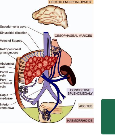
1.Intrahepatic portal hypertension. Cirrhosis is by far the commonest cause of portal hypertension. Other less frequent intrahepatic causes are metastatic tumours, non-cirrhotic nodular regenerative conditions, hepatic venous obstruction (Budd-Chiari syndrome), veno-occlusive disease, schistosomiasis, diffuse granulomatous diseases and extensive fatty change. In cirrhosis and other conditions, there is obstruction to the portal venous flow by fibrosis, thrombosis and pressure by regenerative nodules. About 3060% patients of cirrhosis develop significant portal hypertension.
2.Posthepatic portal hypertension. This is uncommon and results from obstruction to the blood flow through hepatic vein into inferior vena cava. The causes are neoplastic occlusion and thrombosis of the hepatic vein or of the inferior vena cava (including Budd-Chiari syndrome). Prolonged congestive heart failure and constrictive pericarditis may also cause portal hypertension by transmitting the elevated pressure through the hepatic vessels into the portal vein.
3.Prehepatic portal hypertension. Blockage of portal flow before portal blood reaches the hepatic sinusoids results in prehepatic portal hypertension. Such conditions are thrombosis and neoplastic obstruction of the portal vein before it ramifies in the liver, myelofibrosis, and congenital absence of portal vein.
MAJOR SEQUELAE OF PORTAL HYPERTENSION.
Irrespective of the mechanisms involved in the pathogenesis of portal hypertension, there are 4 major clinical conse- quences—ascites, varices (collateral channels or portosystemic shunts), splenomegaly and hepatic encephalopathy (Fig. 21.30).
1. Ascites. Ascites is the accumulation of excessive volume of fluid within the peritoneal cavity. It frequently accompanies cirrhosis and other diffuse liver diseases. The development of ascites is associated with haemodilution, oedema and decreased urinary output. Ascitic fluid is generally transudate with specific gravity of 1.010, protein content below 3 gm/dl and electrolyte concentrations like those of other extracellular fluids. It may contain a few mesothelial cells and mononuclear cells. Presence of neutrophils is suggestive of secondary infection and red blood cells in ascitic fluid points to disseminated intraabdominal cancer. However, some cases of ascites may develop serious complication of spontaneous bacterial peritonitis characterised by sponateneous infection of the ascitic fluid without any intrabdminal infection.
Pathogenesis. The ascites becomes clinically detectable when more than 500 ml of fluid has accumulated in the peritoneal cavity. The mechanisms involved in its formation were discussed in Chapter 5. Briefly, the systemic and local factors favouring ascites formation are as under (Fig. 21.31):
A. Systemic Factors:
i) Decreased plasma colloid oncotic pressure. There is hypoalbuminaemia from impaired hepatic synthesis of plasma proteins including albumin, as well as from loss of albumin from the blood plasma into the peritoneal cavity. Hypoalbuminaemia, in turn, causes reduced plasma oncotic pressure and leads to loss of water into extravascular space.
Figure 21.30 
 Major clinical consequences of portal hypertension.
ii)Hyperaldosteronism. In cirrhosis, there is increased aldosterone secretion by the adrenal gland, probably due to reduced renal blood flow, and impaired hepatic metabolism and excretion of aldosterone.
iii)Impaired renal excretion. Reduced renal blood flow and excessive release of antidiuretic hormone results in renal retention of sodium and water and impaired renal excretion.
B.Local Factors:
i)Increased portal pressure. Portal venous pressure is not directly related to ascites formation but portal hypertension in combination with other factors contributes to the formation and localisation of the fluid retention in the peritoneal cavity.
ii)Increased hepatic lymph formation. Obstruction of hepatic vein such as in Budd-Chiari syndrome and increased intrasinusoidal pressure found in cirrhotic patients stimulates hepatic lymph formation that oozes through the surface of the liver.
2.Varices (Collateral channels or Porto-systemic shunts).
As a result of rise in portal venous pressure and obstruction in the portal circulation within or outside the liver, the blood tends to bypass the liver and return to the heart by development of porto-systemic collateral channels (or shunts or varices). These varices develop at sites where the systemic and portal circulations have common capillary beds. The principal sites are as under:
i)Oesophageal varices: The development of oesophagogastric varices which is frequently manifested by massive
631
Pancreas Exocrine and Tract Biliary Liver, The 21 CHAPTER
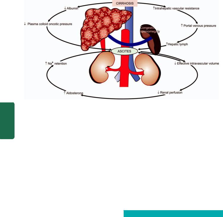
632
Pathology Systemic III SECTION
Figure 21.31 
 Mechanisms of ascites formation in cirrhosis.
haematemesis is the most important consequence of portal hypertension (page 539).
ii)Haemorrhoids: Development of collaterals between the superior, middle and inferior haemorrhoidal veins resulting in haemorrhoids is another common accompaniment. Bleeding from haemorrhoids is usually not as serious a complication as haematemesis from oesophageal varices.
iii)Caput medusae: Anastomoses between the portal and
systemic veins may develop between the hilum of the liver and the umbilicus along the paraumbilical plexus of veins resulting in abdominal wall collaterals. These appear as dilated subcutaneous veins radiating from the umbilicus and are termed caput medusae (named after the snake-haired
Medusa).
iv) Retroperitoneal anastomoses: In the retroperitoneum, portocaval anastomoses may be established through the veins of Retzius and the veins of Sappey.
3.Splenomegaly. The enlargement of the spleen in prolonged portal hypertension is called congestive splenomegaly (page 387). The spleen may weigh 500-1000 gm and is easily palpable. The spleen is larger in young people and in macronodular cirrhosis than in micronodular cirrhosis.
4.Hepatic encephalopathy. Porto-systemic venous shunting may result in a complex metabolic and organic syndrome of the brain characterised by disturbed consciousness, neurologic signs and flapping tremors. Hepatic encephalopathy is particularly associated with advanced hepatocellular disease such as in cirrhosis.
HEPATIC TUMOURS AND TUMOUR-LIKE LESIONS
The liver is the site for benign tumours, tumour-like lesions, and both primary and metastatic malignant tumours.
However, metastatic tumours are much more common than primary tumours and tumour-like lesions. Primary hepatic tumours may arise from hepatic cells, bile duct epithelium, or mesodermal structures (Table 21.11).
TUMOUR-LIKE LESIONS
These include cysts in the liver and focal nodular hyperplasia.
Hepatic Cysts
Cysts in the liver may be single or multiple. These cysts are mainly of 3 types—congenital, simple (nonparasitic) and hydatid (Echinococcus) cysts.
1. CONGENITAL CYSTS. These are uncommon. They are usually small (less than 1 cm in diameter) and are lined by biliary epithelium. They may be single, or occur as polycystic liver disease, often associated with polycystic kidney. On occasions, these cysts have abundant connective tissue and
TABLE 21.11: Classification of Primary Hepatic Tumours.
|
Benign |
Malignant |
A. |
Hepatocellular tumours |
|
|
Hepatocellular (liver cell) |
Hepatocellular (liver cell) |
|
adenoma |
carcinoma |
|
|
Hepatoblastoma (Embryoma) |
B. |
Biliary tumours |
|
|
Bile duct adenoma |
Cholangiocarcinoma |
|
(Cholangioma) |
Combined hepatocellular and |
|
|
cholangiocarcinoma |
|
|
Cystadenocarcinoma |
C. |
Mesodermal tumours |
|
|
Haemangioma |
Angiosarcoma |
|
|
Embryonal sarcoma |
|
|
|

numerous ducts, warranting the designation of congenital hepatic fibrosis.
2. SIMPLE (NON-PARASITIC) CYSTS. Simple cysts are solitary non-parasitic cysts seen more frequently in middleaged women. The cyst is usually large (up to 20 cm in diameter), lying underneath the Glisson’s capsule and filled with serous fluid. The cyst produces a palpable mass and may be associated with jaundice.
Histologically, the cyst wall is composed of compact fibrous tissue and is lined by low columnar to cuboid epithelium and occasionally by squamous lining.
3. HYDATID (ECHINOCOCCUS) CYSTS. Hydatid cyst has already been discussed on page 616.
Focal Nodular Hyperplasia
The etiology of focal nodular hyperplasia is not known but these lesions are more common in women taking oral contraceptives.
Grossly, Focal nodular hyperplasia is a well-demarcated tumour-like nodule occurring underneath the Glisson’s capsule. The nodules may be single or multiple, measuring about 5 cm in diameter. It may be tan-yellow or bile-stained. The sectioned surface shows a central fibrous scar.
Histologically, it is composed of collagenous septa radiating from the central fibrous scar which separate nodules of normal hepatocytes without portal triads or central hepatic veins. The fibrous septa contain prominent lymphocytic infiltrate.
BENIGN HEPATIC TUMOURS
These are uncommon and some of them are incidental autopsy findings. These include hepatocellular (liver cell) adenoma, bile duct adenoma (cholangioma) and haemangioma.
Hepatocellular (Liver Cell) Adenoma
Adenomas arising from hepatocytes are rare and are reported in women in reproductive age group in association with use of oral contraceptives, sex hormone therapy and with pregnancy. The tumour presents as intrahepatic mass that may be mistaken for hepatocellular carcinoma and may rupture causing severe intraperitoneal haemorrhage.
MORPHOLOGIC FEATURES. Grossly, the tumour usually occurs singly but about 10% are multiple. It is partly or completely encapsulated and slightly lighter in colour than adjacent liver or may be bile-stained. The tumours vary from a few centimetres up to 30 cm in diameter. On cut section, many of the tumours have varying degree of infarction and haemorrhage.
Histologically, liver cell adenomas are composed of sheets and cords of hepatocytes which may be normal-looking
or may show slight variation in size and shape but no |
633 |
|
|
mitoses. The hepatocytes in adenomas contain greater |
|
amount of glycogen than the surrounding liver cells and |
|
may sometimes show fatty change. Hepatocellular |
|
adenomas lack portal tracts and bile ducts but bile |
|
canaliculi containing bile-plugs may be present. |
|
Numerous blood vessels are generally present in the |
|
tumour which may be thrombosed. Thrombosis leads to |
|
infarction and may result in rupture with intraperitoneal |
|
haemorrhage. |
|
|
|
Bile Duct Adenoma (Cholangioma) |
|
||
Intrahepatic or extrahepatic bile duct adenoma is a rare |
|
||
benign tumour. The tumour may be small, composed of acini |
|
||
lined by biliary epithelium and separated by variable amount |
|
||
of connective tissue, or are larger cystadenomas having loculi |
|
||
lined by biliary epithelium. |
|
||
Haemangioma |
|
||
Haemangioma is the commonest benign tumour of the liver. |
|
||
Majority of them are asymptomatic and discovered |
CHAPTER |
||
incidentally. Rarely, a haemangioma may rupture into the |
|||
peritoneal cavity. |
|||
|
|
||
MORPHOLOGIC FEATURES. Grossly, haemangiomas |
|
||
appear as solitary or multiple, circumscribed, red-purple |
|
||
lesions, commonly subcapsular and varying from a few |
|
21 |
|
millimetres to a few centimetres in diameter. They are |
|
||
|
|
||
commonly cavernous type giving the sectioned surface a |
|
The |
|
spongy appearance. |
|
||
Histologically, haemangioma of the liver shows |
|
||
|
Liver, |
||
characteristic large, cavernous, blood-filled spaces, lined |
|
||
by a single layer of endothelium and separated by |
|
||
connective tissue (Fig. 21.32). Some haemangiomas |
|
||
|
Biliary |
||
may undergo progressive fibrosis and may later get |
|
||
calcified. |
|
||
|
|
||
MALIGNANT HEPATIC TUMOURS |
Tract |
||
Among the primary malignant tumours of the liver, |
|||
and |
|||
hepatocellular (liver cell) carcinoma accounts for approxi- |
|||
mately 85% of all primary malignant tumours, |
|||
Exocrine |
|||
cholangiocarcinoma for about 5-10%, and infrequently mixed |
|||
pattern is seen. The remainder are rare tumours that include |
|||
hepatoblastoma, haemangiosarcoma (angiosarcoma) and |
|||
embryonal sarcoma. Hepatic haemangiosarcoma and |
|||
embryonal sarcoma resemble in morphology with their |
Pancreas |
||
counterparts elsewhere in the body. |
|||
Hepatocellular Carcinoma |
|||
|
|
||
Hepatocellular carcinoma (HCC) or liver cell carcinoma, also |
|
||
trmed as hepatoma, is the most common primary malignant |
|
||
tumour of the liver. The tumour shows marked geographic |
|
||
variations in incidence which is closely related to HBV and |
|
||
HCV infection in the region. Whereas the prevalence of HCC |
|
||
is less than 1% of all autopsies in the United States and |
|
||
Europe, the incidence in sub-Saharan Africa and South-East |
|
||
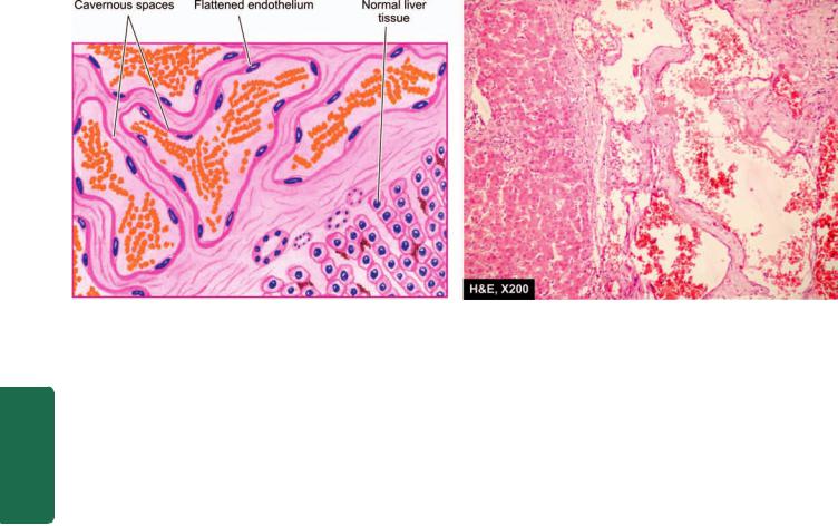
634
Pathology Systemic III SECTION
Figure 21.32 
 Cavernous haemangioma of the liver.
Asia (particularly China) is high (2-8%). Liver cell cancer is more common in males than in females in the ratio of 4:1. The peak incidence occurs in 5th to 6th decades of life but in high incidence areas where HBV and HCV infection is prevalent, it occurs a decade or two earlier. The tumour supervenes on cirrhosis in 70-80% of cases.
ETIOPATHOGENESIS. A number of etiologic factors are implicated in the etiology of HCC, most important being HBV
and HCV infection, and association with cirrhosis.
1. Relation to HBV infection. Genesis of HCC is linked to prolonged infection with HBV. The evidence in support is both epidemiologic and direct.
i)The incidence of HBsAg positivity is higher in HCC patients. For example, in Taiwan, HBsAg-positive carriers have more than 200 times greater risk of developing HCC than HBsAg-negative patients, particularly when the infection is acquired in early life.
ii)In African and Asian patients, 95% cases of HCC have anti-HBc.
iii)There is more direct evidence of integration of HBV-DNA genome in the genome of tumour cells of HCC.
2. Relation to HCV infection. Long-standing HCV infection has emereged as a major factor in the etiology of HCC, generally after more than 30 years of infection. The evidences in support are as under:
i)In developed countries where higher incidence of HCC was earlier attributed to endemic HBV infection (e.g. in Japan) has shown a remarkable shift to HCV infection. However, in developing countries HBV is still the predominant etiologic factor in the pathogenesis of HCC.
ii)The patients having anti-HCV and anti-HBc antibodies together have three times higher risk of developing HCC than in those with either antibody alone.
iii)HCV infection after a long interval produces cirrohosis more often prior to development of HCC, while in HCC following HBV infection half the cases have cirrohosis and remainder have chronic hepatitis.
iv) It is also possible that HBV and HCV infection act synergistically to predispose to HCC.
3.Relation to cirrhosis. Cirrhosis of all etiologic types is more commonly associated with HCC but the most frequent association is with macronodular post-necrotic cirrhosis. The mechanism of progression to HCC appears to be chronic regenerative activity in cirrhosis, or that the damaged liver in cirrhosis is rendered vulnerable to carcinogenic influences. Liver cell dysplasia identified by cellular enlargement, nuclear hyperchromatism and multinucleate cells, is found in 60% of cirrhotic livers with HCC and in only 10% of non-cirrhotic livers.
4.Relation to alcohol. It has been observed that alcoholics have about four-fold increased risk of developing HCC. It is possible that alcohol may act as co-carcinogen with HBV or HCV infection, but alcohol does not appear to be a hepatic carcinogen per se.
5.Mycotoxins. An important mycotoxin, aflatoxin B1, produced by a mould Aspergillus flavus, can contaminate poorly stored wheat grains or groundnuts, especially in developing countries. Aflatoxin B1 is carcinogenic; it may act as a co-carcinogen with hepatitis B or may suppress the cellular immune response.
6.Chemical carcinogens. A number of chemical carcinogens can induce liver cancer in experimental animals. These include butter-yellow and nitrosamines used as common food additives.
7.Miscellaneous factors. Limited role of various other factors in HCC has been observed. These include the following:
i)haemochromatosis;
ii)α-1-antitrypsin deficiency;
iii)prolonged immunosuppressive therapy in renal transplant patients;
iv)other types of viral hepatitis;
v)tobacco smoking; and
vi)parasitic infestations such as clonorchiasis and schistosomiasis.
635
Figure 21.33 
 Macroscopic patterns of hepatocellular carcinoma.
Pathogenesis of hepatocellular carcinoma can be explained on the basis of genetic mutations induced by one of the above major etiologic factors. In many cases, this mutated gene has been identified as inactivation of tumour suppressor oncogene p53 by HBV that results in disruption of normal growth control. In this regards, the role of X-protein (HBxAg) generated from X-gene of HBV has been found to contribute to carcinogenesis by binding to p53.
MORPHOLOGIC FEATURES. Grossly, HCC may form one of the following 3 patterns of growth, in decreasing order of frequency (Fig. 21.33):
i)Expanding type: Most frequently, it forms a single, yellow-brown, large mass, most often in the right lobe of the liver with central necrosis, haemorrhage and occasional bile-staining (Fig. 21.34). It may be deceptively encapsulated.
ii)Multifocal type: Less often, multifocal, multiple masses,
3-5 cm in diameter, scattered throughout the liver are seen.
iii)Infiltrating (Spreading) type: Rarely, the HCC forms diffusely infiltrating tumour mass.
Microscopically, the tumour cells in the typical HCC resemble hepatocytes but vary with the degree of differentiation, ranging from well-differentiated to highly anaplastic lesions. Most of the HCC have trabecular growth pattern. The tumour cells have a tendency to
invade and grow along blood vessels. Thus important diagnostic features are the patterns of tumour cells and their
cytologic features:
1. Histologic patterns: These include the following:
i)Trabecular or sinusoidal pattern is the most common. The trabeculae are made up of 2-8 cell wide layers of tumour cells separated by vascular spaces or sinusoids which are endothelium-lined (Fig. 21.35).
ii)Pseudoglandular or acinar pattern is seen sometimes. The tumour cells are disposed around central cystic space formed by degeneration and breakdown in solid trabeculae.
iii)Compact pattern resembles trabecular pattern but the tumour cells form large solid masses with inconspicuous sinusoids.
iv)Scirrhous pattern is characterised by more abundant fibrous stroma.
2 Cytologic features: The typical cytologic features in the HCC consist of cells resembling hepatocytes having vesicular nuclei with prominent nucleoli. The cytoplasm is granular and eosinophilic but becomes increasingly basophilic with increasing malignancy. Aside from these features, a few other cytologic variants are: pleomorphism, bizarre giant cell formation, spindle-shaped cells, tumour cells with clear cytoplasm, presence of bile within dilated canaliculi, and intracytoplasmic Mallory’s hyalin.
Immunohistochemically, hepatocellular carcinoma cells stain positively with AFP, EMA, keratin etc.
FIBROLAMELLAR CARCINOMA. A clinicopathologic variant of the HCC is fibrolamellar carcinoma of the liver found in young people of both sexes. The tumour forms a
Figure 21.34 
 Hepatocellular carcinoma. Sectioned surface shows a single, large mass (arrow) with irregular borders and having central areas of necrosis, while rest of the hepatic parenchyma in the upper part of the picture shows many nodules of variable sizes owing to co-existent macronodular cirrhosis.
Pancreas Exocrine and Tract Biliary Liver, The 21 CHAPTER
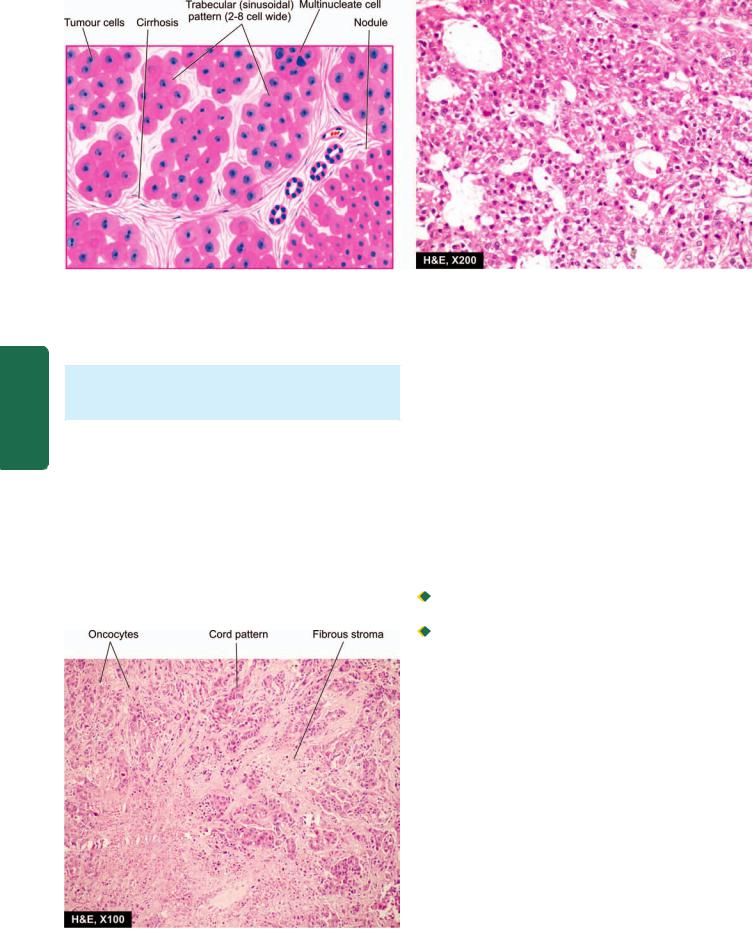
636
Pathology Systemic III SECTION
Figure 21.35 
 Hepatocellular carcinoma, typical microscopic pattern. The tumour cells resembling hepatocytes show pleomorphism and are seen forming 2-8 cell wide trabeculae which are separated by endothelium-lined sinusoidal spaces.
single large mass which may be encapsulated and occurs in the absence of cirrhosis.
Histologically, the tumour is composed of eosinophilic polygonal cells (oncocytes) forming cords and nests which are separated by bands of fibrous stroma (Fig. 21.36).
The prognosis of fibrolamellar carcinoma is better than other forms of HCC.
CLINICAL FEATURES. Hepatic cancer may remain undetected initially because it often occurs in patients with underlying cirrhosis. The usual features consist of hepatomegaly with palpable mass in the liver, right upper quadrant pain or tenderness, and less often, jaundice, fever and haemorrhage from oesophageal varices. Ascites with RBCs and malignant cells is found in about half the patients. Rarely, systemic endocrine manifestations due to paraneo-
Figure 21.36 
 Fibrolamellar carcinoma liver.
plastic syndrome are eobsereved such as hypercalcaemia, hypoglycaemia, gynaecomastia and acquired porphyria.
Laboratory findings yield nonspecific results like anaemia, markedly elevated serum alkaline phosphatase as found in cirrhosis, and high serum alpha-foetoprotein (AFP). Elevated AFP level is quite specific; very high levels of AFP (above 500 ng/ml) are observed in 70-80% cases of HCC but lacks sensitivity since AFP is also found elevated in yolk sac tumour, cirrhosis, chronic hepatitis, massive liver necrosis and normal pregnancy. Ultrasound of the liver has been reported to be more sensitive than elevated AFP level. An abnormal type of prothrombin, des-γ-carboxy prothrombin, is also elevated and correlates well with AFP levels.
SPREAD. The HCC can have both intrahepatic and extrahepatic spread which faithfully reproduces the structure of the primary tumour:

 Intrahepatic spread occurs by haematogenous route and forms multiple metastases in the liver.
 Extrahepatic spread occurs via hepatic or portal veins to different sites, chiefly to lungs and bones, and by lymphatic route to regional lymph nodes at the porta hepatis and to mediastinal and cervical lymph nodes.
The causes of death from the HCC are cachexia, massive bleeding from oesophageal varices, and liver failure with hepatic coma.
Cholangiocarcinoma
Cholangiocarcinoma is the designation used for carcinoma arising from bile duct epithelium within the liver (peripheral cholangiocarcinoma). Carcinomas arising from the large hilar ducts (hilar cholangiocarcinoma) and from extrahepatic ducts are termed bile duct carcinomas (page 644). None of the etiologic factors related to HCC have any role in the genesis of cholangiocarcinoma. However, the etiological factors involved in it are exposure to radio-opaque dye thorotrast, anabolic steroids, clonorchiasis and fibrocystic disease. The tumour affects older people and the clinical features are those of HCC but with prominence of jaundice.
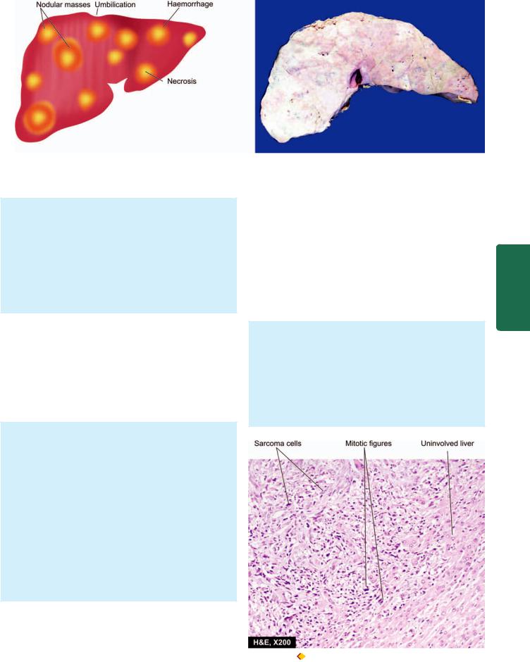
637
Figure 21.37 
 Metastatic tumour deposits in the liver as seen on sectioned surface. Characteristic features include multiple, variable-sized, nodular masses, often under the capsule, producing umbilication on the surface.
MORPHOLOGIC FEATURES. Grossly, the tumour is firm to hard and whitish.
Microscopically, the tumour has glandular structure. The tumour cells resemble biliary epithelium but without bile secretion. They form various patterns such as tubular, ductular or papillary. The stroma consists of fibrous tissue with little or no capillary formation. Occasionally, mucinous, signet-ring and adenosquamous type of patterns are found. An uncommon variant is combined hepatocellular-cholangiocarcinoma.
Hepatoblastoma (Embryoma)
Hepatoblastoma is a rare malignant tumour arising from primitive hepatic parenchymal cells. It presents before the age of 2 years as progressive abdominal distension with anorexia, failure to thrive, fever and jaundice. It is more common in boys. The concentration of serum AFP is high. The tumour grows rapidly and causes death by haemorrhage, hepatic failure or widespread metastases.
MORPHOLOGIC FEATURES, Grossly, the tumour is circumscribed and lobulated mass measuring 5-25 cm in size, having areas of cystic degeneration, haemorrhage and necrosis.
Microscopically, hepatoblastoma consists of 2 components:
i)Epithelial component contains 2 types of cells— ‘embryonal’ hepatocytes are small with dark-staining, hyperchromatic nuclei and scanty cytoplasm, and ‘foetal’ hepatocytes are larger with more cytoplasm that may be granular or clear. The epithelial cells are organised in trabeculae, ribbons or rosettes.
ii)Mesenchymal component includes fibrous connective tissue, cartilage and osteoid of variable degree of maturation. Extramedullary haematopoiesis is a frequent accompaniment.
Secondary Hepatic Tumours
Metastatic tumours in the liver are more common than the primary hepatic tumours. Most frequently, they are bloodborne metastases, irrespective of whether the primary tumour is drained by portal vein or systemic veins. Most
frequent primary tumours metastasising to the liver, in descending order of frequency, are those of stomach, breast, lungs, colon, oesophagus, pancreas, malignant melanoma and haematopoietic malignancies. Sarcomas rarely metastasise to the liver. Occasionally, metastatic involvement may be present in the absence of obvious evidence of primary tumour. Aside from general features of disseminated malignancy such as anorexia, cachexia and anaemia, the patients have hepatomegaly with nodular free margin. There is little hepatic dysfunction until late in the course of hepatic metastatic disease.
MORPHOLOGIC FEATURES. Grossly, most metastatic carcinomas form multiple, spherical, nodular masses which are of variable size. Liver is enlarged and heavy, weighing 5 kg or more. The tumour deposits are white, well-demarcated, soft or haemorrhagic. The surface of the liver shows characteristic umbilication due to central necrosis of nodular masses (Fig. 21.37).
Histologically, the metastatic tumours generally reproduce the structure of the primary lesions (Fig. 21.38).
Figure 21.38 
 Metastatic deposits from undifferentiated sarcoma in the liver.
Pancreas Exocrine and Tract Biliary Liver, The 21 CHAPTER

638
Pathology Systemic III SECTION
BILIARY SYSTEM
NORMAL STRUCTURE
ANATOMY. The gallbladder is a pear-shaped organ, 9 cm in length and has a capacity of approximately 50 ml. It consists of the fundus, body and neck that tapers into the cystic duct. The two hepatic ducts from right and left lobes of the liver unite at the porta hepatis to form the common hepatic duct which is joined by the cystic duct from the gallbladder to form the common bile duct. The common bile duct enters the second part of the duodenum posteriorly. In about 70% of cases, it is joined by the main pancreatic duct to form the combined opening in the duodenum (ampulla of Vater). In 30% cases, the common bile duct and the pancreatic duct open separately into the duodenum (see Fig. 21.1). The common bile duct in its duodenal portion is surrounded by longitudinal and circular muscles derived from the duodenum forming sphincter of Oddi.
HISTOLOGY. Histologically, the gallbladder, unlike the rest of gastrointestinal tract, lacks the muscularis mucosae and submucosa. The wall of the gallbladder is composed of the following 4 layers:
1.Mucosal layer: It has a single layer of tall columnar epithelium which is thrown into permanent folds that are larger and more numerous in the neck of the gallbladder. Beneath the epithelium is delicate lamina propria that contains capillaries, and in the region of the neck, a few acinar glands are present.
2.Smooth muscle layer: External to the lamina propria are smooth muscle bundles in layers—inner longitudinal, middle oblique, and outer circular.
3.Perimuscular layer: Outer to the muscle layer is a zone of fibrous connective tissue with some interspersed fat cells.
4.Perimuscular layer: The perimuscular layer is covered by serosa on the peritoneal surface of the gallbladder. The peritoneum covers the gallbladder except in the region of gallbladder fossa where it is embedded in the liver.
The extrahepatic bile ducts are also lined by tall columnar epithelium that overlies the lamina propria. It is surrounded by dense layer of fibromuscular tissue. The ducts which lie between the lobules of the liver and receive bile from the canaliculi are lined by cuboidal or flattened cells.
FUNCTIONS. The main function of the gallbladder is to store and concentrate the bile secreted by the liver and then deliver it into the intestine for digestion and absorption of fat. The concentrating ability of the gallbladder is due to its absorptive mucosal surface that has numerous folds. Normally, the liver secretes approximately 500 ml of bile per day and the gallbladder concentrates it 5-10 times. The motility, concentration and relaxation of the gallbladder are under the influence of a peptide hormone, cholecystokinin, released from neuroendocrine cells of the duodenum and jejunum.
CONGENITALANOMALIES
Several uncommon congenital anomalies of the biliary system have been described. These include: agenesis,
duplication and heterotopic tissue. However, congenital cystic lesions of the bile ducts (as also of the liver) are more frequently diagnosed. These conditions include: congenital intrahepatic biliary dilatation (Caroli’s disease), choledochal cysts, polycystic liver disease and congenital hepatic fibrosis. They are found in various combinations and are usually inherited. All of them may be complicated by malignant change.
CHOLELITHIASIS (GALLSTONES)
Gallstones are formed from constituents of the bile (viz. cholesterol, bile pigments and calcium salts) along with other organic components. Accordingly, the gallstones commonly contain cholesterol, bile pigment and calcium salts in varying proportions. They are usually formed in the gallbladder, but sometimes may develop within extrahepatic biliary passages, and rarely in the larger intrahepatic bile duct.
RISK FACTORS. The incidence of gallstones varies markedly in different geographic areas, age, gender, diet and various other risk factors. These factors which largely pertain to cholesterol stones can be summed up in the old saying that gallstones are common in 4F’s acronym for—‘fat, female, fertile (multipara) and forty’. Some of the risk factors in lithogenesis are explained below:
1.Geography. Gallstones are quite prevalent in almost the entire Western world. American Indians have the highest known prevalence. Black Africans and populations in the Eastern world are relatively free of cholelithiasis.
2.Genetic factors. There is increased frequency of gallstones in first-degree relatives of patients with cholelithiasis. Patients of gallstones disease have increased secretion of dietary cholesterol in bile than in non-gallstone patients inspite of high-cholesterol diet. Recently, mutation in CYP7A1 gene has been found that results in deficiency of enzyme, cholesterol 7-hydroxylase, which has a role in bile acid synthesis. This mutation is associated with hypercholesterolaemia and gallstones.
3.Age. There is steady increase in the prevalence of gallstones with advancing age which may be related to increased cholesterol content in the bile. The incidence increases above the age of 40 and presentation is usually in the 50s and 60s.
4.Sex. Gallstones are twice more frequent in women than in men. In the United States, autopsy series have shown gallstones in about 20% of women and 8% of men above the age of 40. The incidence is higher in multiparous women than in nulliparous women.
5.Drugs. Women on oestrogen therapy or on birth control pills have higher incidence of gallstones. This is considered to be due to production of more lithogenic bile as a result of cholestatic effect of oestrogen. Similar is the influence of clofibrate used for lowering blood cholesterol.
6.Obesity. Obesity is associated with increased cholesterol synthesis and its excretion resulting in higher incidence of gallstones in obese patients.
7.Diet. Deficiency of dietary fibre content is linked to higher prevalence of gallstones. A moderate consumption of alcohol, however, seems to protect against gallstones.
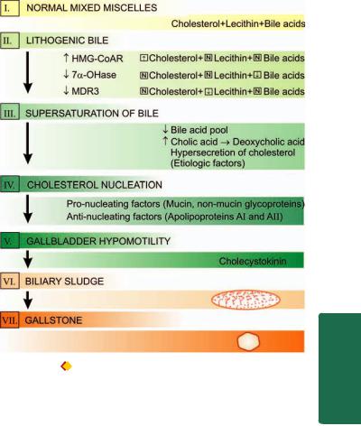
8.Gastrointestinal diseases. Certain gastrointestinal disorders such as Crohn’s disease, ileal resection, ileal bypass surgery etc are associated with interruption in enterohepatic circulation followed by gallstone formation.
9.Factors in pigment gallstones. All the above factors apply largely to cholesterol stones. Pigment stones, whether pure or mixed type, are more frequently associated with haemolytic anaemias which lead to increased content of unconjugated bilirubin in the bile. Pigment stones are also more frequent in cirrhosis and hepatocellular disease.
PATHOGENESIS. The mechanism of gallstone formation (i.e. lithogenesis) is explained separately below under 2 headings: firstly for cholesterol, mixed gallstones and biliary sludge; and, secondly for pigment gallstones as under:
PATHOGENESIS OF CHOLESTEROL, MIXED GALLSTONES AND BILIARY SLUDGE. Cholesterol is essentially insoluble in water and can be solublised by another lipid. Normally, cholesterol and phospholipids (lecithin) are secreted into bile as ‘bilayered vesicles’ but are converted into ‘mixed miscelles’ by addition of bile acids, the third constituent. If there is excess of cholesterol compared to the other two constituents, unstable cholesterol-rich vesicles remain behind which aggregate and form cholesterol crystals. Formation of such lithogenic (stone-forming) bile is explained by the following mechanisms (Fig. 21.39):
1.Supersaturation of bile: Several etiologic factors listed above favour increased secretion of cholesterol in the presence of normal bile acids and lecithin in the bile as the major mechanism for initiation of gallstone formation. These factors cause enhanced activity of enzyme, HMG-CoA reductase, that normally regulates cholesterol synthesis and its hepatic uptake. Two other disturbances which may contribute to supersaturation of the bile with cholesterol are as under:
i)Reduced bile acid pool: This causes rapid loss of the available bile acids into the small intestine and then into the colon, resulting in supersaturation of the bile with cholesterol.
ii)Increased conversion of cholic acid to deoxycholic acid: This causes increased secretion of deoxycholate in the bile which is associated with hypersecretion of cholesterol into the bile.
Mutation in MDR3 gene has been found that causes defect in phospholipid secretion from bile, resulting in cholesterol supersaturation of bile and cholesterol gallstone formation. Although supersaturation of the bile with cholesterol is an important pre-requisite for lithogenesis, this in itself is not sufficient for cholesterol precipitation.
2.Cholesterol nucleation. Initiation of cholesterol stones occurs by nucleation of cholesterol monohydrate crystals. Accelerated nucleation of cholesterol monohydrate may occur either from pro-nucleating factors or from deficiency of anti-nucleating factors:
i)Pro-nucleating factors are mucin and non-mucin glycoproteins secreted by epithelial cells of the gallbladder.
ii)Anti-nucleating factors are apolipoproteins AI and AII, and some glycoproteins.
Cholesterol monohydrate nucleation probably occurs in the mucin gel layer of the gallbladder followed by continued
Figure 21.39 |
Schematic pathogenesis of gallstone formation. (HMG- |
CoAR = hydroxy methyl glutaryl-coenzyme A reductase; 7α-OHase = |
|
cholesterol 7 α-OHase hydroxylase; MDR3 = multidrug resistanceassociated protein 3).
addition and precipitation of more crystals resulting in solid state crystals.
3. Gallbladder hypomotility. Normally, the gallbladder is capable of emptying and clearing any sludge or debris which might initiate stone formation. This takes place under the influence of cholecystokinin secreted from small intestine. However, the motility of gallbladder may be impaired due to decrease in cholecystokinin receptors in the gallbladder resulting in stasis of biliary sludge and lithogenesis. A defect in gallbladder emptying has been found to play a role in recurrence of gall-stone formation in patients who undergo biliary lithotripsy.
PATHOGENESIS OF PIGMENT GALLSTONES. The mechanism of pigment stone formation is explained on the basis of following factors:
i)Chronic haemolysis resulting in increased level of unconjugated bilirubin in the bile.
ii)Alcoholic cirrhosis.
iii)Chronic biliary tract infection e.g. by parasitic infestations
of the biliary tract such as by Clonorchis sinensis and Ascaris
lumbricoides.
iv) Demographic and genetic factors e.g. in rural setting and prevalence in Asian countries.
TYPES OF GALLSTONES. As stated before, gallstones contain cholesterol, bile pigment and calcium carbonate, either in pure form or in various combinations. Accordingly, gallstones are of 3 major types—pure gallstones, mixed gallstones and combined gallstones. Mixed gallstones are the
639
Pancreas Exocrine and Tract Biliary Liver, The 21 CHAPTER

640 TABLE 21.12: Features of Gallstones.
|
Type |
Frequency |
Composition |
Gallbladder Changes |
Appearance |
|
1. |
Pure |
10% |
i) |
Cholesterol |
Cholesterolosis |
Solitary, oval, large, smooth, yellow- |
|
gallstones |
|
|
|
|
white; on C/S radiating glistening |
|
|
|
|
|
|
crystals |
|
|
|
ii) |
Bile pigment |
No change |
Multiple, small, jet-black, mulberry- |
|
|
|
|
|
|
shaped; on C/S soft black |
|
|
|
iii) Calcium carbonate |
No change |
Multiple, small, grey-white, faceted; |
|
|
|
|
|
|
|
C/S hard |
2. |
Mixed |
80% |
Cholesterol, bile pigment |
Chronic cholecystitis |
Multiple, multifaceted, variable size, |
|
|
gallstones |
|
and calcium carbonate in |
|
on C/S laminated alternating dark- |
|
|
|
|
varying combination |
|
pigment layer and pale-white layer |
|
3. |
Combined |
10% |
Pure gallstone nucleus with |
Chronic cholecystitis |
Solitary, large, smooth; on C/S |
|
|
gallstones |
|
mixed gallstone shell, or |
|
central nucleus of pure gallstone |
|
|
|
|
mixed gallstone nucleus with |
|
with mixed shell or vice versa |
|
|
|
|
pure gallstone shell |
|
|
|
|
|
|
|
|
|
|
Pathology Systemic III SECTION
most common (80%) while pure and combined gallstones comprise 10% each. In general, gallstones are formed most frequently in the gallbladder but may occur in extrahepatic as well as intrahepatic biliary passages. Gallbladder containing pure stones shows no significant inflammatory reaction, whereas chronic cholecystitis is invariably present in gallbladder with either mixed or combined gallstones. Presence of calcium salts renders gallstones radio-opaque, while cholesterol stones appear as radiolucent filling defects in the gallbladder.
The salient features of various types of gallstones are summarised in Table 21.12 and presented below:
1. Pure gallstones. They constitute about 10% of all gallstones. They are further divided into 3 types according to the component of bile forming them. These are as under
(Fig. 21.40):
i)Pure cholesterol gallstones: They are usually solitary, oval and fairly large (3 cm or more) filling the gallbladder. Their surface is hard, smooth, whitish-yellow and glistening. On cut section, the pure cholesterol stone shows radiating glistening crystals. It may result in deposition of cholesterol within the mucosal macrophages of the gallbladder producing cholesterolosis which is an asymptomatic condition (Fig. 21.41). Pure cholesterol stones are radiolucent but 1020% of them have calcium carbonate in them which renders them opaque.
ii)Pure pigment gallstones: These stones composed primarily of bile pigment, calcium bilirubinate, and contain less than 20% cholesterol. They are generally multiple, jet-black and small (less than 1 cm in diameter). They have mulberry like external surface. They are soft and can be easily crushed. The gallbladder usually appears uninvolved.
iii)Pure calcium carbonate gallstones: They are rare. Calcium carbonate gallstones are usually multiple, grey-white, small (less than 1 cm in diameter), faceted and fairly hard due to calcium content. They, too, do not produce any change in the gallbladder wall.
2. Mixed gallstones. Mixed gallstones are the most common
(80%) and contain more than 50% cholesterol monohydarate plus an admixture of calcium salts, bile pigments and fatty
acids. They are always multiple, multifaceted so that they fit together and vary in size from as tiny as sand-grain to 1 cm or more in diameter. On section, they have distinct laminated structure with alternating dark pigment layer and pale-white layer revealing different combinations of cholesterol, bilirubin pigment and calcium carbonate, laid down in layers at different times (Fig. 21.42, A).
Mixed gallstones are invariably accompanied by chronic cholecystitis.
3. Combined gallstones. They comprise about 10% of all gallstones. Combined gallstones are usually solitary, large and smooth-surfaced. It has a pure gallstone nucleus (cholesterol, bile pigment or calcium carbonate) and outer shell of mixed gallstone; or a mixed gallstone nucleus with pure gallstone shell (Fig. 21.42, B).
Combined gallstones, too, are associated with chronic cholecystitis.
CLINICAL MANIFESTATIONS AND COMPLICATIONS. In about 50% cases, gallstones cause no symptoms and may be diagnosed by chance during investigations for some other condition (silent gallstones). The future course in such asymptomatic silent cases is controversial, most surgeons advocating cholecystectomy while physicians advising watchful waiting. Follow-up studies, however, show that only about 10% of such cases develop symptoms. Symptomatic gallstone disease appears only when complications develop. These are as under:
1.Cholecystitis. The relationship between cholelithiasis and cholecystitis is well known but it is not certain which of the two comes first. The patients with gallstones develop symptoms due to cholecystitis which include typical biliary colic precipitated by fatty meal, nausea, vomiting, fever alongwith leucocytosis and high serum bilirubin.
2.Choledocholithiasis. Gallstones may pass down into the extrahepatic biliary passages and the small bowel, or less often they may be formed in the biliary tree. Patients with gallstone in the common bile duct frequently develop pain and obstructive jaundice. Fever may develop due to bacterial ascending cholangitis.
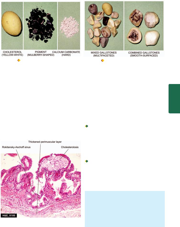
641
Figure 21.40 |
Pure gallstones of various types. |
Figure 21.42 |
Mixed and combined gallstones. |
3.Mucocele. Mucocele or hydrops of the gallbladder is distension of the gallbladder by clear, watery mucinous secretion resulting from impacted stones in the neck of the gallbladder.
4.Biliary fistula. An uncommon complication of cholelithiasis is formation of fistulae between one part of the biliary system and the bowel, and rarely between the gallbladder and the skin.
5.Gallstone ileus. A gallstone in the intestine may be passed in the faeces without causing symptoms. Occasionally, however, gallstones in the intestine may cause intestinal obstruction called gallstone ileus.
6.Gallbladder cancer. There is a small and doubtful risk of development of cancer of the gallbladder in cases with cholelithiasis (page 643).
Figure 21.41 
 Cholesterolosis of the gallbladder. The lamina propria of the mucosa shows foamy macrophages.
CHOLECYSTITIS
Cholecystitis or inflammation of the gallbladder may be acute, chronic, or acute superimposed on chronic. Though chronic cholecystitis is more common, acute cholecystitis is a surgical emergency.
Acute Cholecystitis
In many ways, acute cholecystitis is similar to acute appendicitis. The condition usually begins with obstruction, followed by infection later.
ETIOPATHOGENESIS. Based on the initiating mechanisms, acute cholecystitis occurs in two types of situations—acute
calculous and acute acalculous cholecystitis.
 Acute calculous cholecystitis. In 90% of cases, acute cholecystitis is caused by obstruction in the neck of the gallbladder or in the cystic duct by a gallstone. The commonest location of impaction of a gallstone is in Hartmann’s pouch. Obstruction results in distension of the gallbladder followed by acute inflammation which is initially due to chemical irritation. Later, however, secondary bacterial infection, chiefly by E. coli and Streptococcus faecalis, supervenes.

 Acute acalculous cholecystitis. The remaining 10% cases of acute cholecystitis do not contain gallstones. In such cases, a variety of causes have been assigned such as previous nonbiliary surgery, multiple injuries, burns, recent childbirth, severe sepsis, dehydration, torsion of the gallbladder and diabetes mellitus. Rare causes include primary bacterial infection like salmonellosis and cholera and parasitic infestations.
MORPHOLOGIC FEATURES. Except for the presence or absence of calculi, the two forms of acute cholecystitis are morphologically similar.
Grossly, the gallbladder is distended and tense. The serosal surface is coated with fibrinous exudate with congestion and haemorrhages. The mucosa is bright red. The lumen is filled with pus mixed with green bile. In calculous cholecystitis, a stone is generally impacted in the neck or in the cystic duct. When obstruction of the
Pancreas Exocrine and Tract Biliary Liver, The 21 CHAPTER
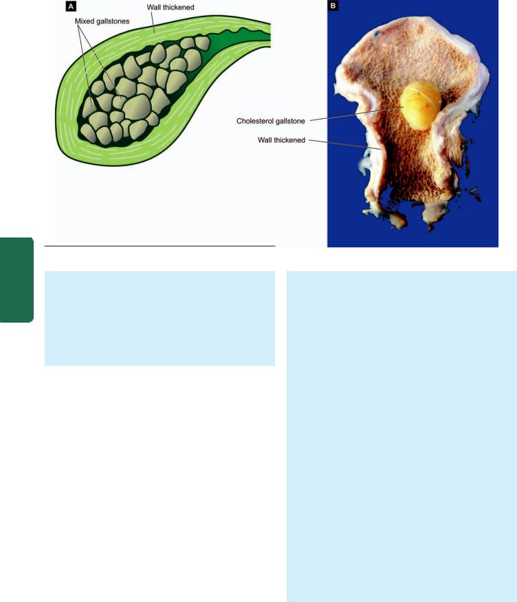
642
Pathology Systemic III SECTION
Figure 21.43 
 Chronic cholecystitis with cholelithiasis. A, Mixed gallstones, diagrammatic view. The wall of the gallbladder is thickened and the lumen is packed with well-fitting, multiple, multi-faceted, mixed gallstones. B, Chronic cholecystitis with cholesterol cholelithiasis. The wall of gallbladder is thickened. The lumen contains a single, large, oval, and hard yellow-white gallstone.
cystic duct is complete, the lumen is filled with purulent exudate and the condition is known as empyema of the
gallbladder.
Microscopically, wall of the gallbladder shows marked inflammatory oedema, congestion and neutrophilic exudate. There may be frank abscesses in the wall and gangrenous necrosis with rupture into the peritoneal
cavity (gangrenous cholecystitis).
CLINICAL FEATURES. The patients of acute cholecystitis of either type have similar clinical features. They present with severe pain in the upper abdomen with features of peritoneal irritation such as guarding and hyperaesthesia. The gallbladder is tender and may be palpable. Fever, leucocytosis with neutrophilia and slight jaundice are generally present. Early cholecystectomy within the first three days has a mortality of less than 0.5% and risk of complications such as perforation, biliary fistula, recurrent attacks and adhesions is avoided. However, medical treatment brings about resolution in a fairly large proportion of cases though chances of recurrence of attack persist.
Chronic Cholecystitis
Chronic cholecystitis is the commonest type of clinical gallbladder disease. There is almost constant association of chronic cholecystitis with cholelithiasis.
ETIOPATHOGENESIS. The association of chronic cholecystitis with mixed and combined gallstones is virtually always present. However, it is not known what initiates the inflammatory response in the gallbladder wall. Possibly, supersaturation of the bile with cholesterol predisposes to both gallstone formation and inflammation. In some patients, repeated attacks of mild acute cholecystitis result in chronic cholecystitis.
MORPHOLOGIC FEATURES. Grossly, the gallbladder is generally contracted but may be normal or enlarged (Fig. 21.43). The wall of the gallbladder is thickened which on cut section is grey-white due to dense fibrosis or may be even calcified. The mucosal folds may be intact, thickened, or flattened and atrophied. The lumen commonly contains multiple mixed stones or a combined stone.
Histologically, the features are as under (Fig. 21.44):
1.Thickened and congested mucosa but occasionally mucosa may be totally destroyed.
2.Penetration of the mucosa deep into the wall of the gallbladder up to muscularis layer to form Rokitansky-
Aschoff’sinuses.
3.Variable degree of chronic inflammatory reaction, consisting of lymphocytes, plasma cells and macrophages, present in the lamina propria and subserosal layer.
4.Variable degree of fibrosis in the subserosal and subepithelial layers.
A few morphologic variants of chronic cholecystitis are considered below:
 Cholecystitis glandularis, when the mucosal folds fuse together due to inflammation and result in formation of crypts of epithelium buried in the gallbladder wall.
 Porcelain gallbladder is the pattern when the gallbladder wall is calcified and cracks like an egg-shell.
 Acute on chronic cholecystitis is the term used for the morphologic changes of acute cholecystitis superimposed on changes of chronic cholecystitis.
CLINICAL FEATURES. Chronic cholecystitis has ill-defined and vague symptoms. Generally, the patient—a fat, fertile, female of forty or fifty, presents with abdominal distension or epigastric discomfort, especially after a fatty meal. There is

Figure 21.44 
 Chronic cholecystitis, microscopic appearance. There is penetration of epithelium-lined spaces into the gallbladder wall (RokitanskyAschoff sinus) in an area. There is subepithelial and subserosal fibrosis and hypertrophy of muscularis. Mononuclear inflammatory cell infiltrate is present in subepithelial and perimuscular layers.
a constant dullache in the right hypochondrium and epigastrium and tenderness over the right upper abdomen. Nausea and flatulence are common. Biliary colic may occasionally occur due to passage of stone into the bile ducts. Cholecystography usually allows radiologic visualisation of the gallstones.
TUMOURS OF BILIARY SYSTEM
BENIGN TUMOURS
Benign tumours such as papilloma, adenoma, adenomyoma, fibroma, lipoma, myxoma, and haemangioma have been described in the biliary tract but all of them are exceedingly rare. Adenomyoma is more common benign tumour than the rest. All these tumours resemble their counterparts in morphology elsewhere in the body.
MALIGNANT TUMOURS
Carcinoma of the gallbladder and carcinoma of the bile ducts and ampulla of Vater are among the more frequent malignant tumours of the biliary tract.
Carcinoma of the Gallbladder
Primary carcinoma of the gallbladder is more prevalent than other cancers of the extrahepatic biliary tract. Like cholelithiasis and cholecystitis, it is more frequent in women than in men (ratio 4:1) with a peak incidence in 7th decade of life. It may remain undetected until the time it is widely spread and rendered inoperable.
ETIOLOGY. A number of etiologic factors have been implicated.
1.Cholelithiasis and cholecystitis. The most significant association of cancer of the gallbladder is with cholelithiasis and cholecystitis, though there is no definite evidence of causal relationship. Cholelithiasis and cholecystitis are present in about 75% cases of gallbladder cancer. On the other hand, incidence of documented gallbladder cancer in the presence of cholelithiasis and cholecystitis is about 0.5% only. Porcelain gallbladdder is particularly likely to become cancerous.
2.Chemical carcinogens. A number of chemical carcinogens structurally similar to naturally-occurring bile acids have been considered to induce gallbladder cancer. These include methyl cholanthrene, various nitrosamines and pesticides. Workers engaged in rubber industry have higher incidence of gallbladder cancer.
3.Genetic factors. There is higher incidence of cancer of the gallbladder in certain populations living in the same geographic region suggesting a strong genetic component in the disease. Japanese immigrants and Native Americans of the South-Western America have increased frequency while American Indians and Mexicans have lower incidence.
4.Miscellaneous. Patients who have undergone previous surgery on the biliary tract have higher incidence of subsequent gallbladder cancer. Patients with inflammatory bowel disease (ulcerative colitis and Crohn’s disease) have high incidence of gallbladder cancer.
MORPHOLOGIC FEATURES. The commonest site is the fundus, followed next in frequency by the neck of the gallbladder (Fig. 21.45).
Grossly, cancer of the gallbladder is of 2 types—infil- trating and fungating type (Fig. 21.46):
643
Pancreas Exocrine and Tract Biliary Liver, The 21 CHAPTER
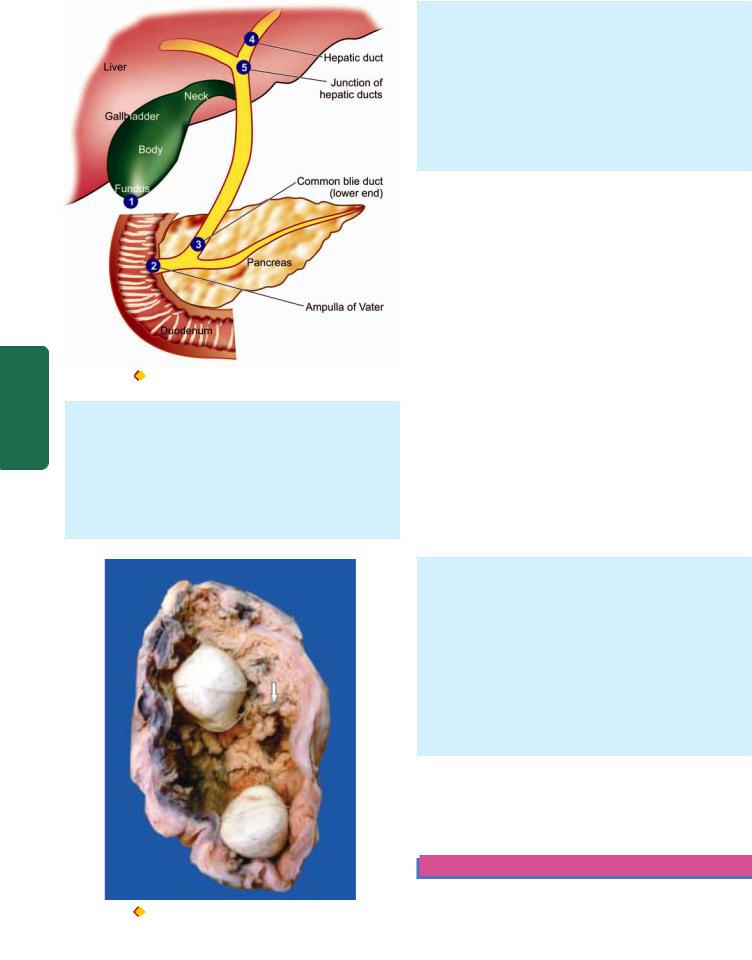
644
Pathology Systemic III SECTION
Figure 21.45 Frequency of cancer in the biliary system.
1.Infiltrating type appears as an irregular area of diffuse thickening and induration of the gallbladder wall. It may have deep ulceration causing direct invasion of the gallbladder wall and liver bed. On section, the gallbladder wall is firm due to scirrhous growth.
2.Fungating type grows like an irregular, friable, papillary or cauliflower-like growth into the lumen as well as into the wall of the gallbladder and beyond.
Histologically, the following patterns are observed:
1.Most gallbladder cancers are adenocarcinomas (90%). They may be papillary or infiltrative, well-differentiated or poorly-differentiated. Most are non-mucin secreting but some are colloid carcinomas forming mucus pools.
2.About 5% of gallbladder cancers are squamous cell carcinomas arising from squamous metaplastic epithelium.
3.A few cases show both squamous and adenocarcinoma pattern of growth called adenosquamous
carcinoma.
CLINICAL FEATURES. Carcinoma of the gallbadder is slow-growing and causes symptoms late in the course of disease. Quite often, the diagnosis is made when gallbladder is removed for cholelithiasis. The symptomatic cases have pain, jaundice, noticeable mass, anorexia and weight loss. In such case, the growth has usually invaded the liver and other adjacent organs and has metastasised to regional lymph nodes and more distant sites such as the lung, peritoneum and gastrointestinal tract.
Carcinoma of Extrahepatic Bile Ducts and
Ampulla of Vater
This is an infrequent neoplasm but is more common than the rare benign tumours of the biliary tract. Unlike other diseases of the biliary passages, it is more common in males with peak incidence in 6th decade of life.
ETIOLOGY. There is no association between bile duct carcinoma and gallstones. Bile duct cancers are associated with a number of other conditions such as ulcerative colitis, sclerosing cholangitis, parasitic infestations of the bile ducts
with Fasciola hepatica (liver fluke), Ascaris lumbricoides and Clonorchis sinensis.
MORPHOLOGIC FEATURES. Extrahepatic bile duct carcinoma may arise anywhere in the biliary tree but the most frequent sites, in descending order of frequency, are: the ampulla of Vater, lower end of common bile duct, hepatic ducts, and the junction of hepatic ducts to form common bile duct (see Fig. 21.45).
Grossly, bile duct carcinoma is usually small, extending for 1-2 cm along the duct, producing thickening of the affected duct.
Histologically, the tumour is usually well-differentiated adenocarcinoma which may or may not be mucinsecreting. Perineural invasion is frequently present.
CLINICAL FEATURES. Obstructive jaundice is the usual presenting feature which is characterised by intense pruritus. Pain, steatorrhoea, weight loss and weakness may be present. The tumour usually metastasises to the regional lymph nodes.
EXOCRINE PANCREAS
Figure 21.46 |
Carcinoma gallbladder. The lumen of the gallbladder |
contains irregular, friable papillary growth arising from mucosa (arrow).
Two multi-faceted gallstones (mixed) are also present in the lumen.
NORMAL STRUCTURE
The human pancreas, though anatomically a single organ, histologically and physiologically has 2 distinct parts—the
exocrine and endocrine parts. The endocrine part of the gland is dealt with in Chapter 27 while the exocrine gland is considered here. The whole of pancreas, exocrine and endocrine, is embryologically derived from the foregut endoderm.
ANATOMY. The pancreas lies obliquely in the concavity of the duodenum as an elongated structure about 15 cm in length and 100 gm in weight (see Fig. 21.1). It is subdivided into 3 topographic zones:
1.The head lying in the concavity of the duodenum and the uncinate process projecting from the head.
2.The body comprises the main part of the gland.
3.The tail is the thin, tapering part of the gland towards the hilum of the spleen.
HISTOLOGY. The exocrine pancreas constitutes 80 to 85% of the total gland, while the endocrine pancreas comprises the remaining part.
The exocrine part is divided into rhomboid lobules separated by thin fibrous tissue septa containing blood vessels, lymphatics, nerves and ducts. Each lobule is composed of numerous acini. The acini are lined by pyramidshaped columnar epithelial cells. These secretory epithelial cells have microvilli projecting into the lumen from their surface. The apical portions of these cells contain zymogen granules in their cytoplasm, while the basal region is deeply basophilic and free of zymogen granules. The zymogen granules are membrane-bound sacs which fuse with the plasma membrane and are then released into the lumina of the acini. The secretions are carried from the acini by fine ductal branches into the small ducts in the lobules and eventually into the main pancreatic duct. The main pancreatic duct is formed by fusion of the ventral duct with the dorsal duct; the latter also called the duct of Wirsung, provides the main drainage for pancreatic secretions into the duodenum. The pancreatic secretions are delivered into the second part of the duodenum either by a combined opening of the pancreatic and bile ducts in the ampulla of Vater, or less often both open separately into the duodenum. Occasionally, the proximal part of the dorsal duct persists
as the duct of Santorini.
FUNCTIONS. The main functions of the exocrine pancreas is the alkaline secretion of digestive enzymes prominent among which are trypsin, chymotrypsin, elastase, amylase, lipase and phospholipase.
DEVELOPMENTAL ANOMALIES
The significant developmental anomalies of the pancreas are ectopic or aberrant pancreatic tissue in Meckel’s diverticulum (page 561), anomalies of the ducts, and cystic fibrosis. Only the last named requires elaboration here.
Cystic Fibrosis
Cystic fibrosis of the pancreas or fibrocystic disease is a hereditary disorder characterised by viscid mucous secretions in all the exocrine glands of the body (muco-
viscidosis) and associated with increased concentrations of electrolytes in the eccrine glands. The terms ‘cystic fibrosis’ and ‘fibrocystic disease’ are preferable over ‘mucoviscidosis’ in view of the main pathologic change of fibrosis produced as a result of obstruction of the passages by viscid mucous secretions.
The disease is transmitted as an autosomal recessive trait with apparent clinical features in homozygotes only. The defect is a genetic mutation in CFTR gene, acronym for coding protein for cystic fibrosis transmembrane conductance regulator located on chromosome 7. It is quite common in the whites (1 per 2000 livebirths). The clinical manifestations may appear at birth or later in adolescence and pertain to multiple organs and systems such as pancreatic insufficiency, intestinal obstruction, steatorrhoea, malnutrition, hepatic cirrhosis and respiratory complications.
MORPHOLOGIC FEATURES. Depending upon the severity of involvement and the organs affected, the pathologic changes are variable. Most of the changes are produced as a result of obstruction by viscid mucous.
1.Pancreas. The pancreas is almost invariably involved in cystic fibrosis.
Grossly, pancreatic lobules are ovoid rather than rhomboid. Fatty replacement of the pancreas and grossly visible cysts may be seen.
Microscopically, the lobular architecture of pancreatic parenchyma is maintained. There is increased interlobular fibrosis. The acini are atrophic and many of the acinar ducts contain laminated, eosinophilic concretions. Rarely, inflammation, fat necrosis and cyst formation may be seen. The islet tissue (endocrine pancreas) generally remains intact. Atrophy of the exocrine pancreas may cause impaired fat absorption, steatorrhoea, intestinal obstruction and avitaminosis A.
2.Liver. The bile canaliculi are plugged by viscid mucous which may cause diffuse fatty change, portal fibrosis and ductular proliferation. More severe involvement may cause biliary cirrhosis (page 625).
3.Respiratory tract. Changes in the respiratory passages are seen in almost all typical cases of cystic fibrosis. The viscid mucous secretions of the submucosal glands of the respiratory tract cause obstruction, dilatation and infection of the airways. The changes include chronic bronchitis, bronchiectasis, bronchiolitis, bronchiolectasis, peribronchiolar pneumonia and inflammatory nasal polyps.
4.Salivary glands. Pathologic changes in the salivary glands are similar to those in pancreas and include obstruction of the ducts, dilatation, fibrosis and glandular atrophy.
5.Sweat glands. Hypersecretion of sodium and chloride in the sweat observed in these patients may be reflected pathologically by diminished vacuolation of the cells of eccrine glands.
645
Pancreas Exocrine and Tract Biliary Liver, The 21 CHAPTER

646 PANCREATITIS
Pancreatitis is inflammation of the pancreas with acinic cell injury. It is classified into acute and chronic forms both of which are distinct entities.
|
Acute Pancreatitis |
|
|
Acute pancreatitis is an acute inflammation of the pancreas |
|
|
presenting clinically with ‘acute abdomen’. The severe form |
|
|
of the disease associated with macroscopic haemorrhages |
|
|
and fat necrosis in and around the pancreas is termed acute |
|
|
haemorrhagic pancreatitis or acute pancreatic necrosis. The |
|
|
condition occurs in adults between the age of 40 and 70 years |
|
|
and is commoner in females than in males. |
|
|
The onset of acute pancreatitis is sudden, occurring after |
|
|
a bout of alcohol or a heavy meal. The patient presents with |
|
|
abdominal pain, vomiting and collapse and the condition |
|
|
must be differentiated from other diseases producing acute |
|
|
abdomen such as acute appendicitis, perforated peptic ulcer, |
|
|
acute cholecystitis, and infarction of the intestine following |
|
|
sudden occlusion of the mesenteric vessels. Characteri- |
|
|
stically, there is elevation of serum amylase level within the |
|
SECTION |
first 24 hours and elevated serum lipase level after 3 to 4 days, |
|
the latter being more specific for pancreatic disease. |
||
Glucosuria occurs in 10% of cases. |
||
|
||
|
ETIOLOGY. The two leading causes associated with acute |
|
|
pancreatitis are alcoholism and cholelithiasis, both of which |
|
III |
are implicated in more than 80% of cases. Less common |
|
causes of acute pancreatitis include trauma, ischaemia, shock, |
||
|
||
|
extension of inflammation from the adjacent tissues, blood- |
|
Systemic |
borne bacterial infection, viral infections, certain drugs (e.g. |
|
thiazides, sulfonamides, oral contraceptives), hypothermia, |
||
|
||
|
hyperlipoproteinaemia and hypercalcaemia from hyperpara- |
|
|
thyroidism. Rarely, familial pancreatitis is encountered. In a |
|
Pathology |
proportion of cases of acute pancreatitis, the etiology remains |
|
unknown (idiopathic pancreatitis). |
||
|
||
|
PATHOGENESIS. The destructive changes in the pancreas |
|
|
are attributed to the liberation and activation of pancreatic |
|
|
enzymes. Though more than 20 enzymes are secreted by |
|
|
exocrine pancreas, 3 main groups of enzymes which bring |
|
|
about destructive effects on the pancreas are as under: |
|
|
1. Proteases such as trypsin and chymotrypsin play the most |
|
|
important role in causing proteolysis. Trypsin also activates |
|
|
the kinin system by converting prekallikrein to kallikrein, |
|
|
and thereby the clotting and complement systems are |
|
|
activated. This results in inflammation, thrombosis, tissue |
|
|
damage and haemorrhages found in acute haemorrhagic |
|
|
pancreatitis. |
|
|
2. Lipases and phospholipases degrade lipids and membrane |
|
|
phospholipids. |
|
|
3. Elastases cause destruction of the elastic tissue of the blood |
|
|
vessels. |
|
|
The activation and release of these enzymes is brought |
|
|
about by one of the following mechanisms: |
|
|
Acinic cell damage caused by the etiologic factors such as |
|
|
alcohol, viruses, drugs, ischaemia and trauma result in |
|
|
release of intracellular enzymes. |
|
|
Duct obstruction caused by cholelithiasis, chronic |
|
|
alcoholism and other obstructing lesions is followed by |
leakage of pancreatic enzymes from the ductules into the interstitial tissue.

 Block in exocytosis of pancreatic enzymes occurring from nutritional causes results in activation of these intracellular enzymes by pancreatic lysosomal hydrolases.
MORPHOLOGIC FEATURES. Grossly, in the early stage, the pancreas is swollen and oedematous. Subsequently, in a day or two, characteristic variegated appearance of grey-white pancreatic necrosis, chalky-white fat necrosis and blue-black haemorrhages are seen. In typical case, the peritoneal cavity contains blood-stained ascitic fluid and white flecks of fat necrosis in the omentum, mesentery and peripancreatic tissue. The resolved lesions show areas of fibrosis, calcification and ductal dilatation.
Microscopically, the following features in varying grades are noticeable:
1.Necrosis of pancreatic lobules and ducts.
2.Necrosis of the arteries and arterioles with areas of haemorrhages.
3.Fat necrosis.
4.Inflammatory reaction, chiefly by polymorphs, around the areas of necrosis and haemorrhages.
COMPLICATIONS. A patient of acute pancreatitis who survives may develop a variety of systemic and local complications.
Systemic complications:
1.Chemical and bacterial peritonitis.
2.Endotoxic shock.
3.Acute renal failure.
Local sequelae:
1.Pancreatic abscess.
2.Pancreatic pseudocyst.
3.Duodenal obstruction.
Mortality in acute pancreatitis is high (20-30%). Patients
succumb to hypotensive shock, infection, acute renal failure, and DIC.
Chronic Pancreatitis
Chronic pancreatitis or chronic relapsing pancreatitis is the progressive destruction of the pancreas due to repeated mild and subclinical attacks of acute pancreatitis. Most patients present with recurrent attacks of severe abdominal pain at intervals of months to years. Weight loss and jaundice are often associated. Later manifestations include associated diabetes mellitus and steatorrhoea. Abdominal radiographs show calcification in the region of pancreas and presence of pancreatic calculi in the ducts.
ETIOLOGY. Most cases of chronic pancreatitis are caused by the same factors as for acute pancreatitis. Thus, most commonly, chronic pancreatitis is related to chronic alcoholism with protein-rich diet, and less often to biliary tract disease. Familial hereditary pancreatitis, though uncommon, is more frequently chronic than the acute form. Other rare causes of chronic pancreatitis are hypercalcaemia, hyperlipidaemia
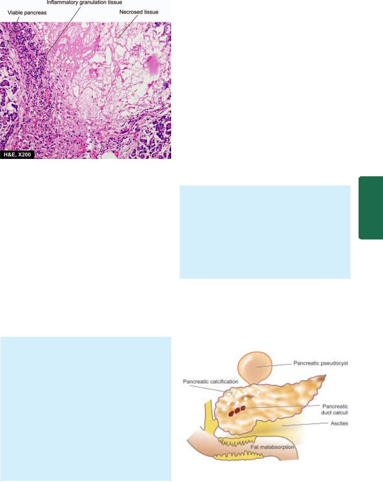
Figure 21.47 
 Chronic pancreatitis. There is destruction of acinar tissue and presence of dystrophic calcification. The necrotic tissue is surrounded by mixed inflammatory infiltrate with granulation tissue formation.
and developmental failure of fusion of dorsal and ventral pancreatic ducts.
PATHOGENESIS. Acute haemorrhagic pancreatitis seldom develops into chronic pancreatitis, but instead develops pancreatic pseudocysts following recovery. Pathogenesis of alcoholic and non-alcoholic chronic pancreatitis is explained by different mechanisms:
1.Chronic pancreatitis due to chronic alcoholism accompanied by a high-protein diet results in increase in protein concentration in the pancreatic juice which obstructs the ducts and causes damage.
2.Non-alcoholic cases of chronic pancreatitis seen in tropical countries (tropical chronic pancreatitis) result from protein-calorie malnutrition. Genetic factors play a role in some cases of chronic pancreatitis.
MORPHOLOGIC FEATURES. Grossly, the pancreas is enlarged, firm and nodular. The cut surface shows a smooth grey appearance with loss of normal lobulation. Foci of calcification and tiny pancreatic concretions to larger visible stones are frequently found. Pseudocysts may be present.
Microscopically, depending upon the stage of development, the following changes are seen (Fig. 21.47):
1.Obstruction of the ducts by fibrosis in the wall and protein plugs or stones in the lumina.
2.Squamous metaplasia and dilatation of some interand intralobular ducts.
3.Chronic inflammatory infiltrate around the lobules as well as the ducts.
4.Atrophy of the acinar tissue with marked increase in interlobular fibrous tissue.
5.Islet tissue is involved in late stage only.
COMPLICATIONS. Late stage of chronic pancreatitis may be complicated by diabetes mellitus, pancreatic insufficiency with steatorrhoea and malabsorption and formation of pancreatic pseudocysts (Fig. 21.48).
TUMOURS AND TUMOUR-LIKE LESIONS
Tumour-like masses of the exocrine pancreas include congenital cystic disease (involving the pancreas, liver and kidney) and pancreatic pseudocysts. True pancreatic tumours are classified into benign (e.g. serous cystadenoma, fibroma, lipoma and adenoma) and malignant (i.e. carcinoma of the pancreas). Out of all these, only two pancreatic lesions— pseudocyst and carcinoma of the pancreas, are common and are discussed below.
Pancreatic Pseudocyst
Pancreatic pseudocyst is a localised collection of pancreatic juice, necrotic debris and haemorrhages. It develops following either acute pancreatitis or trauma. The patients generally present with abdominal mass producing pain, intraperitoneal haemorrhage and generalised peritonitis.
MORPHOLOGIC FEATURES. Grossly, the pseudocyst may be present within or adjacent to the pancreas. Usually it is solitary, unilocular, measuring up to 10 cm in diameter with thin or thick wall (Fig. 21.48).
Microscopically, the cyst wall is composed of dense fibrous tissue with marked inflammatory reaction. There is evidence of preceding haemorrhage and necrosis in the form of deposits of haemosiderin pigment, calcium and cholesterol crystals. The lumen of the cyst contains serous or turbid fluid. The cyst does not show any epithelial lining.
Carcinoma of Pancreas
Pancreatic cancer is the term used for cancer of the exocrine pancreas. It is one of the common cancers, particularly in the Western countries and Japan. In the United States, cancer of the pancreas is the second most common cancer of the alimentary tract after colorectal cancer, is more common in
Figure 21.48 
 Complications of chronic pancreatitis.
647
Pancreas Exocrine and Tract Biliary Liver, The 21 CHAPTER
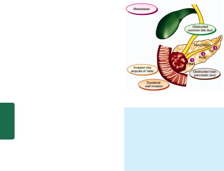
648 African Americans and accounts for 5% of all cancer deaths in that country. It is commoner in males than in females and the incidence increases progressively after the age of 50 years.
|
ETIOLOGY. A significant increase in the incidence of |
|
|
pancreatic cancer has been observed in the UK and US during |
|
|
the last 50 years. Little is known about etiology of pancreatic |
|
|
cancer. However, following factors have been implicated in |
|
|
its etiology: |
|
|
1. Smoking: Heavy cigarette smokers have higher incidence |
|
|
than the non-smokers. However, it is not known whether |
|
|
tobacco metabolites have a direct carcinogenic effect on the |
|
|
pancreas or by some other unknown mechanism. |
|
|
2. Diet and obesity: Diet with high total caloric value and |
|
|
high consumption of animal proteins and fats is related to |
|
|
higher incidence of pancreatic cancer. Obesity is a risk factor |
|
|
for pancreatic cancer. |
|
|
3. Chemical carcinogens: Individuals exposed to β-naph- |
|
|
thylamine, benzidine and nitrosamines have higher incidence |
|
|
of cancer of the pancreas. |
|
|
4. Diabetes mellitus: Patients of long-standing diabetes |
|
|
mellitus have a higher incidence. |
|
SECTION |
5. Chronic pancreatitis patients are at increased risk. |
|
6. H. pylori infection has been reported to have association |
||
|
||
|
with pancreatic cancer. |
|
|
7. Genetic factors have been ound to have association with |
|
|
pancreatic cancer e.g. its occurrence in first-degree relatives |
|
III |
in 10% cases, occurrence in certain hereditary syndromes |
|
(Lynch, FPC, HNPCC). |
||
|
However, excessive consumption of alcohol or coffee, and |
|
Systemic |
cholelithiasis are not risk factors for pancreatic cancer. A |
|
MORPHOLOGIC FEATURES. The most common loca- |
||
|
combination of mutations in K-RAS gene and CDKN2A gene |
|
|
have been found in almost all cases of cancer of the pancreas. |
|
Pathology |
tion of pancreatic cancer is the head of pancreas (70%), |
|
small, homogeneous, poorly-defined, grey-white mass |
||
|
followed in decreasing frequency, by the body and the |
|
|
tail of pancreas (Fig. 21.49). |
|
|
Grossly, carcinoma of the head of pancreas is generally |
|
|
without any sharp demarcation between the tumour and |
|
|
the surrounding pancreatic parenchyma. The tumour of |
|
|
the head extends into the ampulla of Vater, common bile |
|
|
duct and duodenum, producing obstructive biliary |
|
|
symptoms and jaundice early in the course of illness. |
|
|
Carcinomas of the body and tail of the pancreas, on the |
|
|
other hand, are fairly large and irregular masses and |
|
|
frequently infiltrate the transverse colon, stomach, liver, |
|
|
spleen and regional lymph nodes. |
|
|
Microscopically, most pancreatic carcinomas arise from |
|
|
the ductal epithelium which normally comprises less than |
|
|
4% of total pancreatic cells, whereas carcinoma of the acini |
|
|
|
Figure 21.49 
 Distribution of carcinoma of the pancreas (numbered serially) and its major effects.
constitutes less than 1% of pancreatic cancers. The following histologic patterns of pancreatic carcinoma are seen:
1.Well-differentiated adenocarcinoma, both mucinous and non-mucin secreting type, is the most common pattern. Perineural invasion is commonly present and is diagnostic of malignancy.
2.Adenoacanthoma consisting of glandular carcinoma and benign squamous elements is seen in a proportion of cases.
3.Rarely, peculiar tumour giant cell formation is seen with marked anaplasia, pleomorphism and numerous mitoses.
4Acinar cell carcinoma occurs rarely and reproduces the pattern of acini in normal pancreas.
CLINICAL FEATURES. Clinical symptoms depend upon the site of origin of the tumour. Generally, the following features are present:
1.Obstructive jaundice. more often and early in the course of disease in cases with carcinoma head of the pancreas (80%), and less often in cancer of the body and tail of the pancreas. It is characterised by: dark urine, clay-like stools, pruritus, and very high serum alkaline phosphatase.
2.Other features. These include: abdominal pain, anorexia, weight loss, cachexia, weakness and malaise, nausea and vomiting, and migratory thrombophlebitis (Trousseau’s syndrome), GI bleeding and splenomegaly.
The prognosis of pancreatic cancer is dismal: median survival is 6 months from the time of diagnosis. Approximately 10% patients survive 1 year and the 5-year survival is poor 1 to 2%.
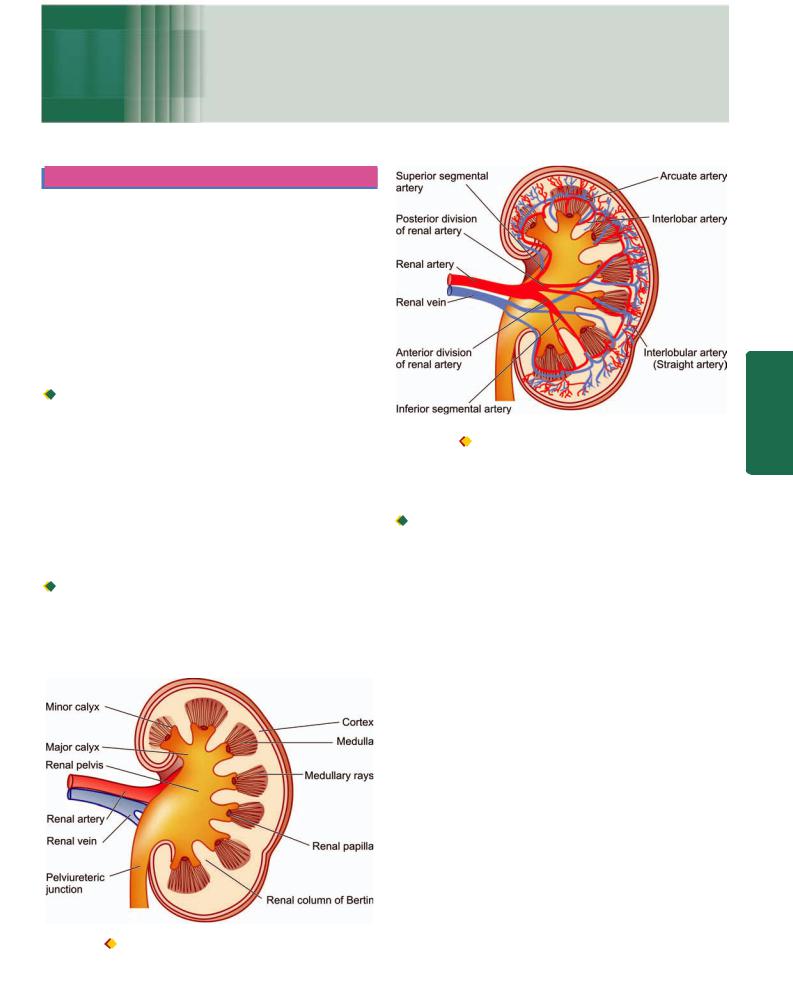
649
Chapter 22
The Kidney and
Lower Urinary Tract
KIDNEY
NORMAL STRUCTURE
ANATOMY. The kidneys are bean-shaped paired organs, each weighing about 150 gm in the adult male and about 135 gm in the adult female. The hilum of the kidney is situated at the midpoint on the medial aspect where the artery, vein, lymphatics and ureter are located. The kidney is surrounded by a thin fibrous capsule which is adherent at the hilum.
Cut surface of the kidney shows 3 main structures: welldemarcated peripheral cortex, inner medulla and the innermost renal pelvis (Fig. 22.1):
 The renal cortex forms the outer rim of the kidney and is about 1 cm in thickness. It contains all the glomeruli and about 85% of the nephron tubules. Remaining 15% nephrons consisting of collecting tubules, collecting ducts, loops of Henle and vasa recta send their loops into the medulla, and are therefore called juxtamedullary nephrons. This latter part of the cortex forms faint striations called medullary rays, a misnomer since theses structures are located in the cortex but are destined for medulla. Columns of renal cortical tissue that extend into the space between adjacent pyramids are called the renal column (septa) of Bertin; they contain the interlobar arteries.

 The renal medulla is composed of 8-18 cone-shaped renal pyramids. The base of a renal pyramid lies adjacent to the outer cortex and forms the cortico-medullary junction, while the apex of each called the renal papilla contains the opening
Figure 22.1 
 Cross-section of the kidney showing gross structures.
Figure 22.2  Cross-section of the kidney showing arterial blood supply.
of each renal pyramid for passage of urine collected from collecting ducts and goes down into minor calyces.

 The renal pelvis is the funnel-shaped collection area of the urine for drainage into the ureter. The minor calyces (8- 18 in number in a normal kidney) collect urine from renal papillae and drain into major calyces (2-3 in a normal kidney).
HISTOLOGY. The parenchyma of each kidney is composed of approximately one million microstructures called nephrons. A nephron, in turn, consists of 5 major parts, each having a functional role in the formation of urine: the glomerular capsule (glomerulus and Bowman’s capsule), the proximal convoluted tubule (PCT), the loop of Henle, the distal convoluted tubule (DCT), and the collecting ducts. From point of view of diseases of the kidneys, 4 components of renal parenchyma require further elaboration: renal vasculature, glomeruli, tubules and interstitium.
1. Renal vasculature. Each kidney is supplied with blood by a main renal artery which arises from the aorta at the level of the 2nd lumbar vertebra. It usually divides into anterior and posterior divisions at the hilum although occasionally these divisions may even arise directly from the aorta. The anterior and posterior divisions divide into segmental branches from which interlobar arteries arise which course between the lobes. Along their course, they give off the arcuate arteries which arch between the cortex and medulla. The arcuate arteries, in turn, give off interlobular arteries which lie in the cortex perpendicular to the capsular surface in the part overlying the pyramids and, therefore, are also called straight arteries (Fig. 22.2). It is from the interlobular arteries that the afferent
Tract Urinary Lower and Kidney The 22 CHAPTER
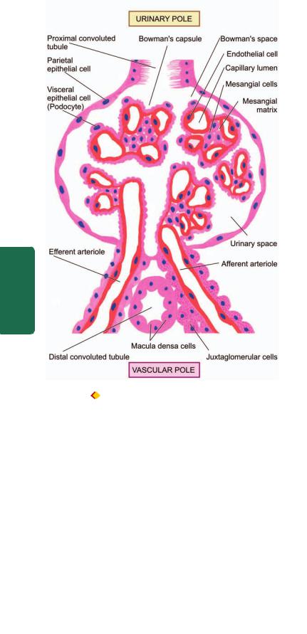
650
Pathology Systemic III SECTION
Figure 22.3 
 A schematic illustration of the structure of a nephron (in longitudinal section) and associated blood supply .
arterioles take their origin, each one supplying a single glomerulus. From the glomerulus emerge the efferent arterioles. Up to this stage, the arteries and arterioles are endvessels. The efferent arterioles leaving the glomerulus supply peritubular capillary plexus which anastomoses with the capillary plexus of another nephron.
The juxtamedullary glomeruli, however, give off a series of parallel vessels called vasa recta which descend to the inner medulla supplying the loop of Henle and collecting ducts and anastomose at all levels throughout the medulla with the ascending vasa recta. These drain into arcuate veins and then into the veins that accompany the corresponding arteries and finally through a single renal vein into the inferior vena cava (Fig. 22.3). Lymphatic drainage likewise occurs through lymphatics associated with the intrarenal vasculature leaving the kidney at the hilum and draining to lateral aortic lymph nodes.
The following important inferences can be drawn from the peculiarities of the renal vasculature:
i) The renal cortex receives about 90% of the total renal blood supply and that the pressure in the glomerular
capillaries is high. Therefore, renal cortex is more prone to the effects of hypertension.
ii)The renal medulla, on the other hand, is poorly perfused and any interference in blood supply to it results in medullary necrosis.
iii)The divisions and subdivisions of the renal artery up to arterioles are end-arteries and have no anastomoses. Thus, occlusion of any of the branches results in infarction of the renal parenchyma supplied by it.
iv)Since the tubular capillary beds are derived from the efferent arterioles leaving the glomeruli, diseases affecting the blood flow through glomerular tuft have significant effects on the tubules as well.
2.Glomerulus. The glomerulus consists of invagination of the blind end of the proximal tubule and contains a capillary tuft fed by the afferent arteriole and drained by efferent arteriole. The capillary tuft is covered by visceral epithelial cells (podocytes) which are continuous with those of the parietal epithelium at the vascular pole. The transition to proximal tubular cells occurs at the urinary pole of the glomerulus. The visceral and parietal epithelial cells are separated by the urinary space or Bowman’s space, into which glomerular filtrate passes (Fig. 22.4).
Subdivisions of capillaries derived from the afferent arterioles result in the formation of lobules (up to 8 in number) within a glomerulus. Each lobule of a glomerular tuft consists of a centrilobular supporting stalk composed of mesangium containing mesangial cells and mesangial matrix. The mesangium is continuous at the hilum with the lacis cells of the juxtaglomerular apparatus. Besides their role as supportive cells, mesangial cells are involved in the production of mesangial matrix and glomerular basement membrane; they function in endocytosis of leaked macromolecules and also possibly in the control of glomerular blood flow through contractile elements present in these cells.
The major function of glomerulus is complex filtration from the capillaries to the urinary space. Glomerular filtrate is quite similar in composition to plasma but lacks proteins and cells. Normally, glomerular filtration rate (GFR) is about
125ml/minute. The barrier to glomerular filtration consists of the following 3 components (Fig. 22.5):
i)Fenestrated endothelial cells lining the capillary loops.
ii)Glomerular basement membrane (GBM) on which the endothelial cells rest. It further consists of 3 layers—the central lamina densa, bounded by lamina rara interna on endothelial side of the capillary and lamina rara externa on visceral epithelial side of the capillary.
iii)Filtration slit pores between the foot processes of the visceral epithelial cells (podocytes) external to GBM.
The barrier to filtration of macromolecules of the size and molecular weight of albumin and larger depends upon the following:

 A normal lamina densa.

 Maintenance of negative charge on both lamina rarae.

 A healthy covering of glomerular epithelial cells.
Juxtaglomerular apparatus. The juxtaglomerular apparatus (JGA) is situated at the vascular pole of the glomerulus and is made up of 3 parts (Fig. 22.4):
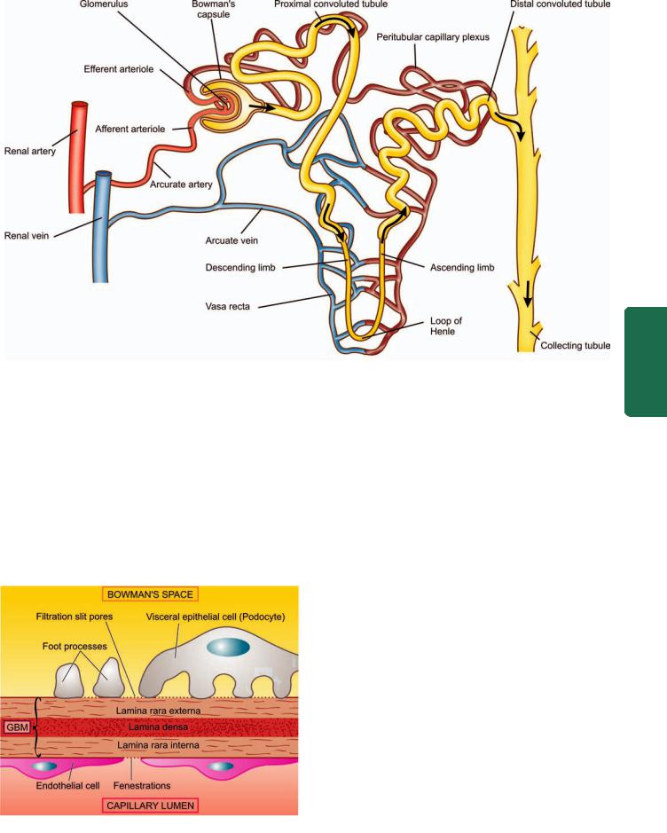
Figure 22.4 
 Structure of a nephron.
i)The juxtaglomerular cells are modified granular smooth muscle cells in the media of the afferent arteriole and contain the hormone, renin.
ii)The macula densa is comprised by specialised region of the distal tubule when it returns to the vascular pole of its parent glomerulus. The tubular cells here are taller and narrower than elsewhere with the nuclei lying close together.
iii)The lacis cells or non-granular cells occupy the space between the macula densa and the arterioles and merge with the glomerular mesangium.
The JGA is intimately concerned with sodium metabolism and is the principal source of renin production. The
Figure 22.5 
 Ultrastructure of glomerular filtration barrier.
mechanism of the release of renin and its role in hypertension are discussed on page 686.
3. Tubules. The tubules of the kidney account for the greatest amount of the renal parenchyma. The structure of renal tubular epithelium varies in different parts of the nephron and is correlated with the functional capacity of that part of the tubule (see Fig. 22.3).
i)Proximal convoluted tubule (PCT). This is the first part arising from the glomerulus and is highly specialised part functionally. It is lined by cuboidal cells with a brush border composed of microvilli and contains numerous mitochondria, Golgi apparatus and endoplasmic reticulum. The major functions of PCT are: active reabsorption of filtered sodium, potassium, glucose, amino acids, proteins, vitamins, bicarbonate, phosphate, calcium and uric acid, and passive reabsorption of 80% of filtered water.
ii)Loop of Henle. The PCT drains into the straight part of loop of Henle that consists of thin descending, and thin and thick ascending limbs, both of which have different structure and function. The descending limb is continuation of PCT, while ascending limb continues further into distal convoluted tubule (DCT). The descending segment of loop is lined by simple epithelium while the ascending limb is lined by columnar cells. The major function of loop of Henle is active reabsorption of sodium, potassium and chloride, and passive diffusion of water resulting in concentrated filtrate of urine.
iii)Distal convoluted tubule (DCT). The DCT represents a transition from thick ascending limb from the point where the ascending limb meets the vascular pole of the glomerulus of its origin, to the early collecting ducts. The lining cells in
651
Tract Urinary Lower and Kidney The 22 CHAPTER

652DCT are cuboidal. The epithelial cells at the point of beginning of DCT are taller, narrower and more closely packed to form the macula densa of JGA as already described. The DCT further contributes to urinary concentration and acidification, while the macula densa of JGA is the source of renin and has a role in sodium metabolism.
iv)Collecting ducts. The system of collecting ducts is the final pathway by which urine reaches the tip of renal papilla. The cells lining the collecting ducts are cuboidal but lack the brush border. Collecting ducts reabsorb water under control of ADH, and secrete H+ and K+ ions.
4. Interstitium. In health, the renal cortical interstitium is scanty and consists of a small number of fibroblast-like cells. But the medullary interstitium is more plentiful and contains stellate interstitial cells which are considered to produce an anti-hypertensive agent and are involved in the metabolism of prostaglandins.
|
RENAL FUNCTION TESTS |
||
|
In general, the kidney performs the following vital functions |
||
SECTION |
in the body: |
||
1. |
Excretion of waste products resulting from protein |
||
metabolism. |
|||
|
|||
|
2. |
Regulation of acid-base balance by excretion of H+ ions |
|
|
(acidification) and bicarbonate ions. |
||
III |
3. |
Regulation of salt-water balance by hormones secreted both |
|
intraand extra-renally. |
|||
|
4. |
Formation of renin and erythropoietin and thereby playing |
|
Systemic |
a role in the regulation of blood pressure and erythropoiesis |
||
parameters: |
|||
|
respectively. |
||
|
|
In order to assess renal function, a number of tests have |
|
|
been devised which give information regarding the following |
||
Pathology |
a) |
Renal blood flow |
|
b) |
Glomerular filtration |
||
|
|||
|
c) |
Renal tubular function |
|
|
d) |
Urinary outflow unhindered by any obstruction. |
|
|
|
Renal function tests are broadly divided into 4 groups |
|
|
(Table 22.1): |
||
|
1. |
Urine analysis. |
|
|
2. |
Concentration and dilution tests. |
|
|
3. |
Blood chemistry. |
|
|
4. |
Renal clearance tests. |
|
In addition, renal biopsy is performed to confirm the diagnosis of renal disease. Renal biopsy is ideally fixed in alcoholic Bouin’s solution and examined by routine morphology combined with special stains and further studies as under:
1.Periodic acid-Schiff stain for highlighting glomerular basement membrane.
2.Silver impregnation to outline the glomerular and tubular basement membrane.
3.Immunofluorescence to localise the antigens, complements and immunoglobulins.
4.Electron microscopy to see the ultrastructure of glomerular changes.
TABLE 22.1: Renal Function Tests.
1.URINE ANALYSIS:
i)Physical examination
(output, colour, specific gravity, pH, osmolality)
ii)Chemical constituents
(protein, glucose, red cells, haemoglobin)
iii)Bacteriologic examination
iv)Microscopy
2.CONCENTRATION AND DILUTION TESTS:
i)Concentration test (fluid deprivation test)
ii)Dilution test (excess fluid intake test)
3.BLOOD CHEMISTRY:
i)Urea
ii)Blood urea nitrogen (BUN)
iii)Creatinine
iv)β2-microglobulin
4.RENAL CLEARANCE TEST:
i)Inulin or mannitol clearance test
ii)Creatinine clearance
iii)Urea clearance
iv)Para-aminohippuric acid (PAH) clearance
1.URINE ANALYSIS. The simplest diagnostic tests for renal function is the physical, chemical, bacteriologic and microscopic examination of the urine.
i)The physical examination includes 24-hour urinary output, colour, specific gravity and osmolality. Normally urine is clear, pale or straw-coloured due to pigment urochrome and 700-2500 ml (average 1200 ml) of urine is passed in 24 hours, mostly during day time. Specific gravity is used to measure the concentrating and diluting power of the kidneys.
ii)The chemical tests are carried out to detect the presence of protein, glucose, red cells and haemoglobin to assess the permeability of glomerular membrane. A number of convenient dipstick tests are available for testing these chemical substances and pH. These consist of paper strips impregnated with appropriate reagents and indicator dyes.
ii)The bacteriologic examination of the urine is done by proper and aseptic collection of midstream specimen of urine.
iv)Urine microscopy is undertaken on a fresh unstained sample. Various components observed on microscopic examination of the urine in renal disease are red cells, pus cells, epithelial cells, crystals and urinary casts. The casts are moulded into cylindrical shapes by passage along tubules in which they are formed. They are the result of precipitation of proteins in the tubule that includes not only albumin but also the tubular secretion of the Tamm Horsfall protein. The latter is a high molecular weight glycoprotein normally secreted by ascending loop of Henle and DCT and probably has body defense function normally. Its secretion is increased in glomerular and tubular diseases. Casts may be hyaline type consisting of only proteins indicating a non-inflammatory etiology of glomerular filtration of proteins, leucocyte casts inflammatory in origin, or red cell casts from haematuria.
2. CONCENTRATION AND DILUTION TESTS. Concentration and dilution tests are designed to evaluate functional capacity of the renal tubules. The ability of the nephron to concentrate or dilute urine is dependent upon both functional activity of the tubular cells in the renal medulla and the

presence of antidiuretic hormone (ADH). Failure to achieve adequate urinary concentration can be due to either defects within the renal medulla (nephrogenic diabetes insipidus), or due to the lack of ADH (central diabetes insipidus).
Traditionally, urinary concentration is determined by specific gravity of the urine (normal range 1.003 to 1.030, average 1.018) which in cases of tubular disease remains constant at approximately 1.010 regardless of changing levels of plasma hydration. However, determination of urinary specific gravity provides only a rough estimate of osmolarity of the urine. The tubular disease can be diagnosed in its early stage by water deprivation (concentration) or water excess
(dilution) tests.
i)In concentration test, an artificial fluid deprivation is induced in the patient for more than 20 hours. If the nephron is normal, water is selectively reabsorbed resulting in excretion of urine of high solute concentration (specific gravity of 1.025 or more). However, if the tubular cells are nonfunctional, the solute concentration of the urine will remain constant regardless of stress of water deprivation.
ii)In dilution test, an excess of fluid is given to the patient. Normally, renal compensation should result in excretion of urine with high water content and lower solute concentration (specific gravity of 1.003 or less). If the renal tubules are diseased, the concentration of solutes in the urine will remain constant irrespective of the excess water intake.
3.BLOOD CHEMISTRY. Impairment of renal function results in elevation of end-products of protein metabolism. This includes increased accumulation of certain substances in the blood, chiefly urea (normal range 20-40 mg/dl), blood urea nitrogen (BUN) (normal range 10-20 mg/dl) and creatinine (normal range 0.6-1.2 mg/dl). An increase of these end-products in the blood is called azotaemia.
High levels of creatinine are associated with high levels of β2-microglobulin in the serum as well as urine, a lowmolecular weight protein filtered excessively in the urine due to glomerular disease or due to increased production by the liver.
4.RENAL CLEARANCE TESTS. A clearance test is employed to assess the rate of glomerular filtration and the renal blood flow. The rate of this filtration can be measured by determining the excretion rate of a substance which is filtered through the glomerulus but subsequently is neither reabsorbed nor secreted by the tubules. The glomerular filtration rate (normal 120 ml/minute in an average adult) is usually equal to clearance of that substance and is calculated from the following equation:
UV
C = _______ where
P
C is the clearance of the substance in ml/ minute;
Uis the concentration of the substance in the urine;
Vis the volume of urine passed per minute; and
Pis the concentration of the substance in the plasma. The substances which are used for clearance tests include
inulin, mannitol, creatinine and urea.
i) In inulin or mannitol clearance tests, an intravenous infusion of the substance inulin or mannitol is given to
maintain constant plasma concentration and accurately timed urine samples are collected. Inulin, a mixture of fructose polymers, is considered the ideal substance for the clearance test since it is filtered from the glomerulus and is excreted unchanged in the urine.
ii)In creatinine clearance test, there is no need of intravenous infusion of creatinine since creatinine is normally released into plasma by muscle metabolism and a very small fraction of this substance is secreted by the tubules. The clearance of creatinine is determined by collecting urine over 24-hour period and a blood sample is withdrawn during the day. In spite of disadvantages like poor reproducibility and secretion of creatinine by the tubules, the ‘endogenous’ creatinine clearance test is easy and routinely employed method of estimating GFR.
iii)In urea clearance test, the sensitivity is much less than the creatinine or inulin clearance because plasma concentration of urea is affected by a number of factors (e.g. dietary protein, fluid intake, infection, trauma, surgery, and corticosteroids) and is partly reabsorbed by the tubules. Like in creatinine clearance, there is no need for intravenous infusion of urea.
iv)Para-aminohippuric acid (PAH) clearance test is employed to measure renal blood flow (unlike the preceding tests which measure GFR). PAH when infused intravenously is both filtered at the glomerulus as well as secreted by the tubules and its clearance is measured by determining its concentration in arterial blood and urine. Normally, renal blood flow is about 1200 ml per minute in an average adult.
PATHOPHYSIOLOGY OF RENAL DISEASE:
RENAL FAILURE
Traditionally, diseases of the kidneys are divided into 4 major groups according to the predominant involvement of corresponding morphologic components:
1.Glomerular diseases: These are most often immuno- logically-mediated and may be acute or chronic.
2.Tubular diseases: These are more likely to be caused by toxic or infectious agents and are often acute.
3.Interstitial diseases: These are likewise commonly due to toxic or infectious agents and quite often involve interstitium as well as tubules (tubulo-interstitial diseases).
4.Vascular diseases: These include changes in the nephron as a consequence of increased intra-glomerular pressure such as in hypertension or impaired blood flow.
In addition, other diseases described in this chapter include: congenital anomalies, obstructive uropathy (including urolithiasis) and tumours of the kidneys.
The major morphologic involvements of the kidneys in the initial stage is confined to one component (glomeruli, tubules, interstitium or blood vessels), but eventually all components are affected leading to end-stage kidneys.
Regardless of cause, renal disease usually results in the evolution of one of the two major pathological syndromes:
acute renal failure and chronic renal failure. The term ‘azotaemia’ is used for biochemical abnormality characterised by elevation of the blood urea nitrogen (BUN) and creatinine levels, while ‘uraemia’ is defined as association of these
653
Tract Urinary Lower and Kidney The 22 CHAPTER

654 biochemical abnormalities with clinical signs and symptoms. The pathophysiological aspects of acute and chronic renal failure are briefly discussed below.
|
Acute Renal Failure (ARF) |
|
|
Acute renal failure (ARF) is a syndrome characterised by |
|
|
rapid onset of renal dysfunction, chiefly oliguria or anuria, |
|
|
and sudden increase in metabolic waste-products (urea and |
|
|
creatinine) in the blood with consequent development of |
|
|
uraemia. |
|
|
ETIOPATHOGENESIS. The causes of ARF may be classified |
|
|
as pre-renal, intra-renal and post-renal in nature. |
|
|
1. Pre-renal causes. Pre-renal diseases are those which cause |
|
|
sudden decrease in blood flow to the nephron. Renal |
|
|
ischaemia ultimately results in functional disorders or |
|
|
depression of GFR, or both. These causes include inadequate |
|
|
cardiac output and hypovolaemia or vascular disease causing |
|
|
reduced perfusion of the kidneys. |
|
|
2. Intra-renal causes. Intra-renal disease is characterised by |
|
|
disease of renal tissue itself. These include vascular disease |
|
SECTION |
of the arteries and arterioles within the kidney, diseases of |
|
glomeruli, acute tubular necrosis due to ischaemia, or the |
||
|
||
|
effect of a nephrotoxin, acute tubulointerstitial nephritis and |
|
|
pyelonephritis. |
|
|
3. Post-renal causes. Post-renal disease is characteristically |
|
III |
caused by obstruction to the flow of urine anywhere along |
|
the renal tract distal to the opening of the collecting ducts. |
||
|
This may be caused by a mass within the lumen or from wall |
|
Systemic |
of the tract, or from external compression anywhere along |
|
infection, eventually leads to intra-renal disease. Thus, full- |
||
|
the lower urinary tract—ureter, bladder neck or urethra. |
|
|
It is important to note that ARF originating in preand |
|
|
post-renal disease, such as by renal ischaemia or renal |
|
Pathology |
blown ARF reflects some degree of nephron damage. |
|
CLINICAL FEATURES. The clinical features will depend |
||
|
||
|
to a large extent on the underlying cause of ARF and on the |
|
|
stage of the disease at which the patient presents. However, |
|
|
one of the following three major patterns usually emerge: |
|
|
1. Syndrome of acute nephritis. This is most frequently |
|
|
associated with acute post-streptococcal glomerulonephritis |
|
|
and rapidly progressive glomerulonephritis. Renal |
|
|
dysfunction results from extensive proliferation of epithelial |
|
|
cells in the glomeruli with consequent mild increase in |
|
|
glomerular permeability and decrease in GFR. The |
|
|
characteristic features are: mild proteinuria, haematuria, |
|
|
oedema and mild hypertension. Fluid retention in acute |
|
|
nephritis syndrome appears to be due to both diminished |
|
|
GFR and increased salt and water reabsorption in distal |
|
|
nephron. |
|
|
2. Syndrome accompanying tubular pathology. When the |
|
|
ARF is caused by destruction of the tubular cells of the |
|
|
nephron as occurs in acute tubular necrosis (page 679), the |
|
|
disease typically progresses through 3 characteristic stages |
|
|
from oliguria to diuresis to recovery. |
|
|
i) Oliguric phase: The initial oliguric phase lasting on an |
|
|
average from 7 to 10 days is characterised by urinary output |
of less than 400 ml per day. The decline in formation of the urine leads to accumulation of waste products of protein metabolism in the blood and resultant azotaemia, metabolic acidosis, hyperkalaemia, hypernatraemia and hypervolaemia due to secondary effects of circulatory overload and pulmonary oedema. The specific gravity of the urine is low but the concentration of sodium in urine tends to be elevated.
ii)Diuretic phase: With the onset of healing of tubules, there is improvement in urinary output. This is believed to occur due to drawing of water and sodium by preceding high levels of creatinine and urea as they move through the nephron so as to be excreted. Since tubular cells have not regained normal functional capacity, the urine is of low or fixed specific gravity.
iii)Phase of recovery: Full recovery with healing of tubular epithelial cells occurs in about half the cases, while others terminate in death. The process of healing may take up to one year with restoration of normal tubular function.
3. Pre-renal syndrome. The ARF occurring secondary to disorders in which neither the glomerulus nor the tubules are damaged, results in pre-renal syndrome. Typically, this pattern is seen in marginal ischaemia caused by renal arterial obstruction, hypovolaemia, hypotension or cardiac insufficiency. Due to depressed renal blood flow, there is decrease in GFR causing oliguria, azotaemia (elevation of BUN and creatinine) and possible fluid retention and oedema. Since the tubular cells are functioning normally, the nephron retains its ability to concentrate the glomerular filtrate according to the adaptive needs.
Chronic Renal Failure (CRF)
Chronic renal failure is a syndrome characterised by progressive and irreversible deterioration of renal function due to slow destruction of renal parenchyma, eventually terminating in death when sufficient number of nephrons have been damaged. Acidosis is the major problem in CRF with development of biochemical azotaemia and clinical uraemia syndrome.
ETIOPATHOGENESIS. All chronic nephropathies can lead to CRF. The diseases leading to CRF can generally be classified into two major groups: those causing glomerular
pathology, and those causing tubulointerstitial pathology. Though this classification is useful to facilitate study, the disease rarely remains confined to either glomeruli or tubulointerstitial tissue alone. In the final stage of CRF, all parts of the nephron are involved.
1. Diseases causing glomerular pathology. A number of glomerular diseases associated with CRF have their pathogenesis in immune mechanisms (page 662). Glomerular destruction results in changes in filtration process and leads to development of the nephrotic syndrome characterised by proteinuria, hypoalbuminaemia and oedema. The important examples of chronic glomerular diseases causing CRF are covered under two headings: primary and systemic.
i) Primary glomerular pathology: The major cause of CRF is chronic glomerulonephritis, usually initiated by various types of glomerulonephritis such as membranous glomerulo-
nephritis, membranoproliferative glomerulonephritis, lipoid nephrosis (minimal change disease) and anti-glomerular basement membrane nephritis.
ii) Systemic glomerular pathology: Certain conditions originate outside the renal system but induce changes in the nephrons secondarily. Major examples of this type are systemic lupus erythematosus, serum sickness nephritis and diabetic nephropathy.
2. Diseases causing tubulointerstitial pathology. Damage to tubulointerstitial tissues results in alterations in reabsorption and secretion of important constituents leading to excretion of large volumes of dilute urine. Tubulointerstitial diseases can be categorised according to initiating etiology into 4 groups: vascular, infectious, toxic and obstructive.
i)Vascular causes: Long-standing primary or essential hypertension produces characteristic changes in renal arteries and arterioles referred to as nephrosclerosis (page 687). Nephrosclerosis causes progressive renal vascular occlusion terminating in ischaemia and necrosis of renal tissue.
ii)Infectious causes: A good example of chronic renal infection causing CRF is chronic pyelonephritis. The chronicity of process results in progressive damage to increasing number of nephrons leading to CRF.
iii)Toxic causes: Some toxic substances induce slow tubular injury, eventually culminating in CRF. The most common example is intake of high doses of analgesics such as phenacetin, aspirin and acetaminophen (chronic analgesic nephritis). Other substances that can cause CRF after prolonged exposure are lead, cadmium and uranium.
iv)Obstructive causes: Chronic obstruction in the urinary tract leads to progressive damage to the nephron due to fluid backpressure. The examples of this type of chronic injury are stones, blood clots, tumours, strictures and enlarged prostate.
Regardless of the initiating cause, CRF evolves progressively through 4 stages:
1.Decreased renal reserve. At this stage, damage to renal parenchyma is marginal and the kidneys remain functional. The GFR is about 50% of normal, BUN and creatinine values are normal and the patients are usually asymptomatic except at times of stress.
2.Renal insufficiency. At this stage, about 75% of functional renal parenchyma has been destroyed. The GFR is about 25% of normal accompanied by elevation in BUN and serum creatinine. Polyuria and nocturia occur due to tubulointerstitial damage. Sudden stress may precipitate uraemic syndrome.
3.Renal failure. At this stage, about 90% of functional renal tissue has been destroyed. The GFR is approximately 10% of normal. Tubular cells are essentially nonfunctional. As a result, the regulation of sodium and water is lost resulting in oedema, metabolic acidosis, hypocalcaemia, and signs and symptoms of uraemia.
4.End-stage kidney. The GFR at this stage is less than 5% of normal and results in complex clinical picture of uraemic syndrome with progressive primary (renal) and secondary systemic (extra-renal) symptoms.
CLINICAL FEATURES. Clinical manifestations of fullblown CRF culminating in uraemic syndrome are described under 2 main headings: primary (renal) uraemic manifestations and secondary (systemic or extra-renal) uraemic manifestations.
A. Primary uraemic (renal) manifestations. Primary symptoms of uraemia develop when there is slow and progressive deterioration of renal function. The resulting imbalances cause the following manifestations:
1.Metabolic acidosis. As a result of renal dysfunction, acidbase balance is progressively lost. Excess of hydrogen ions occurs, while bicarbonate level declines in the blood, resulting in metabolic acidosis. The clinical symptoms of metabolic acidosis include: compensatory Kussmaul breathing, hyperkalaemia and hypercalcaemia.
2.Hyperkalaemia. A decreased GFR results in excessive accumulation of potassium in the blood since potassium is normally excreted mainly in the urine. Hyperkalaemia is further worsened by metabolic acidosis. The clinical features of hyperkalaemia are: cardiac arrhythmias, weakness, nausea, intestinal colic, diarrhoea, muscular irritability and flaccid paralysis.
3.Sodium and water imbalance. As GFR declines, sodium and water cannot pass sufficiently into Bowman’s capsule leading to their retention. Release of renin from juxtaglomerular apparatus further aggravates sodium and water retention. The main symptoms referable to sodium and water retention are: hypervolaemia and circulatory overload with congestive heart failure.
4.Hyperuricaemia. Decreased GFR results in excessive accumulation of uric acid in the blood. Uric acid crystals may be deposited in joints and soft tissues resulting in gout.
5.Azotaemia. The waste-products of protein metabolism fail to be excreted resulting in elevation in the blood levels of urea, creatinine, phenols and guanidines causing biochemical abnormality, azotaemia. The secondary manifestations of uraemia are related to toxic effects of these metabolic waste-products.
B. Secondary uraemic (extra-renal) manifestations. A number of extra-renal systemic manifestations develop secondarily following fluid-electrolyte and acid-base imbalances. These include the following:
1.Anaemia. Decreased production of erythropoietin by diseased kidney results in decline in erythropoiesis and anaemia. Besides, gastrointestinal bleeding may further aggravate anaemia.
2.Integumentary system. Deposit of urinary pigment such as urochrome in the skin causes sallow-yellow colour. The urea content in the sweat as well as in the plasma rises. On evaporation of the perspiration, urea remains on the facial skin as powdery ‘uraemic frost’.
3.Cardiovascular system. Fluid retention secondarily causes cardiovascular symptoms such as increased workload on the heart due to the hypervolaemia and eventually congestive heart failure.
4.Respiratory system. Hypervolaemia and heart failure cause pulmonary congestion and pulmonary oedema due
655
Tract Urinary Lower and Kidney The 22 CHAPTER
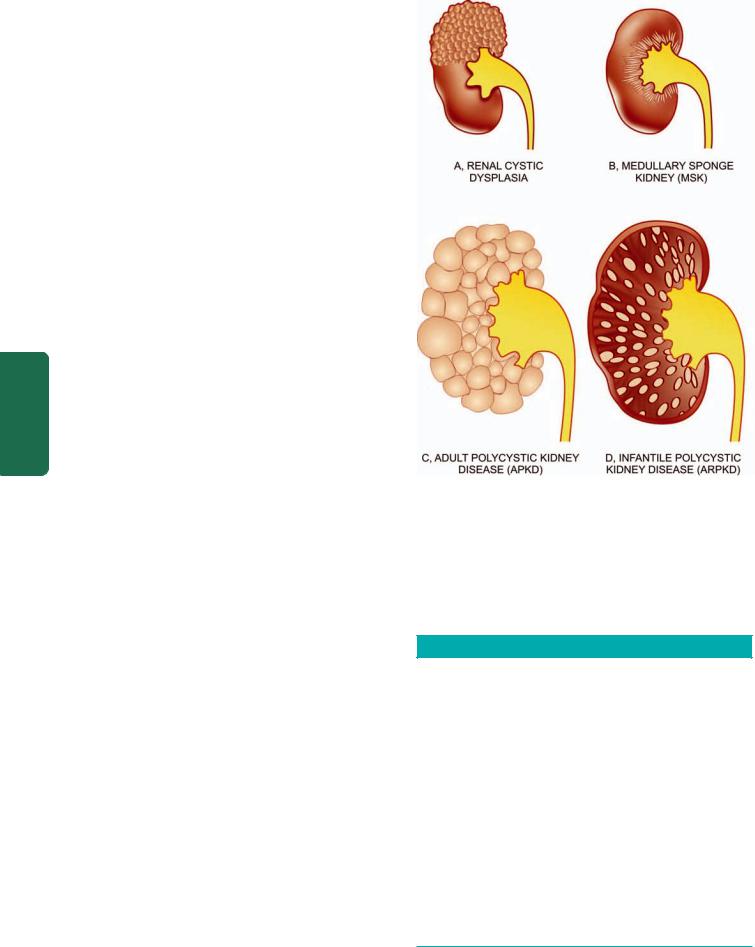
656to back pressure. Radiologically, uraemic pneumonitis shows characteristic central, butterfly-pattern of oedema and congestion in the chest radiograph.
5.Digestive system. Azotaemia directly induces mucosal ulcerations in the lining of the stomach and intestines. Subsequent bleeding can aggravate the existing anaemia. Gastrointestinal irritation may cause nausea, vomiting and diarrhoea.
6.Skeletal system. The skeletal manifestations of renal failure are referred to as renal osteodystrophy (Chapter 28). Two major types of skeletal disorders may occur:
|
i) Osteomalacia occurs from deficiency of a form of vitamin |
|
|
D which is normally activated by the kidney (page 248). Since |
|
|
vitamin D is essential for absorption of calcium, its deficiency |
|
|
results in inadequate deposits of calcium in bone tissue. |
|
|
ii) Osteitis fibrosa occurs due to elevated levels of |
|
|
parathormone. How parathormone excess develops in CRF |
|
|
is complex. As the GFR is decreased, increasing levels of |
|
|
phosphates accumulate in the extracellular fluid which, in |
|
|
turn, cause decline in calcium levels. Decreased calcium level |
|
|
triggers the secretion of parathormone which mobilises |
|
SECTION |
calcium from bone and increases renal tubular reabsorption |
|
of calcium thereby conserving it. However, if the process of |
||
|
||
|
resorption of calcium phosphate from bone continues for |
|
|
sufficient time, hypercalcaemia may be induced with |
|
|
deposits of excess calcium salts in joints and soft tissues and |
|
III |
weakening of bones (renal osteodystrophy). |
|
CONGENITAL MALFORMATIONS |
||
|
||
Systemic |
Approximately 10% of all persons are born with potentially |
|
significant malformations of the urinary system. These range |
||
|
||
|
in severity from minor anomalies which may not produce |
|
|
clinical manifestations to major anomalies which are |
|
Pathology |
incompatible with extrauterine life. About half of all patients |
|
with malformations of the kidneys have coexistent anomalies |
||
|
||
|
either elsewhere in the urinary tract or in other organs. |
|
|
Malformations of the kidneys are classified into 3 broad |
|
|
groups: |
|
|
I. Abnormalities in amount of renal tissue. These include: |
|
|
anomalies with deficient renal parenchyma (e.g. unilateral |
|
|
or bilateral renal hypoplasia) or with excess renal tissue (e.g. |
|
|
renomegaly, supernumerary kidneys). |
|
|
II. Anomalies of position, form and orientation. These are: |
|
|
renal ectopia (pelvic kidney), renal fusion (horseshoe kidney) |
|
|
and persistent foetal lobation. |
|
|
III. Anomalies of differentiation. This group consists of the |
|
|
more important and common morphologic forms covered |
|
|
under the heading of ‘cystic diseases of the kidney’ described |
|
|
in detail below. |
|
|
CYSTIC DISEASES OF KIDNEY |
|
|
Cystic lesions of the kidney may be congenital or acquired, |
|
|
non-neoplastic or neoplastic. Majority of these lesions are |
|
|
congenital non-neoplastic. Cystic lesions in the kidney may |
|
|
occur at any age, extending from foetal life (detected on |
|
|
ultrasonography) to old age. Their clinical presentation may |
|
|
include: abdominal mass, infection, respiratory distress (due |
Figure 22.6 
 Cystic diseases of kidney.
to accompanied pulmonary hypoplasia), haemorrhage, and neoplastic transformation.
Potter divided developmental renal cystic lesions into three types—I, II and III. A simple classification including all cystic lesions of the kidney is given in Table 22.2 and Fig. 22.6. Non-neoplastic lesions are discussed below while
TABLE 22.2: Classification of Cystic Lesions of the Kidney.
A.NON-NEOPLASTIC CYSTIC LESIONS
I. Renal multicystic dysplasia (Potter type II)
II.Polycystic kidney disease (PKD)
1.Adult (autosomal dominant) polycystic kidney disease (ADPKD) (Potter type III)
2.Infantile (autosomal recessive) polycystic kidney disease (ARPKD) (Potter type I)
III.Medullary cystic disease
1.Medullary sponge kidney (MSK)
2.Nephronophthiasis-medullary cystic disease complex
IV. Simple renal cysts
V.Acquired renal cysts VI. Para-renal cysts
B. NEOPLASTIC CYSTIC LESIONS
I.Cystic nephroma (page 694)
II.Cystic partially-differentiated nephroblastoma (CPDN)
III.Multifocal cystic change in Wilms’ tumour (page 696)
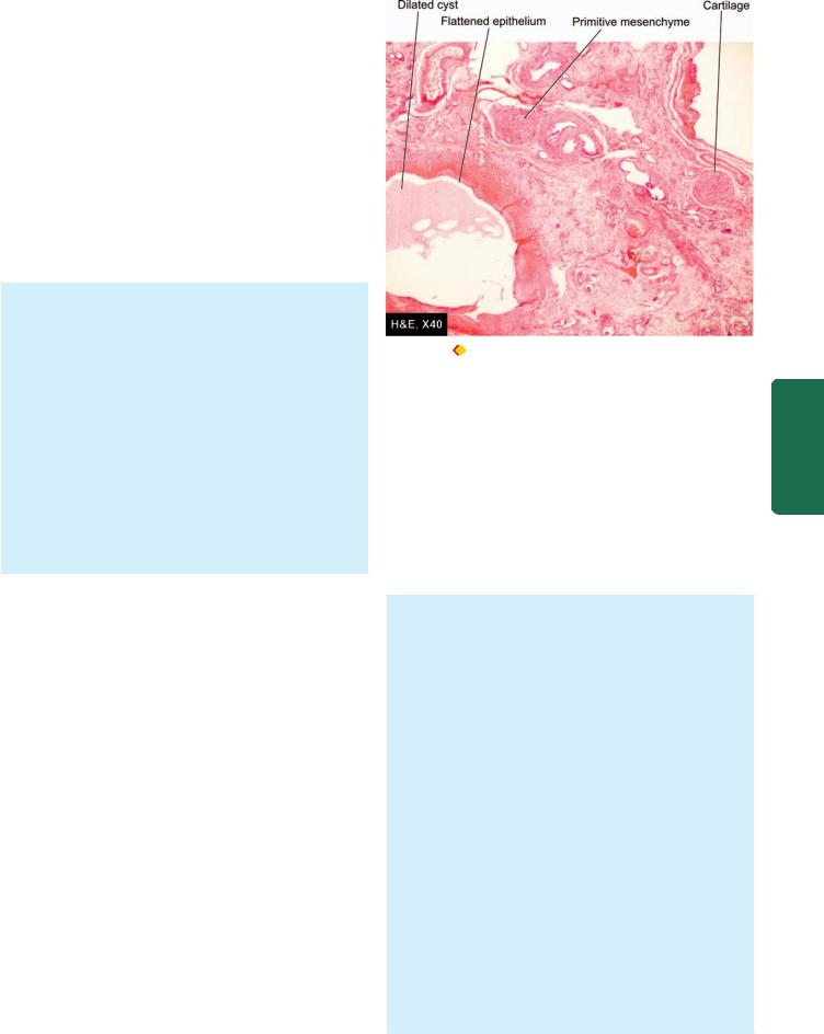
neoplastic cystic lesions of the kidney are described later (page 694).
I.Multicystic Renal Dysplasia
The term ‘multicystic renal dysplasia’ or Potter type II is used for disorganised metanephrogenic differentiation with persistence of structures in the kidney which are not represented in normal nephrogenesis. Renal dysplasia is the most common form of cystic renal disease in the newborn and infants. The condition may occur sporadically or maybe familial and part of a syndrome of other anomalies. It is commonly associated with obstructive abnormalities of the ureter and lower urinary tract such as obstruction of pelviureteric junction (PUJ), ureteral atresia and urethral obstruction.
MORPHOLOGIC FEATURES. Renal dysplasia may be unilateral or bilateral. The dysplastic process may involve the entire renal mass or a part of it.
Grossly, the dysplastic kidney is almost always cystic. The kidney or its affected part is replaced by disorderly mass of multiple cysts resembling a bunch of grapes. Normal renal parenchyma is almost totally obscured by the mass while calyces and pelvis may not be recognised. The ureter is invariably abnormal, being either absent or atretic.
Histologically, the characteristic feature is the presence of undifferentiated mesenchyme that contains smooth muscle, cartilage and immature collecting ducts. The cysts in the mass represent dilated tubules lined by flattened epithelium which are surrounded by concentric layers of connective tissue (Fig. 22.7). Glomeruli and tubules are scanty, primitive or absent.
CLINICAL FEATURES. Unilateral renal dysplasia is frequently discovered in newborn or infants as a flank mass. Often, renal dysplasia is associated with other congenital malformations and syndromes such as ventricular septal defect, tracheo-esophageal fistula, lumbosacral meningomyelocele and Down’s syndrome.
The prognosis of unilateral renal dysplasia following removal of the abnormal kidney is excellent while bilateral renal dysplasia results in death in infancy unless renal transplant is done.
II. Polycystic Kidney Disease
Polycystic disease of the kidney (PKD) is a disorder in which major portion of the renal parenchyma is converted into cysts of varying size. The disease occurs in two forms:
A.An adult type inherited as an autosomal dominant disease; and
B.An infantile type inherited as an autosomal recessive disorder.
A. ADULT POLYCYSTIC KIDNEY DISEASE
Adult (autosomal dominant) polycystic kidney disease (ADPKD) is relatively common (incidence 1:400 to 1: 1:1000) and is the cause of end-stage renal failure in approximately 4% of haemodialysis patients. The pattern of inheritance is
Figure 22.7 Renal cystic dysplasia.There are cysts lined by flattened
epithelium while the intervening parenchyma consists of primitive connective tissue and cartilage.
autosomal dominant with mutation in PKD gene: mutation in PKD-1 gene located on chromosome 16 in over 85% cases (ADPKD-1) while remainder 15% cases have mutation in PKD-2 gene located on chromosome 4 (ADPKD-2). Family history of similar renal disease may be present. The true adult polycystic renal disease is always bilateral and diffuse. Though the kidneys are abnormal at birth, renal function is retained, and symptoms appear in adult life, mostly between the age of 30 and 50 years.
MORPHOLOGIC FEATURES. Grossly, kidneys in ADPKD are always bilaterally enlarged, usually symmetrically, heavy (weighing up to 4 kg) and give it a lobulated appearance on external surface due to underlying cysts . The cut surface shows cysts throughout the renal parenchyma varying in size from tiny cysts to 4-5 cm in diameter (Fig. 22.8,A). The contents of the cysts vary from clear straw-yellow fluid to reddish-brown material. The renal pelvis and calyces are present but are greatly distorted by the cysts and may contain concretions (Fig. 22.9). The cysts, however, do not communicate with the pelvis of the kidney—a feature that helps to distinguish polycystic kidney from hydronephrosis of the kidney on sectioned surface (page 692).
Histologically, the cysts arise from all parts of nephron. It is possible to find some cysts containing recognisable glomerular tufts reflecting their origin from Bowman’s capsule, while others have epithelial lining like that of distal or proximal tubules or collecting ducts. The intervening tissue between the cysts shows some normal renal parenchyma. With advancement of age of the patient, acquired lesions such as pyelonephritis, nephrosclerosis, fibrosis and chronic inflammation are seen with increasingly frequency.
657
Tract Urinary Lower and Kidney The 22 CHAPTER
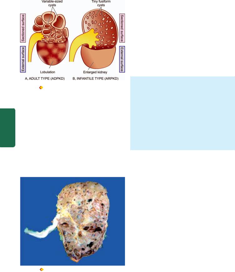
658
Pathology Systemic III SECTION
Figure 22.8 
 Polycystic kidney disease. Diagrammatic representation of comparison of gross appearance of the two main forms.
CLINICAL FEATURES. The condition may become clinically apparent at any age but most commonly manifests in 3rd to 5th decades of life. The most frequent and earliest presenting feature is a dull-ache in the lumbar regions. In others, the presenting complaints are haematuria or passage of blood clots in urine, renal colic, hypertension, urinary tract infections and progressive CRF with polyuria and proteinuria.
ADPKD is considered a systemic disease. About a third of patients with ADPKD have cysts of the liver (Chapter 21). Other associated congenital anomalies seen less frequently are cysts in the pancreas, spleen, lungs and other organs. Approximately 15% of patients have one or more intracranial
Figure 22.9  Adult (autosomal dominant) polycystic kidney disease
(ADPKD). The kidney is enlarged and heavy. Sectioned surface shows loss of demarcation between cortex and medulla and replacement of the entire renal parenchyma by cyst s varying in diameter from a few millimeters to 4-5 cm. These cysts are not communicating with the pelvicalyceal system. The renal pelvis and calyces are distorted due to cystic change.
berry aneurysms of the circle of Willis. Any acquired renal disease is more prone to occur in polycystic kidneys.
B. INFANTILE POLYCYSTIC KIDNEY DISEASE
The infantile (autosomal recessive) form of polycystic kidney disease (ARPKD) is distinct from the adult form and is less common (incidence 1:20,000 births). It is transmitted as an autosomal recessive trait and the family history of similar disease is usually not present. The condition occurs due to a mutation in chromosome 6—6p21, PKHD1 (polycystic kidney and hepatic disease 1). It is invariably bilateral. The age at presentation may be perinatal, neonatal, infantile or juvenile, but frequently serious manifestations are present at birth and result in death from renal failure in early childhood.
MORPHOLOGIC FEATURES. Grossly, the kidneys are bilaterally enlarged with smooth external surface and retained normal reniform shape. Cut surface reveals small, fusiform or cylindrical cysts radiating from the medulla and extend radially to the outer cortex. This gives the sectioned surface of the kidney sponge-like appearance (Fig. 22.8,B). No normal renal parenchyma is grossly recognised. Pelvis, calyces and ureters are normal.
Histologically, the total number of nephrons is normal. Since the cysts are formed from dilatation of collecting tubules, all the collecting tubules show cylindrical or saccular dilatations and are lined by cuboidal to low columnar epithelium. Many of the glomeruli are also cystically dilated.
CLINICAL FEATURES. The clinical manifestations depend on age of the child. In severe form, the gross bilateral cystic renal enlargement may interfere with delivery. In infancy, renal failure may manifest early. Almost all cases of infantile polycystic kidney disease have associated multiple epithelium-lined cysts in the liver or proliferation of portal bile ductules. In older children, associated hepatic changes evelop into what is termed congenital hepatic fibrosis which may lead to portal hypertension and splenomegaly.
The contrasting features of the two main forms of the polycystic kidney disease are presented in Table 22.3.
III. Medullary Cystic Disease
Cystic disease of the renal medulla has two main types:
A.Medullary sponge kidney, a relatively common and innocuous condition; and
B.Nephronophthiasis-medullary cystic disease complex, a common cause of chronic renal failure in juvenile age group.
A. MEDULLARY SPONGE KIDNEY
Medullary sponge kidney consists of multiple cystic dilatations of the papillary ducts in the medulla. It has an autosomal dominant transmission. The condition occurs in adults and may be recognised as an incidental radiographic finding in asymptomatic cases, or the patients may complain of colicky flank pain, dysuria, haematuria and passage of sandy material in the urine. Renal function remains largely

TABLE 22.3. Contrasting Features of Autosomal Dominant (Adult) and Autosomal Recessive (Infantile) Polycystic Kidney Disease |
659 |
|||
|
(ADPKD versus ARPKD). |
|
|
|
|
|
|
|
|
|
Feature |
ADPKD |
ARPKD |
|
|
|
|
|
|
1. |
Inheritance |
Autosomal dominant |
Autosomal recessive |
|
2. |
Cytogenetic defect |
Chromosome 16 (85%): ADPKD-1 |
Chromosome 6 |
|
|
|
Chromosome 4 (15%): ADPKD-2 |
|
|
3. |
Mutations |
PKD 1 gene (85%) |
6p21 PKHD1 |
|
|
|
PKD 2 gene (15%) |
|
|
4. |
Incidence |
1:400 to 1:1000 |
1:20,000 |
|
5. |
Age at presentation |
Adults (3rd to 5th decades) |
Infancy, perinatal |
|
6. |
G/A |
Symmetric bilateral enlargement |
Microand macrocysts radiating from medulla to |
|
|
|
|
outer cortex |
|
7. |
M/E |
Macrocysts |
Enlarged, sometimes asymmetric, sponge-like |
|
|
|
Cysts derived from all parts of nephron |
Cysts from dilated collecting ducts |
|
|
|
(glomeruli, tubules) |
|
|
8. |
Other manifestations |
Intracranial aneurysms, cysts of other organs |
None |
|
|
|
|
|
|
normal or may be mildly impaired in long-standing disease with secondary complications of infection and calculus formation.
MORPHOLOGIC FEATURES. Grossly, the kidneys may be enlarged, normal or shrunken in size depending upon the extent of secondary pyelonephritis. On cut surface, the characteristic feature is the presence of several, small (less than 0.5 cm diameter), cystically dilated papillary ducts, which may contain spherical calculi.
Microscopically, the cysts are lined by tall columnar, cuboidal, transitional or squamous epithelium. Renal cortex may show secondary pyelonephritis but cortical cysts are never a component of medullary sponge kidney.
B.NEPHRONOPHTHIASIS-MEDULLARY CYSTIC DISEASE COMPLEX
This form of medullary cystic disease, also called juvenile nephronophthiasis or uraemic sponge kidney, is a progressive renal disease. It is classified into infantile, juvenile and adolescent type depending upon the age at presentation, juvenile form being the most common. It is the most common form of genetic cause of end-stage renal disease in children and adolescents. The condition has an autosomal recessive inheritance. Familial occurrence is common. The clinical manifestations are due to impaired urinary concentration consequent upon the medullary lesions and consist of polyuria, polydipsia and enuresis. Other features include renal osteodystrophy, growth retardation, anaemia and progressive renal failure leading to uraemia.
MORPHOLOGIC FEATURES. Grossly, the kidneys are moderately reduced in size and granular and have narrow cortices. Cut surface reveals minute cysts, majority of which are present at the cortico-medullary junction.
Microscopically, the cysts are lined by flattened or cuboidal epithelium. There is widespread nonspecific chronic inflammatory infiltrate and interstitial fibrosis. Many glomeruli are hyalinised but tubular atrophy is more pronounced due to marked thickening of tubular basement membrane.
IV. Simple Renal Cysts
Simple renal cysts are a very common postmortem finding. They are seen in about half of all persons above the age of 50 years. Since these cysts are rare in infants and children, they appear to be acquired rather than congenital lesions. Simple cysts of the kidneys are rarely responsible for symptoms. However, symptoms may result from rupture, haemorrhage or infection. The association between simple cysts and hypertension is common.
MORPHOLOGIC FEATURES. Grossly, simple renal cysts are usually solitary but may be multiple. They are commonly located in the cortex. Their size varies from a few millimeters to 10 cm in diameter. The wall of cyst is characteristically yellowish-white and translucent. The cyst usually contains clear straw-coloured fluid which may become rust-coloured due to haemorrhage.
Microscopically, the lining of the cyst is by flattened epithelium. The cyst wall contains variable amount of collagenised fibrous tissue which may occasionally have deposits of haemosiderin or calcium salts.
V. Acquired Renal Cysts
A number of acquired conditions give rise to renal cysts. These include the following:
1.Patients with end-stage renal disease on prolonged dialysis (dialysis-associated cystic disease).
2.Hydatid (echinococcal) cyst.
3.Tuberculosis of the kidney.
4.Cystic degeneration in carcinoma of kidney.
5.Traumatic intrarenal haematoma.
6.Drug-induced cystic disease in experimental animals.
VI. Pararenal Cysts
Cysts occurring adjacent to a kidney are termed pararenal cysts. These include the following:
1.Pyelocalyceal cysts
2.Hilar lymphangiectatic cysts
3.Retroperitoneal cysts
4.Perinephric pseudocysts from trauma.
Tract Urinary Lower and Kidney The 22 CHAPTER

660GLOMERULAR DISEASES DEFINITION AND CLASSIFICATION
Glomerular diseases encompass a large and clinically significant group of renal diseases. Glomerulonephritis (GN) or Bright’s disease is the term used for diseases that primarily involve the renal glomeruli. It is convenient to classify glomerular diseases into 2 broad groups:
I. Primary glomerulonephritis in which the glomeruli are the predominant site of involvement.
II. Secondary glomerular diseases include certain systemic and hereditary diseases which secondarily affect the glomeruli.
Though this division is widely followed, it is somewhat arbitrary since many primary forms of glomerulonephritis have systemic effects, and many systemic diseases may initially present with glomerular involvement. Many classifications of different types of glomerulonephiritis have been described, but most widely accepted classification is based on clinical presentation and pathologic changes in the glomeruli given in Table 22.4.
SECTION |
CLINICAL MANIFESTATIONS |
|||
The clinical presentation of glomerular disease is quite |
||||
|
||||
|
variable but in general four features—proteinuria, haema- |
|||
|
turia, hypertension and disturbed excretory function, are |
|||
III |
present in varying combinations depending upon the |
|||
|
|
|
||
|
|
|||
|
TABLE 22.4: Clinicopathologic Classification of Glomerular |
|||
Systemic |
|
Diseases. |
||
I. |
PRIMARY GLOMERULONEPHRITIS |
|||
|
||||
|
1. |
Acute GN |
||
|
|
i) |
Post-streptococcal |
|
Pathology |
|
ii) |
Non-streptococcal |
|
2. |
Rapidly progressive GN |
|||
|
||||
|
3. |
Minimal change disease |
||
|
4. |
Membranous GN |
||
|
5. |
Membrano-proliferative GN |
||
|
6. |
Focal proliferative GN |
||
|
7. |
Focal segmental glomerulosclerosis (FSGS) |
||
|
8. |
IgA nephropathy |
||
|
9. |
Chronic glomerulonephritis |
||
II. SECONDARY SYSTEMIC GLOMERULAR DISEASES
1.Lupus nephritis (SLE)
2.Diabetic nephropathy
3.Amyloidosis (page 82)
4.Polyarteritis nodosa (page 402)
5.Wegener’s granulomatosis (page 403)
6.Goodpasture’s syndrome (page 494)
7.Henoch-Schönlein purpura (page 331)
8.Systemic infectious diseases (bacterial e.g. bacterial endocarditis, syphilis, leprosy; viral e.g. HBV, HCV, HIV; parasitic e.g. falciparum malaria, filariasis)
9.Idiopathic mixed cryoglobulinaemia
III. HEREDITARY NEPHRITIS
1.Alport’s syndrome
2.Fabry’s disease
3.Nail-patella syndrome
underlying condition. A firm diagnosis, however, can be established by examination of renal biopsy under light, electron and immunofluorescence microscopy.
A number of clinical syndromes are recognised in glomerular diseases. The following are six major glomerular syndromes commonly found in different glomerular diseases: 
 nephritic and nephrotic syndromes;

 acute and chronic renal failure;

 asymptomatic proteinuria and haematuria. These are briefly described below.
I. ACUTE NEPHRITIC SYNDROME. This is the acute onset of haematuria, proteinuria, hypertension, oedema and oliguria following an infective illness about 10 to 20 days earlier.
1.The haematuria is generally slight giving the urine smoky appearance and erythrocytes are detectable by microscopy or by chemical testing for haemoglobin. Appearance of red cell casts is another classical feature of acute nephritic syndrome.
2.The proteinuria is mild (less than 3 gm per 24 hrs) and is usually non-selective (nephritic range proteinuria).
3.Hypertension is variable depending upon the severity of the glomerular disease but is generally mild.
4.Oedema in nephritic syndrome is usually mild and results from sodium and water retention (page 97).
5.Oliguria is variable and reflects the severity of glomerular involvement.
The underlying causes of acute nephritic syndrome may be primary glomerulonephritic diseases (classically acute glomerulonephritis and rapidly progressive glomerulonephritis) or certain systemic diseases (Table 22.5).
II. NEPHROTIC SYNDROME Nephrotic syndrome is a constellation of features in different diseases having varying pathogenesis; it is characterised by findings of massive proteinuria, hypoalbuminaemia, oedema, hyperlipidaemia, lipiduria, and hypercoagulability.
1.Heavy proteinuria (protein loss of more than 3 gm per
24hrs) is the chief characteristic of nephrotic syndrome
(nephrotic range proteinuria). In children, protein loss is correspondingly less. A small amount of protein (20 to 150 mg/day) normally passes through the glomerular filtration barrier and is reabsorbed by the tubules. But in case of increased glomerular permeability to plasma proteins, excess of protein is filtered out exceeding the capacity of tubules for reabsorption and, therefore, appears in the urine. Another feature of protein loss is its ‘selectivity’. A highly-selective proteinuria consists mostly of loss of low molecular weight proteins, while a poorly-selective proteinuria is loss of high molecular weight proteins in the urine. In nephrotic syndrome, proteinuria mostly consists of loss of albumin (molecular weight 66,000) in the urine.
2.Hypoalbuminaemia is produced primarily consequent to urinary loss of albumin, and partly due to increased renal catabolism and inadequate hepatic synthesis of albumin.

TABLE 22.5: Causes of Acute Nephritic Syndrome.
I.PRIMARY GLOMERULONEPHRITIS
1.Acute GN
i)Post-streptococcal
ii)Non-streptococcal
2.Rapidly progressive GN
3.Membranoproliferative GN
4.Focal GN
5.IgA nephropathy
II.SYSTEMIC DISEASES
1.SLE
2.Polyarteritis nodosa
3.Wegener’s granulomatosis
4.Henoch-Schonlein purpura
5.Cryoglobulinaemia
Often, the plasma albumin level is 1 to 3 gm/dl (normal 3.5 to 5.5 gm/dl) and there is reversed albumin-globulin ratio. The concentration of other proteins in the plasma such as immunoglobulins, clotting factors and antithrombin may fall rendering these patients more vulnerable to infections and thrombotic and thromboembolic complications.
3.Oedema in nephrotic syndrome appears due to fall in colloid osmotic pressure consequent upon hypoalbuminaemia. Sodium and water retention further contribute to oedema. Nephrotic oedema is usually peripheral but in children facial oedema may be more prominent (page 99).
4.Hyperlipidaemia is a frequent accompaniment of nephrotic syndrome. The exact mechanism of its genesis is not clear. It is hypothesised that the liver faced with the stress of massive protein synthesis in response to heavy urinary protein loss, also causes increased synthesis of lipoproteins. There are increased blood levels of total lipids, cholesterol, triglycerides, VLDL and LDL but decrease in HDL. Low blood level of HDL is partly due to its loss in the urine.
5.Lipiduria occurs following hyperlipidaemia due to excessive leakiness of glomerular filtration barrier.
6.Hypercoagulability. Patients with nephrotic syndrome may develop spontaneous arterial or venous thrombosis, renal vein thrombosis and pulmonary embolism due to various factors. These include: increased urinary loss of antithrombin III, hyperfibrinogenaemia from increased synthesis in the liver, decreased fibrinolysis, increased platelet aggregation and altered levels of protein C and S.
The causes of nephrotic syndrome are diverse and are listed in Table 22.6. The morphology of individual types is described later. But it must be mentioned here that:

 in children, primary glomerulonephritis is the cause in majority of cases of the nephrotic syndrome; most frequent
is lipoid nephrosis (65%); and

 in adults, on the other hand, systemic diseases (diabetes, amyloidosis and SLE) are more frequent causes of nephrotic syndrome. The most common primary glomerular disease
in adults is membranous glomerulonephritis (40%).
Features of nephritic and nephroic syndromes have been contrasted in Table 22.7.
III.ACUTE RENAL FAILURE. As already described above, acute renal failure (ARF) is characterised by rapid decline in renal function. ARF has many causes including glomerular disease, principally rapidly progressive GN and acute diffuse proliferative GN.
IV.CHRONIC RENAL FAILURE. Glomerular causes of chronic renal failure (CRF) have already been described. These cases have advanced renal impairment progressing over years and is detected by significant proteinuria, haematuria, hypertension and azotaemia. Such patients generally have small contracted kidneys due to chronic glomerulonephritis.
V. ASYMPTOMATIC PROTEINURIA. Presence of proteinuria unexpectedly in a patient may be unrelated to renal disease (e.g. exercise-induced, extreme lordosis and orthostatic proteinuria), or may indicate an underlying mild glomerulonephritis. Association of asymptomatic haematuria, hypertension or impaired renal function with asymptomatic proteinuria should raise strong suspicion of underlying glomerulonephritis.
VI. ASYMPTOMATIC HAEMATURIA. Asymptomatic microscopic haematuria is common in children and young
TABLE 22.6: Causes of Nephrotic Syndrome.
I. PRIMARY GLOMERULONEPHRITIS
1.Minimal change disease (most common in children)
2.Membranous GN (most common in adults)
3.Membranoproliferative GN
4.Focal segmental glomerulosclerosis
5.Focal GN
6.IgA nephropathy
II.SYSTEMIC DISEASES
1.Diabetes mellitus
2.Amyloidosis
3.SLE
III.SYSTEMIC INFECTIONS
1.Viral infections (HBV, HCV, HIV)
2.Bacterial infections (bacterial endocarditis, syphilis, leprosy)
3.Protozoa and parasites (P. falciparum malaria, filariasis)
IV. HYPERSENSITIVITY REACTIONS
1.Drugs (heavy metal compounds like gold and mercury, other drugs like penicillamine, trimethadione and tolbutamide, heroin addiction)
2.Bee stings, snake bite, poison ivy
V.MALIGNANCY
1.Carcinomas
2.Myeloma
3.Hodgkin’s disease
VI. PREGNANCY
Toxaemia of pregnancy
VII. CIRCULATORY DISTURBANCES
1.Renal vein thrombosis
2.Constrictive pericarditis
VIII. HEREDITARY DISEASES
1.Alport’s disease
2.Fabry’s disease
3.Nail-patella syndrome
661
Tract Urinary Lower and Kidney The 22 CHAPTER

662 TABLE 22.7: Contrasting Features of Acute Nephritic and Nephrotic Syndromes.
|
Feature |
Acute Nephritic Syndrome |
Nephrotic Syndrome |
|
|
|
|
1. |
Proteinuria |
Mild (< 3 gm per 24 hrs) |
Heavy (> 3 gm per 24 hrs) |
2. |
Hypoalbuminaemia |
Uncommon |
Present |
3. |
Oedema |
Mild, in loose tissue |
Marked, generalised peripheral |
4. |
Mechanism of Oedema |
Na+ and water retention |
↓ plasma osmotic pressure, Na+ and |
|
|
|
water retention |
5. |
Haematuria |
Present, microscopic |
Absent |
6. |
Hypertension |
Present |
Present in advanced disease |
7. |
Hyperlipidaemia |
Absent |
Present |
8. |
Lipiduria |
Absent |
Present |
9. |
Oliguria |
Present |
Present in advanced disease |
10. |
Hypercoagulability |
Absent |
Present |
|
|
|
|
Pathology Systemic III SECTION
adolescents and has many diverse causes such as diseases |
sis of some forms of glomerular diseases in human beings |
||
of the glomerulus, renal interstitium, calyceal system, ureter, |
(Table 22.9). |
||
bladder, prostate, urethra, and underlying bleeding disorder, |
I. |
IMMUNOLOGIC MECHANISMS |
|
congenital abnormalities of the kidneys or neoplasia. |
|||
Glomerular haematuria is indicated by the presence of red |
Experimental studies and observations in humans have |
||
blood cells, red cell casts and haemoglobin in the urine. |
|||
revealed that immunologic mechanisms, most importantly |
|||
Glomerular haematuria is frequently associated with |
|||
antigen-antibody complexes, underlie most forms of |
|||
asymptomatic proteinuria. |
|||
glomerular injury. The general principles of these |
|||
|
|||
PATHOGENESIS OF GLOMERULAR INJURY |
mechanisms in different forms of glomerular diseases are |
||
discussed below, while specific features pertaining to |
|||
|
|||
Most forms of primary GN and many of the secondary |
individual types of GN are described separately later. |
||
glomerular diseases in human beings have immunologic |
|
|
|
pathogenesis. This view is largely based on immuno- |
A. Antibody-Mediated Glomerular Injury |
||
fluorescence studies of GN in humans which have revealed |
1. IMMUNE COMPLEX DISEASE. Majority of cases of |
||
glomerular deposits of immunoglobulins and complement |
|||
in patterns that closely resemble those of experimental |
glomerular disease result from deposits of immune |
||
models. The consequences of injury at different sites within |
complexes (antigen-antibody complexes). The immune |
||
the glomerulus in various glomerular diseases can be |
complexes are represented by irregular or granular glomer- |
||
assessed when compared with the normal physiologic role |
ular deposits of immunoglobulins (IgG, IgM and IgA) and |
||
of the main cells involved i.e. endothelial, mesangial, visceral |
complement (mainly C3). Based on the experimental models |
||
epithelial, and parietal epithelial cells as well as of the GBM as |
and studies in human beings, the following 3 patterns of |
||
summed up in Table 22.8. |
glomerular deposits of immune complexes in various |
||
glomerular diseases have been observed as illustrated in |
|||
Immunologic mechanisms underlying glomerular |
|||
Fig. 22.10: |
|||
injury are primarily antibody-mediated (immune-complex |
|||
|
|
||
disease). There is evidence to suggest that cell-mediated |
i) Exclusive mesangial deposits are characterised by very mild |
||
immune reactions in the form of delayed type hypersensiti- |
form of glomerular disease. |
||
vity can also cause glomerular injury in some situations. |
ii) |
Extensive subendothelial deposits along the GBM are |
|
In addition, a few secondary mechanisms and some non- |
accompanied by severe hypercellular sclerosing glomerular |
||
immunologic mechanisms are involved in the pathogene- |
lesions. |
||
TABLE 22.8: Relationship of Physiologic Role of Glomerular Components with Consequences in Glomerular Injury.
|
Component |
|
Physiologic Function |
Consequence of Injury |
Related Glomerular Disease |
|
|
|
|
|
|
1. |
Endothelial cells |
i) |
Maintain glomerular perfusion |
Vasoconstriction |
Acute renal failure |
|
|
ii) |
Prevent leucocyte adhesion |
Leucocyte infiltration |
Focal/diffuse proliferative GN |
|
|
iii) |
Prevent platelet aggregation |
Intravascular microthrombi |
Thrombotic microangiopathies |
2. |
Mesangial cells |
Control glomerular filtration |
Proliferation and increased matrix |
Membranoproliferative GN |
|
3. |
Visceral epithelial cells |
Prevent plasma protein filtration |
Proteinuria |
Minimal change disease, FSGS |
|
4. |
GBM |
Prevents plasma protein filtration |
Proteinuria |
Membranous GN, ? MPGN |
|
5. |
Parietal epithelial cells |
Maintain Bowman’s space |
Crescent formation |
RPGN |
|
|
|
|
|
|
|
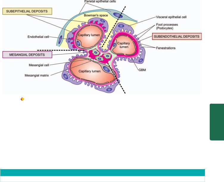
Figure 22.10  Diagrammatic representation of ultrastructure of a portion of glomerular lobule. It shows three patterns of irregular or granular glomerular deposits in immune-complex disease.
iii) Subepithelial deposits are seen between the outer surface of the GBM and the podocytes.
Deposits may be located at one or more of the above sites in any case of glomerular injury.
It was widely believed earlier that glomerular deposits result from circulating immune complexes. Now, it has been shown that glomerular deposits are formed by one of the following two mechanisms:
i) Local immune complex deposits. Formation of glomerular deposits of immune complex in situ occurs as a
result of combination of antibodies with autologous nonbasement membrane antigens or nonglomerular antigens planted on glomeruli. Currently, this mechanism is considered responsible for most cases of immune complex GN. Classic experimental model of in situ immune complex GN is Heymann nephritis (autologous immune complex nephritis) induced in rats by immunising animals with homologous preparations of proximal tubular brush border. The rats develop antibodies to brush border antigens and thereby membranous GN that closely resembles human
TABLE 22.9: Pathogenetic Mechanisms in Glomerular Diseases.
Mechanism |
Related Glomerular Disease |
|
|
I.IMMUNOLOGIC MECHANISMS
A. |
Antibody-mediated glomerular injury |
|
1. |
Immune-complex disease |
Immune-complex mediated GN (Acute diffuse proliferative GN, |
|
|
membranous GN, membranoproliferative GN, IgA nephropathy; |
|
|
secondary glomerular disease in SLE, malaria etc.) |
2. |
Anti-glomerular basement membrane (Anti-GBM) disease |
Goodpasture's disease |
3. |
Alternate pathway disease |
Membranoproliferative GN type II |
4. |
Other mechanisms (anti-neutrophil cytoplasmic antibodies— |
Vasculitis |
|
ANCA, anti-endothelial cell antibodies—AECA) |
|
B. |
Cell-mediated glomerular injury |
Pauci-immune GN (type III RPGN) |
C. |
Secondary pathogenetic mechanisms |
Mediate glomerular injury in various primary and |
|
|
secondary glomerular diseases |
II.NON-IMMUNOLOGIC MECHANISMS
1. Metabolic |
Diabetic nephropathy, Fabry's disease |
2. Haemodynamic |
Hypertensive nephrosclerosis, FSGS |
3. Deposition |
Amyloid nephropathy |
4. Infectious |
HIV-nephropathy, immune-complex GN in SABE |
5. Drugs |
NSAIDs-associated minimal change disease |
6. Inherited |
Alport's syndrome, nail-patella syndrome |
|
|
663
Tract Urinary Lower and Kidney The 22 CHAPTER

664membranous GN. The examples of planted non-glomerular antigens are cationic proteins, lectins, DNA, bacterial products (e.g. a protein of group A streptococci), viral and parasitic products and drugs.
ii)Circulating immune complex deposits. This mechanism used to be considered very important for glomerular injury but now it is believed that circulating immune complexes cause glomerular damage under certain circumstances only. These situations are: their presence in high concentrations for prolonged periods, or when they possess special properties that cause their binding to glomeruli, or when host mechanisms are defective and fail to eliminate immune
|
complexes. The antigens evoking antibody response may be |
|
|
endogenous (e.g. in SLE) or may be exogenous (e.g. Hepatitis |
|
|
B virus, Treponema pallidum, Plasmodium falciparum and |
|
|
various tumour antigens). The antigen-antibody complexes |
|
|
are formed in the circulation and then trapped in the |
|
|
glomeruli where they produce glomerular injury after |
|
|
combining with complement. |
|
|
Immune complex GN is observed in the following human |
|
|
diseases: |
|
SECTION |
i) Primary GN e.g. acute diffuse proliferative GN, memb- |
|
ranous GN, membranoproliferative GN, IgA nephropathy |
||
mixed cryoglobulinaemia. |
||
|
and some cases of rapidly progressive GN and focal GN. |
|
|
ii) Systemic diseases e.g. glomerular disease in SLE, malaria, |
|
|
syphilis, hepatitis, Henoch-Schonlein purpura and idiopathic |
|
III |
2. ANTI-GBM DISEASE. Less than 5% cases of human GN |
|
|
||
|
are associated with anti-GBM antibodies. The constituent of |
|
Systemic |
GBM acting as antigen appears to be a component of collagen |
|
IV of the basement membrane. The experimental model of |
||
|
||
|
anti-GBM disease is Masugi nephritis (nephrotoxic serum |
|
|
nephritis) produced in rats by injection of heterologous |
|
Pathology |
antibodies against GBM prepared in rabbits by immunisation |
|
with rat kidney glomerular tissue. |
||
|
||
|
Anti-GBM disease is classically characterised by |
|
|
interrupted linear deposits of anti-GBM antibodies (mostly IgG; |
|
|
rarely IgA and IgM) and complement (mainly C3) along the |
|
|
glomerular basement membrane. These deposits are detected |
|
|
by immunofluorescence microscopy or by electron |
|
|
microscopy. |
|
|
Anti-GBM disease is characteristically exemplified by |
|
|
glomerular injury in Goodpasture’s syndrome in some cases |
|
|
of rapidly progressive GN. About half to two-third of the |
|
|
patients with renal lesions in Goodpasture’s syndrome have |
|
|
pulmonary haemorrhage mediated by cross-reacting |
|
|
autoantibodies against alveolar basement membrane (page |
|
|
494). |
|
|
3. ALTERNATE PATHWAY DISEASE. As apparent from |
|
|
the above mechanisms, the complement system, in |
|
|
particular C3, contributes to glomerular injury in most |
|
|
forms of GN. Deposits of C3 are associated with the early |
|
|
components C1, C2 and C4 which are evidence of classic |
|
|
pathway activation of complement. But in alternate |
|
|
pathway activation, there is decreased serum C3 level, |
|
|
decreased serum levels of factor B and properdin, normal |
|
|
serum levels of C1, C2 and C4 but C3 and properdin are |
|
|
found deposited in the glomeruli without immunoglobulin |
deposits, reflecting activation of alternate pathway of complement. Such patients have circulating anti-comple- mentary nephritic factor (C3NeF) which is an IgG antibody and acts as an autoantibody to the alternate C3 convertase, leading to persistent alternate pathway activation.
The deposits in alternate pathway disease are characteristically electron-dense under electron microscopy; glomerular lesions in such cases are referred to as dense-
deposit disease.
Alternate pathway disease occurs in most cases of type II membranoproliferative GN, some patients of rapidly progressive GN, acute diffuse proliferative GN, IgA nephropathy and in SLE.
4. OTHER MECHANISMS OF ANTIBODY-MEDIATED INJURY. A few autoantibodies have been implicated in some patients of focal segmental glomerulosclerosis and few other types of GN. These antibodies include the following:
i)Anti-neutrophil cytoplasmic antibodies (ANCA). About 40% cases of rapidly progressive GN are deficient in immunoglobulins in glomeruli (pauciimmune GN) and are positive for ANCA against neutrophil cytoplasmic antigens in their circulation. ANCA causes endothelial injury by generation of reactive oxygen radicals. ANCA-mediated vasculitis is also seen in Wegenger’s granulomatosis and Churg-Strauss syndrome.
ii)Anti-endothelial cell antibodies (AECA). Autoantibodies against endothelial antigens have been detected in circulation in several inflammatory vasculitis and glomerulonephritis. These antibodies increase the adhesiveness of leucocytes to endothelial cells.
B.Cell-mediated Glomerular Injury (Delayed-type Hypersensitivity)
There is evidence to suggest that cell-mediated immune reactions may be involved in causing glomerular injury, particularly in cases with deficient immunoglobulins (e.g. in pauci-immune type glomerulonephritis in RPGN). Cytokines and other mediators released by activated T cells stimulate cytotoxicity, recruitment of more leucocytes and fibrogenesis. CD4+ T lymphocytes recruit more macrophages while CD8+ cytotoxic T lymphocytes and natural killer cells cause further glomerular cell injury by antibody-dependent cell toxicity. Soluble factor derived from T lymphocytes is implicated in proteinuria in minimal change disease and focal GS.
However, cell-mediated injury is yet less clear than antibody-mediated glomerular injury.
C.Secondary Pathogenetic Mechanisms (Mediators of Immunologic Injury)
Secondary pathogenetic mechanisms are a number of mediators of immunologic glomerular injury operating in man and in experimental models. These include the following:
1. NEUTROPHILS. Neutrophils are conspicuous in certain forms of glomerular disease such as in acute diffuse proliferative GN, and may also be present in membranoproliferative
GN and lupus nephritis. Neutrophils can mediate glomerular injury by activation of complement as well as by release of proteases, arachidonic acid metabolites and oxygen-derived free radicals. These agents cause degradation of GBM and cell injury.
2.MONONUCLEAR PHAGOCYTES. Many forms of human and experimental proliferative GN are associated with glomerular infiltration by monocytes and macrophages. Accumulation of mononuclear phagocytes is considered an important constituent of hypercellularity in these forms of GN aside from proliferation of mesangial and endothelial cells. Activated macrophages release a variety of biologically active substances which take part in glomerular injury.
3.COMPLEMENT SYSTEM. The pathogenetic role of classical and alternate pathway of activation of complement has already been highlighted above. Besides the components of complement which mediate glomerular injury via neutrophils already mentioned, C5bC6789 (MAC, acronym for membrane attack complex, also called terminal complex) is capable of inducing damage to GBM directly.
4.PLATELETS. Platelet aggregation and release of mediators play a role in the evolution of some forms of GN. Increased intrarenal platelet consumption has been found to occur in some forms of glomerular disease.
5.MESANGIAL CELLS. There is evidence to suggest that mesangial cells present in the glomeruli may be stimulated to produce mediators of inflammation and take part in glomerular injury.
6.COAGULATION SYSTEM. The presence of fibrin in early crescents in certain forms of human and experimental GN suggests the role of coagulation system in glomerular damage. Fibrinogen may leak into Bowman’s space and act as stimulus for cell proliferation. Crescents usually transform into scar tissue under the influence of fibronectin which is regularly present in crescents in human glomerular disease.
II. NON-IMMUNOLOGIC MECHANISMS
Though most forms of GN are mediated by immunologic mechanisms, a few examples of glomerular injury by nonimmunologic mechanisms are found:
1.Metabolic glomerular injury e.g. in diabetic nephropathy (due to hyperglycaemia), Fabry’s disease (due to sulfatidosis).
2.Haemodynamic glomerular injury e.g. systemic hypertension, intraglomerular hypertension in FSGS.
3.Deposition diseases e.g. amyloidosis.
4.Infectious diseases e.g. HBV, HCV, HIV, E. coli-derived nephrotoxin.
5.Drugs e.g. minimal change disease due to NSAIDs.
6.Inherited glomerular diseases e.g. Alport’s syndrome, nailpatella syndrome.
The evolution of end-stage renal failure in glomerular injury is explained on the basis of adaptive glomerular hypertrophy of unaffected glomeruli that results in increased glomerular blood flow and increased glomerular capillary pressure inducing intraglomerular hypertension. These events lead
to increased deposition of mesangial matrix and proliferation of mesangial cells, endothelial and epithelial cell injury, and eventually to progressive glomerulosclerosis and end-stage renal failure.
SPECIFIC TYPES OF GLOMERULAR DISEASES
Classification of different forms of glomerular diseases is already presented in Table 22.4. Features of individual types are described below and a summary of major forms of primary glomerulonephritis is given in Table 22.11 at the end of this discussion.
I.PRIMARY GLOMERULONEPHRITIS
Acute Glomerulonephritis
(Synonyms: Acute Diffuse Proliferative GN,
Diffuse Endocapillary GN)
Acute GN is known to follow acute infection and characteristically presents as acute nephritic syndrome. Based on etiologic agent, acute GN is subdivided into 2 main groups: acute post-streptococcal GN and acute non-streptococcal GN, the former being more common.
ACUTE POST-STREPTOCOCCAL GN
Acute post-streptococcal GN, though uncommon and sporadic in the Western countries, is a common form of GN in developing countries, mostly affecting children between 2 to 14 years of age but 10% cases are seen in adults above 40 years of age. The onset of disease is generally sudden after 1-2 weeks of streptococcal infection, most frequently of the throat (e.g. streptococcal pharyngitis) and sometimes of the skin (e.g. streptococcal impetigo).
ETIOPATHOGENESIS. The relationship between streptococcal infection and this form of GN is now well established. Particularly nephritogenic are types 12,4,1 and Red Lake of group A β-haemolytic streptococci (compare the etiologic agent with that of RHD, page 438). The glomerular lesions appear to result from deposition of immune complexes in the glomeruli. The evidences cited in support are as under:
i)There is epidemiological evidence of preceding streptococcal sore throat or skin infection about 1-2 weeks prior to the attack.
ii)The latent period between streptococcal infection and onset of clinical manifestations of the disease is compatible with the period required for building up of antibodies.
iii)Streptococcal infection may be identified by culture or may be inferred from elevated titres of antibodies against streptococcal antigens. These include the following:

 anti-streptolysin O (ASO);

 anti-deoxyribonuclease B (anti-DNAse B);

 anti-streptokinase (ASKase);
 anti-nicotinyl adenine dinucleotidase (anti-NADase); and
 anti-hyaluronidase (AHase).
iv)There is usually hypocomplementaemia indicating involvement of complement in the glomerular deposits.
v)It has also been possible to identify antigenic component of streptococci which is cytoplasmic antigen, endostreptosin.
665
Tract Urinary Lower and Kidney The 22 CHAPTER
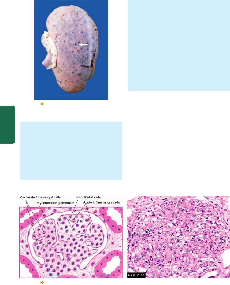
666
Pathology Systemic III SECTION
Figure 22.11 
 Flea-bitten kidney. The kidney is enlarged in size and weight. The cortex shows tiny petechial haemorrhages visible through the capsule (arrow).
MORPHOLOGIC FEATURES. Grossly, the kidneys are symmetrically enlarged, weighing one and a half to twice the normal weight. The cortical as well as sectioned surface show petechial haemorrhages giving the characteristic appearance of flea-bitten kidney (Fig. 22.11).
Light microscopic findings are as under (Fig. 22.12):
i) Glomeruli—The glomeruli are affected diffusely. They are enlarged and hypercellular. The diffuse hypercellularity of the tuft is due to proliferation of mesangial, endothelial and occasionally epithelial cells (acute proliferative lesions) as well as by infiltration of leucocytes,
chiefly polymorphs and sometimes monocytes (acute exudative lesion). There may be small deposits of fibrin within the capillary lumina and in the mesangium.
ii)Tubules—Tubular changes are not very striking. There may be swelling and hyaline droplets in tubular cells, and tubular lumina may contain red cell casts.
iii)Interstitium—There may be some degree of interstitial oedema and leucocytic infiltration.
iv)Vessels—Changes in arteries and arterioles are seldom present in acute GN.
Electron microscopic findings, aside from confirming the light microscopic findings, demonstrate the characteristic electron-dense irregular deposits (‘humps’) on the epithelial side of the GBM. These deposits represent the immune complexes (Fig. 22.13).
Immunofluorescence microscopy reveals that the irregular deposits along the GBM consist principally of IgG and complement C3.
CLINICAL FEATURES. Typically, the patient is a young child, presenting with acute nephritic syndrome (page 660), having sudden and abrupt onset following an episode of sore throat or skin infection 1-2 weeks prior to the development of symptoms. The features include microscopic or intermittent haematuria, red cell casts, mild non-selective proteinuria (less than 3 gm per 24 hrs), hypertension, periorbital oedema and variably oliguria. Less often, the presentation may be as nephrotic syndrome. In adults, the features are atypical and include sudden hypertension, oedema and azotaemia. Development of hypertension in either case is a poor prognostic sign.
Prognosis varies with the age of the patient. Children almost always (95%) recover completely with reversal of proliferative glomerular changes. Complications arise more often in adults and occasionally in children. These include development of rapidly progressive GN, chronic GN, uraemia and chronic renal failure.
Figure 22.12 
 Acute post-streptococcal GN, light microscopic appearance. There is increased cellularity due to proliferation of mesangial cells, endothelial cells and some epithelial cells and infiltration of the tuft by neutrophils and monocytes.
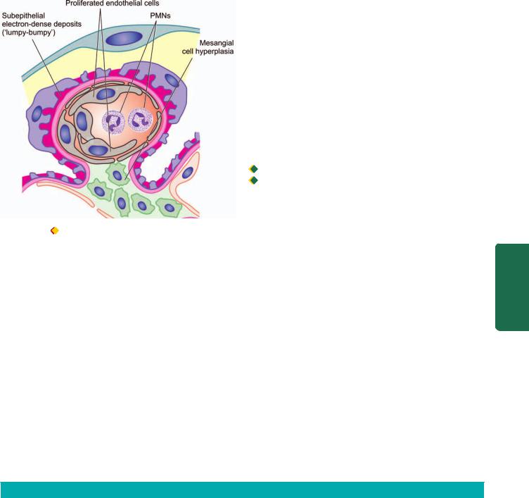
Figure 22.13 Acute glomerulonephritis, diagrammatic represen-
tation of ultrastructure of a portion of glomerular lobule showing characteristic electron-dense irregular deposits or ‘humps’ on the epithelial side of the GBM.
ACUTE NON-STREPTOCOCCAL GN
About one-third cases of acute GN are caused by organisms other than haemolytic streptococci. These include other bacteria (e.g. staphylococci, pneumococci, meningococci, Salmonella and Pseudomonas), viruses (e.g. hepatitis B virus, mumps, infectious mononucleosis and varicella), parasitic infections (e.g. malaria, toxoplasmosis and schistosomiasis) and syphilis. The appearance of renal biopsy by light microscopy, EM and immunofluorescence microscopy is similar to that seen in acute post-streptococcal GN. The prognosis of non-streptococcal GN is not as good as that of streptococcal GN.
Rapidly Progressive Glomerulonephritis
(Synonyms: RPGN, Crescentic GN, Extracapillary GN)
RPGN presents with an acute reduction in renal function resulting in acute renal failure in a few weeks or months. It
is characterised by formation of ‘crescents’ (crescentic GN) outside the glomerular capillaries (extracapillary GN). ‘Crescents’ are formed from the proliferation of parietal epithelial cells lining Bowman’s capsule with contribution from visceral epithelial cells and the invading mononuclear cells. The stimulus for crescent formation appears to be the presence of fibrin in the capsular space. RPGN occurs most frequently in adults, with a slight male preponderance. Prognosis of RPGN in general is dismal.
ETIOPATHOGENESIS. A number of primary glomerular and systemic diseases are characterised by formation of crescents. Based on the etiologic agents and pathogenetic mechanism, patients with RPGN are divided into 3 groups
(Table 22.10):

 RPGN in systemic diseases (anti-GBM type);

 post-infectious RPGN (immune-complex type); and

 pauci-immune RPGN.
Following three serologic markers are used for categorising RPGN:
i)serum C3 level,
ii)anti-GBM antibody; and
iii)anti-neutrophil cytoplasmic antibody (ANCA).
Type I RPGN: Anti-GBM disease. A number of systemic diseases such as Goodpasture’s syndrome, SLE, vasculitis, Wegener’s granulomatosis, Henoch-Schonlein purpura and idiopathic mixed cryoglobulinaemia are associated with crescentic GN. Goodpasture’s syndrome is the characteristic example of anti-GBM disease and is described below:
Goodpasture’s syndrome. Goodpasture’s syndrome is characterised by acute renal failure due to RPGN and pulmonary haemorrhages (page 494). The condition is more common in males in 3rd decade of life. The disease results from damage to the glomeruli by anti-GBM antibodies which cross-react with alveolar basement membrane and hence, produce renal as well as pulmonary lesions. The evidences in support are the characteristic linear deposits of anti-GBM antibodies consisting of IgG and complement along the GBM, detection of circulating anti-GBM antibodies and induction of glomerular lesions with injection of anti-GBM antibodies experimentally in monkeys. Pulmonary lesions can be experi-
TABLE 22.10: Distinguishing Features of Three Main Categories of Rapidly Progressive Glomerulonephritis.
|
Feature |
Type I RPGN |
Type II RPGN |
Type III RPGN |
|
|
(Anti-GBM Disease) |
(Immune Complex Disease) |
(Pauci-immune GN) |
1. |
Clinical syndrome |
Nephritic |
Nephritic |
Nephritic |
2. |
Pathogenetic type |
Anti-GBM |
lmmune-complex |
Pauci-immune |
3. |
Immunofluorescence |
Linear Ig and C3 |
Granular Ig and C3 |
Sparse or absent Ig and C3 |
4.Serologic markers
i) |
Serum C3 level |
Normal |
Low-to-normal |
Normal |
ii) |
Anti-GBM antibody |
Positive |
Negative |
Negative |
iii) |
ANCA |
Negative |
Negative |
Positive |
5. Underlying cause |
Idiopathic |
Idiopathic |
Idiopathic |
|
|
|
Goodpasture’s syndrome, SLE, |
Post-infectious |
Polyarteritis nodosa, |
|
|
vasculitis, Wegener’s granulomatosis, |
(post-streptococcal GN) |
Wegener’s granulomatosis |
|
|
Henoch-Schonlein purpura |
|
|
|
|
|
|
|
667
Tract Urinary Lower and Kidney The 22 CHAPTER
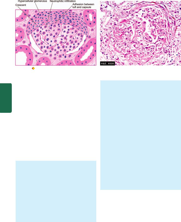
668
Pathology Systemic III SECTION
Figure 22.14 
 RPGN (post-infectious type), light microscopic appearance.There are crescents in Bowman’s space forming adhesions between the glomerular tuft and Bowman’s capsule. The tuft shows hypercellularity and leucocytic infiltration.
mentally induced if the lungs are previously injured by viral or bacterial infection or exposed to hydrocarbons. The Goodpasture’s antigen appears to be a component of collagen type IV.
Type II RPGN: Immune complex disease. A small proportion of cases of post-streptococcal GN, particularly in adults and sometimes of non-streptococcal origin, develop RPGN. The evidences in support of post-infectious RPGN having immune complex pathogenesis are granular deposits of immune complexes of IgG and C3 along the glomerular capillary walls, lowering of blood complement levels and demonstration of circulating complexes.
Type III RPGN: Pauci-immune GN. These include cases of Wegener’s granulomatosis and microscopic polyarteritis nodosa. The pathogenesis of pauci-immune GN is yet not fully defined. However, majority of these patients are ANCApositive, implying a defect in humoral immunity. Serum complement levels are normal and anti-GBM antibody is negative. There is little or no glomerular immune deposit (i.e. pauci-immune).
MORPHOLOGIC FEATURES. Grossly, the kidneys are usually enlarged and pale with smooth outer surface (large white kidney). Cut surface shows pale cortex and congested medulla.
Light Microscopic findings vary according to the cause but in general following features are present (Fig. 22.14):
i) Glomeruli—Irrespective of the underlying etiology, all forms of RPGN show pathognomonic ‘crescents’ on the inside of Bowman’s capsules. These are collections of palestaining polygonal cells which commonly tend to be elongated. Eventually, crescents obliterate the Bowman’s space and compress the glomerular tuft. Fibrin deposition is invariably present alongside crescents. Besides the crescents, glomerular tufts may show increased cellularity as a result of proliferation of endothelial and mesangial
cells and leucocytic infiltration. Fibrin thrombi are frequently present in the glomerular tufts.
ii)Tubules—Tubular epithelial cells may show hyaline droplets. Tubular lumina may contain casts, red blood cells and fibrin.
iii)Interstitium—The interstitium is oedematous and may show early fibrosis. Inflammatory cells, usually lymphocytes and plasma cells, are commonly distributed in the interstitial tissue.
iv)Vessels—Arteries and arterioles may show no change, but cases associated with hypertension usually show severe vascular changes.
Electron microscopic findings vary according to the type of RPGN. Post-infectious RPGN cases show electron-dense subepithelial granular deposits similar to those seen in acute GN, while cases of RPGN in Goodpasture’s syndrome show characteristic linear deposits along the GBM (Fig. 22.15).
Immunofluorescence microscopy shows following patterns in various types of RPGN:
 linear pattern of RPGN in Goodpasture’s syndrome (type I RPGN), containing IgG accompanied by C3 along the capillaries.
 Granular pattern of post-infectious RPGN (type II RPGN) consisting of IgG and C3 along the capillary wall.
 Scanty or no deposits of immunoglobulin and C3 in pauci-immune GN (type III RPGN).
CLINICAL FEATURES. Generally, the features of postinfectious RPGN are similar to those of acute GN, presenting as acute renal failure. The patients of Goodpasture’s syndrome may present as acute renal failure and/or associated intrapulmonary haemorrhage producing recurrent haemoptysis. Prognosis of all forms of RPGN is poor. However, post-infectious cases have somewhat better outcome and may show recovery.
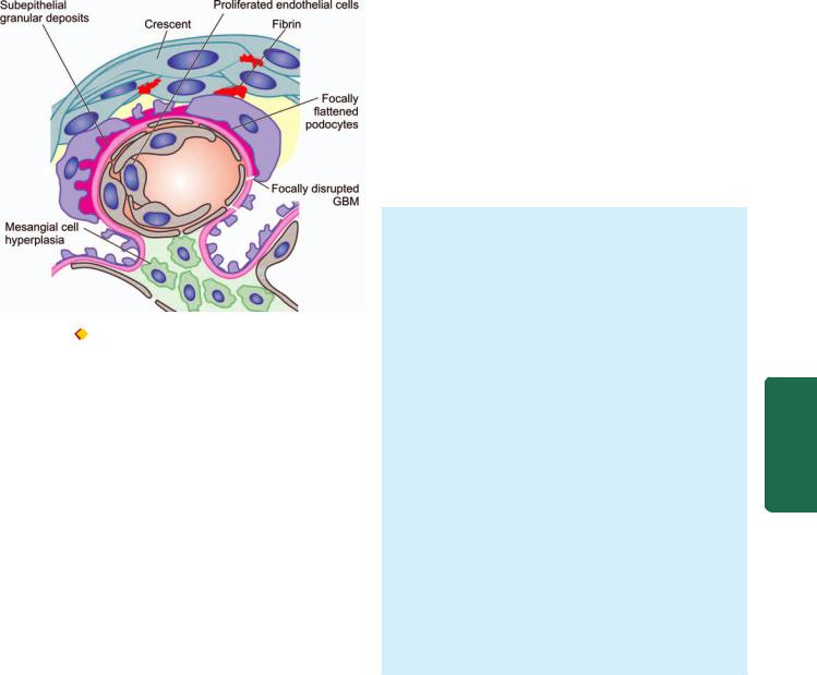
Figure 22.15  RPGN, diagrammatic representation of ultrastructure of a portion of glomerular lobule showing epithelial crescent formation and subepithelial granular deposits.
Minimal Change Disease
(Synonyms: MCD, Lipoid Nephrosis, Foot Process
Disease, Nil Deposit Disease)
Minimal change disease (MCD) is a condition in which the nephrotic syndrome is accompanied by no apparent change in glomeruli by light microscopy. Its other synonyms, lipoid nephrosis and foot process disease, are descriptive terms for fatty changes in the tubules and electron microscopic appearance of flattened podocytes respectively. Minimal change disease accounts for 80% cases of nephrotic syndrome in children under 16 years of age with preponderance in boys (ratio of boys to girls 2:1). In fact, historically, lipoid nephrosis was the first condition associated with nephrotic sndrome.
ETIOPATHOGENESIS. The etiology of MCD remain elusive. However, following two groups have been identified:
i)Idiopathic (majority of cases).
ii)Cases associated with systemic diseases (Hodgkin’s disease, HIV infection) and drug therapy (e.g. NSAIDs, rifampicin, interferon-α).
The following features point to possible immunologic pathogenesis for MCD:
i)Absence of deposits by immunofluorescence microscopy.
ii)Normal circulating levels of complement but presence of circulating immune complexes in many cases.
iii)Universal satisfactory response to steroid therapy.
iv)Evidence of increased suppressor T cell activity with elaboration of cytokines (interleukin-8, tumour necrosis factor) which probably cause foot process flattening and altered charge on the GBM.
v)Detection of a mutation in nephrin gene in cases of congenital MCD has focused attention on genetic basis.
Nephrotic syndrome in MCD in children is characterised by selective proteinuria containing mainly albumin, and
minimal amounts of high molecular weight proteins such as α2-macroglobulin. The basis for selective proteinuria appears to be as under:
i)Reduction of normal negative charge on GBM (page 650) due to loss of heparan sulfate proteoglycan from the GBM.
ii)Change in the shape of epithelial cells producing foot process flattening due to reduction of sialoglycoprotein cell coat.
Adults having MCD, however, have non-selective proteinuria, suggesting more extensive membrane permeability defect.
MORPHOLOGIC FEATURES. Grossly, the kidneys are of normal size and shape.
By light microscopy, the findings are as under
(Fig. 22.16,A):
i)Glomeruli—The most characteristic feature is no apparent abnormality in the glomeruli except for slight increase in the mesangial matrix at the most (minimal change disease or nil lesion).
ii)Tubules—There is presence of fine lipid vacuolation and hyaline droplets in the cells of proximal convoluted tubules and, hence, the older name of the condition as ‘lipoid nephrosis’.
iii)Interstitium—There may be oedema of the interstitium.
iv)Vessels—Blood vessels do not show any significant change.
By electron microscopy, the most characteristic feature of the disease is identified which is diffuse flattening of foot processes of the visceral epithelial cells (podocytes) and, hence, the name foot process disease or podocytopathy (Fig. 22.16,B). Unlike other forms of GN, no deposits are seen and the GBM is normal.
By immunofluorescence microscopy, no deposits of complement or immunoglobulins are recognised (nil deposit disease).
CLINICAL FEATURES. The classical presentation of MCD is of fully-developed nephrotic syndrome with massive and highly selective proteinuria, but hypertension is unusual. Most frequently, the patients are children under 16 years (peak incidence at 6-8 years of age).
The onset may be preceded by an upper respiratory infection, atopic allergy or immunisation.
The disease characteristically responds to steroid therapy. In spite of remissions and relapses, long-term prognosis is very good and most children become free of albuminuria after several years.
Membranous Glomerulonephritis
(Synonym: Epimembranous Nephropathy)
Membranous GN is characterised by widespread thickening of the glomerular capillary wall and is the most common cause of nephrotic syndrome in adults. In majority of cases (85%), membranous GN is truly idiopathic, while in about 15% of cases it is secondary to an underlying condition (e.g.
669
Tract Urinary Lower and Kidney The 22 CHAPTER
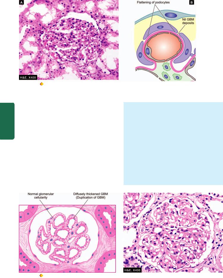
670
Figure 22.16  Minimal change disease. A, Light microscopy shows a normal glomerulus while tubules show cytoplasmic vacuolation and proteinaceous material. B, Diagrammatic representation of ultrastructure of a portion of glomerular lobule showing diffuse fusion or flattening of foot processes of visceral epithelial cells (podocytes). The GBM is normal and there are no deposits.
Pathology Systemic III SECTION
SLE, malignancies, infections such as chronic hepatitis B and C, syphilis, malaria and drugs).
ETIOPATHOGENESIS. Idiopathic membranous GN is an immune complex disease. The deposits of immune complex are formed locally because circulating immune complexes are detected in less than a quarter of cases. Since leucocytic infiltration is not a feature of membranous GN, damage to the GBM is mediated directly by complement. While nephritogenic antigen against which autoantibodies are formed in idiopathic membranous GN is not known yet, the antigen in cases of secondary membranous GN is either an endogenous (e.g. DNA in SLE) or exogenous one (e.g. hepatitis B virus, tumour antigen, treponema antigen, drug therapy with penicillamine). Currently, pathogenesis of membrane alteration in membranous GN is believed to be by MAC (membrane attack complex i.e. C35b-C9) terminal complex on podocytes.
MORPHOLOGIC FEATURES. Grossly, the kidneys are enlarged, pale and smooth.
Light microscopy shows the following findings
(Fig. 22.17):
i)Glomeruli—The characteristic finding is diffuse thickening of the glomerular capillary walls with all the glomeruli being affected more or less uniformly. As the disease progresses, the deposits are incorporated into enormously thickened basement membrane, producing ‘duplication’ of GBM which is actually formation of a new basement membrane. These basement membrane changes are best appreciated by silver impregnation stains (black colour) or by periodic acid-Schiff stain (pink colour). There is no cellular proliferation in the glomerular tufts.
ii)Tubules—The renal tubules remain normal except in the early stage when lipid vacuolation of the proximal convoluted tubules may be seen.
Figure 22.17 
 Membranous GN, light microscopic appearance. Glomeruli are normocellular but the capillary walls are diffusely thickened due to duplication of the GBM.
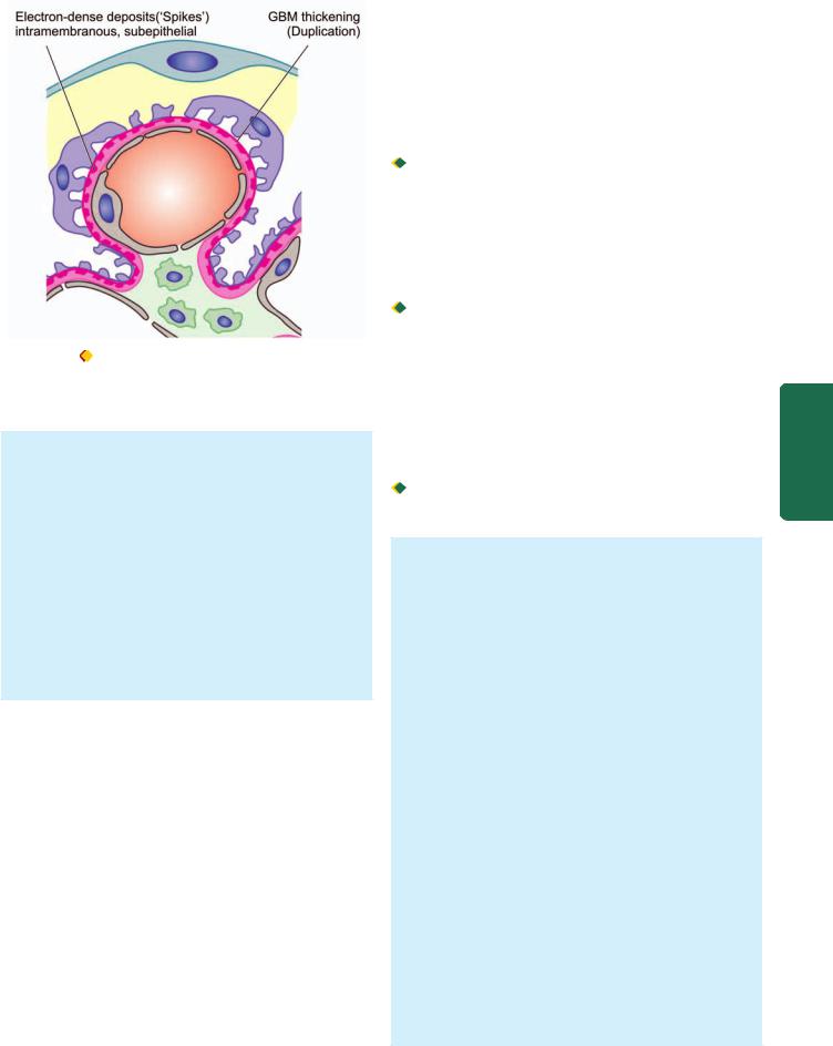
Figure 22.18 Membranous GN, diagrammatic represent ation of
ultrastructure of a portion of glomerular lobule showing subepithelial deposits of electron-dense material so that the basement membrane material protrudes between these deposit s.
iii)Interstitium—The interstitium may show fine fibrosis and scanty chronic inflammatory cells.
iv)Vessels—In the early stage, vascular changes are not prominent, while later hypertensive changes of arterioles may occur.
Electron microscopy shows characteristic electron-dense deposits in subepithelial location. The basement membrane material protrudes between deposits as ‘spikes’
(Fig. 22.18).
Immunofluorescence microscopy reveals granular deposits of immune complexes consisting of IgG associated with complement C3. In secondary cases of membranous GN the relevant antigen such as hepatitis B or tumour antigen may be seen.
CLINICAL FEATURES. The presentation in majority of cases is insidious onset of nephrotic syndrome in an adult. The proteinuria is usually of non-selective type. In addition, microscopic haematuria and hypertension may be present at the onset or may develop during the course of the disease. The changes in membranous GN are irreversible in majority of patients. Progression to impaired renal function and endstage renal disease with progressive azotaemia occurs in approximately 50% cases within a span of 2 to 20 years. Renal vein thrombosis has been found to develop in patients with membranous GN due to hypercoagulability. The role and beneficial effects of steroid therapy with or without the addition of immunosuppressive drugs is debatable.
Membranoproliferative Glomerulonephritis
(Synonyms: MPGN, Mesangiocapillary GN)
Membranoproliferative GN is another important cause of nephrotic syndrome in children and young adults. As the name implies, it is characterised by two histologic features—
increase in cellularity of the mesangium associated with increased lobulation of the tuft, and irregular thickening of the capillary wall.
ETIOPATHOGENESIS. Etiology of MPGN is unknown though in some cases there is evidence of preceding streptococcal infection. Based on ultrastructural, immunofluorescence and pathogenetic mechanisms, three types of MPGN are recognised:

 Type I or classic form is an example of immune complex disease and comprises more than 70% cases. It is characterised by immune deposits in the subendothelial position. Immune-complex MPGN is seen in association with systemic immune-complex diseases (e.g. SLE, mixed cryoglobulinaemia, Sjögren’s syndrome), chronic infections (e.g. bacterial endocarditis, HIV, hepatitis B and C) and malignancies (e.g. lymphomas and leukaemias).
 Type II or dense deposit disease is the example of alternate pathway disease (page 664) and constitutes about 30% cases. The capillary wall thickening is due to the deposition of electron-dense material in the lamina densa of the GBM. Type II MPGN is an autoimmune disease in which patients have IgG autoantibody termed C3 nephritic factor. Type II cases have an association with partial lipodystrophy, an unusual condition of unknown pathogenesis characterised by symmetrical loss of subcutaneous fat from the upper half of the body.

 Type III is rare and shows features of type I MPGN and membranous nephropathy in association with systemic diseases or drugs.
MORPHOLOGIC FEATURES. Grossly and by light microscopy, all the three types of MPGN are similar.
Grossly, the kidneys are usually pale in appearance and firm in consistency.
By light microscopy, the features are as under (Fig. 22.19):
i)Glomeruli—Glomeruli show highly characteristic changes. They are enlarged with accentuated lobular pattern. The enlargement is due to variable degree of mesangial cellular proliferation and increase in mesangial matrix. The GBM is considerably thickened, which with silver stains shows two basement membranes with a clear zone between them. This is commonly referred to as
‘double contour’, splitting, or ‘tram track’ appearance.
ii)Tubules—Tubular cells may show vacuolation and hyaline droplets.
iii)Interstitium—There may be scattered chronic inflammatory cells and some finely granular foam cells in the interstitium.
iv)Vessels—Vascular changes are prominent in cases in which hypertension develops.
By electron microscopy and immunofluorescence microscopy, the changes are different in the three types of MPGN
(Fig. 22.20):
Type I: It shows electron-dense deposits in subendothelial location conforming to immune-complex character of the disease. These deposits reveal positive fluorescence for C3 and slightly fainter staining for IgG.
671
Tract Urinary Lower and Kidney The 22 CHAPTER
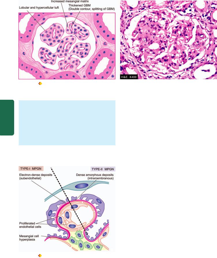
672
Pathology Systemic III SECTION
Figure 22.19  Membranoproliferative GN, light microscopic appearance. The glomerular tufts show lobulation and mesangial hypercellularity.
There is increase in the mesangial matrix between the capillaries. There is widespread thickening of the GBM.
Type II: The hallmark of type II MPGN is the presence of dense amorphous deposits within the lamina densa of the GBM and in the mesangium. Immunofluorescence studies reveal the universal presence of C3 and properdin in the deposits but the immunoglobulins are usually absent.
Type III: This rare form has electron-dense deposits within the GBM as well as in subendothelial and subepithelial regions of the GBM. Immunofluorescence studies show the presence of C3, IgG and IgM.
CLINICAL FEATURES. Clinically, there are many similarities between the main forms of MPGN. The most common age at diagnosis is between 15 and 20 years.
Figure 22.20  MPGN, diagrammatic representation of ultrastructure of a portion of glomerular lobule showing features of type I (left half) and type II (right half) MPGN. Type I (classic form) shows the characteristic subendothelial electron-dense deposit s, while type II (dense deposit disease) is characterised by intramembranous dense deposit s.
Approximately 50% of the patients present with nephrotic syndrome; about 30% have asymptomatic proteinuria; and 20% have nephritic syndrome at presentation. The proteinuria is non-selective. Haematuria and hypertension are frequently present. Hypocomplementaemia is a common feature. With time, majority of patients progress to renal failure, while some continue to have proteinuria, haematuria and hypertension with stable renal function.
Prognosis of type I is relatively better and majority of patients survive without clinically significant impairment of GFR, while type II cases run a variable clinical course.
Focal Proliferative Glomerulonephritis
(Synonym: Mesangial Proliferative GN)
Focal proliferative GN is characterised by pathologic changes in certain number of glomeruli (focal), and often confined to one or two lobules of the affected glomeruli (segmental), while other glomeruli are normal. Focal GN is, thus, a pathologic diagnosis.
ETIOPATHOGENESIS. It may occur under following diverse clinical settings:

 As an early manifestation of a number of systemic diseases such as SLE, Henoch-Schonlein purpura, subacute bacterial endocarditis, Wegener’s granulomatosis, and polyarteritis nodosa, Goodpasture’s syndrome.

 As a component of a known renal disease such as in IgA nephropathy.

 As a primary idiopathic glomerular disease unrelated to systemic or other renal disease.
The diverse settings under which focal GN is encountered make it unlikely that there are common etiologic agents or pathogenetic mechanisms. However, the observation of mesangial deposits of immunoglobulins and complement suggest immune complex disease and participation of the mesangium.
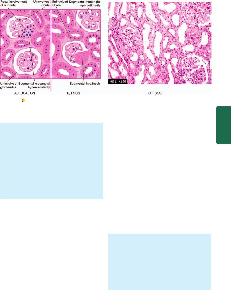
673
Figure 22.21 A, Focal GN. The characteristic feature is the cellular proliferation in some glomeruli and in one or two lobules of the affected
glomeruli i.e. focal and segmental proliferative change. B, C, Focal segmental glomerulosclerosis.The features are focal and segmental involvement of the glomeruli by sclerosis and hyalinosis and mesangial hypercellularity.
MORPHOLOGIC FEATURES. By light microscopy, the single most important feature in focal GN is the abnormality seen in certain number of glomeruli and generally confined to one or two lobules of the affected glomeruli i.e. focal and segmental glomerular involvement
(Fig. 22.21,A). The pathologic change most frequently consists of focal and segmental cellular proliferation of mesangial cells and endothelial cells but sometimes necrotising changes can be seen. The condition must be distinguished from focal and segmental glomerulosclerosis (discussed below).
By immunofluorescence microscopy, widespread mesangial deposits of immunoglobulins (mainly IgA with or without IgG), complement (C3) and fibrin are demonstrated in most cases of focal GN.
CLINICAL FEATURES. The clinical features vary according to the condition causing it. Haematuria is one of the most common clinical manifestation. Proteinuria is frequently mild to moderate but hypertension is uncommon.
Focal Segmental Glomerulosclerosis
(Synonyms: Focal Sclerosis, Focal Hyalinosis)
Focal segmental glomerulosclerosis (FSGS) is a condition in which there is sclerosis and hyalinosis of some glomeruli and portions of their tuft (less than 50% in a tissue section), while the other glomeruli are normal by light microscopy i.e. involvement is focal and segmental. The incidence of FSGS has increased over the last decades and is currently responsible for about one-third cases of nephrotic syndrome in the adults.
ETIOPATHOGENESIS. FSGS was previously believed to be a variant of MCD with accentuation of epithelial damage
in the form of hyalinosis and sclerosis. Currently, the condition is divided into 3 groups:
i)Idiopathic type. This group comprises majority of cases. It is found in children and young adults with presentation of nephrotic syndrome. It differs from minimal change disease in having non-selective proteinuria, in being steroidresistant, and may progress to chronic renal failure. Immunofluorescence microscopy reveals deposits of IgM and C3 in the sclerotic segment.
ii)With superimposed primary glomerular disease. There may be cases of FSGS with superimposed MCD or IgA nephropathy. Those associated with MCD show good response to steroid therapy and progression to chronic renal failure may occur after a long time.
iii)Secondary type. This group consists of focal segmental sclerotic lesions as a secondary manifestation of certain diseases such as HIV, diabetes mellitus, reflux nephropathy, heroin abuse and analgesic nephropathy.
The hallmark of pathogenesis of FSGS is injury to visceral epithelial cells that results in disruption of visceral epithelial cells and resultant nephron loss.
MORPHOLOGIC FEATURES. By light microscopy, depending upon the severity of the disease, variable number of glomeruli are affected focally and segmentally, while others are normal. The affected glomeruli show solidification or sclerosis of one or more lobules of the tuft. Hyalinosis refers to collection of eosinophilic, homogeneous, PAS-positive, hyaline material present on the inner aspect of a sclerotic peripheral capillary loop. Mesangial hypercellularity is present in appreciable number of cases. In addition, to glomerular changes, there is interstitial fibrosis and infiltration by mononuclear
Tract Urinary Lower and Kidney The 22 CHAPTER

674leucocytes, and tubular epithelial cell atrophy and degeneration (Fig. 22.21,B, C).
Besides the lesions of focal and segmental scarring, a variant of FSGS, collapsing glomerulopathy, has been described in HIV patients. It is segmental or global glomerular collapse of the tuft along with the presence of hyperpasia and hypertrophy of podocytes producing a pseudo-crescent and a rapid decline in renal function.
By electron microscopy, diffuse loss of foot processes as seen in minimal change disease is evident but, in addition, there are electron-dense deposits in the region of hyalinosis and sclerosis which are believed to be immune complexes.
By Immunofluorescence microscopy, the deposits in the lesions are shown to contain IgM and C3.
CLINICAL FEATURES. The condition may affect all ages including children and has male preponderance. The most common presentation is in the form of nephrotic syndrome with heavy proteinuria. Haematuria and hypertension tend to occur more frequently than in minimal change disease. Evidence of renal failure may be present at the onset.
SECTION |
IgA Nephropathy |
|
|
||
|
(Synonyms: Berger’s Disease, IgA GN) |
|
|
IgA nephropathy is emerging as the most common form of |
|
III |
glomerulopathy worldwide and its incidence has been rising. |
|
It is characterised by aggregates of IgA, deposited principally |
||
|
in the mesangium. The condition was first described by |
|
Systemic |
Berger, a French physician in 1968 (Not to be confused with |
|
Buerger’s disease or thromboangiitis obliterans described by |
||
|
||
|
an American pathologist in 1908 and discussed on page 404). |
|
|
ETIOPATHOGENESIS. The etiology of IgA nephropathy |
|
Pathology |
remains unclear: |
|
i) It is idiopathic in most cases. |
||
|
||
|
ii) Seen as part of Henoch-Schonlein purpura. |
|
|
iii) Association with chronic inflammation in various body |
|
|
systems (e.g. chronic liver disease, inflammatory bowel |
|
|
disease, interstitial pneumonitis, leprosy, dermatitis |
|
|
herpetiformis, uveitis, ankylosing spondylitis, Sjögren’s |
|
|
syndrome, monoclonal IgA gammopathy). |
|
|
Pathogenesis of IgA nephropathy is explained on the basis |
|
|
of following mechanisms: |
|
|
i) In view of exclusive mesangial deposits of IgA and |
|
|
elevated serum levels of IgA and IgA-immune complexes, |
|
|
IgA nephropathy has been considered to arise from |
|
|
entrapment of these complexes in the mesangium. |
|
|
ii) There is absence of early components of the complement |
|
|
but presence of C3 and properdin in the mesangial deposits, |
|
|
which point towards activation of alternate complement |
|
|
pathway. |
|
|
iii) Since there is close association between mucosal |
|
|
infections (e.g. of the respiratory, gastrointestinal or urinary |
|
|
tract), it is suggested that IgA deposited in the mesangium |
|
|
could be due to increased mucosal secretion of IgA. |
|
|
iv) HLA-B35 association has been reported in some cases. |
|
|
Another possibility is genetically-determined abnormality of |
|
|
the immune system producing an increase in circulating IgA. |
MORPHOLOGIC FEATURES. By light microscopy, the pattern of involvement varies. These include: focal proliferative GN, focal segmental glomerulosclerosis, membranoproliferative GN, and rarely RPGN.
By electron microscopy, finely granular electron-dense deposits are seen in the mesangium.
By immunofluorescence microscopy, the diagnosis is firmly established by demonstration of mesangial deposits of IgA, with or without IgG, and usually with C3 and properdin.
CLINICAL FEATURES. The disease is common in children and young adults. The clinical picture is usually characterised by recurrent bouts of haematuria that are often precipitated by mucosal infections. Mild proteinuria is usually present and occasionally nephrotic syndrome may develop.
Chronic Glomerulonephritis
(Synonym: End-Stage Kidney)
Chronic GN is the final stage of a variety of glomerular diseases which result in irreversible impairment of renal function. The conditions which may progress to chronic GN, in descending order of frequency, are as under:
i)Rapidly progressive GN (90%)
ii)Membranous GN (50%)
iii)Membranoproliferative GN (50%)
iv)Focal segmental glomerulosclerosis (50%)
v)IgA nephropathy (40%)
vi)Acute post-streptococcal GN (1%).
However, about 20% cases of chronic GN are idiopathic without evidence of preceding GN of any type.
MORPHOLOGIC FEATURES. Grossly, the kidneys are usually small and contracted weighing as low as 50 gm each. The capsule is adherent to the cortex. The cortical surface is generally diffusely granular (Fig. 22.22). On cut section, the cortex is narrow and atrophic, while the medulla is unremarkable.
Microscopically, the changes vary greatly depending upon the underlying glomerular disease. In general, the following changes are seen (Fig. 22.23).
i)Glomeruli—Glomeruli are reduced in number and most of those present show completely hyalinised tufts, giving the appearance of acellular, eosinophilic masses which are PAS-positive. Evidence of underlying glomerular disease may be present.
ii)Tubules—Many tubules completely disappear and there may be atrophy of tubules close to scarred glomeruli. Tubular cells show hyaline-droplets, degeneration and tubular lumina frequently contain eosinophilic, homogeneous casts.
iii)Interstitium—There is fine and delicate fibrosis of the interstitial tissue and varying number of chronic inflammatory cells are often seen.
iv)Vessels—Advanced cases which are frequently associated with hypertension show conspicuous arterial and arteriolar sclerosis.
675
Figure 22.22  End-stage kidney, gross appearance of short contract kidney in chronic glomerulonephritis. The kidney is small and contracted.
The capsule is adherent to the cortex and has diffusely granular cortical surface.
Patients of end-stage kidney disease on dialysis show a variety of dialysis associated changes that include acquired cystic disease (page 659), occurrence of adenomas and adenocarcinomas of the kidney, calcification of tufts and deposition of calcium oxalate crystals in tubules.
CLINICAL FEATURES. The patients are usually adults. The terminal stage of chronic GN is characterised by hypertension, uraemia and progressive deterioration of renal function. Besides the primary changes due to chronic renal failure, there are a variety of systemic manifestations of uraemia (page 655). These patients eventually die if they do not receive a renal transplant.
The salient features of various types of primary glomerulonephritis are summarised in Table 22.11.
II. SECONDARY GLOMERULAR DISEASES
Glomerular involvement may occur secondary to certain systemic diseases or a few hereditary diseases. In some of
these, renal involvement may be the initial presentation, while in others clinical evidence of renal disease appears long after other manifestations have appeared. A list of these conditions has already been given in Table 22.4. The important examples are described below.
Lupus Nephritis
Renal manifestations of systemic lupus erythematosus (SLE) are termed lupus nephritis. Other clinical manifestations, etiology and pathogenesis of this multi-system autoimmune disease are described in Chapter 4 (page 78). The incidence of renal involvement in SLE ranges from 40 to 75%. The two cardinal clinical manifestations of lupus nephritis are proteinuria and haematuria. In addition, hypertension and casts of different types such as red cell casts, fatty casts and leucocyte casts in the urinary sediment are found.
Pathogenesis of lesions in lupus nephritis is linked to genes related to major histocompatibility complex and B-cell signaling pathways such as TNF superfamily members.
Tract Urinary Lower and Kidney The 22 CHAPTER
Figure 22.23 
 End-stage kidney in chronic GN, light microscopy. Glomerular tufts are acellular and completely hyalinised. Blood vessels in the interstitium are hyalinised and thickened while the interstitium shows fine fibrosis and a few chronic inflammatory cells.

676
Pathology Systemic III SECTION
TABLE 22.11: Comparative Features of Major Forms of Primary Glomerulonephritis.
|
Type |
Clinical |
Pathogenesis |
Pathology |
|
|
|
|
Features |
|
LM |
EM |
IFM |
1. |
Acute GN |
Acute nephrotic |
Immune complex |
Diffuse proliferation, |
Subepithelial |
Irregular IgG, |
|
|
syndrome |
disease (local or |
leucocytic infiltration |
deposits |
C3 |
|
|
|
circulating) |
|
(‘humps’) |
|
2. |
RPGN |
Acute renal |
i) Type I: |
Proliferation, crescents |
i) Linear deposits |
i) Linear IgG, C3 |
|
|
failure |
anti-GBM type |
|
along GBM |
|
|
|
|
ii) Type II: |
|
ii) Subepithelial |
ii) Granular IgG, |
|
|
|
immune complex type |
|
deposits |
C3 |
|
|
|
iii) Type III: |
|
iii) No deposits |
iii) Negative |
|
|
|
pauci-immune RPGN |
|
|
|
3. |
Minimal |
Nephrotic |
Reduction of normal |
Normal glomeruli, |
Loss of foot |
Negative |
|
change |
syndrome |
negative charge on |
lipid vacuolation in |
processes, no |
|
|
disease |
(highly selective |
GBM |
tubules |
deposits |
|
|
|
proteinuria) |
?Cell-mediated |
|
|
|
|
|
|
mechanism |
|
|
|
4. |
Membranous |
Nephrotic |
Immune complex |
Diffuse thickening |
Subepithelial |
Granular IgG, C3 |
|
GN |
syndrome |
disease (local) |
of capillary wall |
deposits (‘spikes’) |
|
5. |
Membrano- |
Nephrotic |
Type I: immune |
Lobular proliferation |
Type I: Subendothelial |
Type I: IgG, C3 |
|
proliferative |
syndrome |
complex disease |
of mesangial cells, |
deposits |
|
|
GN |
|
Type II: dense |
increased mesangial |
Type II: Dense |
Type II: C3 |
|
|
|
deposit disease |
matrix, double |
intramembranous |
properdin |
|
|
|
(alternate pathway |
contour of GBM |
deposits |
Type III: C3 |
|
|
|
activation) |
|
|
IgG, IgM |
|
|
|
Type III: rare, |
|
Type III: |
|
|
|
|
with systemic |
|
Subendothelial |
|
|
|
|
diseases and drugs |
|
and subepithelial |
|
|
|
|
|
|
deposits |
|
6. |
Focal GN |
Variable, haema- |
Variable, possibly |
Focal and segmental |
Mesangial deposits |
IgA ± IgG, C3 |
|
|
turia common |
immune complex |
proliferation |
|
and fibrin |
|
|
|
disease |
|
|
|
7. |
Focal |
Nephrotic |
i) Idiopathic |
Focal and segmental |
Loss of foot |
IgM, C3 |
|
Segmental |
syndrome |
ii) With superimposed |
sclerosis and hyalinosis |
processes, electron |
|
|
glomerulo- |
|
primary glomerular |
|
dense deposits in |
|
|
sclerosis |
|
disease |
|
regions of sclerosis |
|
|
|
|
iii) Secondary type |
|
and hyalinosis |
|
8. |
IgA |
Recurrent |
Unknown, possibly |
Variable, commonly |
Mesangial |
IgA ± IgG, C3, |
|
nephropathy |
haematuria, |
alternate pathway |
focal proliferative GN |
electron-dense |
properdin |
|
|
mild proteinuria |
disease |
|
deposits |
|
9. |
Chronic GN |
Chronic renal |
Variable |
Hyalinised glomeruli |
Variable |
Variable |
|
|
failure |
|
|
|
|
(GN = glomerulonephritis; LM = light microscopy; EM = electron microscopy; IFM = immunofluorescence microscopy)
MORPHOLOGIC FEATURES. According to the WHO, six patterns of mutually-merging renal lesions are seen in lupus nephritis (also refer to Table 4.9):
Class I: Minimal lesions. On light microscopy, these cases do not show any abnormality. But examination by electron microscopy and immunofluorescence microscopy shows deposits within the mesangium which consist of IgG and C3.
Class II: Mesangial lupus nephritis. These cases have mild clinical manifestations. By light microscopy, there is increase in the number of mesangial cells and of mesangial matrix. Ultrastructural and immunofluorescence studies
reveal granular mesangial deposits of IgG and C3; sometimes IgA and IgM are also present in the deposits.
Class III: Focal segmental lupus nephritis. This is characterised by focal and segmental proliferation of endothelial and mesangial cells, together with infiltration by macrophages and sometimes neutrophils. Haematoxylin bodies of Gross may be present. Subendothelial and subepithelial deposits of IgG, often with IgM or IgA and C3, are seen.
Class IV: Diffuse proliferative lupus nephritis. In this type, all the morphologic manifestations of lupus are present in most advanced form. This is the most severe
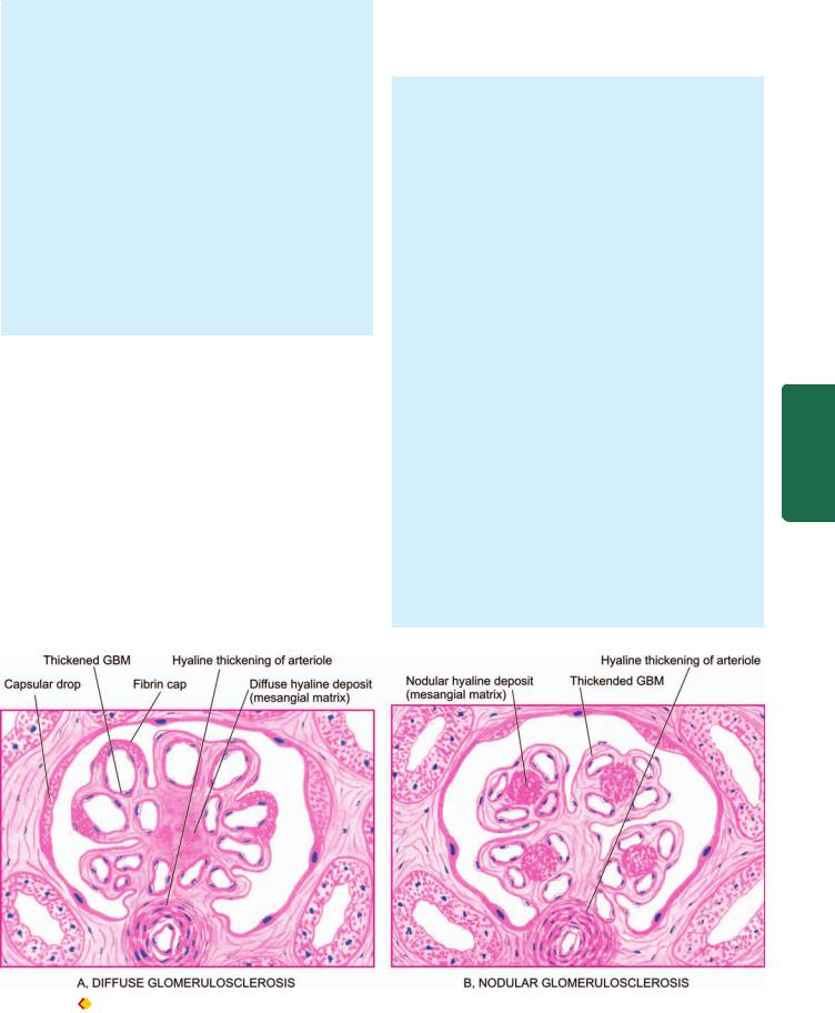
and the most common form of lupus nephritis. There is diffuse proliferation of endothelial, mesangial, and sometimes epithelial cells, involving most or all glomeruli. Electron microscopy shows large electron-dense deposits in the mesangium and in the subendothelial region which on immunofluorescence are positive for IgG; sometimes also for IgA or IgM, and C3.
Class V: Membranous lupus nephritis. These lesions resemble those of idiopathic membranous GN. These consist of diffuse thickening of glomerular capillary wall on light microscopy and show subendothelial deposits of immune complexes containing IgG, IgM and C3 on ultrastructural studies. Mesangial hypercellularity is present in some cases.
Class VI: Sclerosing lupus nephritis. This is end-stage kidney of SLE, akin to chronic GN. Most glomeruli are sclerosed and hyalinised and there may be remnants of preceding lesions.
Although in a given case, the lesions in lupus nephririts fit into one of the classes described above, it is not unusual to find overlapping and progressive transformation of lupus lesions during the course of disease.
Diabetic Nephropathy
Renal involvement is an important complication of diabetes mellitus. End-stage kidney with renal failure accounts for deaths in more than 10% of all diabetics. Renal complications are more severe, develop early and more frequently in type 1 (earlier called insulin-dependent) diabetes mellitus (30-40% cases) than in type 2 (earlier termed non-insulin- dependent) diabetics (about 20% cases). A variety of clinical syndromes are associated with diabetic nephropathy that includes asymptomatic proteinuria, nephrotic syndrome, progressive renal failure and hypertension. Cardiovascular
disease is 40 times more common in patients of end-stage renal disease in diabetes mellitus than in non-diabetics and more diabetics die from cardiovascular complications than from uraemia.
MORPHOLOGIC FEATURES. Diabetic nephropathy encompasses 4 types of renal lesions in diabetes mellitus: diabetic glomerulosclerosis, vascular lesions, diabetic pyelonephritis and tubular lesions (Armanni-Ebstein lesions).
1. DIABETIC GLOMERULOSCLEROSIS. Glomerular lesions in diabetes mellitus are particularly common and account for majority of abnormal findings referable to the kidney.
Pathogenesis of these lesions in diabetes mellitus is explained by following sequential changes: hyperglycaemia → glomerular hypertension → renal hyperperfusion → deposition of proteins in the mesangium → glomerulosclerosis → renal failure. In addition, cellular infiltration in renal lesions in diabetic glomerular lesions is due to growth factors, particularly transforming growth factor-β. Strict control of blood glucose level and control of systemic hypertension in these patients retards progression to diabetic nephropathy.
Glomerulosclerosis in diabetes may take one of the 2 forms: diffuse or nodular lesions:
i) Diffuse glomerulosclerosis. Diffuse glomerular lesions are the most common. There is involvement of all parts of glomeruli. The pathologic changes consist of thickening of the GBM and diffuse increase in mesangial matrix with mild proliferation of mesangial cells. Various exudative lesions such as capsular hyaline drops and fibrin caps may also be present (Fig. 22.24,A) Capsular drop is an eosinophilic hyaline thickening of the parietal layer of Bowman’s capsule and bulges into the glomerular space.
677
Tract Urinary Lower and Kidney The 22 CHAPTER
Figure 22.24  Diabetic glomerulosclerosis. A, Diffuse lesions. The characteristic features are dif fuse involvement of the glomeruli showing thickening of the GBM and diffuse increase in the mesangial matrix with mild proliferation of mesangial cells and exudative lesions (fibrin caps and capsular drops). B, Nodular lesion (Kimmelstiel-Wilson Lesion). There are one or more hyaline nodules within the lobules of glomeruli, surrounded peripherally by glomerular capillaries with thickened walls.

678
Figure 22.25 |
Diabetic nephropathy—nodular (Kimmelstiel-W ilson |
or KW) lesions. |
|
SECTION |
Fibrin cap is homogeneous, brightly eosinophilic material |
|
appearing on the wall of a peripheral capillary of a lobule. |
||
ii) Nodular glomerulosclerosis. Nodular lesions of |
||
diabetic glomerulosclerosis are also called as Kimmelstiel- |
||
Wilson (KW) lesions or intercapillary glomerulosclerosis. |
||
III |
These lesions are specific for type 1 diabetes (juvenile- |
|
onset diabetes) or islet cell antibody-positive diabetes |
||
|
||
Systemic |
mellitus. The pathologic changes consist of one or more |
|
nodules in a few or many glomeruli. Nodule is an ovoid or |
||
|
||
|
spherical, laminated, hyaline, acellular mass located |
|
|
within a lobule of the glomerulus. The nodules are |
|
|
surrounded peripherally by glomerular capillary loops |
|
Pathology |
which may have normal or thickened GBM (Fig. 22.24,B). |
|
The nodules are PAS-positive and contain lipid and fibrin. |
||
|
||
|
As the nodular lesions enlarge, they compress the glomer- |
|
|
ular capillaries and obliterate the glomerular tuft |
|
|
(Fig. 22.25). As a result of glomerular and arteriolar |
|
|
involvement, renal ischaemia occurs leading to tubular |
|
|
atrophy and interstitial fibrosis and grossly small, |
|
|
contracted kidney. |
2.VASCULAR LESIONS. Atheroma of renal arteries is very common and severe in diabetes mellitus. Hyaline arteriolosclerosis (Chapter 15) affecting the afferent and efferent arterioles of the glomeruli is also often severe in diabetes. These vascular lesions are responsible for renal ischaemia that results in tubular atrophy and interstitial fibrosis.
3.DIABETIC PYELONEPHRITIS. Poorly-controlled diabetics are particularly susceptible to bacterial infections. Papillary necrosis (necrotising papillitis) (page 682) is an important complication of diabetes that may result in acute pyelonephritis. Chronic pyelonephritis is 10 to
20times more common in diabetics than in others.
4.TUBULAR LESIONS (ARMANNI-EBSTEIN LESIONS). In untreated diabetics who have extremely
high blood sugar level, the epithelial cells of the proximal convoluted tubules develop extensive glycogen deposits appearing as vacuoles. These are called Armanni-Ebstein lesions. The tubules return to normal on control of hyperglycaemic state.
Hereditary Nephritis
A group of hereditary diseases principally involving the glomeruli are termed hereditary nephritis. These include the following:
1.Alport’s syndrome
2.Fabry’s disease
3.Nail-patella syndrome
1.Alport’s syndrome. Out of various hereditary nephritis, Alport’s syndrome is relatively more common and has been extensively studied. This is an X-linked dominant disorder having mutation in α−5 chain of type IV collagen located on X-chromosome. It affects males more severely than females. The syndrome consists of sensori-neural deafness and ophthalmic complications (lens dislocation, posterior cataracts and corneal dystrophy) associated with hereditary nephritis. The condition is slowly progressive, terminating in end-stage kidney in the 2nd to 3rd decades of life. The common presenting features are persistent or recurrent haematuria accompanied by erythrocyte casts, proteinuria and hypertension.
By light microscopy, the glomeruli have predominant involvement and show segmental proliferation of mesangial cells with increased mesangial matrix and occasional segmental sclerosis. Another prominent feature is the presence of lipid-laden foam cells in the interstitium. As the disease progresses, there is increasing sclerosis of glomeruli, tubular atrophy and interstitial fibrosis.
Electron microscopy reveals characteristic basement membrane splitting or lamination in the affected parts of glomeruli.
Immunofluorescence studies fail to show deposits of immunoglobulins or complement components.
2.Fabry’s disease, another hereditary nephritis is characterised by accumulation of neutral glycosphingolipids in lysosomes of glomerular, tubular, vascular and interstitial cells.
3.Nail-patella syndrome or osteonychodysplasia is a rare hereditary disease having abnormality in α-1 chain of collagen V on chromosome 9 associated with multiple osseous defects of elbows, knees and nail dysplasia. About half the cases develop nephropathy.
TUBULAR AND TUBULOINTERSTITIAL DISEASES
It is difficult to separate the involvement of the tubules and the interstitium since most forms of tubular diseases also involve the interstitium, while the tubules and interstitium may be involved secondarily as a part of the diseases of other renal components. For the purpose of present discussion, this group of diseases is discussed under 2 headings:
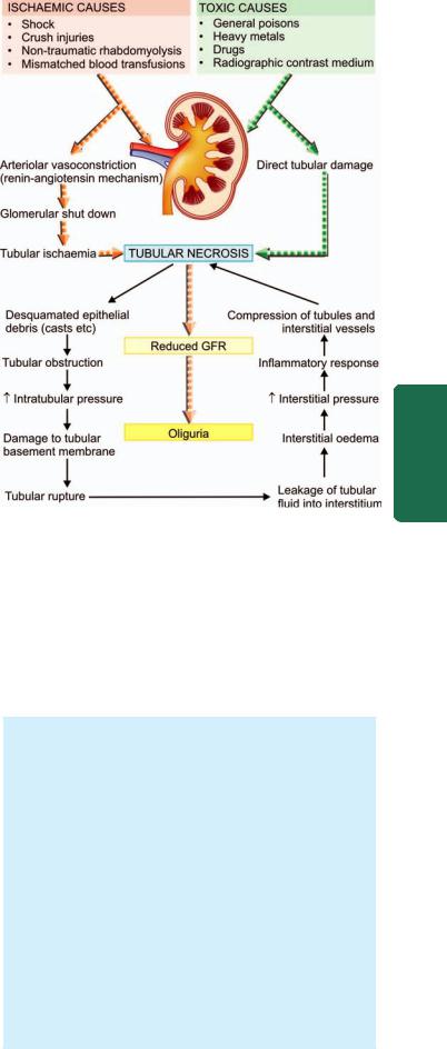
I. Primary tubular diseases that include tubular injury by ischaemic or toxic agents i.e. acute tubular necrosis.
II. Tubulointerstitial diseases that include inflammatory involvement of the tubules and the interstitium i.e. tubulointerstitial nephritis.
ACUTE TUBULAR NECROSIS
Acute tubular necrosis (ATN) is the term used for acute renal failure (ARF) resulting from destruction of tubular epithelial cells. ATN is the most common and most important cause of ARF characterised by sudden cessation of renal function. Various other causes of ARF (pre-renal, intra-renal and postrenal) as well as the clinical syndrome accompanying ATN (oliguric phase, diuretic phase and phase of recovery) are described already on page 654. Based on etiology and morphology, two forms of ATN are distinguished— ischaemic and toxic; however both forms have a somewhat common pathogenesis.
Pathogenesis of ATN
The pathogenesis of both types of ATN resulting in ARF is explained on the basis of the following sequential mechanism and is illustrated in Fig. 22.26:
i)Renal tubules are highly susceptible to injury by ischaemia and toxic agents.
ii)Tubular damage in ischaemic ATN is initiated by arteriolar vasoconstriction induced by renin-angiotensin system, while in toxic ATN by direct damage to tubules by the agent.
iii)Debris of the desquamated epithelium due to necrosis causes tubular obstruction and may block urinary outflow with consequent reduction of GFR and also produce casts in the urine.
iv)These events cause increased intratubular pressure resulting in damage to tubular basement membrane.
v)Due to increased intratubular pressure, there is tubular rupture.
vi)Damage to tubules is accompanied with leakage of fluid into the interstitium causing interstitial oedema.
vii)Leakage of tubular fluid into the interstitium increases interstitial pressure.
viii)Leaked fluid incites host inflammatory response.
ix)Increased interstitial pressure causes compression of tubules and blood vessels and setting up a vicious cycle of accentuated ischaemia and necrosis.
x)Ultimately, it leads to reduced GFR and consequently oliguria.
Ischaemic ATN
Ischaemic ATN, also called tubulorrhectic ATN, lower (distal) nephron nephrosis, anoxic nephrosis, or shock kidney, occurs due to hypoperfusion of the kidneys resulting in focal damage to the distal parts of the convoluted tubules.
ETIOLOGY. Ischaemic ATN is more common than toxic ATN and accounts for more than 80% cases of tubular injury. Ischaemia may result from a variety of causes as follows:
Figure 22.26 
 Pathogenesis of ATN.
1.Shock (post-traumatic, surgical, burns, dehydration, obstetrical and septic type).
2.Crush injuries.
3.Non-traumatic rhabdomyolysis induced by alcohol, coma, muscle disease or extreme muscular exertion (myoglobinuric nephrosis).
4.Mismatched blood transfusions, black-water fever (haemoglobinuric nephrosis).
MORPHOLOGIC FEATURES. Grossly, the kidneys are enlarged and swollen. On cut section, the cortex is often widened and pale, while medulla is dark.
Histologically, predominant changes are seen in the tubules, while glomeruli remain unaffected. The interstitium shows oedema and mild chronic inflammatory cell infiltrate. Tubular changes are as follows
(Fig. 22.27):
1.Dilatation of the proximal and distal convoluted tubules.
2.Focal tubular necrosis at different points along the nephron.
3.Flattened epithelium lining the tubules suggesting epithelial regeneration.
4.Eosinophilic hyaline casts or pigmented haemoglobin and myoglobin casts in the tubular lumina (Fig. 22.28).
5.Disruption of tubular basement membrane adjacent to the cast may occur (tubulorrhexis).
679
Tract Urinary Lower and Kidney The 22 CHAPTER
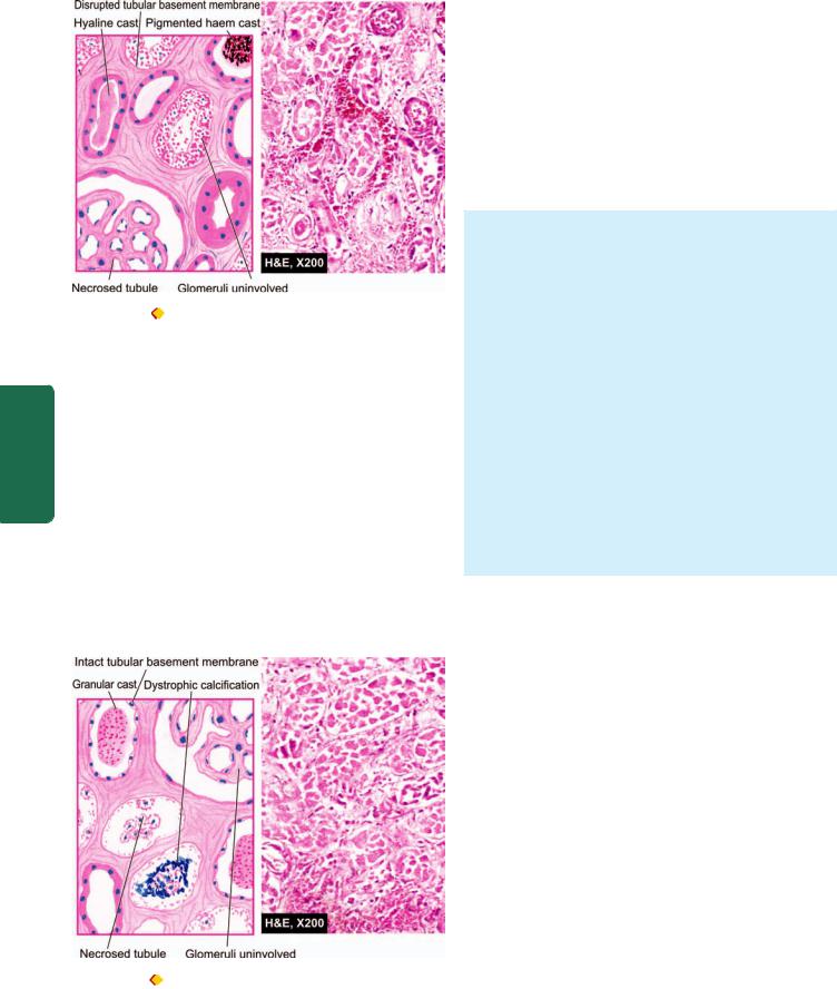
680
Pathology Systemic III SECTION
Figure 22.27 Ischaemic ATN. There is focal necrosis along the
nephron involving proximal convoluted tubule (PCT) as well as distal convoluted tubule (DCT). The affected tubules are dilated, their lumina contain casts (hyaline or pigmented haem) and the affected regions are lined by regenerating thin and flat epithelium.
Prognosis of ischaemic ATN depends upon the underlying etiology. In general, cases that follow severe trauma, surgical procedures, extensive burns and sepsis have much worse outlook than the others.
Toxic ATN
Toxic ATN, also called nephrotoxic ATN or toxic nephrosis or upper (proximal) nephron nephrosis, occurs as a result of direct damage to tubules, more marked in proximal portions, by ingestion, injection or inhalation of a number of toxic agents.
ETIOLOGY. The toxic agents causing toxic ATN are as under:
1.General poisons such as mercuric chloride, carbon tetrachloride, ethylene glycol, mushroom poisoning and insecticides.
2.Heavy metals (mercury, lead, arsenic, phosphorus and gold).
3.Drugs such as sulfonamides, certain antibiotics (gentamycin, cephalosporin), anaesthetic agents (methoxyflurane, halothane), barbiturates, salicylates.
4.Radiographic contrast material.
MORPHOLOGIC FEATURES. Poisoning with mercuric chloride provides the classic example that produces widespread and readily discernible tubular necrosis (acute
mercury nephropathy).
Grossly, the kidneys are enlarged and swollen. On cut section, the cortex is pale and swollen, while the medulla is slightly darker than normal.
Histologically, the appearance varies according to the cause of toxic ATN but, in general, involves the segment of tubule diffusely (unlike ischaemic ATN where the involvement of nephron is focal). In mercuric chloride poisoning, the features are as follows (Fig. 22.28):
1.Epithelial cells of mainly proximal convoluted tubules are necrotic and desquamated into the tubular lumina.
2.The desquamated cells may undergo dystrophic calcification.
3.Tubular basement membrane is generally intact.
4.The regenerating epithelium, which is flat and thin with few mitoses, may be seen lining the tubular basement membrane.
Prognosis of toxic ATN is good if there is no serious damage to other organs such as heart and liver.
The contrasting features of the two forms of ATN are presented in Table 22.12.
TUBULOINTERSTITIAL DISEASES
The term tubulointerstitial nephritis is used for inflammatory process that predominantly involves the renal interstitial tissue and is usually accompanied by some degree of tubular damage. A number of primary glomerular, tubular, vascular and obstructive diseases are secondarily associated with interstitial reaction. However, the term interstitial nephritis is reserved for those cases where there is no primary involvement of glomeruli, tubules or blood vessels. The older nomenclature, interstitial nephritis, is currently used syno-
nymously with tubulointerstitial nephritis or tubulointerstitial
nephropathy.
A number of bacterial and non-bacterial, acute and chronic conditions may produce tubulointerstitial nephritis and are listed in Table 22.13. The important and common examples among these are discussed below.
Figure 22.28 |
Toxic ATN. There is extensive necrosis of epithelial |
Acute Pyelonephritis |
cells involving predominantly proximal convoluted tubule (PCT) diffusely. |
||
The necrosed cells are desquamated into the tubular lumina and may |
Acute pyelonephritis is an acute suppurative inflammation |
|
undergo dystrophic calcification. The tubular lumina cont ain casts |
||
(granular) and the regenerating flat epithelium lines the necrosed tubule. |
of the kidney caused by pyogenic bacteria. |
|
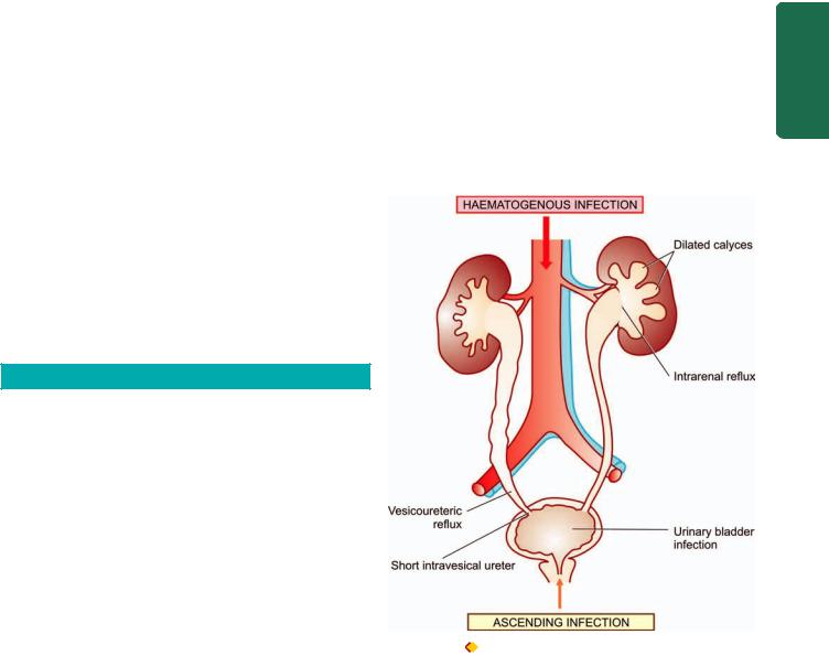
TABLE 22.12. Contrasting Features of Ischaemic and Toxic ATN. |
|
681 |
|||
|
Feature |
Ischaemic ATN |
Toxic ATN |
|
|
|
|
|
|
|
|
1. |
Synonyms |
Tubulorrhectic ATN, lower (distal) nephron |
Nephrotoxic ATN, upper (proximal) nephron |
||
|
|
nephrosis, anoxic nephrosis, shock kidney |
nephrosis, toxic ATN |
||
2. |
Frequency |
More common (80% cases) |
Less common |
||
3. |
Major etiologies |
Shock, crush injuries, mismatched blood |
Poisons, heavy metals, certain drugs |
||
|
|
transfusion |
|
|
|
4. |
G/A |
Kidneys enlarged, swollen, cut section |
Similar to ischaemic ATN |
||
|
|
cortex pale, medulla dark |
|
|
|
5. |
M/E |
i) Distal tubular damage more prominent |
i) Proximal tubular damage more prominent |
||
|
|
ii) |
Focal tubular necrosis |
ii) More diffuse tubular injury |
|
|
|
iii) |
Regenerating epithelium |
iii) Regenerating epithelium |
|
|
|
iv) Casts: Hyaline, pigment, myoglobin |
iv) Tubular lumina may contained dystrophic |
||
|
|
|
|
calcification |
|
|
|
v) |
Basement membrane disrupted |
v) Basement membrane generally intact |
|
6. |
Prognosis |
Worse |
Good |
||
|
|
|
|
|
|
ETIOPATHOGENESIS. Most cases of acute pyelonephritis follow infection of the lower urinary tract. The most common pathogenic organism in urinary tract infection (UTI) is Escherichia coli (in 90% of cases), followed in decreasing frequency, by Enterobacter, Klebsiella, Pseudomonas and Proteus. The bacteria gain entry into the urinary tract, and thence into the kidney by one of the two routes: ascending infection and haematogenous infection
(Fig. 22.29):
1. Ascending infection. This is the most common route of infection. The common pathogenic organisms are inhabitants of the colon and may cause faecal contamination of the urethral orifice, especially in females in reproductive age group. This has been variously attributed to shorter urethra in females liable to faecal contamination, hormonal influences facilitating bacterial adherence to the mucosa, absence of prostatic secretions which have antibacterial properties, and urethral trauma during sexual intercourse. The last named produces what is appropriately labelled as ‘honeymoon pyelitis’. Ascending infection may occur in a normal individual but the susceptibility is increased in patients with
diabetes mellitus, pregnancy, urinary tract obstruction or instrumentation. Bacteria multiply in the urinary bladder and produce asymptomatic bacteriuria found in many of these cases. After having caused urethritis and cystitis, the bacteria in susceptible cases ascend further up into the ureters against the flow of urine, extend into the renal pelvis and then the renal cortex. The role of vesico-ureteral reflux is not of a great significance in the pathogenesis of acute chronic pyelonephritis as it is in chronic pyelonephritis.
2. Haematogenous infection. Less often, acute pyelonephritis may result from blood-borne spread of infection. This occurs more often in patients with obstructive lesions
TABLE 22.13: Tubulointerstitial Diseases.
A. INFECTIVE
1.Acute pyelonephritis
2.Chronic pyelonephritis
3.Tuberculous pyelonephritis
4.Other infections (viruses, parasites etc)
B.NON-INFECTIVE
1.Acute hypersensitivity interstitial nephritis
2.Analgesic abuse (phenacetin) nephropathy
3.Myeloma nephropathy
4.Balkan nephropathy
5.Urate nephropathy
6.Gout nephropathy
7.Radiation nephritis
8.Transplant rejection (page 65)
9.Nephrocalcinosis
10. |
Idiopathic interstitial nephritis |
Figure 22.29 |
Pathogenesis of reflux nephropathy. |
|
|
Tract Urinary Lower and Kidney The 22 CHAPTER
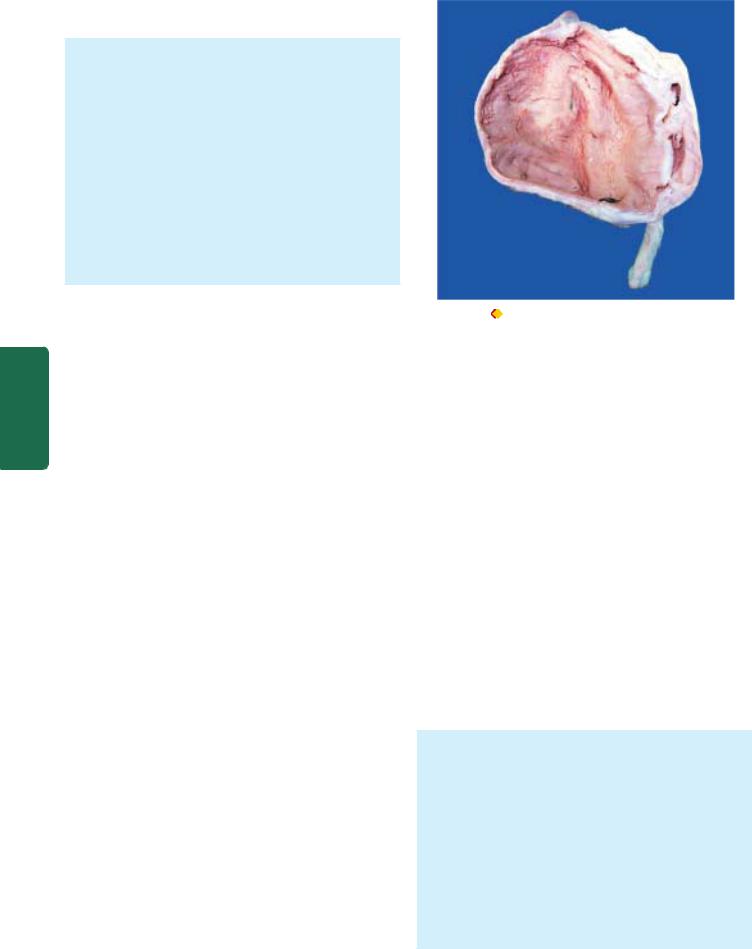
682in the urinary tract, and in debilitated or immunosuppressed patients.
MORPHOLOGIC FEATURES. Grossly, well-developed cases of acute pyelonephritis show enlarged and swollen kidney that bulges on section. The cut surface shows small, yellow-white abscesses with a haemorrhagic rim. These abscesses may be several millimetres across and are situated mainly in the cortex.
Microscopically, acute pyelonephritis is characterised by extensive acute inflammation involving the interstitium and causing destruction of the tubules. Generally, the glomeruli and renal blood vessels show considerable resistance to infection and are spared. The acute inflammation may be in the form of large number of neutrophils in the interstitial tissue and bursting into tubules, or may form focal neutrophilic abscesses in the renal parenchyma.
|
CLINICAL FEATURES. Classically, acute pyelonephritis has |
|
|
an acute onset with chills, fever, loin pain, lumbar tenderness, |
|
|
dysuria and frequency of micturition. Urine will show |
|
SECTION |
bacteria in excess of 100,000/ml, pus cells and pus cell casts |
|
in the urinary sediment. Institution of specific antibiotics, |
||
after identification of bacteria by culture followed by |
||
|
||
|
sensitivity test, eradicates the infection in majority of patients. |
|
|
COMPLICATIONS. Complications of acute pyelonephritis |
|
III |
are encountered more often in patients with diabetes mellitus |
|
or with urinary tract obstruction. Following are the three |
||
|
important complications of acute pyelonephritis: |
|
Systemic |
1. Papillary necrosis. Papillary necrosis or necrotising |
|
papillitis develops more commonly in analgesic abuse |
||
|
||
|
nephropathy and in sickle cell disease but may occur as a |
|
|
complication of acute pyelonephritis as well. It may affect |
|
Pathology |
one or both kidneys. |
|
|
||
Microscopically, necrotic tissue is separated from the |
||
|
Grossly, the necrotic papillae are yellow to grey-white, |
|
|
sharply-defined areas with congested border and resemble |
|
|
infarction. The pelvis may be dilated. |
|
|
viable tissue by a dense zone of polymorphs. The necrotic |
|
|
area shows characteristic coagulative necrosis as seen in |
|
|
renal infarcts. |
|
|
2. Pyonephrosis. Rarely, the abscesses in the kidney in |
|
|
acute pyelonephritis are extensive, particularly in cases with |
|
|
obstruction. This results in inability of the abscesses to drain |
|
|
and this transforms the kidney into a multilocular sac |
|
|
filled with pus called as pyonephrosis or renal carbuncle |
|
|
(Fig. 22.30). |
|
|
3. Perinephric abscess. The abscesses in the kidney may |
|
|
extend through the capsule of the kidney into the perinephric |
|
|
tissue and form perinephric abscess. |
|
|
Chronic Pyelonephritis |
|
|
Chronic pyelonephritis is a chronic tubulointerstitial disease |
|
|
resulting from repeated attacks of inflammation and scarring. |
|
|
ETIOPATHOGENESIS. Depending upon the etiology and |
|
|
pathogenesis, two types of chronic pyelonephritis are descri- |
Figure 22.30 Pyonephrosis. The kidney is enlarged and has
increased perinephric fat in the hilum. Sectioned surface shows markedly dilated pelvis and calyces having irregular and ragged inner surface and containing necrotic debris and pus.
bed—reflux nephropathy and obstructive pyelonephritis
(Fig. 22.31):
1.Reflux nephropathy. Reflux of urine from the bladder into one or both the ureters during micturition is the major cause of chronic pyelonephritis. Vesicoureteric reflux is particularly common in children, especially in girls, due to congenital absence or shortening of the intravesical portion of the ureter so that ureter is not compressed during the act of micturition. Reflux results in increase in pressure in the renal pelvis so that the urine is forced into renal tubules which is eventually followed by damage to the kidney and scar formation (Fig. 22.29). Vesicoureteric reflux is more common in patients with urinary tract infection, whether symptomatic or asymptomatic, but reflux of sterile urine can also cause renal damage.
2.Obstructive pyelonephritis. Obstruction to the outflow of urine at different levels predisposes the kidney to infection (page 690). Recurrent episodes of such obstruction and infection result in renal damage and scarring. Rarely, recurrent attacks of acute pyelonephritis may cause renal damage and scarring.
MORPHOLOGIC FEATURES. Grossly, the kidneys show rather characteristic appearance. The kidneys are usually small and contracted (weighing less than 100 gm) showing unequal reduction, which distinguishes it from other forms of contracted kidney. The surface of the kidney is irregularly scarred; the capsule can be stripped off with difficulty due to adherence to scars. These scars are of variable size and show characteristic U-shaped depressions on the cortical surface. There is generally blunting and dilatation of calyces (calyectasis) and dilated pelvis of the kidney (Fig. 22.32).
Microscopically, predominant changes are seen in interstitium and (Fig. 22.33):
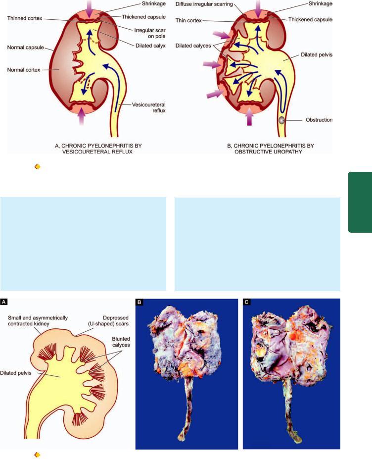
683
Figure 22.31 |
The two major types of mechanisms in chronic pyelonephritis. A,Vesicoureteric reflux causing infection of peripheral papillae |
and consequent scars at the poles of the kidney. B, Obstructive pyelonephritis due to obstruction of the urinary tract causing high pressure backflow of urine and infection of all the papillae and consequent dif fuse scarring of the kidney and thinning of the cortex.
i) Interstitium—There is chronic interstitial inflammatory reaction, chiefly composed of lymphocytes, plasma cells and macrophages with pronounced
interstitial fibrosis. Xanthogranulomatous pyelonephritis is an uncommon variant characterised by collection of foamy macrophages admixed with other inflammatory cells and giant cells.
ii) Tubules—The tubules show varying degree of atrophy and dilatation. Dilated tubules may contain eosinophilic colloid casts producing thyroidisation of tubules. A few tubules may contain neutrophils.
iii)Pelvicalyceal system—The renal pelvis and calyces are dilated. The walls of pelvis and calyces show marked chronic inflammation and fibrosis. Lymphoid follicles with germinal centres may be present in the pelvicalyceal walls. The lining epithelium may undergo squamous metaplastic change.
iv)Blood vessels—Blood vessels entrapped in the scarred areas show obliterative endarteritis. There may be changes of hypertensive hyaline arteriolosclerosis.
v)Glomeruli—Though glomerular tuft in the scarred area is usually intact, there is often periglomerular fibrosis.
Tract Urinary Lower and Kidney The 22 CHAPTER
Figure 22.32  Small contracted kidney in chronic pyelonephritis with calyectasis. A, Diagrammatic representation. B, External surface of small and contracted kidney. The kidney is small in size and contracted. The capsule is adherent to the cortex and has irregular scars on the surface.
C, Sectioned surface shows dilated pelvi-calyceal system with atrophied and thin peripheral cortex and increased hilar fat extending inside.
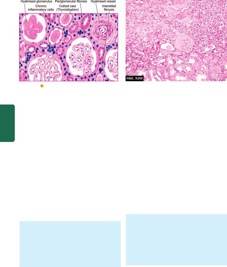
684
Pathology Systemic III SECTION
Figure 22.33 
 Chronic pyelonephritis. The scarred area shows atrophy of some tubules and dilat ation of others which contain colloid cast s (thyroidisation). The tubules are surrounded by abundant fibrous tissue and chronic interstitial inflammatory reaction. The blood vessels included are thick-walled and the glomeruli show periglomerular fibrosis.
In advanced cases, there may be hyalinisation of glomeruli.
CLINICAL FEATURES. Chronic pyelonephritis often has an insidious onset. The patients present with clinical picture of chronic renal failure or with symptoms of hypertension. Sometimes, the patients may present with features of acute recurrent pyelonephritis with fever, loin pain, lumbar tenderness, dysuria, pyouria, bacteriuria and frequency of micturition. Diagnosis is made by intravenous pyelography (IVP). Culture of the urine may give positive results. Longstanding cases of chronic pyelonephritis may develop secondary systemic amyloidosis.
Tuberculous Pyelonephritis
Tuberculosis of the kidney occurs due to haematogenous spread of infection from another site, most often from the lungs. Less commonly, it may result from ascending infection from tuberculosis of the genitourinary system such as from epididymis or Fallopian tubes. The renal lesions in tuberculosis may be in the form of tuberculous pyelonephritis or appear as multiple miliary tubercles.
MORPHOLOGIC FEATURES. Grossly, the lesions in tuberculous pyelonephritis are often bilateral, usually involving the medulla with replacement of the papillae by caseous tissue. Obstruction may result in tuberculous pyonephrosis in which thinned out renal parenchyma surrounds dilated pelvis and calyces filled with caseous material.
Histologically, typical granulomatous reaction is seen. Acid-fast bacilli can often be demonstrated in the lesions.
CLINICAL FEATURES. Most patients are young to middleaged adults. The clinical presentation is extremely variable but it should always be considered as a possibility in a patient
in whom there is persistent sterile pyouria, microscopic haematuria and mild proteinuria after effective antibiotic therapy for urinary tract infection. The diagnosis rests on identification of M. tuberculosis by repeated culture of urine on L.J. media.
Myeloma Nephropathy
Renal involvement in multiple myeloma (Chapter 14) is referred to as myeloma nephropathy or myeloma kidney. Functional renal impairment in multiple myeloma is a common manifestation, developing in about 50% of patients. The pathogenesis of myeloma kidney is related to excess filtration of Bence Jones proteins through the glomerulus, usually kappa (κ) light chains. These light chain proteins are precipitated in the distal convoluted tubules in combination with Tamm-Horsfall proteins, the urinary glycoproteins. The precipitates form tubular casts which are eosinophilic and often laminated. These casts may induce peritubular interstitial inflammatory reaction. Not all light chains are nephrotoxic and their toxicity occurs under acidic pH of the tubular fluid.
MORPHOLOGIC FEATURES. Grossly, the kidneys may be normal or small and shrunken.
Histologically, there are some areas of tubular atrophy while many other tubular lumina are dilated and contain characteristic bright pink laminated cracked or fractured casts consisting of Bence-Jones proteins called fractured casts. These casts are surrounded by peritubular interstitial inflammatory reaction including the presence of nonspecific inflammatory cells and some multinucleate giant cells induced by tubular casts.
Nephrocalcinosis
Nephrocalcinosis is a diffuse deposition of calcium salts in renal tissue in a number of renal diseases, in hypercalcaemia,
hyperphosphataemia and renal tubular acidosis. Most commonly, it develops as a complication of severe hypercalcaemia such as due to hyperparathyroidism, hypervitaminosis D, excessive bone destruction in metastatic malignancy, hyperthyroidism, excessive calcium intake such as in milk-alkali syndrome and sarcoidosis (page 53). Clinically, patients of hypercalcaemia and nephrocalcinosis may have renal colic, band keratopathy due to calcium deposits in the cornea, visceral metastatic calcification, polyuria and renal failure.
MORPHOLOGIC FEATURES. Nephrocalcinosis due to hypercalcaemia characteristically shows deposition of calcium in the tubular epithelial cells in the basement membrane, within the mitochondria and in the cytoplasm. These concretions may produce secondary tubular atrophy, interstitial fibrosis and nonspecific chronic inflammation in the interstitium. As the calcification occurs intracellularly, radiological evidence is usually not present until fairly late in the disease. The calcium deposits are first visible as small opacities in the renal papillae.
RENAL VASCULAR DISEASES
Renal blood vessels which enormously perfuse the kidney are affected secondarily in majority of renal diseases. Renal blood flow is controlled by systemic and local haemodynamic, hormonal and intrinsic intra-renal mechanisms. Diseases which disturb these controlling mechanisms give rise to primary renal vascular lesions. These diseases are as under:
I. Most importantly, hypertensive vascular disease and its consequent renal manifestations in the form of benign and malignant nephrosclerosis.
II. Thrombotic microangiopathy. III. Renal cortical necrosis.
IV. Renal infarcts.
Renal infarcts are already described in Chapter 5; other conditions are discussed here.
HYPERTENSIVE VASCULAR DISEASE
An elevated arterial blood pressure is a major health problem, particularly in developed countries. A persistent and sustained high blood pressure has damaging effects on the heart (e.g. hypertensive heart disease, Chapter 16), brain (e.g. cerebrovascular accident or stroke, Chapter 30) and kidneys (benign and malignant nephrosclerosis).
Definition and Classification
Hypertension is a common disease in industrialised countries and accounts for 6% of death worldwide. Epidemiologic studies have revealed that with elevation in systolic and diastolic blood pressure above normal in adults, there is a continuous increased risk of cardiovascular disease, stroke and renal disease—cardiovascular risk doubles with every 20 mmHg increase in systolic and 10 mmHg increase in
TABLE 22.14: Clinical Classification of Hypertension*. |
|
685 |
|||
Category |
Systolic |
Diastolic |
|
|
|
|
(mmHg) |
(mmHg) |
|
|
|
|
|
|
|
|
|
Normal |
< 120 |
and < 80 |
|
||
Prehypertension |
120-139 |
or 80-89 |
|
||
Hypertension |
|
|
|
|
|
Stege 1 |
140-159 |
or 90-99 |
|
||
Stege 2 |
>160 |
or >100 |
|
||
Isolated systolic hypertension |
>140 |
and < 90 |
|
||
Malignant hypertension |
> 200 |
> 140 |
|
|
|
(sudden onset) |
(sudden onset) |
|
|||
|
|
|
|
|
|
*Chobanian et al, JAMA, 2003. |
|
|
|
|
|
diastolic blood pressure above normal levels. Recently, |
|
||||
criteria for normal blood pressure, prehypertension and |
|
||||
hypertension (stage 1 and stage 2) have been laid by the |
|
||||
National Institutes of Health (NIH), US (Table 22.14). |
|
||||
According to this criteria, normal cut-off values for systolic |
|
||||
and diastolic blood pressure are taken as < 120 and < 80 |
|
||||
mmHg respectively. As per this criteria, arterial or systemic |
|
||||
hypertension in adults is defined clinically as persistently |
CHAPTER |
||||
elevated systolic blood pressure of 140-159 mmHg, or |
|||||
diastolic pressure of 90-99 mmHg as stage 1 hypertension, |
|||||
|
|||||
and corresponding values above 160 or above 100 mmHg |
|
||||
as stage 2 hypertension. Cases falling between upper normal |
|
||||
values for systolic and diastolic blood pressure (i.e. above |
|
||||
120/80 mmHg) and those for stage 1 hypertension (120- |
22 |
||||
139/80-89 mmHg) are grouped under prehypertension |
|||||
requiring monitoring anf follow-up. The diastolic pressure |
|
||||
is often considered more significant. Since blood pressure |
The |
||||
varies with many factors such as age of the patient, exercise, |
|||||
|
|||||
emotional disturbances like fear and anxiety, it is important |
Kidney |
||||
to measure blood pressure at least twice during two |
|||||
separate examinations under least stressful conditions. |
|||||
Hypertension is generally classified into 2 types: |
|||||
1. Primary or essential hypertension in which the cause of |
and |
||||
|
|||||
increase in blood pressure |
is unknown. Essential |
Lower |
|||
hypertension constitutes about 80-95% patients of |
|||||
|
|||||
hypertension. |
|
|
|
|
|
2. Secondary hypertension, in which the increase in blood |
Urinary |
||||
pressure is caused by diseases of the kidneys, endocrines or |
|||||
|
|||||
some other organs. Secondary hypertension comprises |
|
||||
remaining 5-20% cases of hypertension. |
|
|
|
||
According to the clinical course, both essential and |
Tract |
||||
secondary hypertension may be benign or malignant. |
|||||
|
|||||
Benign hypertension is moderate elevation of blood |
|
||||
pressure and the rise is slow over the years. About 90-95% |
|
||||
patients of hypertension have benign hypertension. |
|
||||

 Malignant hypertension is marked and suden increase of blood pressure to 200/140 mmHg or more in a known case of hypertension or in a previously normotensive individual; the patients develop papilloedema, retinal haemorrhages and hypertensive encephalopathy. Less than 5% of hypertensive patients develop malignant hypertension and life expectancy after diagnosis in these patients is generally less than 2 years if not treated effectively.

686 TABLE 22.15: Etiologic Classification of Hypertension.
A.ESSENTIAL HYPERTENSION (90%)
1.Genetic factors
2.Racial and environmental factors
3.Risk factors modifying the course
B.SECONDARY HYPERTENSION (10%)
1.Renal
i)Renovascular
ii)Renal parenchymal diseases
2.Endocrine
i)Adrenocortical hyperfunction
ii)Hyperparathyroidism
iii)Oral contraceptives
3.Coarctation of Aorta
4.Neurogenic
|
Etiology and Pathogenesis |
|
|
The etiology and pathogenesis of secondary hypertension |
|
|
that comprises less than 10% cases has been better |
|
|
understood, whereas the mechanism of essential |
|
|
hypertension that constitutes about 90% of cases remains |
|
SECTION |
largely obscure. In general, normal blood pressure is |
|
regulated by 2 haemodynamic forces—cardiac output and total |
||
|
||
|
peripheral vascular resistance. Factors which alter these two |
|
|
factors result in hypertension. The role of kidney in |
|
|
hypertension, particularly in secondary hypertension, by |
|
III |
elaboration of renin and subsequent formation of angiotensin |
|
II, is well established (renin-angiotensin system). |
||
|
With this background knowledge, we next turn to the |
|
Systemic |
mechanisms involved in the two forms of hypertension |
|
(Table 22.15). |
||
|
||
|
ESSENTIAL (PRIMARY) HYPERTENSION. By definition, |
|
|
the cause of essential hypertension is unknown but a number |
|
Pathology |
of factors are related to its development. These are as under: |
|
1. Genetic factors. The role of heredity in the etiology of |
||
|
||
|
essential hypertension has long been suspected. The |
|
|
evidences in support are the familial aggregation, occurrence |
|
|
of hypertension in twins, epidemiologic data, experimental |
|
|
animal studies and identification of hypertension |
|
|
susceptibility gene (angiotensinogen gene). |
|
|
2. Racial and environmental factors. Surveys in the US |
|
|
have revealed higher incidence of essential hypertension in |
|
|
African Americans than in whites. A number of environ- |
|
|
mental factors have been implicated in the development of |
|
|
hypertension including salt intake, obesity, skilled |
|
|
occupation, higher living standards and individuals under |
|
|
high stress. |
|
|
3. Risk factors modifying the course of essential hyper- |
|
|
tension. There is sufficient evidence to show that the course |
|
|
of essential hypertension that begins in middle life is |
|
|
modified by a number of factors. These are as under: |
|
|
i) Age. Younger the age at which hypertension is first noted |
|
|
but left untreated, lower the life expectancy. |
|
|
ii) Sex. Females with hypertension appear to do better than |
|
|
males. |
|
|
iii) Atherosclerosis. Accelerated atherosclerosis invariably |
|
|
accompanies essential hypertension. This could be due to |
contributory role of other independent factors like cigarette smoking, elevated serum cholesterol, glucose intolerance and obesity.
iv) Other risk factors. Other factors which alter the prognosis in hypertension include: smoking, excess of alcohol intake, diabetes mellitus, persistently high diastolic pressure above normal and evidence of end-organ damage (i.e. heart, eyes, kidney and nervous system).
The pathogenetic mechanism in essential hypertension is explained by many theories. These are as under:
1.High plasma level of catecholamines.
2.Increase in blood volume i.e. arterial overfilling (volume hypertension) and arteriolar constriction (vasoconstrictor hypertension).
3.Increased cardiac output.
4.Low-renin essential hypertension found in approximately 20% patients due to altered responsiveness to renin release.
5.High renin essential hypertension seen in about 15% cases due to decreased adrenal responsiveness to angiotensin II.
SECONDARY HYPERTENSION. Though much less common than essential hypertension, mechanisms underlying secondary hypertension with identifiable cause have been studied more extensively. Based on the etiology, these are described under four headings: renal hypertension, endocrine hypertension, hypertension associated with coarctation of aorta and neurogenic causes.
1. RENAL HYPERTENSION. Hypertension produced by renal diseases is called renal hypertension. Renal hypertension is subdivided into 2 groups:
i)Renal vascular hypertension e.g. in occlusion of a major renal artery, pre-eclampsia, eclampsia, polyarteritis nodosa and fibromuscular dysplasia of renal artery.
ii)Renal parenchymal hypertension e.g. in various types of glomerulonephritis, pyelonephritis, interstitial nephritis, diabetic nephropathy, amyloidosis, polycystic kidney disease and renin-producing tumours.
In either case, renal hypertension can be produced by one of the following 3 inter-related pathogenetic mechanisms:
a)Activation of renin-angiotensin system. Renin is a proteolytic enzyme produced and stored in the granules of the juxtaglomerular cells surrounding the afferent arterioles of glomerulus (page 651). The release of renin is stimulated by renal ischaemia, sympathetic nervous system stimulation, depressed sodium concentration, fluid depletion and decreased potassium intake. Released renin is transported through blood stream to the liver where it acts upon substrate
angiotensinogen, an α2-globulin synthesised in the liver, to form angiotensin I, a decapeptide. Angiotensin I is converted into angiotensin II, an octapeptide, by the action of convertase in the lungs. Angiotensin II is the most potent naturallyoccurring vasoconstrictor substance and its pressor action is mainly attributed to peripheral arteriolar vasoconstriction. The other main effect of angiotensin II is to stimulate the adrenal cortex to secrete aldosterone that promotes reabsorption of sodium and water.
Thus, the renin-angiotensin system is concerned mainly with 3 functions:
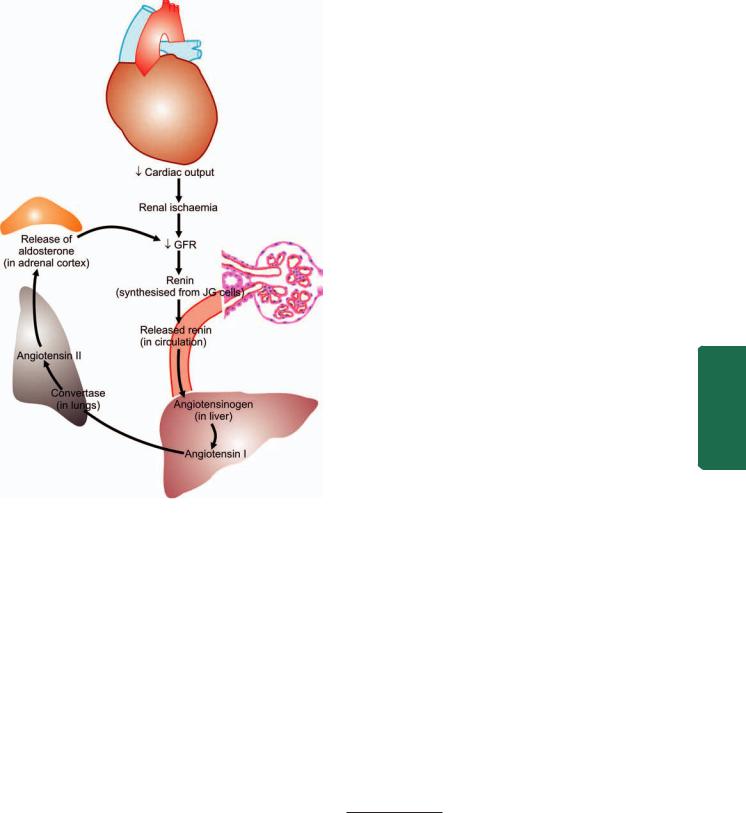
Figure 22.34 
 The renin-angiotensin mechanism.
i)Control of blood pressure by altering plasma concentration of angiotensin II and aldosterone.
ii)Regulation of sodium and water content.
iii)Regulation of potassium balance.
The renin-angiotensin mechanism is summarised in
Fig. 22.34.
b)Sodium and water retention. Blood volume and cardiac output, both of which have a bearing on blood pressure, are regulated by blood levels of sodium which is significant for maintaining extracellular fluid volume. Blood concentration of sodium is regulated by 3 mechanisms:
i)Release of aldosterone from activation of renin-angiotensin system, as already explained.
ii)Reduction in GFR due to reduced blood flow as occurs in reduced renal mass or renal artery stenosis. This results in proximal tubular reabsorption of sodium.
iii)Release of atriopeptin hormone from atria of the heart in response to volume expansion. These peptides cause increased GFR and inhibit sodium reabsorption.
c)Release of vasodepressor material. A number of vasodepressor materials and antihypertensives counterbalance the vasopressor effect of angiotensin II. These substances include: prostaglandins (PGE2, PGF2, PGA or medullin) released from interstitial cells of the medulla, urinary kallikrein-kinin system and platelet-activating factor.
2. ENDOCRINE HYPERTENSION. A number of hormonal |
687 |
secretions may produce secondary hypertension. These are: |
|
i)Adrenal gland—e.g. in primary aldosteronism, Cushing’s syndrome, adrenal virilism and pheochromocytoma.
ii)Parathyroid gland—e.g. hypercalcaemia in hyperparathyroidism.
iii)Oral contraceptives—Oestrogen component in the oral contraceptives stimulates hepatic synthesis of renin substrate.
3.COARCTATION OF AORTA. Coarctation of the aorta causes systolic hypertension in the upper part of the body due to constriction itself (Chapter 15). Diastolic hypertension results from changes in circulation.
4.NEUROGENIC. Psychogenic, polyneuritis, increased intracranial pressure and section of spinal cord are all uncommon causes of secondary hypertension.
EFFECTS OF HYPERTENSION |
|
||
Systemic hypertension causes major effects in three main |
|
||
organs—heart and its blood vessels, nervous system, and |
|
||
kidneys. The renal effects in the form of benign and malignant |
CHAPTER |
||
nephrosclerosis are discussed below, whereas hypertensive |
|||
effects on other organs are described elsewhere in the |
|||
|
|||
respective chapters. An important and early clinical marker |
|
||
for renal injury from hypertension and risk factor for |
|
||
cardiovascular disease is macroalbuminuria (i.e. albuminuria |
|
||
> 150 mg/day or random urine albumin/creatinine ratio of |
22 |
||
>300 mg/gm creatinine), or microalbuminuria estimated by |
|||
radioimmunoassay (i.e. microalbumin 30-300 mg/day or |
|
||
random urine microalbumin/creatinine ratio of |
The |
||
30-300 mg/gm creatinine). |
|||
Kidney |
|||
Benign Nephrosclerosis |
|||
|
|||
Benign nephrosclerosis is the term used to describe the |
|
||
kidney of benign phase of hypertension. Mild benign |
and |
||
nephrosclerosis is the most common form of renal disease in |
|||
|
|||
persons over 60 years of age but its severity increases in the |
Lower |
||
presence of hypertension and diabetes mellitus. |
|||
|
|||
|
|
|
|
MORPHOLOGIC FEATURES. Grossly, both the kidneys |
|
Urinary |
|
are affected equally and are reduced in size and weight, |
|
||
|
|
||
often weighing about 100 gm or less. The capsule is often |
|
|
|
adherent to the cortical surface. The surface of the kidney |
|
|
|
is finely granular and shows V-shaped areas of scarring.* |
|
Tract |
|
The cut surface shows firm kidney and narrowed cortex |
|
||
(Fig. 22.35). |
|
||
Microscopically, there are primarily diffuse vascular |
|
|
|
changes which produce parenchymal changes secondarily |
|
|
|
|
|
|
|
*The various acquired causes of ‘small contracted kidney’ and their characteristic gross macroscopic appearance may be recollected here. These are: 1. Chronic GN (granular appearance); 2. Chronic pyelonephritis (U-shaped scars); and 3. Benign nephrosclerosis (V-shaped scars). Although granular, U- and V-shaped scars correspond to the respective macroscopic patterns, acronym to remember is: ‘granular’ for glomerular scars of chronic GN; ‘U-scars’ for uneven scars of chronic pyelonephritis; and ‘V-scars’ for vascular scars of benign nephrosclerosis. Less common causes are: amyloidosis of the kidney, myeloma kidney and diabetic nephropathy.
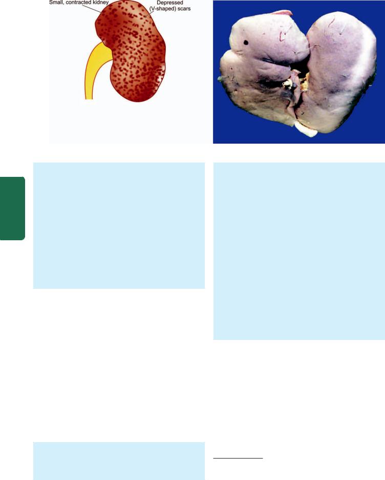
688
Figure 22.35 Small, contracted kidney in chronic hypertension
(benign nephrosclerosis).The kidney is small and contracted.The capsule is adherent to the cortex and has granular depressed scars on the surface.
Pathology Systemic III SECTION
as a result of ischaemia. The histologic changes are, thus, described as vascular and parenchymal (Fig. 22.36,A): i) Vascular changes: Changes in blood vessels involve arterioles and arteries up to the size of arcuate arteries. There are 2 types of changes in these blood vessels:
a)Hyaline arteriolosclerosis that results in homogeneous and eosinophilic thickening of the wall of small blood vessels.
b)lntimal thickening due to proliferation of smooth muscle cells in the intima.
ii)Parenchymal changes: As a consequence of ischaemia, there is variable degree of atrophy of parenchyma. This includes: glomerular shrinkage, deposition of collagen in Bowman’s space, periglomerular fibrosis, tubular atrophy and fine interstitial fibrosis.
CLINICAL FEATURES. There is variable elevation of the blood pressure with headache, dizziness, palpitation and nervousness. Eye ground changes may be found but papilloedema is absent. Renal function tests and urine examination are normal in early stage. In long-standing cases, there may be mild proteinuria with some hyaline or granular casts. Rarely, renal failure and uraemia may occur.
Malignant Nephrosclerosis
Malignant nephrosclerosis is the form of renal disease that occurs in malignant or accelerated hypertension. Malignant nephrosclerosis is uncommon and usually occurs as a superimposed complication in 5% cases of pre-existing benign essential hypertension or in those having secondary hypertension with identifiable cause such as in chronic renal diseases. However, the pure form of disease also occurs, particularly at younger age with preponderance in males.
MORPHOLOGIC FEATURES. Grossly, the appearance of the kidney varies. In a case of malignant hypertension superimposed on pre-existing benign nephrosclerosis, the kidneys are small in size, shrunken and reduced in weight
and have finely granular surface. However, the kidneys of a patient who develops malignant hypertension in pure form are enlarged, oedematous and have petechial haemorrhages on the surface producing so called ‘fleabitten kidney’.* Cut surface shows red and yellow mottled appearance (Fig. 22.37).
Microscopically, most commonly the changes are superimposed on benign nephrosclerosis. These changes are as under (Fig. 22.36,B):
i) Vascular changes: These are more severe and involve the arterioles. The two characteristic vascular changes seen are as under:
a)Necrotising arteriolitis develops on hyaline arteriolosclerosis. The vessel wall shows fibrinoid necrosis, a few acute inflammatory cells and small haemorrhages.
b)Hyperplastic intimal sclerosis or onionskin proliferation is characterised by concentric laminae of proliferated smooth muscle cells, collagen and basement membranes.
ii)Ischaemic changes: The effects of vascular narrowing on the parenchyma include tubular loss, fine interstitial fibrosis and foci of infarction necrosis.
CLINICAL FEATURES. The patients of malignant nephrosclerosis have malignant or accelerated hypertension with blood pressure of 200/140 mmHg or higher. Headache, dizziness and impaired vision are commonly found. The presence of papilloedema distinguishes malignant from benign phase of hypertension. The urine frequently shows haematuria and proteinuria. Renal function tests show deterioration during the course of the illness. Azotaemia (high BUN and serum creatinine) and uraemia develop soon if malignant hypertension is not treated aggressively. Approximately 90% of patients die within one year from causes such as uraemia, congestive heart failure and cerebrovascular accidents.
*Recall the other causes of flea-bitten kidney: acute post-streptococcal GN, rapidly progressive GN, haemolytic-uraemic syndrome, thrombotic thrombocytopenic purpura and Henoch-Schonlein purpura.

THROMBOTIC MICROANGIOPATHY
Thrombotic renal disease encompasses a group of diseases having in common the formation of thrombi composed by platelets and fibrin in arterioles and glomeruli of the kidney and culminating clinically in acute renal failure. Causes of thrombotic microangiopathy of renal microvasculature are listed in Table 22.16.
TABLE 22.16: Causes of Thrombotic Microangiopathy.
1.Infections
(E.coli, Shigella, Pseudomonas)
2.Drugs
(e.g. mitomycin, cisplatin, cyclosporine)
3.Autoimmune disease (scleroderma, SLE)
4.Thrombotic thrombocytopenic purpura
5.Haemolytic-uraemic syndrome
6.Pregnancy and pre-eclampsia
7.Malignant hypertension
689
|
|
|
22 CHAPTER |
|
Figure 22.36 |
Microscopic changes in kidney in hypertension. A, |
The |
||
Benign nephrosclerosis. The vascular changes are hyaline |
||||
arteriolosclerosis and intimal thickening of small blood vessels in the |
||||
glomerular tuft. The parenchymal changes include sclerosed glomeruli, |
Kidney |
|||
tubular atrophy and fine interstitial fibrosis. B, Malignant nephrosclerosis. |
||||
|
||||
The vascular changes are necrotising arteriolitis and hyperplastic intimal |
|
|||
sclerosis or onion-skin proliferation.The parenchymal changes are tubular |
|
|||
loss, fine interstitial fibrosis and foci of infarction necrosis. |
and |
|||
|
|
|
||
The common clinical manifestations include microangio- |
Lower |
|||
|
||||
pathic haemolytic anaemia, thrombocytopenia, DIC, and |
|
|||
eventually renal failure. |
Urinary |
|||
PATHOGENESIS In all such cases, endothelial injury |
||||
appears to be the trigger for vascular changes. The injured |
||||
endothelial surface causes the following effects: |
||||
Passage of plasma constituents to the subendothelial zone |
Tract |
|||
|
||||
of microvasculature. |
|
|||
Promotes thrombosis. |
|
|||
|
|
|
||
MORPHOLOGIC FEATURES. The lesions closely |
|
|
||
resemble those of malignant nephrosclerosis. The features |
|
|
||
are as under: |
|
|
|
|
Fibrinoid necrosis of arterioles. |
|
|
||
Thrombi in renal microvasculature. |
|
|
||
Oedema of intima of arterioles. |
|
|
||
Consolidation, necrosis and congestion of glomeruli. |
|
|
||
If the renal lesions are massive, the prognosis is generally |
|
|||
lethal. |
|
|
|
|
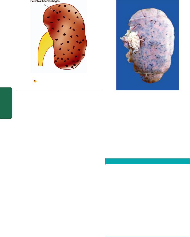
690
Pathology Systemic III SECTION
Figure 22.37 Malignant nephrosclerosis. The kidney is enlarged in
size and weight. The cortex shows characteristic ‘flea bitten kidney’ due to tiny petechial haemorrhages on the surface.
RENAL CORTICAL NECROSIS
Renal cortical necrosis is infarction of renal cortex varying from microscopic foci to a situation where most of the renal cortex is destroyed. The medulla, the juxtamedullary cortex and a rim of cortex under the capsule are usually spared. The condition develops most commonly as an obstetrical emergency (e.g. in eclampsia, pre-eclampsia, premature separation of the placenta). Other causes include septic shock, poisoning, severe trauma etc.
The lesions may be present focally, patchily or diffusely. The gross and microscopic characteristics of infarcts of cortex are present. Patients present with sudden oliguria or anuria and haematuria. If the process has involved renal cortex extensively, acute renal failure and uraemia develop and prognosis is grave.
OBSTRUCTIVE UROPATHY
Obstruction in the urinary tract is common and important because it increases the susceptibility to infection and stone formation. Obstruction can occur at any age and in either sex. The cause of obstruction may lie at any level of the urinary tract—renal pelvis, ureters, urinary bladder and urethra. The obstruction at any of these anatomic locations may be intraluminal, intramural or extramural. Important causes are listed in Table 22.17 and illustrated in Fig. 22.38.
The obstruction may be unilateral or bilateral, partial or complete, sudden or insidious. Complete bilateral obstruction may result in irreversible renal failure, whereas long-standing chronic partial obstruction may cause various functional abnormalities and anatomic changes. There are three important anatomic sequelae of obstruction, namely:
hydronephrosis, hydroureter and hypertrophy of the bladder.
Before describing these conditions, an account of the most
common and important cause of obstructive uropathy, urolithiasis, is given below.
NEPHROLITHIASIS
Nephrolithiasis or urolithiasis is formation of urinary calculi at any level of the urinary tract. Urinary calculi are worldwide in distribution but are particularly common in some geographic locations such as in parts of the United States, South Africa, India and South-East Asia. It is estimated that approximately 2% of the population experiences renal stone disease at sometime in their life with male-female ratio of 2:1. The peak incidence is observed in 2nd to 3rd decades of life. Renal calculi are characterised clinically by colicky pain
TABLE 22.17: Causes of Obstructive Uropathy.
A. INTRALUMINAL
1.Calculi
2.Tumours (e.g. cancer of kidney and bladder)
3.Sloughed renal papilla
4.Blood clots
5.Foreign body
B. INTRAMURAL
1.Pelvi-ureteric junction (PUJ) obstruction
2.Vesicoureteric obstruction
3.Urethral stricture
4.Urethral valves
5.Inflammation (e.g. phimosis, cystitis etc)
6.Neuromuscular dysfunction
C. EXTRAMURAL
1.Pregnant uterus
2.Retroperitoneal fibrosis
3.Tumours (e.g. carcinoma of cervix, rectum, colon, caecum etc)
4.Prostatic enlargement, prostatic carcinoma and prostatitis
5.Trauma
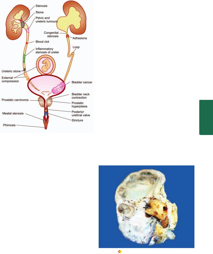
Figure 22.38 
 Causes of obstructive uropathy.
(renal colic) as they pass down along the ureter and manifest by haematuria.
Types of Urinary Calculi
There are 4 main types of urinary calculi—calcium containing, mixed (struvite), uric acid and cystine stones, and a few rare types (Table 22.18).
1. CALCIUM STONES. Calcium stones are the most common comprising about 75% of all urinary calculi. They may be pure stones of calcium oxalate (50%) or calcium phosphate (5%), or mixture of calcium oxalate and calcium phosphate (45%).
Etiology. Etiology of calcium stones is variable.
i)About 50% of patients with calcium stones have idiopathic hypercalciuria without hypercalcaemia.
ii)Approximately 10% cases are associated with hypercalcaemia and hypercalciuria, most commonly due to hyperparathyroidism, or a defect in the bowel (i.e. absorptive hypercalciuria), or in the kidney (i.e. renal hypercalciuria).
iii)About 15% of patients with calcium stones have hyperuricosuria with a normal blood uric acid level and without any abnormality of calcium metabolism.
iv)In about 25% of patients with calcium stones, the cause is unknown as there is no abnormality in urinary excretion
of calcium, uric acid or oxalate and is referred to as ‘idiopathic calcium stone disease’.
Pathogenesis. The mechanism of calcium stone formation is 691 explained on the basis of imbalance between the degree of supersaturation of the ions forming the stone and the concentration of inhibitors in the urine. Most likely site where
the crystals of calcium oxalate and/or calcium phosphate are precipitated is the tubular lining or around some fragment of debris in the tubule acting as nidus of the stone. The stone grows, as more and more crystals are deposited around the nidus. A number of other predisposing factors contributing to formation of calcium stones are alkaline urinary pH, decreased urinary volume and increased excretion of oxalate and uric acid.
Morphology. Calcium stones are usually small (less than a centimeter), ovoid, hard, with granular rough surface. They are dark brown due to old blood pigment deposited in them as a result of repeated trauma caused to the urinary tract by these sharp-edged stones.
2. MIXED (STRUVITE) STONES. About 15% of urinary |
|
|
calculi are made of magnesium-ammonium-calcium |
|
|
phosphate, often called struvite; hence mixed stones are also |
|
|
called as ‘struvite stones’ or ‘triple phosphate stones’. |
CHAPTER |
|
Etiology. Struvite stones are formed as a result of infection |
||
|
||
of the urinary tract with urea-splitting organisms that |
|
|
produce urease such as by species of Proteus, and occasionally |
|
|
Klebsiella, Pseudomonas and Enterobacter. These are, therefore, |
|
|
also known as infection-induced stones. However, E. coli does |
22 |
|
not form urease. |
||
|
||
Morphology. Struvite stones are yellow-white or grey. They |
|
|
tend to be soft and friable and irregular in shape. ‘Staghorn |
The |
|
stone’ which is a large, solitary stone that takes the shape of |
||
|
||
the renal pelvis where it is often formed is an example of |
TractUrinaryLowerandKidney |
|
struvite stone (Fig. 22.39). |
||
|
Figure 22.39 
 Staghorn renal stone with chronic pyelonephritis.The kidney is enlarged and heavy. The capsule is adherent to the cortex and has irregular scars on the surface. Sectioned surface shows dilated pelvicalyceal system with atrophied and thin peripheral cortex. The pelvis of the kidney contains a single, large, sof t yellow white stone t aking the contour of the pelvi-calyceal system (arrow).

692 TABLE 22.18: Salient Features of Urinary Calculi.
|
Type |
Incidence |
Etiology |
Pathogenesis |
|
|
|
|
|
1. |
Calcium stones |
75% |
Hypercalciuria with or |
Supersaturation of ions in urine, alkaline |
|
|
|
without hypercalcaemia; |
pH of urine; low urinary volume, oxaluria |
|
|
|
idiopathic |
and hyperuricosuria |
2. |
Mixed (struvite) |
15% |
Urinary infection with urea- |
Alkaline urinary pH produced by ammonia |
|
stones |
|
splitting organisms like |
from splitting of urea by bacterially |
|
|
|
Proteus |
produced urease |
3. |
Uric acid |
6% |
Hyperuricosuria with or without |
Acidic urine (pH below 6) decreases the |
|
stones |
|
hyperuricaemia (e.g. in |
solubility of uric acid in urine and favours |
|
|
|
primary and secondary gout) |
its precipitation |
4. |
Cystine stones |
2% |
Genetically-determined |
Cystinuria containing least soluble cystine |
|
|
|
defect in cystine transport |
precipitates as cystine crystals |
5. |
Other types |
< 2% |
Inherited abnormalities of |
Xanthinuria |
|
|
|
amino acid metabolism |
|
|
|
|
|
|
Pathology Systemic III SECTION
3. URIC ACID STONES. Approximately 6% of urinary calculi are made of uric acid. Uric acid calculi are radiolucent unlike radio-opaque calcium stones.
Etiology. Uric acid stones are frequently formed in cases with hyperuricaemia and hyperuricosuria such as due to primary gout or secondary gout due to myeloproliferative disorders (e.g. in leukaemias), especially those on chemotherapy, and administration of uricosuric drugs (e.g. salicylates, probenacid). Other factors contributing to their formation are acidic urinary pH (below 6) and low urinary volume.
Pathogenesis. The solubility of uric acid at pH of 7 is 200 mg/dl while at pH of 5 is 15 mg/dl. Thus, as the urine becomes more acidic, the solubility of uric acid in urine decreases and precipitation of uric acid crystals increases favouring the formation of uric acid stones. Hyperuricosuria is the most important factor in the production of uric acid stones, while hyperuricaemia is found in about half the cases.
Morphology. Uric acid stones are smooth, yellowish-brown, hard and often multiple. On cut section, they show laminated structure.
4. CYSTINE STONES. Cystine stones comprise less than
2% of urinary calculi.
Etiology. Cystine stones are associated with cystinuria due to a genetically-determined defect in the transport of cystine and other amino acids across the cell membrane of the renal tubules and the small intestinal mucosa.
Pathogenesis. The resultant excessive excretion of cystine which is least soluble of the naturally-occurring amino acids leads to formation of crystals and eventually cystine calculi.
Morphology. Cystine stones are small, rounded, smooth and often multiple. They are yellowish and waxy.
5. OTHER CALCULI. Less than 2% of urinary calculi consist of other rare types such as due to inherited abnormality of enzyme metabolism e.g. hereditary xanthinuria developing xanthine stones.
HYDRONEPHROSIS
Hydronephrosis is the term used for dilatation of renal pelvis and calyces due to partial or intermittent obstruction to the
outflow of urine (see Fig. 22.38). Hydronephrosis develops if one or both the pelviureteric sphincters are incompetent, as otherwise there will be dilatation and hypertrophy of the urinary bladder but no hydronephrosis. Hydroureter nearly always accompanies hydronephrosis. Hydronephrosis may
be unilateral or bilateral.
Unilateral Hydronephrosis
This occurs due to some form of ureteral obstruction at the level of pelviureteric junction (PUJ). The causes are:
1.Intraluminal e.g. a calculus in the ureter or renal pelvis.
2.Intramural e.g. congenital PUJ obstruction, atresia of ureter, inflammatory stricture, trauma, neoplasm of ureter or bladder.
3.Extramural e.g. obstruction of upper part of the ureter by inferior renal artery or vein, pressure on ureter from outside such as carcinoma cervix, prostate, rectum, colon or caecum and retroperitoneal fibrosis.
Bilateral Hydronephrosis
This is generally the result of some form of urethral obstruction but can occur from the various causes listed above if the lesions involve both sides. Based on this, hydronephrosis may be of following types:
1.Congenital e.g. atresia of the urethral meatus, congenital posterior urethral valve.
2.Acquired e.g. bladder tumour involving both ureteric orifices, prostatic enlargement, prostatic carcinoma and prostatitis, bladder neck stenosis, inflammatory or traumatic urethral stricture and phimosis.
MORPHOLOGIC FEATURES. The pathologic changes vary depending upon whether the obstruction is sudden and complete, or incomplete and intermittent. The latter situation is more common.
Grossly, the kidneys may have moderate to marked enlargement. Initially, there is extrarenal hydronephrosis characterised by dilatation of renal pelvis medially in the form of a sac (Fig. 22.40,A). As the obstruction persists,
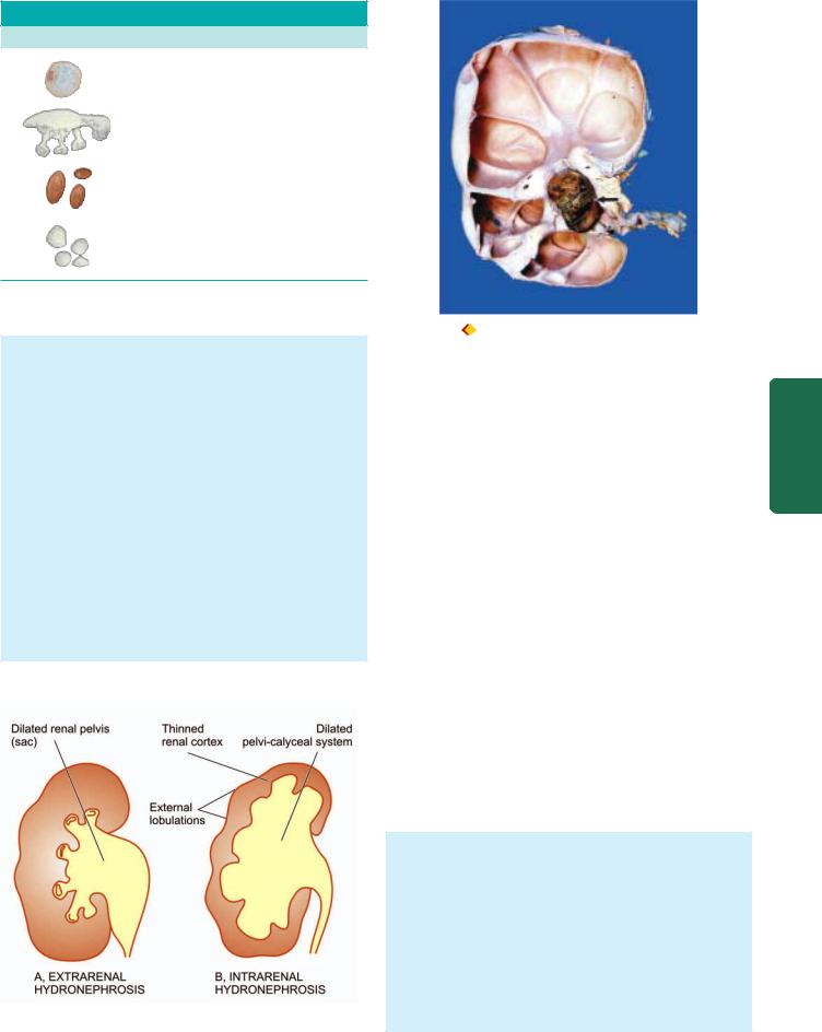
TABLE 22.18: Urinary Calculi (continued)
Morphology
Calcium oxalate stones
Struvite (‘Staghorn’) stone
Uric acid stones
Cystine stones
Figure facing Table 22.18
there is progressive dilatation of pelvis and calyces and pressure atrophy of renal parenchyma. Eventually, the dilated pelvi-calyceal system extends deep into the renal cortex so that a thin rim of renal cortex is stretched over the dilated calyces and the external surface assumes lobulated appearance. This advanced stage is called as intrarenal hydronephrosis (Fig. 22.40,B). An important point of distinction between the sectioned surface of advanced hydronephrosis and polycystic kidney disease (page 657) is the direct continuity of dilated cystic spaces (i.e. dilated calyces) with the renal pelvis in the former (Fig. 22.41). Microscopically, the wall of hydronephrotic sac is thickened due to fibrous scarring and chronic inflammatory cell infiltrate. There is progressive atrophy of tubules and glomeruli alongwith interstitial fibrosis. Stasis of urine in hydronephrosis causes infection (pyelitis) resulting in filling of the sac with pus, a condition called
pyonephrosis.
Figure 22.40 
 Hydronephrosis, stages in its evolution.
Figure 22.41 
 Hydronephrosis with nephrolithiasis. The kidney is enlarged and heavy . On cut section, the renal pelvis and calyces are dilated and cystic and contain a large stone in the pelvis of the kidney (arrow). The cystic change is seen to extend into renal p arenchyma, compressing the cortex as a thin rim at the periphery . Unlike polycystic kidney, however, these cysts are communicating with the pelvi-calyceal system.
TUMOURS OF KIDNEY
Both benign and malignant tumours occur in the kidney, the latter being more common. These may arise from renal tubules (adenoma, adenocarcinoma), embryonic tissue (mesoblastic nephroma, Wilms’ tumour), mesenchymal tissue (angiomyolipoma, medullary interstitial tumour) and from the epithelium of the renal pelvis (urothelial carcinoma). Besides these tumours, the kidney may be the site of the secondary tumours.
Table 22.19 provides a list of kidney tumours; the important forms of renal neoplasms are described below.
BENIGN TUMOURS
Benign renal tumours are usually small and are often an incidental finding at autopsy or nephrectomy.
Cortical Adenoma
Cortical tubular adenomas are more common than other benign renal neoplasms. They are frequently multiple and associated with chronic pyelonephritis or benign nephrosclerosis.
Grossly, these tumours may form tiny nodules up to 3 cm in diameter. They are encapsulated and white or yellow.
Microscopically, they are composed of tubular cords or papillary structures projecting into cystic space. The cells of the adenoma are usually uniform, cuboidal with no atypicality or mitosis. However, size of the tumour rather than histologic criteria is considered more significant parameter to predict the behaviour of the tumour—those larger than 3 cm in diameter are potentially malignant and metastasising.
693
Tract Urinary Lower and Kidney The 22 CHAPTER

694 TABLE 22.19: Classification of Kidney Tumours.
|
Benign |
Malignant |
|
|
|
||
|
A. EPITHELIAL TUMOURS OF RENAL PARENCHYMA |
||
|
Adenoma |
Adenocarcinoma |
|
|
Oncocytoma |
(hypernephroma, renal cell |
|
|
|
carcinoma) |
|
|
B. EPITHELIAL TUMOURS OF RENAL PELVIS |
||
|
Transitional cell papilloma |
Transitional cell carcinoma |
|
|
|
Others (squamous cell carcinoma, |
|
|
|
adenocarcinoma of renal pelvis, |
|
|
|
undifferentiated carcinoma of |
|
|
|
renal pelvis) |
|
|
C. EMBRYONAL TUMOURS |
|
|
|
Mesoblastic nephroma |
Wilms’ tumour (nephroblastoma) |
|
|
Multicystic nephroma |
|
|
|
D. NON-EPITHELIAL TUMOURS |
||
|
Angiomyolipoma |
Sarcomas (rare) |
|
|
Medullary interstitial |
|
|
SECTION |
tumour (fibroma) |
|
|
E. MISCELLANEOUS |
|
||
|
|
||
|
Juxtaglomerular cell |
|
|
|
tumour (Reninoma) |
|
|
III |
F. METASTATIC TUMOURS |
|
|
|
|
||
Oncocytoma |
|
||
Systemic |
|
||
Oncocytoma is a benign epithelial tumour arising from |
|||
|
|||
|
collecting ducts. |
|
|
|
|
||
Pathology |
Grossly, The tumour is encapsulated and has variable size. |
||
Cut section is homogeneous and has characteristic |
|||
|
|||
|
mahogany-brown or tan colour. |
||
|
Microscopically, the tumour cells are plump with |
||
|
abundant, finely granular, acidophilic cytoplasm and |
||
|
round nuclei. Electron microscopy demonstrates |
||
|
numerous mitochondria in the cytoplasm. |
||
|
|
|
|
|
Other Benign Tumours |
|
|
|
Angiomyolipoma is a hamartoma of the kidney that |
||
|
contains differentiated tissue element derived from blood |
||
|
vessels, smooth muscle and fat. Patients of tuberous sclerosis, |
||
|
a multisystem disease characterised by skin lesions, CNS and |
||
|
renal involvement, frequently have bilateral angiomyo- |
||
|
lipomas. |
|
|
|
Mesoblastic nephroma is a congenital benign tumour. |
||
|
|
||
|
Grossly, the tumour resembles a uterine leiomyoma in |
||
|
having whorled appearance. |
||
|
Microscopically, it shows cellular growth of spindle cells |
||
|
derived from secondary mesenchyme. |
||
|
|
||
|
Multicystic nephroma is another uncommon tumour of |
||
|
early infancy. |
|
|
Grossly, it is a solitary, unilateral well demarcated tumour of varying size. Cut surface shows characteristic multilocular appearance.
Microscopically, the cysts are lined by tubular epithelium while the stroma between the cysts contains mesenchymal tissue with some immature blastemal or abortive tubules. Some authors consider this entity as fully-differentiated variant of Wilms’ tumour. However, clinically multicystic nephroma is always benign compared to Wilms’ tumour.
 Medullary interstitial cell tumour is a tiny nodule in the medulla composed of fibroblast-like cells in hyalinised stroma. These tumours used to be called renal fibromas but electron microscopy has revealed that the tumour cells are not fibrocytes but are medullary interstitial cells.

 Juxtaglomerular tumour or reninoma is a rare tumour of renal cortex consisting of sheets of epithelioid cells with many small blood vessels. The tumour secretes excessive quantities of renin and, thus, the patients are likely to have hypertension.
MALIGNANT TUMOURS
The two most common primary malignant tumours of the
kidney are adenocarcinoma and Wilms’ tumour. A third malignant renal tumour is urothelial carcinoma occurring more commonly in the renal pelvis is described in the next section along with other tumours of the lower urinary tract.
Adenocarcinoma of Kidney (Synonyms: Renal cell carcinoma, Hypernephroma, Grawitz tumour)
Hypernephroma is an old misnomer under the mistaken belief that the tumour arises from adrenal rests because of the resemblance of the tumour cells with clear cells of the adrenal cortex. It is now known that the renal cell carcinoma (RCC) is an adenocarcinoma arising from tubular epithelium. This cancer comprises 70 to 80% of all renal cancers and occurs most commonly in 50 to 70 years of age with male preponderance (2:1).
ETIOLOGY AND PATHOGENESIS. Various etiologic factors implicated in the etiology of RCC are as follows:
1.Tobacco. Tobacco is the major risk factor for RCC, whether chewed or smoked and accounts for 20-30% cases of RCC. Cigarette smokers have two-fold higher risk of developing RCC.
2.Genetic factors. Heredity and first-degree relatives of RCC are associated with higher risk. Although majority of cases of RCC are sporadic but about 5% cases are inherited. These cases have following associations:
i)von Hippel-Lindau (VHL) disease: It is an autosomal dominant cancer syndrome that includes: haemangioblastoma of the cerebellum, retinal angiomas, multiple RCC (clear cell type), pheochromocytoma and cysts in different organs. Patients of VHL disease have germline mutations of tumour suppressor VHL gene located on chromosome 3p, commonly as homozygous loss of VHL gene. About 35% cases of VHL develop RCC.
TABLE 22.20: Classification of Renal Cell Carcinoma |
|
695 |
|||
|
Type |
Incidence |
Genetics |
Main Histology |
|
1. |
Clear cell type |
70% |
Sporadic and familial |
Clear cytoplasm (due to glycogen |
|
|
(non-papillary) |
|
(Homozygous loss of VHL gene located |
and lipid), well differentiated |
|
|
|
|
on chromosome 3) |
|
|
2. |
Papillary type |
15% |
Familial and sporadic |
Papillary pattern, psammoma bodies |
|
|
|
|
(Familial cases: mutation in MET gene |
|
|
|
|
|
on chromosome 7; sporadic cases trisomy |
|
|
|
|
|
of chromosome 7, 16, 17 and loss of Y |
|
|
|
|
|
chromosome) |
|
|
3. |
Granular cell type |
8% |
Sporadic and familial |
Abundant acidophilic cytoplasm, |
|
|
|
|
|
marked atypia |
|
4. |
Chromophobe type |
5% |
Multiple chromosome losses, |
Mixture of pale clear cells with |
|
|
|
|
hypodiploidy |
perinuclear halo and granular cells |
|
5. |
Sarcomatoid type |
1.5% |
— |
Whorls of atypical anaplastic spindle |
|
|
|
|
|
cells |
|
6. |
Collecting duct type |
0.5% |
— |
Tubular and papillary pattern |
|
|
|
|
|
|
|
ii)Hereditary clear cell RCC: These are cases of clear cell type RCC confined to the kidney without other manifestations of VHL but having autosomal dominant inheritance.
iii)Papillary RCC: This form of RCC is characterised by bilateral and multifocal cancer with papillary growth pattern. Genetic abnormality in these cases lies in MET gene located on chromosome 7.
iv)Chromophobe RCC: These cases have genetic defects in the form of multiple losses of whole chromosomes i.e. they have extreme degree of hypodiploidy.
3.Cystic diseases of the kidneys. Both hereditary and acquired cystic diseases of the kidney have increased risk of development of RCC. Patients on longterm dialysis develop acquired cystic disease which may evolve into RCC and adenomas. Adult polycystic kidney disease and multicystic nephroma is associated with higher occurrence of papillary RCC.
4.Other risk factors. Besides above, following other factors are associated with higher incidence of RCC:
i)Exposure to asbestos, heavy metals and petrochemical products.
ii)In women, obesity and oestrogen therapy.
iii)Analgesic nephropathy.
v) Tuberous sclerosis.
CLASSIFICATION. Based on cytogenetics of sporadic and familial tumours, RCC has been reclassified into clear cell, papillary, granular cell, chromophobe, sarcomatoid and collecting duct type (Table 22.20).
MORPHOLOGIC FEATURES. Grossly, RCC commonly arises from the poles of the kidney as a solitary and unilateral tumour, more often in the upper pole. The tumour is generally large, golden yellow and circumscribed. Papillary tumours have grossly visible papillae and may be multifocal. About 1% RCC are bilateral. Cut section of the tumour commonly shows large areas of
ischaemic necrosis, cystic change and foci of haemorrhages. Another significant characteristic is the frequent presence of tumour thrombus in the renal vein which may extend into the vena cava (Fig. 22.42).
Histologically, the features of various types of RCC are as under:
1.Clear cell type RCC (70%): This is the most common pattern. The clear cytoplasm of tumour cells is due to removal of glycogen and lipid from the cytoplasm during processing of tissues. The tumour cells have a variety of patterns: solid, trabecular and tubular, separated by delicate vasculature. Majority of clear cell tumours are well differentiated (Fig. 22.43).
2.Papillary type RCC (15%): The tumour cells are arranged in papillary pattern over the fibrovascular stalks. The tumour cells are cuboidal with small round nuclei. Psammoma bodies may be seen.
3.Granular cell type RCC (8%): The tumour cells have abundant acidophilic cytoplasm. These tumours have more marked nuclear pleomorphism, hyperchromatism and cellular atypia.
4.Chromophobe type RCC (5%): This type shows admixture of pale clear cells with perinuclear halo and acidophilic granular cells. The cytoplasm of these tumour cells contains many vesicles.
5.Sarcomatoid type RCC (1.5%): This is the most anaplastic and poorly differentiated form. The tumour is characterised by whorls of atypical spindle tumour cells.
6.Collecting duct type RCC (0.5%): This is a rare type that occurs in the medulla. It is composed of a single layer of cuboidal tumour cells arranged in tubular and papillary pattern.
CLINICAL FEATURES. Renal cell carcinoma is generally a slow-growing tumour and the tumour may have been present for years before it is detected. The classical clinical
Tract Urinary Lower and Kidney The 22 CHAPTER
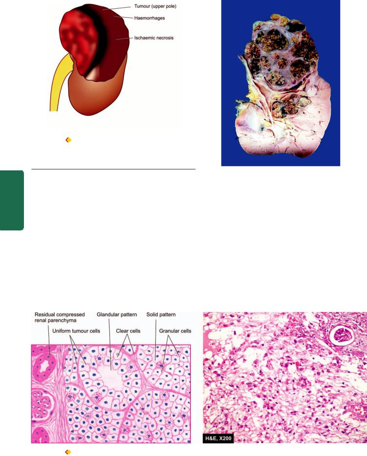
696
Figure 22.42 
 Renal cell carcinoma. The upper pole of the kidney shows a large and tan mass while rest of the kidney has reniform contour.
Sectioned surface shows irregular , circumscribed, yellowish mass with areas of haemorrhages and necrosis.The residual kidney is compressed on one side and shows obliterated calyces and renal pelvis.
SECTION |
evidence for diagnosis of renal cell carcinoma is the triad of |
|
|
||
|
gross haematuria, flank plain and palpable abdominal mass. The |
|
|
most common presenting abnormality is haematuria that |
|
III |
occurs in about 60% of cases. By the time the tumour is |
|
detected, it has spread to distant sites via haematogenous |
||
|
||
|
route to the lungs, brain and bone, and locally to the liver |
|
Systemic |
and perirenal lymph nodes. |
|
paraneoplastic syndromes due to ectopic hormone |
||
|
Systemic symptoms of fatiguability, weight loss, cachexia |
|
|
and intermittent fever unassociated with evidence of infection |
|
|
are found in many cases at presentation. A number of |
|
Pathology |
production by the renal cell carcinoma have been described. |
|
These include polycythaemia (by erythropoietin), hyper- |
||
|
||
|
calcaemia (by parathyroid hormone and prostaglandins), |
hypertension (by renin), effects of feminisation or masculinisation
(by gonadotropins) and Cushing’s syndrome (by glucocorticoids).
The prognosis in renal cell carcinoma depends upon the extent of tumour involvement at the time of diagnosis. The overall 5-year survival rate is about 70%. Presence of metastases, renal vein invasion and higher nuclear grade of the tumour are some of the predictors of poor prognosis.
Wilms’ Tumour
(Synonym: Nephroblastoma)
Nephroblastoma or Wilms’ tumour is an embryonic tumour derived from primitive renal epithelial and mesenchymal components. It is the most common abdominal malignant
Figure 22.43 
 Renal cell carcinoma. The tumour shows solid masses and acini of uniform-appearing tumour cells. Clear cells predominate in the tumour while the stroma is composed of fine and delicate fibrous tissue.
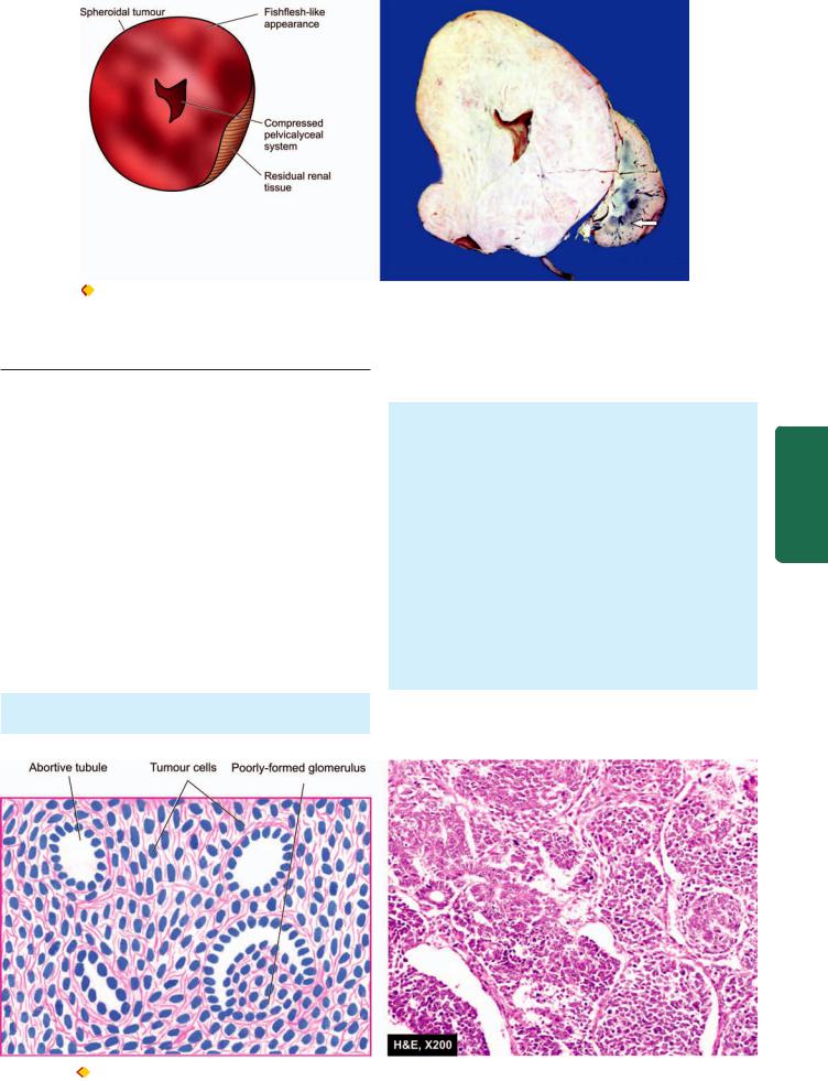
697
Figure 22.44 |
Nephroblastoma (W ilms’ tumour). The kidney is |
enlarged and has ovoid and nodular appearance.The sectioned surface shows replacement of almost whole kidney by the tumour leaving a thin strip of compressed renal tissue at lower end (arrow). Cut section of the tumour is gray white, fleshy and has small areas of haemorrhages and necrosis.
tumour of young children, seen most commonly between 1 to 6 years of age with equal sex incidence.
ETIOLOGY AND PATHOGENESIS. Wilms’ tumour has following etiologic associations:
1.A defect in chromosome 11p13 results in abnormal growth of metanephric blastema without differentiation into normal tubules and glomeruli.
2.A higher incidence has been seen in monozygotic twins and cases with family history.
3.Association of Wilms’ tumour with some other congenital anomalies has been observed, especially of the genitourinary tract.
4.A few other malignancies are known to have higher incidence of Wilms’ tumour. These include osteosarcoma, botyroid sarcoma, retinoblastoma, neuroblastoma etc.
MORPHOLOGIC FEATURES. Grossly, the tumour is usually quite large, spheroidal, replacing most of the
kidney. It is generally solitary and unilateral but 5-10% cases may have bilateral tumour. On cut section, the tumour shows characteristic variegated appearance— soft, fishflesh-like grey-white to cream-yellow tumour with foci of necrosis and haemorrhages and grossly identifiable myxomatous or cartilaginous elements (Fig. 22.44). Invasion into renal vein is grossly evident in half the cases.
Microscopically, nephroblastoma shows mixture of primitive epithelial and mesenchymal elements. Most of the tumour consists of small, round to spindled, anaplastic, sarcomatoid tumour cells. In these areas are present abortive tubules and poorly-formed glomerular structures (Fig. 22.45). Mesenchymal elements such as smooth and skeletal muscle, cartilage and bone, fat cells and fibrous tissue, may be seen.
CLINICAL FEATURES. The most common presenting feature is a palpable abdominal mass in a child. Other
Tract Urinary Lower and Kidney The 22 CHAPTER
Figure 22.45  Wilms’ tumour. There is predominance of small round to spindled sarcomatoid tumour cells. A few abortive tubules and poorlyformed glomerular structures are present in it.

698common abnormalities are haematuria, pain, fever and hypertension. The tumour rapidly spreads via blood, especially to lungs.
The prognosis of the tumour with combination therapy of nephrectomy, post-operative irradiation and chemotherapy, has improved considerably and the 5-year survival now is 80-90%.
Secondary Tumours
Leukaemic infiltration of the kidneys is a common finding, particularly in chronic myeloid leukaemia. Kidney is a common site for blood-borne metastases from different primary sites, chiefly from cancers of the lungs, breast and stomach.
LOWER URINARY TRACT
|
NORMAL STRUCTURE |
|
|
The lower urinary tract consists of ureters, urinary bladder and |
|
|
urethra. |
|
SECTION |
URETERS are tubular structures, 30 cm in length and half a |
|
centimeter in diameter, and extend from the renal pelvis |
||
|
||
|
(pelvi-ureteric junction) to the urinary bladder (vesico- |
|
|
ureteric junction). Normally they enter obliquely into the |
|
|
bladder, so that ureter is compressed during micturition, thus |
|
III |
preventing vesico-ureteric reflux. Ureters lie retroperitoneally |
|
throughout their course. |
||
|
Histologically, ureter has an outer fibrous investing layer |
|
Systemic |
which overlies a thick muscular layer and is lined internally |
|
URINARY BLADDER lies extraperitoneally and the |
||
|
by transitional epithelium or urothelium similar to the lining |
|
|
of the renal pelvis above and bladder below. |
|
Pathology |
peritoneum is reflected on its superior surface. Besides |
|
capacity of bladder is about 400 to 500 ml without over- |
||
|
superior surface (or dome), the bladder has posterior surface |
|
|
(or base) and two lateral surfaces. The trigone is at the base of |
|
|
the bladder and continues as blader neck. Normally, the |
|
|
distension. Micturition is partly a reflex and partly a |
|
|
voluntary act under the control of sympathetic and |
|
|
parasympathetic innervation. |
|
|
Histologically, the greater part of the bladder wall is made |
|
|
up of muscular layer (detrusor muscle) having 3 coats— |
|
|
internal, middle and external. The trigone muscle is derived |
|
|
from the prolongation of the longitudinal muscle layer of |
|
|
each ureter. The inner layer of bladder consists of urothelium |
|
|
6-7 layers in thickness. The superficial epithelial layer is made |
|
|
of larger cells in the form of a row and have abundant |
|
|
eosinphilic cytoplasm; these cells are called umbrella cells. |
|
|
URETHRA runs from the bladder up to the external meatus. |
|
|
The male urethra consists of 3 parts—prostatic, membranous |
|
|
and penile. It is lined in the prostatic part by urothelium but |
|
|
elsewhere by stratified columnar epithelium except near its |
|
|
orifice where the epithelium is stratified squamous. The |
|
|
urethral mucosa rests on highly vascular submucosa and |
|
|
outer layer of striated muscle. There are numerous small |
mucous glands in the urethral mucosa. The female urethra is shorter and runs from the bladder parallel with the anterior wall of the vagina. The mucous membrane in female urethra is lined throughout by columnar epithelium except near the bladder where the epithelium is transitional. The other layers and mucous glands are similar to those in male urethra.
CONGENITAL ANOMALIES
Vesicoureteric reflux is the most common anomaly described already on page 682. A few others are considered below.
DOUBLE URETER. This is a condition in which the entire ureter or only the upper part is duplicated. Double ureter is invariably associated with a double renal pelvis, one in the upper part and the other in the lower part of the kidney. If double ureter affects the entire length, then there are two separate openings into the bladder on one side but more commonly they are joined in the intravesical portion and open by a single ureteric orifice.
URETEROCELE. Ureterocele is cystic dilatation of the terminal part of the ureter which lies within the bladder wall. The cystic dilatation lies beneath the bladder mucosa and can be visualised by cystoscopy.
ECTOPIA VESICAE (EXSTROPHY). This is a rare condition owing to congenital developmental deficiency of anterior wall of the bladder and is associated with splitting of the overlying anterior abdominal wall. This results in exposed interior of the bladder. There may be prolapse of the posterior wall of the bladder through the defect in the anterior bladder and abdominal wall. The condition in males is often associated with epispadias in which the urethra opens on the dorsal aspect of penis. If the defect is not properly repaired, the exposed bladder mucosa gets infected repeatedly and may undergo squamous metaplasia with subsequent increased tendency to develop carcinoma of the bladder.
URACHAL ABNORMALITIES. Rarely, there may be persistence of the urachus in which urine passes from the bladder to the umbilicus. More often, part of urachus remains patent which may be the umbilical end, bladder end, or central portion. Persistence of central portion gives rise to urachal cyst lined by transitional or squamous epithelium. Adenocarcinoma may develop in urachal cyst.
INFLAMMATIONS
Urinary tract infection (UTI) is common, especially in females and has been described already along with its morphologic consequences (page 681). Inflammation of the tissues of lower urinary tract (ureteritis, cystitis and urethritis) are considered here.
Ureteritis
Infection of the ureter is almost always secondary to pyelitis above, or cystitis below. Ureteritis is usually mild but repeated and longstanding infection may give rise to chronic ureteritis.

Cystitis
Inflammation of the urinary bladder is called cystitis. Primary cystitis is rare since the normal bladder epithelium is quite resistant to infection. Cystitis may occur by spread of infection from upper urinary tract as seen following renal tuberculosis, or may spread from the urethra such as in instrumentation. Cystitis is caused by a variety of bacterial and fungal infections as discussed in the etiology of pyelonephritis (page 682). The most common pathogenic organism in UTI is E. coli, followed in decreasing frequency by Enterobacter, Klebsiella, Pseudomonas and Proteus. Infection with Candida albicans may occur in the bladder in immunosuppressed patients. Besides bacterial and fungal organisms, parasitic infestations such as with Schistosoma haematobium is common in the Middle-East countries, particularly in Egypt. Chlamydia and Mycoplasma may occasionally cause cystitis. In addition, radiation, direct exposure to chemical irritant, foreign bodies and local trauma may all initiate cystitis.
Cystitis, like UTI, is more common in females than in males because of the shortness of urethra which is liable to faecal contamination and due to mechanical trauma during sexual intercourse. In males, prostatic obstruction is a frequent cause of cystitis. All forms of cystitis are clinically characterised by a triad of symptoms—frequency (repeated urination), dysuria (painful or burning micturition) and low abdominal pain. There may, however, be systemic manifestations of bacteraemia such as fever, chills and malaise.
MORPHOLOGIC FEATURES. Cystitis may be acute or chronic.
ACUTE CYSTITIS. Grossly, the bladder mucosa is red, swollen and haemorrhagic. There may be suppurative exudate or ulcers on the bladder mucosa.
Microscopically, this form of cystitis is characterised by intense neutrophilic exudate admixed with lymphocytes and macrophages. There is oedema and congestion of mucosa.
CHRONIC CYSTITIS. Repeated attacks of acute cystitis lead to chronic cystitis.
Grossly, the mucosal epithelium is thickened, red and granular with formation of polypoid masses. Longstanding cases result in thickened bladder wall and shrunken cavity.
Microscopically, there is patchy ulceration of the mucosa with formation of granulation tissue in the regions of polypoid masses. Submucosa and muscular coat show fibrosis and infiltration by chronic inflammatory cells. A form of chronic cystitis characterised by formation of lymphoid follicles in the bladder mucosa is termed cystitis
follicularis.
A few other special forms of cystitis having distinct clinical and morphological appearance are described below.
INTERSTITIAL CYSTITIS (HUNNER’S ULCER). This variant of cystitis occurs in middle-aged women. The patients
get repeated attacks of severe and excruciating pain on distension of the bladder, frequency of micturition and great decrease in bladder capacity. Cystoscopy often reveals a localised ulcer. The etiology of the condition is unknown but it is thought to be neurogenic in origin.
Microscopically, the submucosa and muscle coat show increased fibrosis and chronic inflammatory infiltrate, chiefly lymphocytes, plasma cells and eosinophils.
CYSTITIS CYSTICA. As a result of long-standing chronic inflammation, there occurs a downward projection of epithelial nests known as Brunn’s nests from the deeper layer of bladder mucosa. These epithelial cells may appear as small cystic inclusions in the bladder wall, or may actually develop columnar metaplasia with secretions in the lumen of cysts.
MALAKOPLAKIA. This is a rare condition most frequently found in the urinary bladder but can occur in the ureters, kidney, testis and prostate, and occasionally in the gut. The etiology of the condition is unknown but probably results from persistence of chronic inflammation with defective phagocytic process by the macrophages. Malakoplakia occurs more frequently in immunosuppressed patients and recipients of transplants.
Grossly, the lesions appear as soft, flat, yellowish, slightly raised plaques on the bladder mucosa. They may vary from 0.5 to 5 cm in diameter.
Microscopically, the plaques are composed of massive accumulation of foamy macrophages with occasional multinucleate giant cells and some lymphocytes. These macrophages have granular PAS-positive cytoplasm and some of them contain cytoplasmic laminated concretions of calcium phosphate called Michaelis-Gutmann bodies. These bodies ultrastructurally represent lysosomes filled with partly digested debris of bacteria phagocytosed by macrophages which have not been digested fully by them due to defective phagocytosis.
POLYPOID CYSTITIS. Polypoid cystitis is characterised by papillary projections on the bladder mucosa due to submucosal oedema and can be confused with transitional cell carcinoma. The condition occurs due to indwelling catheters and infection.
Urethritis
Urethritis may be gonococcal or non-gonococcal.
 Gonococcal (gonorrhoeal) urethritis is an acute suppurative condition caused by gonococci (Neisseria gonorrhoeae). The mucosa and submucosa are eventually converted into granulation tissue which becomes fibrosed and scarred resulting in urethral stricture.

 Non-gonococcal urethritis is more common and is most frequently caused by E. coli. The infection of urethra often accompanies cystitis in females and prostatitis in males. Urethritis is one of the components in the triad of Reiter’s syndrome which comprises arthritis, conjunctivitis and
699
Tract Urinary Lower and Kidney The 22 CHAPTER
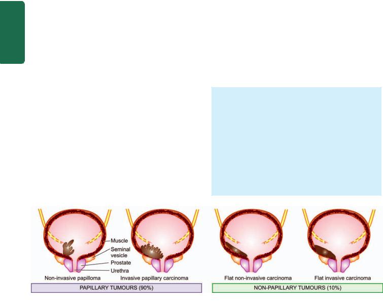
700 urethritis (Chapter 4). The pathologic changes are similar to inflammation of the lower urinary tract elsewhere but strictures are less common than following gonococcal infection of the urethra.
TUMOURS
Majority of lower urinary tract tumours are epithelial. Both benign and malignant tumours occur; the latter being more common. About 90% of malignant tumours of the lower urinary tract occur in the urinary bladder, 8% in the renal pelvis and remaining 2% are seen in the urethra or ureters.
|
TUMOURS OF THE BLADDER |
|
|
Epithelial (Urothelial) Bladder Tumours |
|
|
More than 90% of bladder tumours arise from transitional |
|
|
epithelial (urothelium) lining of the bladder in continuity |
|
|
with the epithelial lining of the renal pelvis, ureters, and the |
|
|
major part of the urethra. Though many workers consider |
|
|
all transitional cell tumours as transitional cell carcinoma, |
|
|
others distinguish true transitional cell papilloma from grade |
|
SECTION |
I transitional cell carcinoma. |
|
Bladder cancer comprises about 3% of all cancers. Most |
||
|
||
|
of the cases appear beyond 5th decade of life with 3-times |
|
|
higher preponderance in males than females. |
|
|
ETIOPATHOGENESIS. Urothelial tumours in the urinary |
|
III |
tract are typically multifocal and the pattern of disease |
|
becomes apparent over a period of years. A number of |
||
|
||
|
environmental and host factors are associated with increased |
|
Systemic |
risk of bladder cancer. These are as under: |
|
1. Industrial occupations. Workers in industries that |
||
|
||
|
produce aniline dyes, rubber, plastic, textiles, and cable have |
|
|
high incidence of bladder cancer. Bladder cancer may occur |
|
Pathology |
in workers in these factories after a prolonged exposure of |
|
2. Schistosomiasis. There is increased risk of bladder cancer, |
||
|
about 20 years. The carcinogenic substances responsible for |
|
|
bladder cancer in these cases are the metabolites of β- |
|
|
naphthylamine and benzene. |
|
|
particularly squamous cell carcinoma, in patients having |
|
|
bilharzial infestation (Schistosoma haematobium) of the |
|
|
bladder. Schistosomiasis is common in Egypt and accounts |
|
|
for high incidence of bladder cancer in that country. It is |
|
|
thought to induce local irritant effect and initiate squamous |
|
|
metaplasia followed by squamous cell carcinoma. |
3.Dietary factors. Certain carcinogenic metabolites of tryptophan are excreted in urine of patients with bladder cancer. These metabolites have been shown to induce bladder cancer in experimental animals. The role of artificial sweeteners like saccharin, coffee or caffeine and chronic alcoholism in the etiology of bladder cancer in man is controversial.
4.Local lesions. A number of local lesions in the bladder predispose to the development of bladder cancer. These include ectopia vesicae (extrophied bladder), vesical diverticulum, leukoplakia of the bladder mucosa and urinary diversion in defunctionalised bladder. All these conditions are associated with squamous metaplasia and high incidence of bladder cancer.
5.Smoking. Tobacco smoking is associated with 2 to 3 fold increased risk of developing bladder cancer, probably due to increased urinary excretion of carcinogenic substances.
6.Drugs. Immunosuppressive therapy with cyclophosphamide and patients having analgesic-abuse (phenacetin-) nephropathy have high risk of developing bladder cancer.
Multicentric nature of urothelial cancer and high rate of recurrence has led to the hypothesis that a field effect in the urothelium is responsible for this form of cancer. This is responsible for polychronotropism in bladder cancer i.e. the tumour tends to recur with time and develops at new locations within the urinary tract.
Several cytogentic abnormalities have been seen in bladder cancer. These include mutations in p53, RB gene and p21 gene, all of which are associated with higher rate of recurrences and metastasis.
MORPHOLOGIC FEATURES. Grossly, urothelial tumours may be single or multiple. About 90% of the tumours are papillary (non-invasive or invasive), whereas the remaining 10% are flat indurated (non-invasive or invasive) (Fig. 22.46). The papillary tumours have free floating fern-like arrangement with a broad or narrow pedicle. The non-papillary tumours are bulkier with ulcerated surface (Fig. 22.47). More common locations for either of the two types are the trigone, the region of ureteral orifices and on the lateral walls.
Histologically, urothelial tumours are of 3 cell types— transitional cell, squamous cell, and glandular
(Table 22.21).
Figure 22.46 
 Gross patterns of epithelial bladder tumours.
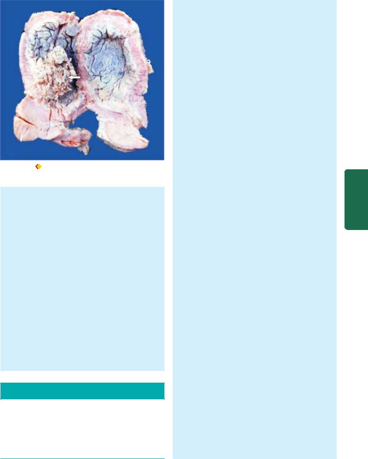
Figure 22.47 
 Carcinoma urinary bladder . The mucosal surface shows papillary tumour floating in the lumen (arrow).
A. TRANSITIONAL CELL TUMOURS. Approximately 90% of all epithelial tumours of the bladder are transitional cell tumours. As stated before, transitional cell papilloma is distinguished by some workers from grade I transitional cell carcinoma (TCC), whereas others do not consider this as a distinct entity.
1.Transitional cell papilloma. Papillomas may occur singly or may be multiple. They are generally small, less than 2 cm in diameter, papillary with branching pattern. Each papilla is composed of fibrovascular stromal core covered by normal-looking transitional cells having normal number of layers (upto 6-7) in thickness. The individual cells resemble the normal transitional cells and do not vary in size and shape. Mitoses are absent and basal polarity is retained. It must be emphasised that the designation transitional cell papilloma is purely a histological diagnosis but does not imply an innocent biologic behaviour. In fact, it may recur and behave in a malignant manner.
2.Carcinoma in situ (CIS). Foci of epithelial hyperplasia, dysplasia and carcinoma in situ are seen in other parts of the bladder in non-invasive as well as in invasive
TABLE 22.21: Histologic Classification of Epithelial (Urothelial) Tumours (Mostofi, 1960).
A.Transitional cell tumours (90%)
1.Transitional cell papilloma
2.Transitional cell carcinoma (grade I, II and III)
B.Carcinoma in situ
C.Squamous cell carcinoma (5%)
D.Adenocarcinoma (Rare)
E.Mixed carcinoma (5%)
carcinomas. Similar foci may be present in the ureters and renal pelvis. The malignant potential of epithelial hyperplasia and dysplasia is uncertain but carcinoma in situ is certainly precancerous and is currently included as grade 0 transitional cell carcinoma. Carcinoma in situ is characterised by anaplastic malignant cells confined to layers superficial to basement membrane of the bladder mucosa. These pathologic changes can be induced in experimental animals by chemical carcinogens. Therefore, it is reasonable to assume that these stages are precursors of invasive bladder cancer.
3. Transitional cell carcinoma (TCC). This is the commonest cancer of the bladder. The widely accepted classification of Mostofi (1960) adopted by the American Bladder Tumour Registry divides TCC into 3 grades. The basis for this grading depends upon 2 features: the degree
of anaplasia and the extent of invasion.
 The criteria for anaplasia are: increased cellularity, nuclear crowding, deranged cellular polarity, failure of normal orientation from base to the surface, variation in cell size and shape, variation in nuclear chromatin pattern, mitotic figures and giant cells.
 The criteria for invasion in papillary as well as nonpapillary tumours are: penetration of the basement membrane of bladder mucosa.
Based on these salient features, the characteristics of three grades of transitional cell carcinoma are as under:
Grade I: The tumour cells are clearly transitional type but show increased number of layers of cells (c.f. transitional cell papilloma). The individual cells are generally regular but are slightly larger and show mild hyperchromatism.
Grade II: The tumour cells are still recognisable as of transitional cell origin and the number of layers of cells is increased. The individual tumour cells are less regular, larger in size, and show pronounced nuclear hyperchromatism, mitotic activity and loss of polarity. The tumour may or may not be invasive (Fig.22.48).
Grade III: This is the anaplastic or undifferentiated grade of the tumour which is always invasive extending into the bladder wall to variable depth depending upon the clinical stage (described later). The tumour cells are no longer recognisable as of transitional origin. The individual tumour cells show pronounced features of anaplasia such as marked pleomorphism, hyperchromatism, total loss of polarity with loosened surface cells exfoliated in the bladder lumen.
There may be foci of squamous or glandular metaplasia in any grade of the tumour.
B. SQUAMOUS CELL CARCINOMA. Squamous cell carcinoma comprises about 5% of the bladder carcinomas. Unlike TCC which is mostly papillary and non-ulcerating, most squamous carcinomas of the bladder are sessile, nodular, infiltrating and ulcerating. Association of squamous carcinoma and schistosomiasis has already been highlighted. The carcinoma may be welldifferentiated with keratin pearl formation, or may be anaplastic.
701
Tract Urinary Lower and Kidney The 22 CHAPTER
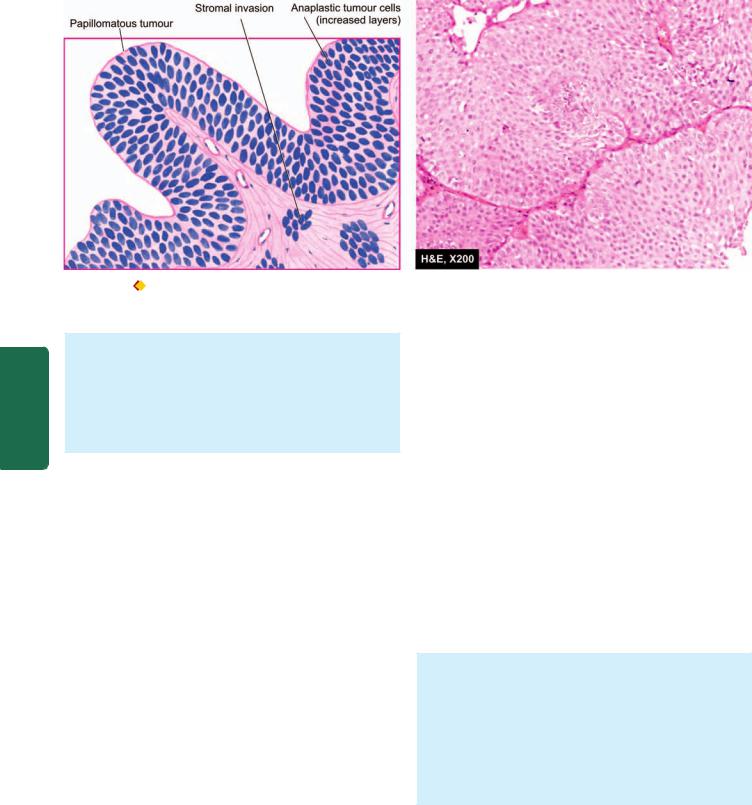
702
Pathology Systemic III SECTION
Figure 22.48 
 Transitional cell carcinoma, grade II. There is increase in the number of layers of epithelium. The cells are still recognisabl as of transitional origin and show features of anaplasia.
C. ADENOCARCINOMA. Adenocarcinoma of the bladder is rare. Adenocarcinoma has association with exostrophy of the bladder with glandular metaplasia, or may arise from urachal rests, periurethral and periprostatic glands, or from cystitis cystica. The tumour is characterised by glandular and tubular pattern with or without mucus production.
STAGING OF BLADDER CANCER. The clinical behaviour and prognosis of bladder cancer can be assessed by the following simple staging system:
Stage 0: Carcinoma confined to the mucosa.
Stage A: Carcinoma invades the lamina propria but not the muscularis.
Stage B1: Carcinoma invades the superficial muscle layer. Stage B2: Carcinoma invades the deep muscle layer. Stage C: Carcinoma invades the perivesical tissues.
Stage D1: Carcinoma shows regional metastases. Stage D2: Carcinoma shows distant metastases.
Non-epithelial Bladder Tumours
These may be benign or malignant.
BENIGN. The most common benign mesenchymal tumour of the bladder is leiomyoma. Other less common examples are neurofibroma, haemangioma and granular cell myoblastoma.
MALIGNANT. Rhabdomyosarcoma is the most frequent malignant mesenchymal tumour. It exists in 2 forms:
Adult form occurring in adults over 40 years of age and resembles the rhabdomyosarcoma of skeletal muscle.
Childhood form occurring in infancy and childhood and appears as large polypoid, soft, fleshy, grapelike mass
and is also called sarcoma botryoides or embryonal rhabdo-
myosarcoma. It is morphologically characterised by masses of embryonic mesenchyme consisting of masses of highly pleomorphic stellate cells in myxomatous background. Similar tumours occur in the female genital tract (Chapter 24).
TUMOURS OF RENAL PELVIS AND URETERS
Almost all the tumours of the renal pelvis and ureters are of epithelial origin. They are of the same types as are seen in the urinary bladder. However, tumours in the ureters are quite rare.
TUMOURS OF URETHRA
Tumours of the urethra are rare except for the urethral caruncle which is a tumour-like lesion.
URETHRAL CARUNCLE. Urethral caruncle is not uncommon. It is an inflammatory lesion present on external urethral meatus in elderly females.
Grossly, the caruncle appears as a solitary, 1 to 2 cm in diameter, pink or red mass, protruding from urethral meatus. It is quite friable and ulcerated.
Microscopically, the mass may be covered by squamous or transitional epithelium or there may be ulcerated surface. The underlying tissues show proliferating blood vessels, fibroblastic connective tissue and intense acute and chronic inflammatory infiltrate. Thus, the histologic appearance closely resembles a pyogenic granuloma.
URETHRAL CARCINOMA. Carcinoma of the urethra is uncommon. In most cases it occurs in the distal urethra near the external meatus and thus is commonly squamous cell carcinoma. Less often, there may be transitional cell carcinoma or adenocarcinoma arising from periurethral glands.

703
Chapter 23 |
The Male Reproductive System |
|
and Prostate |
||
|
TESTIS AND EPIDIDYMIS
NORMAL STRUCTURE
Contents of the scrotal sac include the testicle and epididymis along with lower end of the spermatic cord and the tunica vaginalis that forms the outer serous investing layer. The epididymis is attached to body of the testis posteriorly. Thus, the testicle and epididymis may be regarded as one organ.
Structurally, the main components of the testicle are the seminiferous tubules which when uncoiled are of considerable length.
Histologically, the seminiferous tubules are formed of a lamellar connective tissue membrane and contain several layers of cells (Fig. 23.1,A). In the adult, the cells lining the seminiferous tubules are of 2 types:
1.Spermatogonia or germ cells which produce spermatocytes (primary and secondary), spermatids and mature spermatozoa.
2.Sertoli cells which are larger and act as supportive cells to germ cells, produce mainly androgen (testosterone) and little oestrogen.
The seminiferous tubules drain into collecting ducts which form the rete testis from where the secretions pass into the vasa efferentia. Vasa efferentia opens at the upper end of the epididymis. The lower end of the epididymis is prolonged into a thick muscular tube, the vas deferens, that transports the secretions into the urethra.
The fibrovascular stroma present between the seminiferous tubules contains varying number of interstitial cells of Leydig. Leydig cells have abundant cytoplasm containing lipid granules and elongated Reinke’s crystals. These cells are the main source of testosterone and other androgenic hormones in males. Thus, Sertoli and Leydig cells are hormone-producing cells homologous to their ovarian counterparts (granulosa-theca cells) and are termed specialised stromal cells of the gonads.
Thus, the main functions of the testis are to produce sperms and testosterone.
CONGENITAL ANOMALIES
Cryptorchidism
Cryptorchidism or undescended testis is a condition in which the testicle is arrested at some point along its descent. Its incidence is about 0.2% in adult male population. In 70% of cases, the undescended testis lies in the inguinal ring, in 25% in the abdomen and, in the remaining 5%, it may be present
at other sites along its descent from intra-abdominal location to the scrotal sac.
ETIOLOGY. The exact etiology is not known in majority of cases. However, a few apparent causes associated with cryptorchidism are as under:
1.Mechanical factors e.g. short spermatic cord, narrow inguinal canal, adhesions to the peritoneum.
2.Genetic factors e.g. trisomy 13, maldevelopment of the scrotum or cremaster muscles.
3.Hormonal factors e.g. deficient androgenic secretions.
MORPHOLOGIC FEATURES. Cryptorchidism is unilateral in 80% cases and bilateral in the rest.
Grossly, the cryptorchid testis is small in size, firm and fibrotic.
Histologically, contrary to previous beliefs, the changes of atrophy begin to appear by about 2 years of age. These changes are as under (Fig. 23.1,B):
1.Seminiferous tubules: There is progressive loss of germ cell elements so that the tubules may be lined by only spermatogonia and spermatids but foci of spermatogenesis are discernible in 10% of cases. The tubular basement membrane is thickened. Advanced cases show hyalinised tubules with a few Sertoli cells only, surrounded by prominent basement membrane.
2.Interstitial stroma: There is usually increase in the interstitial fibrovascular stroma and conspicuous presence of Leydig cells, seen singly or in small clusters.
CLINICAL FEATURES. As such, cryptorchidism is completely asymptomatic and is discovered only on physical examination. However, if surgical correction by orchiopexy is not undertaken by about 2 years of age, or certainly in the prepubertal period, significant adverse clinical outcome may result as under:
1.Sterility-infertility. Bilateral cryptorchidism is associated with sterility while unilateral disease may result in infertility.
2.Inguinal hernia. A concomitant inguinal hernia is frequently present along with cryptorchidism.
3.Malignancy. Cryptorchid testis is at 30-50 times increased risk of developing testicular malignancy, most commonly seminoma and embryonal carcinoma, than a normally descended testis. The risk of malignancy is greater in intraabdominal testis than in testis in the inguinal canal for the simple reason that the neoplastic process in the testis in scrotal location is detected early than intra-abdominal site.
Prostate and System Reproductive Male The 23 CHAPTER
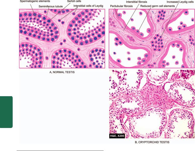
704
Pathology Systemic III SECTION
Figure 23.1 
 Microscopic appearance of normal testis (A) contrasted with that of cryptorchid testis (B).
Male Infertility
The morphologic pattern of testicular atrophy described above for cryptorchidism can result from various other causes of male infertility. These causes can be divided into 3 groups: pre-testicular, testicular and post-testicular.
A. PRE-TESTICULAR CAUSES:
1.Hypopituitarism. Pre-pubertal or post-pubertal hypopituitarism such as from tumour, trauma, infarction, cyst and genetic deficiency of FSH and LH secretion.
2.Oestrogen excess. Endogenous excess such as from hepatic cirrhosis, adrenal tumour, Sertoli and Leydig cell tumour; or exogenous excess such as in the treatment of carcinoma of the prostate.
3.Glucocorticoid excess. Endogenous excess may occur in Cushing’s syndrome while exogenous excess may occur in the treatment of ulcerative colitis, bronchial asthma, rheumatoid arthritis etc.
4.Other endocrine disorders. Hypothyroidism and diabetes mellitus are associated with hypospermatogenesis.
B. TESTICULAR CAUSES:
1.Agonadism i.e. total absence of the testes.
2.Cryptorchidism or undescended testis described above.
3.Maturation arrest i.e. failure of spermatogenesis beyond one of the immature stages.
4.Hypospermatogenesis i.e. presence of all the maturation stages of spermatogenesis but in decreased number.
5.Sertoli cell-only syndrome. Congenital or acquired absence of all germ cells so that the seminiferous tubules are lined by Sertoli cells only.
6.Klinefelter’s syndrome. An XXY intersexuality characterised by primary hypogonadism, azoospermia, gynaecomastia, eunuchoid built and subnormal intelligence.
7.Mumps orchitis occurring as a complication of parotitis (Chapter 19).
8.Irradiation damage resulting in permanent germ cell destruction.
C. POST-TESTICULAR CAUSES:
1. Congenital block e.g. absence or atresia of vas deferens.
2.Acquired block e.g. due to gonorrhoea and surgical intervention.
3.Impaired sperm motility in the presence of normal sperm counts e.g. immotile cilia syndrome (Chapter 17).
INFLAMMATIONS
Inflammation of the testis is termed as orchitis and of epididymis is called as epididymitis; the latter being more common. A combination epididymo-orchitis may also occur. A few important types are described below.
Non-specific Epididymitis and Orchitis
Non-specific epididymitis and orchitis, or their combination, may be acute or chronic. The common routes of spread of infection are via the vas deferens, or via lymphatic and haematogenous routes. Most frequently, the infection is caused by urethritis, cystitis, prostatitis and seminal vesiculitis. Other causes are mumps, smallpox, dengue fever, influenza, pneumonia and filariasis. The common infecting organisms in sexually-active men under 35 years of age are
Neisseria gonorrhoeae and Chlamydia trachomatis, whereas in older individuals the common organisms are urinary tract pathogens like Escherichia coli and Pseudomonas.
MORPHOLOGIC FEATURES. Grossly, in acute stage the testicle is firm, tense, swollen and congested. There may be multiple abscesses, especially in gonorrhoeal infection. In chronic cases, there is usually variable degree of atrophy and fibrosis.
Histologically, acute orchitis and epididymitis are characterised by congestion, oedema and diffuse infiltration by neutrophils, lymphocytes, plasma cells and macrophages or formation of neutrophilic abscesses. Acute inflammation may resolve, or may progress to chronic form. In chronic epididymo-orchitis, there is focal or diffuse chronic inflammation, disappearance of seminiferous tubules, fibrous scarring and destruction of interstitial Leydig cells. Such cases usually result in permanent sterility.
Granulomatous (Autoimmune) Orchitis
Non-tuberculous granulomatous orchitis is a peculiar type of unilateral, painless testicular enlargement in middle-aged men that may resemble a testicular tumour clinically. The exact etiology and pathogenesis of the condition are not known though an autoimmune basis is suspected.
MORPHOLOGIC FEATURES. Grossly, the affected testis is enlarged with thickened tunica. Cut section of the testicle is greyish-white to tan-brown.
Histologically, there are circumscribed non-caseating granulomas lying within the seminiferous tubules. These granulomas are composed of epithelioid cells, lymphocytes, plasma cells, some neutrophils and multinucleate giant cells. The origin of the epithelioid cells is from Sertoli cells lining the tubules. The tubules show peritubular fibrosis which merges into the interstitial tissue that is infiltrated by lymphocytes and plasma cells.
Tuberculous Epididymo-orchitis |
705 |
Tuberculosis invariably begins in the epididymis and spreads to involve the testis. Tuberculous epididymo-orchitis is generally secondary tuberculosis from elsewhere in the body. It may occur either by direct spread from genitourinary tuberculosis such as tuberculous seminal vesiculitis, prostatitis and renal tuberculosis, or may reach by haematogenous spread of infection such as from tuberculosis of the lungs. Primary genital tuberculosis may occur rarely.
MORPHOLOGIC FEATURES. Grossly, discrete, |
|
|
|
yellowish, caseous necrotic areas are seen. |
|
|
|
Microscopically, numerous tubercles which may coalesce |
|
|
|
to form large caseous mass are seen. Characteristics of |
|
|
|
typical tubercles such as epithelioid cells, peripheral |
|
|
|
mantle of lymphocytes, occasional multinucleate giant |
|
|
|
cells and central areas of caseation necrosis are seen |
|
|
|
(Fig. 23.2). Numerous acid-fast bacilli can be |
|
|
|
demonstrated by Ziehl-Neelsen staining. The lesions |
|
|
|
produce extensive destruction of the epididymis and may |
|
|
|
form chronic discharging sinuses on the scrotal skin. In |
|
|
|
late stage, the lesions heal by fibrous scarring and may |
|
CHAPTER |
|
undergo calcification. |
|
||
|
|
||
Spermatic Granuloma |
|
||
Spermatic granuloma is the term used for development of |
|
||
inflammatory lesions due to invasion of spermatozoa into |
23 |
||
the stroma. Spermatic granuloma may develop due to |
|||
|
|||
trauma, inflammation and loss of ligature following |
|
||
vasectomy. |
The |
||
|
|
||
MORPHOLOGIC FEATURES. Grossly, the sperm granu- |
|
||
loma is a small nodule, 3 mm to 3 cm in diameter, firm, |
|
Prostate and System Reproductive Male |
|
white to yellowish-brown. |
|
||
|
|
||
|
|
||
Figure 23.2 
 Tuberculous epididymo-orchitis. The interstitium contains several epithelioid cell granulomas with central areas of caseation necrosis. These granulomas are surrounded by Langhans’ giant cells and mantle of lymphocytes.
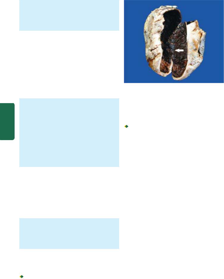
706
Pathology Systemic III SECTION
Histologically, it consists of a granuloma composed of histiocytes, epithelioid cells, lymphocytes and some neutrophils. Characteristically, the centre of spermatic granuloma contains spermatozoa and necrotic debris. The late lesions have fibroblastic proliferation at the periphery and hyalinisation.
Elephantiasis
Elephantiasis is enormous thickening of the scrotal skin resembling the elephant’s hide and results in enlargement of the scrotum. The condition results from filariasis in which the adult worm lives in the lymphatics, while the larvae travel in the blood. The most important variety of filaria is Wuchereria bancrofti. The condition is common in all tropical countries. The vector is generally the Culex mosquito. The patients may remain asymptomatic or may manifest with fever, local pain, swelling, rash, tender lymphadenopathy and blood eosinophilia. An asthma-like respiratory complaint may develop in some cases.
MORPHOLOGIC FEATURES. Grossly, the affected leg and scrotum are enormously thickened with enlargement of regional lymph nodes. The affected area of skin may show dilated dermal lymphatics and varicosities.
Histologically, the changes begin with lymphatic obstruction by the adult worms. The worm in alive, dead or calcified form may be found in the dilated lymphatics or in the lymph nodes. Dead or calcified worm in lymphatics is usually followed by lymphangitis with intense infiltration by eosinophils. Sometimes, granulomatous reaction may be evident. In advanced cases, chronic lymphoedema with tough subcutaneous fibrosis and epidermal hyperkeratosis develops which is termed elephantiasis.
MISCELLANEOUS LESIONS
Torsion of Testis
Torsion of the testicle may occur either in a fully-descended testis or in an undescended testis. The latter is more common and more severe. It results from sudden cessation of venous drainage and arterial supply to the testis, usually following sudden muscular effort or physical trauma. Torsion is common in boys and young men.
MORPHOLOGIC FEATURES. The pathologic changes vary depending upon the duration and severity of vascular occlusion. There may be coagulative necrosis of the testis and epididymis, or there may be haemorrhagic infarction. The inflammatory reaction is generally not so pronounced.
Varicocele
Varicocele is the dilatation, elongation and tortuosity of the veins of the pampiniform plexus in the spermatic cord. It is of 2 types: primary (idiopathic) and secondary.

 Primary or idiopathic form is more frequent and is common in young unmarried men. It is nearly always on
Figure 23.3 
 Haematocele testis. Sectioned surface of the sac shows thick wall coated internally by brownish, tan and necrotic material which is organised blood clot (arrow).
the left side as the loaded rectum presses the left vein. Besides, the left spermatic vein enters the renal vein at right angles while the right spermatic vein enters the vena cava obliquely.

 Secondary form occurs due to pressure on the spermatic vein by enlarged liver, spleen or kidney. It is commoner in middle-aged people.
Hydrocele
A hydrocele is abnormal collection of serous fluid in the tunica vaginalis. It may be acute or chronic, congenital or acquired. The usual causes are trauma, systemic oedema such as in cardiac failure and renal disease, and as a complication of gonorrhoea, syphilis and tuberculosis.
The hydrocele fluid is generally clear and straw-coloured but may be slightly turbid or haemorrhagic. The hydrocele sac may have single loculus or may have multiple loculi. The wall of the hydrocele sac is composed of fibrous tissue infiltrated with lymphocytes and plasma cells.
Haematocele
Haematocele is haemorrhage into the sac of the tunica vaginalis. It may result from direct trauma, from injury to a vein by the needle, or from haemorrhagic diseases.
In recent haematocele, the blood coagulates and the wall is coated with ragged deposits of fibrin. In long-standing cases, the tunica vaginalis is thickened with dense fibrous tissue and occasionally may get partly calcified (Fig. 23.3).
TESTICULAR TUMOURS
Testicular tumours are the cause of about 1% of all cancer deaths. They are more frequent in white male population but are less common in Africans and Asians. They have trimodal age distribution—a peak during infancy, another during late adolescence and early adulthood, and a third peak after 60 years of age.

TABLE 23.1: Classification of Testicular Tumours.
I. GERM CELL TUMOURS
1.Seminoma
2.Spermatocytic seminoma
3.Embryonal carcinoma
4.Yolk sac tumour (Syn. endodermal sinus tumour, orchioblastoma, infantile type embryonal carcinoma)
5.Polyembryoma
6.Choriocarcinoma
7.Teratomas
(i)Mature
(ii)Immature
(iii)With malignant transformation
8.Mixed germ cell tumours
II.SEX CORD-STROMAL TUMOURS
1.Leydig cell tumour
2.Sertoli cell tumour (Androblastoma)
3.Granulosa cell tumour
4.Mixed forms
III.COMBINED GERM CELL-SEX CORD-STROMAL TUMOURS Gonadoblastoma
IV. OTHER TUMOURS
1.Malignant lymphoma (5%)
2.Rare tumours
CLASSIFICATION
The most widely accepted classification is the histogenetic classification proposed by the World Health Organisation (Table 23.1). Based on this, all testicular tumours are divided into 3 groups: germ cell tumours, sex cord-stromal tumours and mixed forms. Vast majority of the testicular tumours (95%) arise from germ cells or their precursors in the seminiferous tubules, while less than 5% originate from sex cord-stromal components of the testis. From clinical point of view, germ cell tumours of the testis are categorised into 2 main groups— seminomatous and non-seminomatous which need to be distinguished (Table 23.2).
ETIOLOGIC FACTORS
Exact etiology of testicular germ cell tumours is unknown, but the following factors have been implicated:
1. Cryptorchidism. The probability of a germ cell tumour developing in an undescended testis is 30-50 times greater than in a normally-descended testis. About 10% of testicular
germ cell tumours are associated with cryptorchidism. The high incidence is attributed to higher temperature to which the undescended testis in the groin or abdomen is exposed. Intra-abdominal testis is at greater risk than the inguinal testis. There is increased incidence of tumour in the contralateral normally-descended testis. There are no data to confirm or deny whether surgical repositioning or orchiopexy of a cryptorchid testis alters the incidence of testicular tumour. However, surgical correction is still helpful since it is easier to detect the tumour in scrotal testis than in an abdominal or inguinal testis.
2.Other developmental disorders. Dysgenetic gonads associated with endocrine abnormalities such as androgen insensitivity syndrome have higher incidence of development of germ cell tumours.
3.Genetic factors. Genetic factors play a role in the development of germ cell tumours supported by the observation of high incidence in first-degree family members, twins and in white male populations while blacks in Africa have a very low incidence. However, no definite pattern of inheritance has been recognised.
4.Other factors. A few less common factors include the following:
i)Orchitis. A history of mumps or other forms of orchitis may be given by the patient with germ cell tumour.
ii)Trauma. Many patients give a history of trauma prior to the development of the tumour but it is not certain how trauma initiates the neoplastic process. Instead, possibly it brings the patient to attention of the physician.
iii)Carcinogens. A number of carcinogens such as use of certain drugs (e.g. LSD, hormonal therapy for sterility, copper, zinc etc), exposure to radiation and endocrine abnormalities may play a role in the development of testicular tumours.
HISTOGENESIS
Pathogenesis of testicular tumours remains controversial except that vast majority of these tumours originate from germ cells. Based on current concepts on histogenesis of testicular tumours, following agreements and disagreements have emerged (Fig. 23.4):
1. Developmental disorders. Disorders such as cryptorchidism, gonadal dysgenesis and androgen insensitivity syndrome are high risk factors for development of testicular germ cell tumours. These observations point to developmental defect in gonadogenesis.
TABLE 23.2: Distinguishing Features of Seminomatous (SGCT) and Non-seminomatous (NSGCT) Germ Cell Tumours of Testis.
|
Feature |
SGCT |
NSGCT |
1. |
Primary tumour |
Larger, confined to testis for sufficient time; |
Smaller, at times indistinct; |
|
|
testicular contour retained |
testicular contour may be distorted |
2. |
Metastasis |
Generally to regional lymph nodes |
Haematogenous spread early |
3. |
Response to radiation |
Radiosensitive |
Radioresistant |
4. |
Serum markers |
hCG; generally low levels |
hCG, AFP, or both; high levels |
5. |
Prognosis |
Better |
Poor |
|
|
|
|
707
Prostate and System Reproductive Male The 23 CHAPTER
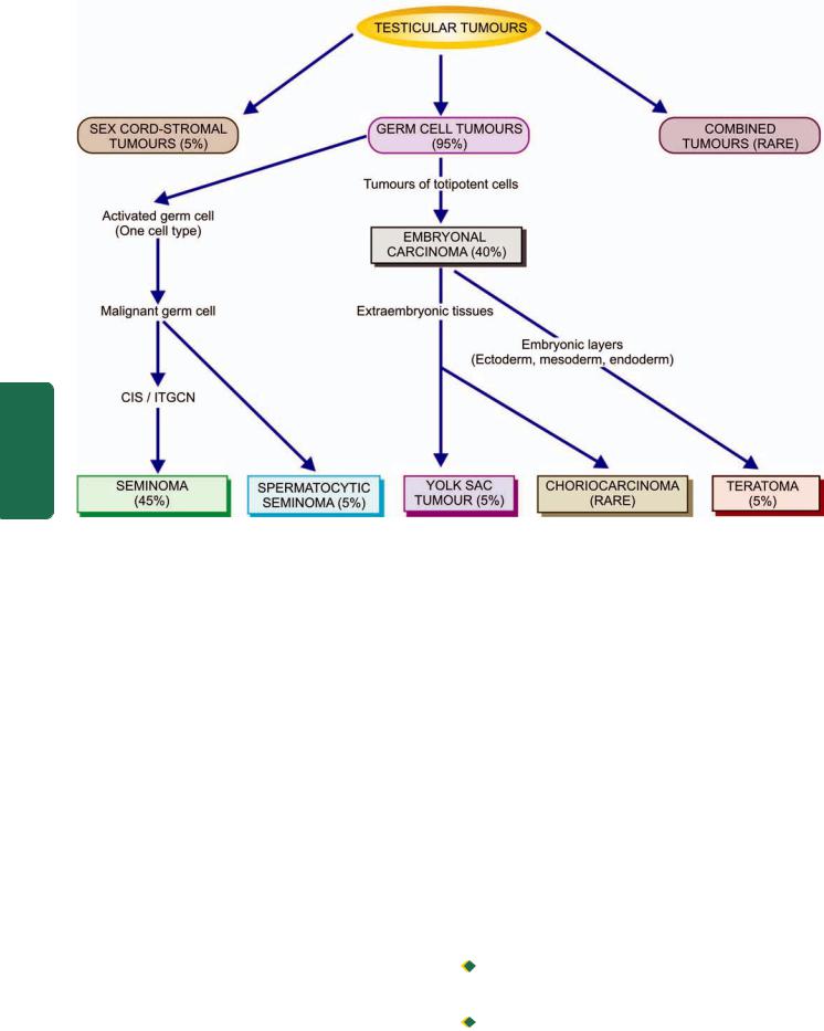
708
III SECTION
Pathology Systemic
Figure 23.4 
 Schematic diagram showing histogenesis of testicular tumours.
2.Molecular genetic features. Testicular germ cell tumours have been found to have several genetic abnormalities suggesting a common molecular pathogenesis of all germ cell tumours:
i) Hyperdiploidy is almost a constant feature of all germ cell tumours of the testis.
ii) In more than 90% of testicular germ cell tumours irrespective of histologic type (as also ovarian germ cell tumours), an isochromosome of short arm of chromosome 12, abbreviated as i(12p), is found.
iii) Similarly, deletion of long arm of chromosome 12 abbreviated as del(12q), is present.
iv) Telomerase activity is present in all germ cell tumours of the testis.
v) Other mutations include p53, cyclin E and FAS gene.
3.CIS/ITGCN. A preinvasive stage of carcinoma in situ
(CIS) termed intratubular germ cell neoplasia (ITGCN) generally precedes the development of most of the invasive testicular germ cell tumours in adults. CIS originates from spermatogenic elements. Areas of CIS are found in seminiferous tubules adjacent to most seminomas, embryonal carcinomas and other mixed germ cell tumours. However, CIS is not found in seminiferous tubules adjoining yolk sac carcinoma of childhood, benign teratoma in children and adolescents, and spermatocytic seminoma indicating their different pathogenetic mechanisms.
4. ‘Three hit’ process. Germ cells in seminiferous tubules undergo activation (‘first hit’) before undergoing malignant transformation confined to seminiferous tubules (CIS) (‘second hit’) and eventually into invasive stage by some epigenetic phenomena (‘third hit’). Though this sequential tumorigenesis explains the development of seminomatous tumours, it is yet not clear whether non-seminomatous germ cell tumours develop directly or through intermediate stage.
CLINICAL FEATURES AND DIAGNOSIS
The usual presenting clinical symptoms of testicular tumours are gradual gonadal enlargement and a dragging sensation in the testis. Metastatic involvement may produce secondary symptoms such as pain, lymphadenopathy, haemoptysis and urinary obstruction.
SPREAD. Since testicular germ cell tumours originate from totipotent germ cells, it is not unusual to find metastases of histologic types different from the primary growth. Testicular tumours may spread by both lymphatic and haematogenous routes:
 Lymphatic spread occurs to retroperitoneal para-aortic lymph nodes, mediastinal lymph nodes and supraclavicular lymph nodes.

 Haematogenous spread primarily occurs to the lungs, liver, brain and bones.
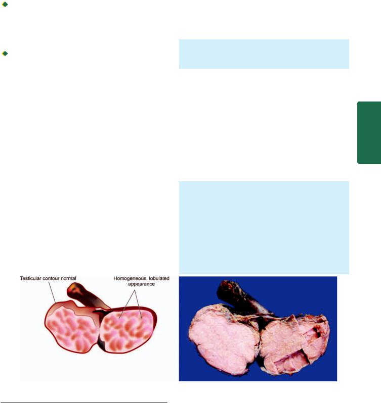
TUMOUR MARKERS. Germ cell tumours of the testis secrete polypeptide hormones and certain enzymes which can be detected in the blood. Two tumour markers widely used in the diagnosis, staging and monitoring the follow-up of patients with testicular tumours are: human chorionic gonadotropin (hCG) and alpha-foetoprotein (AFP). In addition, carcinoembryonic antigen (CEA), human placental lactogen (HPL), placental alkaline phosphatase, testosterone, oestrogen and luteinising hormone may also be elevated.
 hCG is synthesised by placental syncytio-trophoblast such as in various non-seminomatous germ cell tumours of the testis (e.g. in choriocarcinoma, yolk sac tumour and embryonal carcinoma). However, ectopic hCG production may occur in a variety of non-testicular non-germ cell tumours as well.

 AFP is normally synthesised by the foetal liver cells, yolk sac and foetal gut. Its levels are elevated in testicular tumours associated with yolk sac components. However, elevated serum AFP levels are also found in liver cell carcinoma.
PROGNOSIS. For selecting post-orchiectomy treatment (radiation, surgery, chemotherapy or all the three) and for monitoring prognosis, 3 clinical stages are defined:
Stage I: tumour confined to the testis.
Stage II: distant spread confined to retroperitoneal lymph nodes below the diaphragm.
Stage III: distant metastases beyond the retroperitoneal lymph nodes.
Seminomas tend to remain localised to the testis (stage I) while non-seminomatous germ cell tumours more often present with advanced clinical disease (stage II and III). Seminomas are extremely radiosensitive while nonseminomatous germ cell tumours are radio-resistant. In general, seminomas have a better prognosis with 90% cure rate while the non-seminomatous tumours behave in a more aggressive manner and have poor prognosis.
After these general comments, specific testicular tumours are as described below.
GERM CELL TUMOURS
Germ cell tumours comprise approximately 95% of all testicular tumours and are more frequent before the age of
45 years. Testicular germ cell tumours are almost always malignant. Nearly half of them contain more than one histologic type. Germ cell tumours are also found at the extragonadal sites such as the retroperitoneum and mediastinum, besides their counterparts in the female gonads (page 745).
Intratubular Germ Cell Neoplasia
The term intratubular germ cell neoplasia (ITGCN) is used to describe the preinvasive stage of germ cell tumours, notably intratubular seminoma and intratubular embryonal carcinoma. Others have used carcinoma in situ (CIS) stage of germ cell tumours as synonymous term.
Histologically, the malignant atypical tumour cells are restricted to the seminiferous tubules without evident invasion into the interstitium.
Classic Seminoma
Seminoma is the commonest malignant tumour of the testis and corresponds to dysgerminoma in the female (page 747). It constitutes about 45% of all germ cell tumours, and in another 15% comprises the major component of mixed germ cell tumour. Seminoma is divided into 2 main categories: classic and spermatocytic. Classic seminoma comprises about 93% of all seminomas and has a peak incidence in the 4th decade of life and is rare before puberty. Undescended testis harbours seminoma more frequently as compared to other germ cell tumours. About 10% pure seminomas are associated with elevated hCG levels in serum.
MORPHOLOGIC FEATURES. Grossly, the involved testis is enlarged up to 10 times its normal size but tends to maintain its normal contour since the tumour rarely invades the tunica. The larger tumour replaces the entire testis, whereas the smaller tumour appears as circumscribed mass in the testis. Cut section of the affected testis shows homogeneous, grey-white lobulated appearance (Fig. 23.5). Necrosis and haemorrhage in the tumour are rare.
Microscopically, the tumour has the following characteristics (Fig. 23.6):
709
Prostate and System Reproductive Male The 23 CHAPTER
Figure 23.5 
 Seminoma testis. The testis is enlarged but without distorting its contour. Sectioned surface shows replacement of the entire testis by lobulated, homogeneous, grey-white mass.
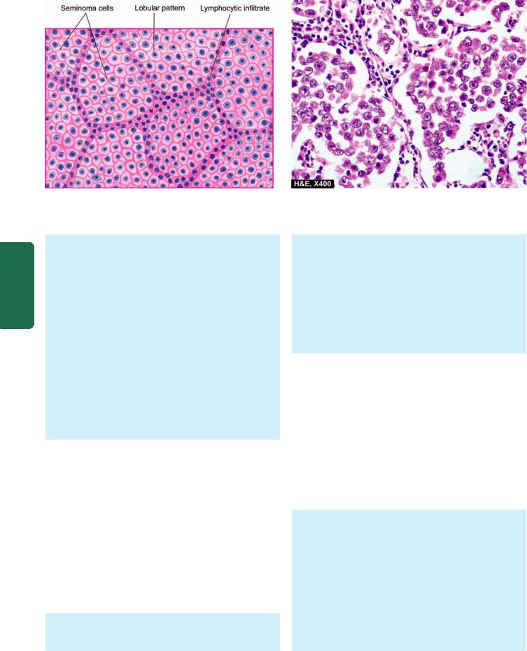
710
Pathology Systemic III SECTION
Figure 23.6 
 Seminoma testis. Microscopy of the tumour shows lobules of monomorphic seminoma cells separated by delicate fibrous stroma containing lymphocytic infiltration.
1. Tumour cells. The seminoma cells generally lie in cords, sheets or columns forming lobules. Typically, in a classic seminoma, the tumour cells are fairly uniform in size with clear cytoplasm and well-defined cell borders. The cytoplasm contains variable amount of glycogen that stains positively with PAS reaction. The nuclei are centrally located, large, hyperchromatic and usually contain 1-2 prominent nucleoli. Tumour giant cells may be present. Mitotic figures are infrequent. However, about 10% of seminomas have increased mitotic activity and have aggressive behaviour and are categorised as anaplastic
seminomas.
2. Stroma. The stroma of seminoma is delicate fibrous tissue which divides the tumour into lobules. The stroma shows a characteristic lymphocytic infiltration, indicative of immunologic response of the host to the tumour. About 20% of the tumours show granulomatous reaction in the stroma.
The prognosis of classic seminoma is better than other germ cell tumours. The tumour is highly radiosensitive. Natural history of anaplastic seminoma, however, is unclear—perhaps it represents an advanced stage of classic seminoma with a more aggressive behaviour.
Spermatocytic Seminoma
Spermatocytic seminoma is both clinically and morphologically a distinctive tumour from classic seminoma and is, therefore, classified separately in the WHO classification. It is an uncommon tumour having an incidence of about 5% of all germ cell tumours. Spermatocytic seminoma usually occurs in older patients, generally in 6th decade of life. The tumour is bilateral in 10% of patients.
MORPHOLOGIC FEATURES. Grossly, spermatocytic seminoma is homogeneous, larger, softer and more yellowish and gelatinous than the classic seminoma.
Histologically, the distinctive features are as under:
1.Tumour cells. The tumour cells vary considerably in size from lymphocyte-like to huge mononucleate or multinucleate giant cells. Majority of the tumour cells are, however, of intermediate size. The cells have eosinophilic cytoplasm devoid of glycogen. The nuclei of intermediate and large cells have filamentous pattern. Mitoses are often frequent.
2.Stroma. The stroma lacks lymphocytic and granulomatous reaction seen in classic seminoma.
The prognosis of spermatocytic seminoma is excellent compared and better than classic seminoma since the tumour is slow-growing and rarely metastasises. The tumour is radiosensitive.
Embryonal Carcinoma
Pure embryonal carcinoma constitutes 30% of germ cell tumours but areas of embryonal carcinoma are present in 40% of germ cell tumours. These tumours are more common in 2nd to 3rd decades of life. About 90% cases are associated with elevation of AFP or hCG or both. They are more aggressive than the seminomas.
MORPHOLOGIC FEATURES. Grossly, embryonal carcinoma is usually a small tumour in the testis. It distorts the contour of the testis as it frequently invades the tunica and the epididymis. Cut surface of the tumour is greywhite, soft with areas of haemorrhages and necrosis.
Microscopically, the following features are seen:
1.The tumour cells are arranged in a variety of patterns— glandular, tubular, papillary and solid.
2.The tumour cells are highly anaplastic carcinomatous cells having large size, indistinct cell borders, amphophilic cytoplasm and prominent hyperchromatic nuclei showing considerable variation in nuclear size. Mitotic figures and
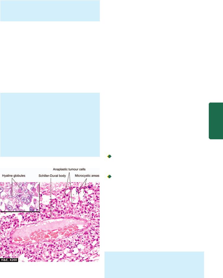
tumour giant cells are frequently present. Haemorrhage and necrosis are common.
3. The stroma is not as distinct as in seminoma and may contain variable amount of primitive mesenchyme.
Embryonal carcinoma is more aggressive and less radiosensitive than seminoma. Chemotherapy is more effective in treating this tumour.
Yolk Sac Tumour
(Synonyms: Endodermal Sinus Tumour,
Orchioblastoma, Infantile Embryonal Carcinoma)
This characteristic tumour is the most common testicular tumour of infants and young children upto the age of 4 years. In adults, however, yolk sac tumour in pure form is rare but may be present as the major component in 40% of germ cell tumours. AFP levels are elevated in 100% cases of yolk sac tumours.
MORPHOLOGIC FEATURES. Grossly, the tumour is generally soft, yellow-white, mucoid with areas of necrosis and haemorrhages.
Microscopically, yolk sac tumour has the following features (Fig. 23.7):
1.The tumour cells form a variety of patterns—loose reticular network, papillary, tubular and solid arrangement.
2.The tumour cells are flattened to cuboid epithelial cells with clear vacuolated cytoplasm.
3.The tumour cells may form distinctive perivascular structures resembling the yolk sac or endodermal sinuses of the rat placenta called Schiller-Duval bodies.
Figure 23.7 
 Yolk sac tumour testis. The tumour has microcystic pattern and has highly anaplastic tumour cells. Several characteristic Schiller-Duval bodies are present. Inset shows intraand extracellular hyaline globules.
4. There may be presence of both intracellular and |
|
711 |
|
|
|
extracellular PAS-positive hyaline globules, many of which |
|
|
contain AFP. |
|
|
|
|
|
Polyembryoma |
|
|
Polyembryoma is defined as a tumour composed predo- |
|
|
minantly of embryoid bodies. Embryoid bodies are structures |
|
|
containing a disc and cavities surrounded by loose |
|
|
mesenchyme simulating an embryo of about 2 weeks’ |
|
|
gestation. Polyembryoma is extremely rare but embryoid |
|
|
bodies may be present with embryonal carcinoma and |
|
|
teratoma. |
|
|
Choriocarcinoma |
|
||
Pure choriocarcinoma is a highly malignant tumour compo- |
|
||
sed of elements consisting of syncytiotrophoblast and |
|
||
cytotrophoblast. |
|
||
However, pure form is extremely rare and occurs more |
|
||
often in combination with other germ cell tumours. The |
|
||
patients are generally in their 2nd decade of life. The primary |
|
||
tumour is usually small and the patient may manifest initially |
CHAPTER |
||
usually small and may appear as a soft, haemorrhagic and |
|||
with symptoms of metastasis. The serum and urinary levels |
|
||
of hCG are greatly elevated in 100% cases. |
|
||
MORPHOLOGIC FEATURES. Grossly, the tumour is |
|
|
|
necrotic mass. |
|
23 |
|
Microscopically, the characteristic feature is the |
|
||
|
|
||
identification of intimately related syncytiotrophoblast |
|
|
|
and cytotrophoblast without formation of definite |
|
The |
|
placental-type villi. |
|
||
Syncytiotrophoblastic cells are large with many irregular |
|
Male |
|
|
|
||
and bizarre nuclei and abundant eosinophilic vacuolated |
|
|
|
cytoplasm which stains positively for hCG. These cells |
|
Reproductive |
|
often surround masses of cytotrophoblastic cells. |
|
||
|
|
||
Cytotrophoblastic cells are polyhedral cells which are |
|
|
|
more regular and have clear or eosinophilic cytoplasm |
|
|
|
with hyperchromatic nuclei. |
|
|
|
|
|
|
|
Teratoma |
System |
||
Teratomas are complex tumours composed of tissues derived |
|||
from more than one of the three germ cell layers—endoderm, |
|||
mesoderm and ectoderm. Testicular teratomas are more |
|||
common in infants and children and constitute about 40% |
and |
||
of testicular tumours in infants, whereas in adults they |
|||
comprise 5% of all germ cell tumours. However, teratomas |
Prostate |
||
are found in combination with other germ cell tumours (most |
|||
|
|||
commonly with embryonal carcinoma) in about 45% of |
|
||
mixed germ cell tumours. About half the teratomas have |
|
||
elevated hCG or AFP levels or both. |
|
||
MORPHOLOGIC FEATURES. Testicular teratomas are classified into 3 types:
1.Mature (differentiated) teratoma
2.Immature teratoma
3.Teratoma with malignant transformation.
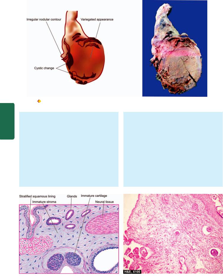
712
Pathology Systemic III SECTION
Figure 23.8 |
Immature teratoma testis. The testis is enlarged and nodular distorting the testicular contour. Sectioned surface shows replacement |
of the entire testis by variegated mass having grey-white solid areas, cystic areas, honey-combed areas and foci of cartilage and bone.
Grossly, most teratomas are large, grey-white masses enlarging the involved testis. Cut surface shows characteristic variegated appearance—grey-white solid areas, cystic and honey-combed areas, and foci of cartilage and bone (Fig. 23.8). Dermoid tumours commonly seen in the ovaries are rare in testicular teratomas.
Microscopically, the three categories of teratomas show different appearances:
1. Mature (differentiated) teratoma. Mature teratoma is composed of disorderly mixture of a variety of welldifferentiated structures such as cartilage, smooth muscle, intestinal and respiratory epithelium, mucus glands, cysts lined by squamous and transitional epithelium, neural tissue, fat and bone. This type of mature or differentiated teratoma is the most common, seen more frequently in
infants and children and has favourable prognosis. However, similar mature and benign-appearing tumour in adults is invariably associated with small hidden foci of immature elements so that their clinical course in adults is unpredictable. It is believed that all testicular teratomas
in the adults are malignant.
As mentioned above, dermoid cysts similar to those of the ovary are rare in the testis.
2. Immature teratoma. Immature teratoma is composed of incompletely differentiated and primitive or embryonic tissues along with some mature elements (Fig. 23.9). Primitive or embryonic tissue commonly present are poorly-formed cartilage, mesenchyme, neural tissues, abortive eye, intestinal and respiratory tissue elements etc. Mitoses are usually frequent.
Figure 23.9 
 Immature teratoma testis. Microscopy shows a variety of incompletely differentiated tissue elements.

3. Teratoma with malignant transformation. This is an extremely rare form of teratoma in which one or more of the tissue elements show malignant transformation. Such malignant change resembles morphologically with typical malignancies in other organs and tissues and commonly includes rhabdomyosarcoma, squamous cell carcinoma and adenocarcinoma.
Mixed Germ Cell Tumours
About 60% of germ cell tumours have more than one of the above histologic types (except spermatocytic seminoma) and are called mixed germ cell tumours. The clinical behaviour of these tumours is worsened by inclusion of more aggressive tumour component in a less malignant tumour. Interestingly, metastases of the mixed germ cell tumours may not exactly reproduce the histologic types present in the primary tumour.
The most common combinations of mixed germ cell tumours are as under:
1.Teratoma, embryonal carcinoma, yolk sac tumour and syncytiotrophoblast.
2.Embryonal carcinoma and teratoma (teratocarcinoma).
3.Seminoma and embryonal carcinoma.
SEX CORD-STROMAL TUMOURS
Tumours arising from specialised gonadal stroma are classified on the basis of histogenesis. The primitive mesenchyme which forms the specialised stroma of gonads in either sex gives rise to theca, granulosa and lutein cells in the female, and Sertoli and interstitial Leydig cells in the male. Since the cell of origin of primitive mesenchyme is identical, Sertoli and interstitial Leydig cell tumours may occur in the ovaries (in addition to theca cell, granulosa cell and lutein cell tumours). Likewise, the latter three tumours may occur in the testis (in addition to Sertoli cell and Leydig cell tumours). All these tumours secrete various hormones. The biologic behaviour of these tumours generally cannot be determined on histological grounds alone but is related to clinical parameters and hormonal elaboration by these tumours.
Leydig (Interstitial) Cell Tumour
Leydig cell tumours are quite uncommon. They may occur at any age but are more frequent in the age group of 20 to 50 years. Characteristically, these cells secrete androgen, or both androgen and oestrogen, and rarely corticosteroids. Bilateral tumours may occur typically in congenital adrenogenital syndrome.
MORPHOLOGIC FEATURES. Grossly, the tumour appears as a small, well-demarcated and lobulated nodule. Cut surface is homogeneously yellowish or brown.
Histologically, the tumour is composed of sheets and cords of normal-looking Leydig cells. These cells contain abundant eosinophilic cytoplasm and Reinke’s crystals and a small central nucleus.
Most of Leydig cell tumours are benign. Only about 10% may invade and metastasise.
Sertoli Cell Tumours (Androblastoma) |
713 |
Sertoli cell tumours correspond to arrhenoblastoma of the ovary. They may occur at all ages but are more frequent in infants and children. These tumours may elaborate oestrogen or androgen and may account for gynaecomastia in an adult, or precocious sexual development in a child.
MORPHOLOGIC FEATURES. Grossly, the tumour is fairly large, firm, round, and well circumscribed. Cut surface of the tumour is yellowish or yellow-grey.
Microscopically, Sertoli cell tumour is composed of benign Sertoli cells arranged in well-defined tubules.
Majority of Sertoli cell tumours are benign but about 10%
may metastasise to regional lymph nodes. |
|
||
Granulosa Cell Tumour |
|
||
This is an extremely rare tumour in the testis and resembles |
|
||
morphologically with its ovarian counterpart (Chapter 24). |
|
||
MIXED GERM CELL-SEX CORD STROMAL TUMOURS |
|
||
An example of combination of both germ cells and sex cord |
CHAPTER |
||
Dysgenetic gonads and undescended testis are predisposed |
|||
stromal components is gonadoblastoma. |
|
||
Gonadoblastoma |
|
||
to develop such combined proliferations of germ cells and |
23 |
||
sex cord-stromal elements. The patients are commonly |
|||
|
|||
intersexuals, particularly phenotypic females. Most of the |
The |
||
gonadoblastomas secrete androgen and therefore produce |
|||
|
|||
virilisation in female phenotype. A few, however, secrete |
Male |
||
oestrogen. |
|||
|
|||
|
|
|
|
MORPHOLOGIC FEATURES. Grossly, the tumour is of |
|
Reproductive |
|
a granulosa cell tumour may be present. |
|
||
variable size, yellowish-white and soft. |
|
|
|
Microscopically, gonadoblastoma is composed of 2 |
|
|
|
principal cell types—large germ cells resembling |
|
|
|
seminoma cells, and small cells resembling immature |
|
|
|
Sertoli, Leydig and granulosa cells. Call-Exner bodies of |
|
|
|
|
|
System |
|
Prognosis largely depends upon the malignant potential |
|||
|
|||
of the type of germ cell components included. |
and |
||
OTHER TUMOURS |
|||
Prostate |
|||
Malignant Lymphoma |
|||
|
|||
Malignant lymphomas comprises 5% of testicular malignan- |
|
||
cies and is the most common testicular tumour in the elderly. |
|
||
Bilaterality is seen in half the cases. Most common are large |
|
||
cell non-Hodgkin’s lymphoma of B cell type. |
|
||
Rare Tumours |
|
||
In addition to the testicular tumours described above, some |
|
||
other uncommon tumours in this location include: plasma- |
|
||
cytoma, leukaemic infiltration, carcinoid tumour, |
|
||
haemangioma, primary sarcomas and metastatic tumours. |
|
||

714 |
|
|
|
|
PENIS |
||
|
|||
|
NORMAL STRUCTURE |
||
|
The penis is covered by skin, foreskin (prepuce) and strati- |
||
|
fied squamous mucosa. The structure of penis consists of 3 |
||
|
masses of erectile tissue— two corpora cavernosa, one on |
||
|
each side dorsally, and the corpus spongiosum ventrally |
||
|
through which the urethra passes. The expanded free end of |
||
|
the corpus spongiosum forms the glans. |
||
|
|
The lumen of the urethra in sectioned surface of the penis |
|
|
appears as an irregular cleft in the middle of the corpus |
||
|
spongiosum. In the prostatic part, it is lined by transitional |
||
|
epithelium, but elsewhere it is lined by columnar epithelium |
||
|
except near its orifice where stratified squamous epithelium |
||
|
lines it. |
||
|
CONGENITAL ANOMALIES |
||
|
Phimosis |
||
|
Phimosis is a condition in which the prepuce is too small to |
||
|
permit its normal retraction behind the glans. It may be |
||
SECTION |
congenital or acquired. Congenital phimosis is a developmental |
||
anomaly whereas acquired phimosis may result from |
|||
|
|||
|
inflammation, trauma or oedema leading to narrowing of |
||
|
preputial opening. In either case, phimosis interferes with |
||
|
cleanliness and predisposes to the development of secondary |
||
III |
infection, preputial calculi and squamous cell carcinoma. |
||
|
Paraphimosis is a condition in which the phimotic pre- |
||
|
|
||
|
puce is forcibly retracted resulting in constriction over the |
||
Systemic |
glans penis and subsequent swelling. |
||
Hypospadias is a developmental defect of the urethra in which |
|||
|
Hypospadias and Epispadias |
||
Pathology |
the urethral meatus fails to reach the end of the penis, but |
||
and epispadias may cause urethral constriction with |
|||
|
instead, opens on the ventral surface of the penis. Similar |
||
|
developmental defect with resultant urethral opening on the |
||
|
dorsal surface of the penis is termed epispadias. Hypospadias |
||
|
consequent infection and may also interfere with normal |
||
|
ejaculation and insemination. Both these urethral anomalies |
||
|
are more frequently associated with cryptorchidism. |
||
|
INFLAMMATIONS |
||
|
Glans and prepuce are frequently involved in inflammation |
||
|
in a number of specific and non-specific conditions. The |
||
|
specific inflammations include various sexually-transmitted |
||
|
diseases such as hard chancre in syphilis, chancroid caused |
||
|
by Haemophilus ducreyi, gonorrhoea caused by gonococci, |
||
|
herpes progenitalis, granuloma inguinale (donovanosis), and |
||
|
lymphopathia venereum caused by Chlamydia trachomatis. |
||
|
Balanoposthitis |
||
|
Balanoposthitis is the term used for non-specific inflam- |
||
|
mation of the inner surface of the prepuce (balanitis) and |
||
|
adjacent surface of the glans (posthitis). It is caused by a |
||
|
variety of microorganisms such as staphylococci, |
||
|
streptococci, coliform bacilli and gonococci. Balanoposthitis |
||
usually results from lack of cleanliness resulting in accumulation of secretions and smegma. It is a common accompaniment of phimosis. The type of inflammation may be acute or chronic, sometimes with ulceration on the mucosal surface of the glans.
Balanitis Xerotica Obliterans
Balanitis xerotica obliterans is a white atrophic lesion on the glans penis and the prepuce and is a counterpart of the lichen sclerosus et atrophicus in the vulva described on page 721.
TUMOURS
Benign and malignant tumours as well as certain premalignant lesions may occur on the penis. These are discussed below:
BENIGN TUMOURS
Condyloma Acuminatum
Condyloma acuminatum or anogenital wart is a benign tumour caused by human papilloma virus (HPV) types 6 and 11. The tumour may occur singly, or there may be conglomerated papillomas. A more extensive, solitary, exophytic and cauliflower-like warty mass is termed giant
condyloma or Buschke-Löwenstein tumour or verrucous carcinoma.
MORPHOLOGIC FEATURES. The condyloma is commonly located on the coronal sulcus on the penis or the perineal area.
Grossly, the tumour consists of solitary or multiple, warty, cauliflower-shaped lesions of variable size with exophytic growth pattern.
Histologically, the lesions are essentially like common warts (verruca vulgaris). The features include formation of papillary villi composed of connective tissue stroma and covered by squamous epithelium which shows hyperkeratosis, parakeratosis, and hyperplasia of prickle cell layer. Many of the prickle cells show clear vacuolisation of the cytoplasm (koilocytosis) indicative of HPV infection.
Giant condyloma shows upward as well as downward growth of the tumour but is otherwise histologically identical to condyloma acuminatum. Though histologically benign, clinically the giant condyloma is associated with recurrences and behaves as intermediate between truly benign condyloma acuminatum and squamous cell carcinoma.
PREMALIGNANT LESIONS (CARCINOMA IN SITU)
In the region of external male genitalia, three lesions display cytological changes of malignancy confined to epithelial layers only without evidence of invasion. These conditions are: Bowen’s disease, erythroplasia of Queyrat and bowenoid papulosis.
Bowen’s Disease
Bowen’s disease is located on the shaft of the penis and the scrotum besides the sun-exposed areas of the skin (page 782).
715
Figure 23.10 
 Carcinoma of the penis. Diagrammatic representation of flat-ulcerating (A) and cauliflower papillary (B) patterns of growth at common locations. C, Amputated specimen of the penis shows a cauliflower growth on the coronal sulcus (arrow). The surface of the growth is ulcerated and is chalky-white in appearance.
Grossly, it appears as a solitary, circumscribed plaque lesion with ulceration.
Histologically, the changes are superficial to the dermoepidermal border. The epithelial cells of the epidermis show hyperplasia, hyperkeratosis, parakeratosis and scattered bizarre dyskeratotic cells.
A fair proportion of cases of Bowen’s disease are associated with internal visceral cancers.
Erythroplasia of Queyrat
The lesions of erythroplasia of Queyrat appear on the penile mucosa.
Grossly, the lesions are pink, shiny and velvety soft. Histologically, the thickened and acanthotic epidermis shows variable degree of dysplasia.
Unlike Bowen’s disease, there is no relationship between erythroplasia of Queyrat and internal malignancy.
Bowenoid Papulosis
The lesions of bowenoid papulosis appear on the penile shaft and adjacent genital skin.
Grossly, they are solitary or multiple, shiny, red-brown papular lesions.
Histologically, there is orderly maturation of epithelial cells in hyperplastic epidermis with scattered hyperchromatic nuclei and dysplastic cells.
MALIGNANT TUMOURS
Squamous Cell Carcinoma
The incidence of penile carcinoma shows wide variation in different populations. In the United States, the overall incidence of penile cancer is less than 1% of all cancers in males but it is 3-4 times more common in blacks than in whites. In some Asian, African and Latin American countries, its incidence is about 10% of all cancers. Relationship of penile cancer with HPV has been well supported; high-risk
Prostate and System Reproductive Male The 23 CHAPTER
Figure 23.11 
 Squamous cell carcinoma penis. Microscopy shows whorls of malignant squamous cells with central keratin pearls.
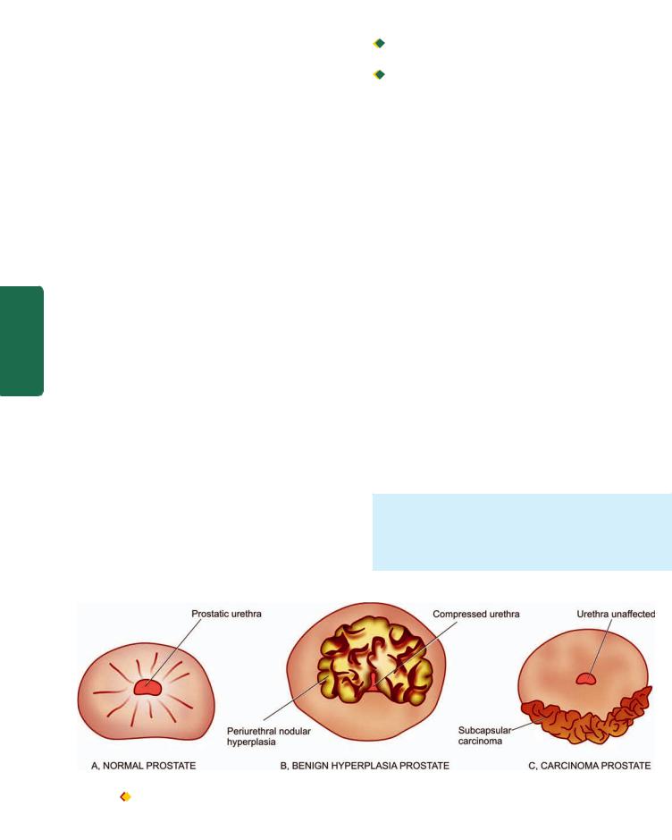
716HPV types 16 and 18 are strongly implicated and their DNA has been documented in the nuclei of malignant cells. Carcinoma of the penis is quite rare in Jews and Muslims who undergo a ritual of circumcision early in life. In India, cancer of the penis is rare in Muslims who practice circumcision as a religious rite in infancy, whereas Hindus who do not normally circumcise have higher incidence. Circumcision provides protection against penile cancer due to prevention of accumulation of smegma which is believed to be carcinogenic. The greatest incidence of penile cancer is between 45 and 60 years.
|
MORPHOLOGIC FEATURES. Grossly, the tumour is |
|
|
located, in decreasing frequency, on frenum, prepuce, |
|
|
glans and coronal sulcus. The tumour may be cauliflower- |
|
|
like and papillary, or flat and ulcerating (Fig. 23.10). |
|
|
Histologically, squamous cell carcinoma of both |
|
|
fungating and ulcerating type is generally well differen- |
|
|
tiated to moderately-differentiated type which resembles |
|
|
in morphology to similar cancer elsewhere in the body |
|
|
(Fig. 23.11). |
|
IIISECTION |
The tumour metastasises via lymphatics to regional |
|
NORMAL STRUCTURE |
||
|
lymph nodes. Visceral metastases by haematogenous route |
|
|
are uncommon and occur in advanced cases only. |
|
|
PROSTATE |
|
|
The prostate gland in the normal adult weighs approximately |
|
Systemic |
20 gm. It surrounds the commencement of the male urethra |
|
and is composed of 5 lobes during embryonic development— |
||
|
||
|
anterior, middle, posterior and two lateral lobes. But at birth, |
|
|
the five lobes fuse to form 3 distinct lobes—two major lateral |
|
Pathology |
lobes and a small median lobe (Fig. 23.12,A). |
|
Histologically, the prostate is composed of tubular alveoli |
||
|
||
|
(acini) embedded in fibromuscular tissue mass. The |
|
|
glandular epithelium forms infoldings and consists of 2 |
|
|
layers—a basal layer of low cuboidal cells and an inner layer |
|
|
of mucus-secreting tall columnar cells. The alveoli are |
|
|
separated by thick fibromuscular septa containing abundant |
|
|
smooth muscle fibres. |
The prostate has numerous blood vessels and nerves. In addition to nervous control, the prostate is an endocrinedependent organ. Based on hormonal responsiveness, the prostate is divided into 2 separate parts:
 the inner periurethral female part which is sensitive to oestrogen and androgen; and

 outer subcapsular true male part which is sensitive to androgen.
Prostate is involved in 3 important pathologic processes: prostatitis, nodular hyperplasia and carcinoma. While benign nodular hyperplasia occurs in the periurethral part distorting and compressing the centrally located urethral lumen, the prostatic carcinoma usually arises from the outer subcapsular part in which case it does not compress the urethra
(Figs. 23.12, B, C).
PROSTATITIS
Inflammation of the prostate i.e. prostatitis, may be acute, chronic and granulomatous types.
Acute Prostatitis
Acute focal or diffuse suppurative inflammation of the prostate is not uncommon. It occurs most commonly due to ascent of bacteria from the urethra, less often by descent from the upper urinary tract or bladder, and occasionally by lymphogenous or haematogenous spread from a distant focus of infection. The infection may occur spontaneously or may be a complication of urethral manipulation such as by catheterisation, cystoscopy, urethral dilatation and surgical procedures on the prostate. The common pathogens are those which cause UTI, most frequently E. coli, and others
such as Klebsiella, Proteus, Pseudomonas, Enterobacter, gonococci, staphylococci and streptococci. The diagnosis is made by culture of urine specimen.
MORPHOLOGIC FEATURES. Grossly, the prostate is enlarged, swollen and tense. Cut section shows multiple abscesses and foci of necrosis.
Histologically, the prostatic acini are dilated and filled with neutrophilic exudate. There may be diffuse acute
Figure 23.12 
 Normal prostate, benign nodular hyperplasia and prostatic carcinoma. The nodule in case of benign nodular hyperplasia (B) is located in the inner periurethral part and compresses the prostatic urethra while prostatic carcinoma (C) generally arises in the peripheral glands and, thus, does not compress the urethra.

inflammatory infiltrate. Oedema, hyperaemia and foci of necrosis frequently accompany acute inflammatory involvement.
Chronic Prostatitis
Chronic prostatitis is more common and foci of chronic inflammation are frequently present in the prostate of men above 40 years of age. Chronic prostatitis is usually asymptomatic but may cause allergic reactions, iritis, neuritis or arthritis.
Chronic prostatitis is of 2 types—bacterial and abacterial.

 Chronic bacterial prostatitis is caused in much the same way and by the same organisms as the acute prostatitis. It is generally a consequence of recurrent UTI. Diagnosis is made by detection of more than 10-12 leucocytes per high power field in expressed prostatic secretions, and by positive culture of urine specimen and prostatic secretions. This condition is more difficult to treat since antibiotics penetrate the prostate poorly.
 Chronic abacterial prostatitis is more common. There is no history of recurrent UTI and culture of urine and prostatic secretions is always negative, though leucocytosis is demonstrable in prostatic secretions. The pathogens implicated are Chlamydia trachomatis and Ureaplasma
urealyticum.
MORPHOLOGIC FEATURES. Pathologic changes in both bacterial and abacterial prostatitis are similar. Grossly, the prostate may be enlarged, fibrosed and shrunken.
Histologically, the diagnosis of chronic prostatitis is made by foci of lymphocytes, plasma cells, macrophages and neutrophils within the prostatic substance. Corpora amylacea, prostatic calculi and foci of squamous metaplasia in the prostatic acini may accompany inflammatory changes. Seminal vesicles are invariably involved.
Granulomatous Prostatitis
Granulomatous prostatitis is a variety of chronic prostatitis, probably caused by leakage of prostatic secretions into the tissue, or could be of autoimmune origin.
MORPHOLOGIC FEATURES. Grossly, the gland is firm to hard, giving the clinical impression of prostatic carcinoma on rectal examination.
Histologically, the inflammatory reaction consists of macrophages, lymphocytes, plasma cells and some multinucleate giant cells. The condition may be confused with tuberculous prostatitis.
NODULAR HYPERPLASIA
Non-neoplastic tumour-like enlargement of the prostate, commonly termed benign nodular hyperplasia (BNH) or benign enlargement of prostate (BEP), is a very common condition in men and considered by some as normal ageing process. It becomes increasingly more frequent above the age
of 50 years and its incidence approaches 75-80% in men above |
717 |
|
80 years. However, symptomatic BEP producing urinary tract |
|
|
obstruction and requiring surgical treatment occurs in 5-10% |
|
|
of cases only. |
|
|
ETIOLOGY. The cause of BEP has not been fully established. |
|
|
However, a few etiologic factors such as endocrinologic, racial, |
|
|
inflammation and arteriosclerosis have been implicated but |
|
|
endocrine basis for hyperplasia has been more fully |
|
|
investigated and considered a strong possibility in its genesis. |
|
|
It has been found that both sexes elaborate androgen and |
|
|
oestrogen, though the level of androgen is high in males and |
|
|
that of oestrogen is high in females. With advancing age, |
|
|
there is decline in the level of androgen and a corresponding |
|
|
rise of oestrogen in the males. The periurethral inner prostate |
|
|
which is primarily involved in BEP is responsive to the rising |
|
|
level of oestrogen, whereas the outer prostate which is mainly |
|
|
involved in the carcinoma is responsive to androgen. A |
|
|
plausible hypothesis suggested is that there is synergistic |
|
|
stimulation of the prostate by both hormones—the oestrogen |
|
|
acting to sensitise the prostatic tissue to the growth |
|
|
promoting effect of dihydroxy-testosterone derived from |
|
|
plasma testosterone. |
CHAPTER |
|
|
|
|
appearance on cut section varies depending upon whether |
|
|
MORPHOLOGIC FEATURES. Grossly, the enlarged pro- |
|
|
state is nodular, smooth and firm and weighs 2-4 times |
|
|
its normal weight i.e. may weigh up to 40-80 gm. The |
|
|
the hyperplasia is predominantly of the glandular or |
|
23 |
fibromuscular tissue (Fig. 23.13). In primarily glandular BEP |
|
|
|
|
|
the tissue is yellow-pink, soft, honey-combed, and milky |
|
The |
fluid exudes, whereas in mainly fibromuscular BEP the cut |
|
|
surface is firm, homogeneous and does not exude milky |
|
|
fluid. The hyperplastic nodule forms a mass mainly in |
|
Male |
the inner periurethral prostatic gland so that the |
|
|
|
|
|
surrounding prostatic tissue forms a false capsule which |
|
ProstateandSystemReproductive |
enables the surgeon to enucleate the nodular masses. The |
|
|
|
|
|
|
|
|
Figure 23.13 
 Nodular enlargement of the prostate. Sectioned surface of enlarged prostate shows soft to firm, grey-white, nodularity with microcystic areas.
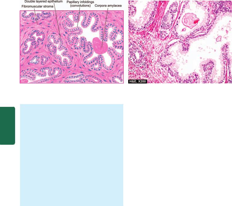
718
Pathology Systemic III SECTION
Figure 23.14 
 Nodular hyperplasia of the prostate. There is hyperplasia of fibromuscular elements. There are areas of intra-acinar papillary infoldings (convolutions) lined by two layers of epithelium with basal polarity of nuclei.
left-over peripheral prostatic tissue may sometimes undergo recurrent nodular enlargement or may develop carcinoma later.
Histologically, in every case, there is hyperplasia of all three tissue elements in varying proportions—glandular, fibrous and muscular (Fig. 23.14):
 Glandular hyperplasia predominates in most cases and is identified by exaggerated intra-acinar papillary infoldings with delicate fibrovascular cores. The lining epithelium is two-layered: the inner tall columnar mucussecreting with poorly-defined borders, and the outer cuboidal to flattened epithelium with basal nuclei.
 Fibromuscular hyperplasia when present as dominant component appears as aggregates of spindle cells forming an appearance akin to fibromyoma of the uterus.
In addition to glandular and/or fibromuscular hyperplasia, other histologic features frequently found include foci of lymphocytic aggregates, small areas of infarction, corpora amylacea and foci of squamous metaplasia.
CLINICAL FEATURES. Clinically, the symptomatic cases develop symptoms due to complications such as urethral obstruction and secondary effects on the bladder (e.g. hypertrophy, cystitis), ureter (e.g. hydroureter) and kidneys (e.g. hydronephrosis). The presenting features include frequency, nocturia, difficulty in micturition, pain, haematuria and sometimes, the patients present with acute retention of urine requiring immediate catheterisation.
CARCINOMA OF PROSTATE
Cancer of the prostate is the second most common form of cancer in males, followed in frequency by lung cancer. It is a disease of men above the age of 50 years and its prevalence increases with increasing age so that more than 50% of men 80 years old have asymptomatic (latent) carcinoma of the prostate. Many a times, carcinoma of the prostate is small
and detected as microscopic foci in a prostate removed for BEP or found incidentally at autopsy. Thus, it is common to classify carcinoma of the prostate into the following 4 types:
1.Latent carcinoma. This is found unexpectedly as a small focus of carcinoma in the prostate during autopsy studies in men dying of other causes. Its incidence in autopsies has been variously reported as 25-35%.
2.Incidental carcinoma. About 15-20% of prostatectomies done for BEP reveal incidental carcinoma of the prostate.
3.Occult carcinoma. This is the type in which the patient has no symptoms of prostatic carcinoma but shows evidence of metastases on clinical examination and investigations.
4.Clinical carcinoma. Clinical prostatic carcinoma is the type detected by rectal examination and other investigations and confirmed by pathologic examination of biopsy of the prostate.
ETIOLOGY. The cause of prostatic cancer remains obscure. However, a few factors have been suspected. These are as under:
1. Endocrinologic factors. Androgens are considered essential for development and maintenance of prostatic epithelium. But how androgens are responsible for causing malignant transformation is not yet clear. However, the etiologic role of androgens is supported by the following indirect evidences:
i)Orchiectomy causes arrest of metastatic prostatic cancer disease (testis being the main source of testosterone).
ii)Administration of oestrogen causes regression of prostatic carcinoma.
iii)Cancer of the prostate is extremely rare in eunuchs and in patients with Klinefelter’s syndrome.
iv)Cancer of the prostate begins at the stage of life when androgen levels are high. However, the cancer may remain latent with decline in androgen level with advancing age.
2.Racial and geographic influences. There are some racial and geographic differences in the incidence of prostatic
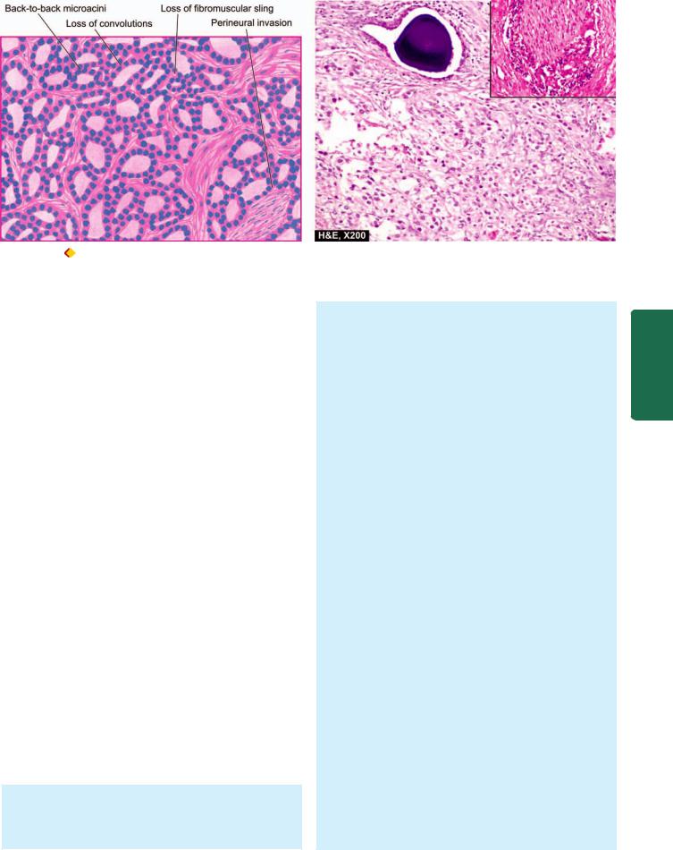
719
Figure 23.15 |
Carcinoma of the prostate. The field shows microacini of small malignant cells infiltrating the prostatic stroma. Inset in the |
photomicrograph shows perineural invasion by prostatic adenocarcinoma.
cancer. It is uncommon in Japanese and Chinese, while the prevalence is high in Americans. African Americans have a markedly higher incidence as compared to whites which may be related to genetic variation in androgen receptor gene.
3.Environmental influences. Some common environmental factors and carcinogens have been identified with high risk to development of prostatic cancer. These include high dietary fat, and exposure to polycyclic aromatic hydrocarbons. Flavonoids, antioxidants and selenium may reduce the risk.
4.Nodular hyperplasia. Though nodular prostatic hyperplasia has been suggested by some as precursor for development of prostatic cancer, it is considered unlikely. Most prostatic cancers develop in the periphery of the gland while BEP occurs in the periurethral part of the gland. Any concomitant occurrence of the two diseases may be considered as aging process. Approximately 15-20% of nodular hyperplastic prostates harbour carcinoma.
5.Heredity. The possibility of genetic basis of prostatic cancer has been suggested by the observations of familial clustering and 2-fold higher frequency in first-degree relatives. Prostatic cancer susceptibility gene has been identified in familial cases.
HISTOGENESIS. Histogenesis of prostatic adenocarcinoma has been documented as a mutistep process arising from premalignant stage of prostatic intraepithelial neoplasia (PIN).
PIN refers to multiple foci of cytologically atypical luminal cells overlying diminished number of basal cells in prostatic ducts and is a forerunner of invasive prostatic carcinoma. Based on cytologic atypia, PIN may be low grade to high grade. PIN of high-grade progresses to prostatic adenocarcinoma.
MORPHOLOGIC FEATURES. Grossly, the prostate may be enlarged, normal in size or smaller than normal. In 95% of cases, prostatic carcinoma is located in the peripheral zone, especially in the posterior lobe. The malignant
prostate is firm and fibrous. Cut section is homogeneous and contains irregular yellowish areas. Microscopically, 4 histologic types are described— adenocarcinoma, transitional cell carcinoma, squamous cell carcinoma and undifferentiated carcinoma. However, adenocarcinoma is the most common type found in 96% of cases and is the one generally referred to as carcinoma of the prostate. The other three histologic types are rare and resemble in morphology with similar malignant tumours elsewhere in the body.
The histologic characteristics of adenocarcinoma of the prostate are as under (Fig. 23.15):
1.Architectural disturbance. In contrast to convoluted appearance of the glands seen in normal and hyperplastic prostate, there is loss of intra-acinar papillary convolutions. The groups of acini are either closely packed in back-to-back arrangement without intervening stroma or are haphazardly distributed.
2.Stroma. Normally, fibromuscular sling surrounds the acini, whereas malignant acini have little or no stroma between them. The tumour cells may penetrate and replace the fibromuscular stroma.
3.Gland pattern. Most frequently, the glands in welldifferentiated prostatic adenocarcinoma are small or medium-sized, lined by a single layer of cuboidal or low columnar cells. Moderately-differentiated tumours have cribriform or fenestrated glandular appearance. Poorlydifferentiated tumours have little or no glandular arrangement but instead show solid or trabecular pattern.
4.Tumour cells. In many cases, the individual tumour cells in prostatic carcinoma do not show usual morphologic features of malignancy. The tumour cells may be clear, dark and eosinophilic cells. Clear cells have foamy cytoplasm, dark cells have homogeneous basophilic cytoplasm, and eosinophilic cells have granular cytoplasm. The cells may show varying degree of anaplasia and nuclear atypia but is generally slight.
Prostate and System Reproductive Male The 23 CHAPTER

720
Pathology Systemic III SECTION
5. Invasion. One of the important diagnostic features of malignancy in prostate is the early and frequent occurrence of invasion of intra-prostatic perineural spaces. Lymphatic and vascular invasion may be present but are difficult to detect.
SPREAD. The tumour spreads within the gland by direct extension, and to distant sites by metastases.
Direct spread. Direct extension of the tumour occurs into the prostatic capsule and beyond. In late stage, the tumour may extend into the bladder neck, seminal vesicles, trigone and ureteral openings.
Metastases. Distant spread occurs by both lymphatic and haematogenous routes. The rich lymphatic network surrounding the prostate is the main mode of spread to the sacral, iliac and para-aortic lymph nodes. The earliest metastasis occur to the obturator lymph node. Haematogenous spread leads most often to characteristic osteoblastic osseous metastases, especially to pelvis, and lumbar spine; other sites of metastases are lungs, kidneys, breast and brain. The route of blood-borne metastases may be retrograde spread by prostatic venous plexus or via systemic circulation.
CLINICAL FEATURES. By the time symptoms appear, the carcinoma of prostate is usually palpable on rectal examination as a hard and nodular gland fixed to the surrounding tissues. In such symptomatic cases, clinical features are: urinary obstruction with dysuria, frequency, retention of urine, haematuria, and in 10% of cases pain in the back due to skeletal metastases.
Clinical staging of carcinoma prostate takes into account the following:
 The tumour found incidentally or a clinically unsuspected cancer in prostate removed for benign disorder (Stage A).

 The tumour palpable by rectal digital examination but confined to the prostate (Stage B).
 The tumour has extended locally beyond the prostate into the surrounding tissues (Stage C).

 The tumour is associated with distant metastases
(Stage D).
Clinical staging has good correlation with histologic grading and, thus, has a prognostic significance. Mostofi’s
(WHO) histologic grading categorising prostate cancer into grade I (well-differentiated), grade II (moderately differentiated) and grade III (poorly differentiated) has largely been replaced with Gleason’s microscopic grading system which is based on two features:
i)Degree of glandular differentiation.
ii)Growth pattern of the tumour in relation to the stroma. These features are assessed by low-power examination
of the prostatic tissue. For clinical staging of prostate cancer, TNM system is considered international standard.
The diagnosis of prostatic carcinoma is made by cytologic, biochemical, radiologic, ultrasonographic and pathologic methods. However, definite diagnosis is established by histopathologic examination of transrectal ultrasound (TRUS)- guided core needle biopsy.
Two serum tumour markers employed commonly for diagnosis and monitoring the prognosis of prostatic carcinoma are as under:

 Prostatic acid phosphatase (PAP) is secreted by prostatic epithelium. Elevation of serum level of PAP is found in cases of prostatic cancer which have extended beyond the capsule or have metastasised. PAP can also be demonstrated in the normal prostatic tissues.
 Prostate-specific antigen (PSA) can be detected by immunohistochemical method in the malignant prostatic epithelium as well as estimated in the serum. A reading between 4 and 10 (normal 0-4 ng/ml) is highly suspicious (10% risk) but value above 10 is diagnostic of prostatic carcinoma. PSA assay is useful in deciding whether the metastasis originated from the prostate or not. PSA assay is also helpful in distinguishing high-grade prostatic cancer from urothelial carcinoma, colonic carcinoma, lymphoma and prostatitis. PSA level is generally higher in low-grade tumours than in high-grade tumours.
Treatment of prostatic carcinoma consists of surgery, radiotherapy and hormonal therapy. The hormonal dependence of prostate cancer consists of depriving the tumour cells of growth-promoting influence of testosterone. This can be achieved by bilateral orchiectomy followed by administration of oestrogen. Surgical approaches for prostate cancer include transurethral resection (TUR), radical prostatectomy and transurethral US-guided laser-induced prostatectomy (TULIP).