

Figure 3.6 Agenesis of the corpus callosum. This midline sagittal magnetic resonance image shows an absent corpus callosum, a dramatic example of a neurodevelopmental anomaly which, while extremely rare, is thought to have an increased incidence in people with schizophrenia
schizophrenic patients compared with normal controls.
FUNCTIONAL BRAIN IMAGING
Functional brain imaging studies have used positron emission tomography (PET), single photon emission tomography (SPET) and, more recently, functional magnetic resonance imaging techniques (fMRI) to investigate regional cerebral blood flow (rCBF) and brain metabolism in schizophrenia (Figure 3.9)5.
It was previously thought that a decrease in frontal blood flow and metabolism (‘hypofrontality’) was a constant feature of schizophrenia. However, this now appears to be a function of the cognitive load involved in the test that patients are carrying out at the time. For example, activation studies using ‘frontal’ tasks such as the Wisconsin
1 |
I |
I |
|
II |
II |
||
|
|||
|
III |
III |
|
2 |
IV |
IV |
|
|
|
||
|
V |
V |
|
3 |
VI |
VI |
|
|
|
4
5
6
7
8
Controls |
|
|
|
Schizophrenics |
|
|
|
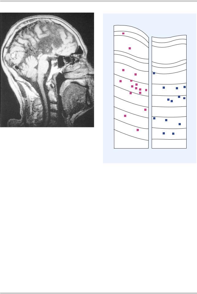
Figure 3.7 These camera lucida drawings compare the distribution of nicotinamide-adenine dinucleotide phosphate-diaphorase-stained neurons (squares) in sections through the superior frontal gyrus of a control and schizophrenic brain. There is a significant shift in the direction of the white matter in the schizophrenic brain. Numbers 1 through 8 indicate compartments of the brain; Roman numerals indicate cortical layers. Figure reproduced with permission from Akbarian S, Bunney WE, Jr, Potkin SG, et al. Altered distribution of nicotinamideadenine dinucleotide phosphate-diaphorase cells in frontal lobe of schizophrenics implies disturbances of cortical development. Arch Gen Psychiatry 1993:50:169–77
card sorting test have shown that healthy volunteers increase blood flow to the dorsolateral prefrontal cortex during the task, while this is not apparent when schizophrenic patients perform the task. Other studies using verbal fluency as an activation task have found impaired frontal blood flow in schizophrenic patients (Figure 3.10)6. However, there are studies on both tasks that have
©2002 CRC Press LLC
DERMATOGLYPHIC ABNORMALITIES
IN SCHIZOPHRENIA
60
difference |
count |
50 |
|
|
|
||
intrapairAbsolute |
ridgetotalin |
40 |
|
30 |
|||
|
|
||
|
|
20 |
10
0
Nonschizophrenic |
Discordant for |
(n = 7) |
schizophrenia |
|
(n = 23) |
Monozygotic twin pairs
Pair discordant for depression
Figure 3.8 Structural abnormalities may be found in schizophrenia outside the CNS; other structures that develop at the same time may also be involved. In monozygotic twins there should be little or no difference between the twins in total finger ridge count. It can be seen, however, that in twin pairs where one twin suffers from schizophrenia, there is a much greater difference in total finger ridge count. This is significant because finger ridges develop in the second trimester and therefore the differences illustrated in this slide may indicate a degree of maldevelopment in affected twins
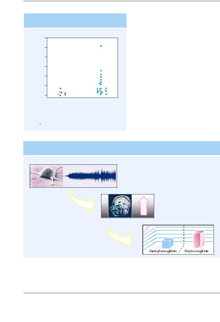
BASIS OF FUNCTIONAL MAGNETIC RESONANCE IMAGING
Neuronal activity
Increased local cerebral blood flow
Change in blood oxygenation
Figure 3.9 Oxyhemoglobin and deoxyhemoglobin have slightly different magnetic properties, and this is used as the basis for the blood oxygenated level dependent (BOLD) method in functional magnetic resonance imaging (MRI). Increases in neuronal activity are accompanied by increases in regional cerebral blood flow, which exceed the increase in cerebral oxygen utilization. As a result the oxygen content of the venous blood is increased, leading to an increase in MRI signal intensity. Figure reproduced with permission from Longworth C, Honey G, Sharma T. Science, medicine, and the future. Functional magnetic resonance imaging in neuropsychiatry. Br Med J 1999;319:1551–4
©2002 CRC Press LLC

CONTROLS
PATIENTS WITH
SCHIZOPHRENIA
Figure 3.10 Verbal fluency and frontal lobe blood flow in schizophrenia. Comparison using functional magnetic resonance imaging between five right-handed male schizophrenic patients and five matched controls performing a covert verbal fluency task. The schizophrenic patients showed a comparatively reduced response (red) in the left dorsal prefrontal cortex and inferior frontal gyrus, and an increased response (orange) in the medial parietal cortex. Figure reproduced with permission from Curtis VA, Bullmore ET, Brammer MJ, et al. Attenuated frontal activation during a verbal fluency task in patients with schizophrenia. Am J Psychiatry 1998; 155:1056–63
MOTOR ACTIVATIONS AT TWO POINTS IN TIME 4–6 WEEKS APART
|
|
Normals at time 2 |
|
Schizophrenics at t1 |
|
Schizophrenics at t2 |
|
Sz t1–t2 |
Normals at time 1 |
||||||||
sagittal |
|
sagittal |
|
sagittal |
|
sagittal |
|
sagittal |
|
|
|
|
|
|
|
|
|
Figure 3.11 Hypofrontality in a motor activation task remitting with recovery from schizophrenic relapse. This study shows changes over time in neuronal response in a positron emission tomography study of willed action using a simple motor task. Prefrontal cortical activation not apparent at time 1 in the schizophrenic subjects (when they were acutely ill) becomes apparent at time 2, 4–6 weeks later. The figures show statistical parametric maps thresholded at p < 0.05, Bonferrroni corrected. Figure reproduced with permission from Spence SA, Hirsch SR, Brooks DJ, Grasby PM. Prefrontal cortext activity in people with schizophrenia and control subjects. Evidence from positron emission tomography for remission of ‘hypofrontality’ with recovery from acute schizophrenia. Br J Psychiatry 1998;172:316–23
©2002 CRC Press LLC

REGIONAL CEREBRAL BLOODFLOW (rCBF)
AND SYNDROMES OF SCHIZOPHRENIA
Psychomotor |
|
Disorganization |
|
Reality distortion |
poverty syndrome |
|
syndrome |
|
syndrome |
|
|
|
|
|
Negative correlations
Positive correlations
Figure 3.12 Statistical parametric maps showing pixels in which there are significant correlations between rCBF and syndrome score, for the three syndromes of psychomotor poverty, disorganization and reality distortion. Different syndromes of schizophrenia may have different patterns of aberrant rCBF. Figure reproduced with permission from Liddle PF, Friston KJ, Frith CD, et al. Patterns of cerebral blood flow in schizophrenia. Br J Psychiatry 1992;160:179–86
not shown these differences between schizophrenics and controls. Although some studies have suggested that differences in rCBF between patients and controls are persistent, others have found that they appear to be state dependent, and remit with treatment (Figure 3.11)7.
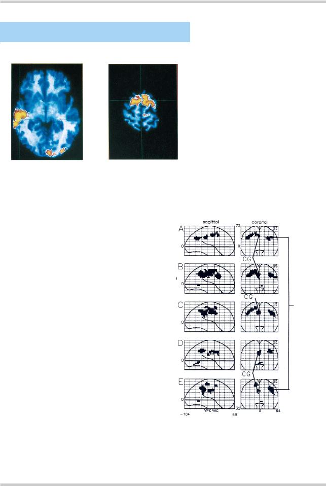
A number of studies have attempted to correlate patterns of brain activation with specific symptoms or syndromes of schizophrenia. Liddle and colleagues8 (Figure 3.12) investigated the three syndromes of psychomotor poverty, disorganization (i.e. inappropriate affect, speech content abnormalities), and reality distortion (i.e. delusions and hallucinations). They found that reduced rCBF in the left and medial prefrontal cortex correlated with psychomotor poverty; the severity of disorganization correlated with increased rCBF in the right medial prefrontal
cortex and decreased perfusion in Broca’s area; and reality distortion correlated with increased rCBF in the left hippocampal formation. Studies of schizophrenic patients with and without auditory hallucinations have shown increased blood flow to Broca’s area when patients are hearing voices. Furthermore, patients prone to auditory hallucinations show abnormal patterns of blood flow when asked to imagine hearing voices, compared with normal controls (Figure 3.13)9, and patients with schizophrenia also appear to demonstrate abnormal patterns of temporal cortex activation in response to external speech. Abnormal patterns of blood flow also appear to be related to other specific symptoms of schizophrenia, including passivity phenomena (Figure 3.14)10 and formal thought disorder (Figure 3.15)11. Interest is focusing increasingly on the
©2002 CRC Press LLC
SEROTONINERGIC PATHWAYS
INNER SPEECH AND AUDITORY HALLUCINATIONS
0 mm |
+60 mm |
|
|
Figure 3.13 This study investigated the hypothesis that a predisposition to verbal hallucinations is associated with a failure to activate areas concerned with the monitoring of inner speech. Subjects, who included patients with schizophrenia both with and without a significant history of hallucinations, as well as normal controls, were asked to imagine sentences being spoken in another person’s voice. The figure illustrates positron emission tomography data superimposed on a normal magnetic resonance imaging scan, and shows reduced activation in the left middle temporal gyrus and the rostral part of the supplementary motor area in hallucinators compared to non-hallucinators. Similar findings were found in the comparison between schizophrenic patients and controls. Figure reproduced with permission from McGuire PK, Silbersweig DA, Wright I. Speech: a physiological basis for auditory hallucinations. Lancet 1995;346:596–600
RELATIVE HYPERACTIVATION IN PATIENTS WITH PASSIVITY
Compared with normals
– free movement minus rest
Compared with other schizophrenic patients
– free movement minus rest
Right inferior parietal lobule
–stereotypic movement minus rest
Compared with themselves at time 2
– free movement minus rest
–stereotypic movement minus rest
Figure 3.14 Positron emission tomography study of schizophrenic patients with passivity phenomena. This study looked at patients with schizophrenia who were also experiencing passivity phenomena (delusions of alien control) during a voluntary movement task. Hyperactivity is seen in these patients compared with normal controls (A), compared with other schizophrenic patients (B and C), and with themselves as their symptoms resolve (D and E). In each case, greater activation is seen in the right inferior parietal lobule (IPL), and at loci within the cingulate gyrus (CG). Figure reproduced with permission from Spence SA, Brookes DJ, Hirsch SR, et al. A PET study of voluntary movement in schizophrenic patients experiencing passivity phenomena (delusions of alien control). Brain 1997;120:1997–2011
©2002 CRC Press LLC