

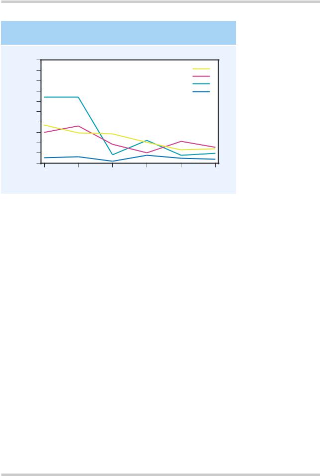
FREQUENCY OF LIFE EVENTS
|
100 |
|
|
|
|
|
subjects |
90 |
|
|
|
Schizophrenia |
|
|
|
|
Mania |
|
||
80 |
|
|
|
|
||
|
|
Depressive psychosis |
|
|||
|
|
|
|
|||
70 |
|
|
|
Controls |
|
|
per100 |
|
|
|
|
||
60 |
|
|
|
|
|
|
50 |
|
|
|
|
|
|
rates |
|
|
|
|
|
|
40 |
|
|
|
|
|
|
|
|
|
|
|
|
|
event |
30 |
|
|
|
|
|
20 |
|
|
|
|
|
|
Life |
|
|
|
|
|
|
10 |
|
|
|
|
|
|
|
0 |
|
|
|
|
|
|
1 |
2 |
3 |
4 |
5 |
6 |
Months before onset/interview
Figure 2.13 The rate of life events is increased in schizophrenia, although the effect is not as great as in depression. Figure reproduced with permission from Bebbington P, Wilkins S, Jones P, et al. Life events and psychosis. Initial results from the Camberwell Collaborative Psychosis Study. Br J Psychiatry 1993;162:72–9
It is estimated that 20–50% of the population with schizophrenia in Western countries may qualify as substance abusers or ‘dual-diagnosis’ patients. Such patients have a higher use of services and worse outcome than patients who are not abusers; they are more likely to be hospitalized and more likely to relapse. Patients with schizophrenia seem to be more vulnerable to significant harm at relatively lower levels of substance use. As substance misuse is so prevalent in the West, this is an area where secondary prevention of relapse could be focused.
Social and psychological factors
Psychosocial factors appear to contribute to both the onset and the relapse of schizophrenia. The best documented are life events (Figure 2.13)20. The effect size is smaller than in depression and the time frame is somewhat shorter than in depression (where adverse life events are well
recognized etiological factors), with the 3 weeks prior to onset seeming to be the most important. Unlike depression, all kinds of life events appear to be important, not just those involving loss.
Many migrant groups show an increased firstinception rate of schizophrenia compared both to the population they have left and to that which they have joined. The most striking example of this is people of African-Caribbean origin living in the UK (Figure 2.14)1. It seems unlikely that the factors are biological. Odegaard21 suggested in 1933 that social isolation and alienation are the crucial factors, and most recent evidence points in this direction.
CONCLUSION
There is no single cause for schizophrenia, rather a number of risk factors (Figure 2.15) interact to propel the individual over a threshold for expression of the disease.
©2002 CRC Press LLC

ETHNICITY AND SCHIZOPHRENIA
|
60 |
|
|
50 |
|
population |
40 |
|
|
|
|
per 100000 |
30 |
|
20 |
|
|
Rate |
|
|
|
|
|
|
10 |
|
|
0 |
|
|
All |
All other |
|
African- |
ethnic |
|
Caribbean |
groups |
Figure 2.14 The differences in incidence in schizophrenia in people of African-Caribbean origin compared with those from other ethnic groups, in one study from Camberwell, South London. Figure reproduced with permission from Castle E, Wessely S, Der G, Murray RM. The incidence of operationally defined schizophrenia in Camberwell 1965–84. Br J Psychiatry 1991;159:790–4
RISK FACTORS AND EFFECT SIZES
Psychotic first-degree relative
(RR = 10)
Obstetric complications
Winter birth
City birth / upbringing
Cannabis use
Member of certain immigrant groups
Life events
Figure 2.15 There is no single cause of schizophrenia. Instead, like other complex disorders such as coronary heart disease, a number of genetic and environmental factors interact to cause the disease
©2002 CRC Press LLC

REFERENCES
1.Castle E, Wessely S, Der G, Murray RM. The incidence of operationally defined schizophrenia in Camberwell 1965–84. Br J Psychiatry 1991;159: 790–4
2.Mortensen PB, Pedersen CB, Westergaard T, et al. Effects of family history and place and season of birth on the risk of schizophrenia. N Engl J Med 1999;340:603–8
3.Gottesman I, Irving I. Schizophrenia Genesis: The Origin of Madness. New York, Oxford: WH Freeman, 1991:203
4.Shields J, Gottesman II. Obstetric complications and twin studies of schizophrenia: clarifications and affirmations. Schizophr Bull 1977;3:351–4
5.Kendell RE, Zealley AK. Companion to Psychiatric Studies. Edinburgh: Churchill Livingstone, 1993
6.Kringlen E. Twin studies in schizophrenia with special emphasis on concordance figures. Am J Med Genet 2000;97:4–11
7.Fischer M. Genetic and environmental factors in schizophrenia. A study of schizophrenic twins and their families. Acta Psychiatr Scand 1973;238 (Suppl.):9–142
8.Tienari P, Sorri A, Lahti I, et al. Genetic and psychosocial factors in schizophrenia: the Finnish Adoptive Family Study. Schizophr Bull 1987;13: 477–84
9.Pollin W, Allen MG, Hoffer A, et al. Psychopathology in 15,909 pairs of veteran twins: evidence for a genetic factor in the pathogenesis of schizophrenia and its relative absence in psychoneurosis. Am J Psychiatry 1969;126:597–610
10.Cardno AG, Marshall EJ, Coid B, et al. Heritability estimates for psychotic disorders: the Maudsley twin psychosis series. Arch Gen Psychiatry 1999;56:162–8
11.Kety SS, Rosenthal D, Wender PH, Schulsinger F, et al. Mental illness in the biological and adoptive
families of adopted schizophrenics. Am J Psychiatry 1971;128:302–6
12.Rosenthal D, Wender PH, Kety SS, et al. The adopted-away offspring of schizophrenics. Am J Psychiatry 1971;128:307–11
13.Pollack M, Woerner MG, Goodman W, Greenberg IM. Childhood development patterns of hospitalized adult schizophrenic and nonschizophrenic patients and their siblings. Am J Orthopsychiatry 1966;36: 510–7
14.Pearlson GD, Garbacz DJ, Moberg PJ, et al. Symptomatic, familial, perinatal, and social correlates of computerized axial tomography (CAT) changes in schizophrenics and bipolars. J Nerv Ment Dis 1985; 173:42–50
15.Owen MJ, Lewis SW, Murray RM. Obstetric complications and schizophrenia: a computed tomographic study. Psychol Med 1988;18:331–9
16.O'Callaghan E, Gibson T, Colohan HA, et al. Risk of schizophrenia in adults born after obstetric complications and their association with early onset of illness: a controlled study. Br Med J 1992;305: 1256–9
17.O'Callaghan E, Larkin C, Kinsella A, Waddington JL. Obstetric complications, the putative familialsporadic distinction, and tardive dyskinesia in schizophrenia. Br J Psychiatry 1990;157:578–84
18.Susser E, Neugebauer R, Hoek HW, et al. Schizophrenia after prenatal famine: further evidence. Arch Gen Psychiatry 1996;53:25–31
19.Andreasson S, Allebeck P, Engstrom A, Rydberg U. Cannabis and schizophrenia. A longitudinal study of Swedish conscripts. Lancet 1987;2:1483–6
20.Bebbington P, Wilkins S, Jones P, et al. Life events and psychosis. Initial results from the Camberwell Collaborative Psychosis Study. Br J Psychiatry 1993; 162:72–9
21.Odegaard S. Emigration and insanity. Acta Psychiatr Scand 1932;Suppl. 4
©2002 CRC Press LLC
CHAPTER 3
Pathogenesis
Pathogenetic theories need to encompass all levels of brain structure and function, from the basic neuroanatomical level, through neurochemical, neurophysiological and neuropsychological findings, and thence through to symptoms. As yet, we have only a very partial understanding of these mechanisms.
STRUCTURAL IMAGING AND ANATOMICAL STUDIES
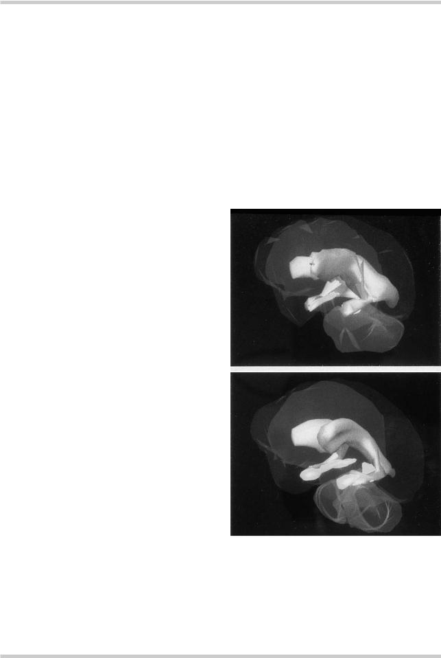
The core brain structural finding in schizophrenia, of lateral ventricular enlargement (Figure 3.1) is now well established, but the degree of enlargement is relatively small (Figure 3.2); about 25% on average1. Monozygotic twins discordant for schizophrenia can be distinguished from their cotwins on the basis of ventriculomegaly and decreased temporal cortical volume (Figure 3.3)2. Numerous other morphological abnormalities have been reported (Figures 3.43 and 3.5). People with schizophrenia appear to have very slightly smaller brains with sulcal widening and reduced cortical volume, particularly in the temporal lobes.
A number of other findings have been reported. Most of these are non-specific and tell us little about pathogenesis, but there are some clues to the processes that might be involved. Normally rare developmental abnormalities, such as agenesis of the corpus callosum (Figure 3.6), aqueduct stenosis, cavum septum pellucidum, cerebral
a
b
Figure 3.1 Three-dimensional reconstruction of the ventricular system in schizophrenia. Structural changes appear in the shrunken hippocampus (yellow) and enlarged fluid-filled ventricles (gray) of the brain of a patient with schizophrenia (a) compared with that of a healthy volunteer
(b). Figure reproduced with kind permission of Professor Nancy C. Andreasen, University of Iowa, USA
©2002 CRC Press LLC
INCREASED VENTRICLE VOLUME
IN SCHIZOPHRENIA
12
VBR |
10 |
|
|
|
|
|
|
|
|
|
|
|
|
|
|
mean |
8 |
|
|
|
|
|
|
|
|
|
|
|
|
|
|
Schizophrenic |
|
|
|
|
|
|
|
|
|
|
|
|
|
|
|
6 |
|
|
|
|
|
|
|
|
|
|
|
|
|
|
|
|
|
|
|
|
|
|
|
|
|
|
|
|
|
|
|
|
4 |
|
|
|
|
|
|
|
|
|
|
|
|
|
|
|
|
|
|
|
|
|
|
|
|
|
|
|
|
|
|
|
2 |
|
|
Line of equality |
|
|
|
|
|
|
|
|
|||
|
|
|
|
|
|
|
|
|
|
|
|||||
|
0 |
|
|
|
|
|
|
|
|
|
|
||||
|
|
|
|
|
|
|
|
|
|
|
|
|
|
|
|
|
|
|
|
|
|
|
|
|
|
|
|
|
|
|
|
|
0 |
2 |
4 |
6 |
8 |
10 |
12 |
||||||||
|
|
|
|
|
|
Control mean VBR |
|
|
|
|
|||||
|
Diagnostic criteria and methods of measurement: |
|
|
||||||||||||
|
Planimetry |
|
|
|
Computerized |
|
|
|
|
||||||
|
|
|
DSM–III |
|
|
|
|
DSM–III |
|
|
|
|
|||
|
|
|
RDC |
|
|
|
|
|
|
RDC |
|
|
|
|
|
|
|
|
DSM–III/RDC |
|
|
|
|
DSM–III/RDC |
|
|
|||||
|
|
|
WUC |
|
|
|
|
|
|
|
|
|
|
|
|
Figure 3.2 Mean ventricle : brain ratio (VBR) in controls and patients with schizophrenia from a total of 39 separate studies. The diagonal line indicates the line of equality. Thus, this figure demonstrates that no matter which diagnostic system or method of measuring brain volume is used, patients with schizophrenia do have larger ventricles than controls. Figure reproduced with permission from Van Horn JD, McManus IC. Ventricular enlargement in schizophrenia. A meta-analysis of studies of the ventricle : brain ration (VBR). Br J Psychiatry 1992;160:687–97
Figure 3.3 Ventricular size in monozygotic twins discordant for schizophrenia. Coronal magnetic resonance images of twins discordant for schizophrenia show lateral ventricular enlargement in the affected twin. Figure reproduced with permission from Suddath RL, Christison GW, Torrey EF, et al. Anatomical abnormalities in the brains of monozygotic twins discordant for schizophrenia. N Engl J Med 1990;322:789–4
©2002 CRC Press LLC
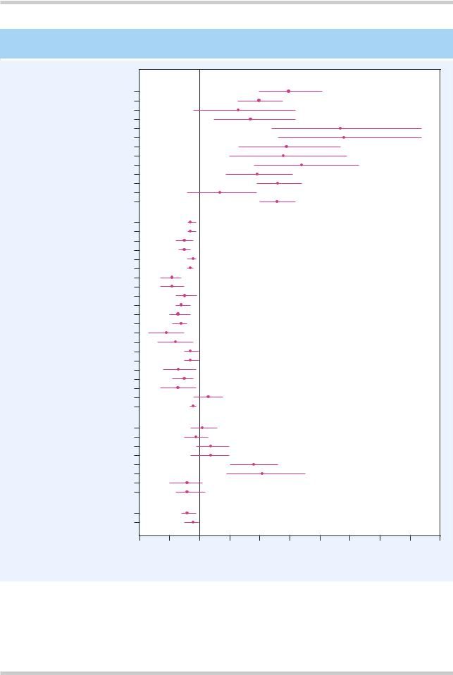
COMPARATIVE MEAN VOLUMES OF BRAIN REGIONS IN SCHIZOPHRENIA
Ventricular structures
Left lateral ventricle
Right lateral ventricle
Left frontal horn
Right frontal horn
Left body ventricle
Right body ventricle
Left occipital horn
Right occipital horn
Left temporal horn
Right temporal horn
Third ventricle
Fourth ventricle
Total ventricles
Cortical/limbic structures
Left hemisphere
Right hemisphere
Left frontal volume
Right frontal volume
Left temporal lobe
Right temporal lobe
Left amygdala
Right amygdala
Left hippocampus-amygdala
Right hippocampus-amygdala
Left hippocampus
Right hippocampus
Left parahippocampus
Right parahippocampus
Left superior temporal gyrus
Right superior temporal gyrus
Left anterior superior temporal gyrus
Right anterior superior temporal gyrus
Left posterior superior temporal gyrus
Right posterior superior temporal gyrus
Whole brain
Subcortical structures
Left caudate
Right caudate
Left putamen
Right putamen
Left globus pallidus
Right globus pallidus
Left thalamus
Right thalamus
Whole brain gray/white matter
Gray matter
White matter
80 |
90 |
100 |
110 |
120 |
130 |
140 |
150 |
160 |
170 |
180 |
Comparative mean volume of subjects with schizophrenia (%)
Figure 3.4 Meta-analysis of absolute regional brain volumes in schizophrenic patients and controls, from a total of 58 studies. This figure shows how the mean volumes of different brain regions from people with schizophrenia differ from those of controls. Figure reproduced with permission from Wright IC, Rabe-Hesketh S, Woodruff PW, et al. Meta-analysis of regional brain volumes in schizophrenia. Am J Psychiatry 2000;157:16–25
©2002 CRC Press LLC

STRUCTURAL BRAIN ABNORMALITIES IN SCHIZOPHRENIA
Enlarged
Gyral abnormalities
Abnormalities
of white matter
Cortical cellular displacement
Reduced hippocampal |
temporal horns |
volume |
of lateral ventricles |
Figure 3.5 Some structural brain abnormalities possibly implicated in the pathogenesis of schizophrenia. Structural abnormalities have been described in many brain areas, and at a variety of anatomical levels, from gross macroscopic changes in whole brain volume, through to subtle cellular displacement or disorganization in the cortex. Increasingly, interest has focused on the distribution of abnormalities, and their structural connectivity: thus, white matter myelination, as well as cortical abnormalities, are targets of investigation
hamartomas and arteriovenous malformations occur with increased frequency in schizophrenia.
At the cellular level, various abnormalities in cytoarchitecture have been reported in several brain regions, although not all of these findings have proved robust. However, evidence of neuronal displacement (Figure 3.7) suggests the possibility of some failure in neuronal migration, a process that occurs mainly during the second trimester of fetal development4.
Several findings weigh against the most likely alternative of a neurodegenerative process. The balance of evidence is that most of the brain
abnormalities seen in schizophrenia are present at first onset and are non-progressive. Furthermore, markers of neurodegeneration, such as proteins associated with glial response are largely absent, although there may be a small degree of periventricular gliosis. Extracerebral markers of abnormal fetal development provide indirect support for the idea that aberrant neurodevelopment is implicated in schizophrenia. Dermatoglyphic abnormalities are thought to reflect fetal maldevelopment and appear to be more common in schizophrenia (Figure 3.8). Minor physical anomalies also occur with greater frequency in
©2002 CRC Press LLC