

DOPAMINE SYNTHESIS
|
|
|
H |
|
|
Phenylalanine |
|
C |
CH |
NH2 |
|
|
H |
|
|
||
|
|
|
COOH |
||
|
|
|
|
||
|
|
|
H |
|
|
Tyrosine |
|
C |
CH |
NH2 |
|
|
H |
|
|
||
|
|
|
COOH |
||
|
|
HO |
|
||
tyrosine |
Rate |
|
|
|
|
|
|
|
|
||
hydroxylase |
limiting |
|
|
|
|
(pterin cofactor) |
step |
HO |
H |
|
|
|
|
|
|
||
|
|
C |
CH |
NH2 |
|
Dopa |
|
|
|||
|
|
H |
|
|
|
|
|
|
|
|
|
Dopa |
|
HO |
|
COOH |
|
|
|
|
|
|
|
decarboxylase |
|
|
|
|
|
(pyridoxal cofactor) |
|
HO |
H |
|
|
|
|
|
|
||
|
|
C CH 2 |
NH2 |
||
Dopamine |
|
||||
|
H |
|
|
||
|
|
|
|
|
|
HO
Dopamine -hydroxylase (copper containing enzyme) Ascorbate and O
HO
H
Norepinephrine C CH 2 NH2
OH
HO
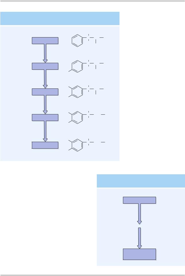
Figure 4.7 Dopamine is synthesized in a common pathway with norepinephrine. In the pathway shown, tyrosine hydroxylase is the rate-limiting enzyme. Dopamine β-hydroxylase is only found in noradrenaline neurons in the CNS
Figure 4.8 |
Dopamine is |
metabolized |
by two enzymes. |
One, monoamine oxidase type B (MAO-B), is intraneuronal, and the other, catechol-O-methyl transferase (COMT), is extraneuronal. The primary metabolite of dopamine is homovanillic acid
DOPAMINE METABOLISM
Dopamine is synthesized as part of the common pathway for catecholamines (Figure 4.7). Dopamine is metabolized by two enzymes; one, monoamine oxidase type B (MAO-B), is intraneuronal, and the other, catechol-O-methyl transferase (COMT), is extraneuronal. The primary metabolite of dopamine is homovanillic acid (HVA; Figure 4.8).
Serotonin
The serotonergic hypothesis of schizophrenia predates the dopaminergic hypothesis and stems from the finding by Woolley and Shaw in 195412 that the hallucinogen LSD acted via serotonin.
Dopamine
|
Catechol-O-methyl |
Extraneural |
transferase (COMT) |
|
Monoamine oxidase |
Intraneural |
type B (MAO-B) |
Homovanillic acid (HVA)
©2002 CRC Press LLC
SPET SCANS OF 5-HT2A RECEPTOR DENSITY
Temporal cortex and cerebellum
Frontal, parietal and occipital cortices
Healthy volunteer |
Clozapine treated |
Risperidone treated |
Figure 4.9 Single photon emission tomography (SPET) scans of 5-HT2A receptor density in a healthy volunteer and two patients with schizophrenia receiving a therapeutic dose of either clozapine (450 mg/day) or risperidone (6 mg/day). In the slice at the level of the frontal, parietal and occipital cortices from the healthy volunteer the ubiquitous distribution of 5-HT2A receptors in the cortical gray matter can be seen. This binding of the tracer is absent from the equivalent slices from the medicated patients. This indicates that both clozapine and risperidone are producing very high or ‘saturation’ levels of 5- HT2A receptor blockade. This high level of blockade may confer some protection against the development of EPS and may be one of the mechanisms of antipsychotic ‘atypicality’. Figure reproduced with permission from Travis MJ, Busatto GF, Pilowsky LS, et al. 5-HT2A receptor blockade in patients with schizophrenia treated with risperidone or clozapine. A SPET study using the novel 5-HT2A ligand 123I-5-I-R-91150. Br J Psychiatry 1998;173:236–41
There is a neuroanatomical and functional interaction of 5-HT and dopaminergic systems such that blocking 5-HT2A receptors enhances dopaminergic transmission. The newer atypical antipsychotics, in contrast with the typical antipsychotics, all have a higher affinity for the 5-HT2A receptor than for the D2 receptor. In terms of treatment response, there is a correlation of serotonergic neuroendocrine responses with symptomatic improvement on clozapine and preliminary data suggesting that allelic variations in the 5-HT2A receptor gene vary with, and may predict, treatment response10.
PET studies of 5-HT2A receptor density in drug-naive patients with schizophrenia have failed to show any difference in comparison with
matched healthy controls13,14. Previous studies with PET and SPET have indicated that the ‘atypical’ and newer antipsychotic medications like clozapine, risperidone, olanzapine and sertindole all lead to almost complete occupancy of cortical 5-HT2A receptors at clinically relevant doses15–17 (Figure 4.9) Full characterization of the effects of the older typical antipsychotics on 5-HT2A receptors remains to be completed, however, preliminary data indicates that broadspectrum typical antipsychotics such as phenothiazines and thioxanthines also lead to a significant occupancy (or reduction in receptor availability) of cortical 5-HT2A receptors, but the level of occupancy is still significantly lower than that seen with the newer medications18.
©2002 CRC Press LLC
SEROTONINERGIC PATHWAYS
F
D
ACaudal raphe nuclei
BRostral raphe nuclei
CDeep cerebellar nuclei
DLimbic structures
EThalamus
FNeocortex
GCingulum
HCingulate gyrus
ITo hippocampus
G
H
E
I 
B
C
A
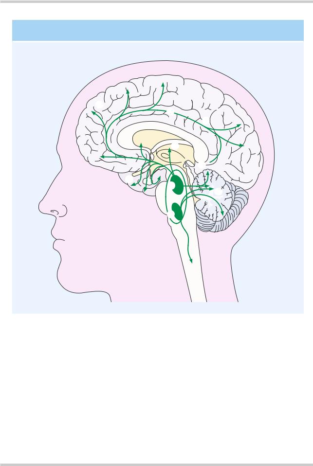
Figure 4.10 Representation of the primary serotonin-containing tracts in the human brain. Arising from the raphe nuclei these cells project to all cortical gray matter, with additional tracts to the basal ganglia and the cerebellum
More than nine distinct serotonin (5-HT) receptors have been identified. The 5-HT1A, 5- HT2A, 5-HT2C, and 5-HT3 receptors have been most extensively studied. The major site of serotonergic cell bodies is in the area of the upper pons and midbrain. The classic areas for 5-HT- containing neurons are the median and dorsal raphe nuclei. The neurons from the raphe nuclei project to the basal ganglia and various parts of the
limbic system, and have a wide distribution throughout the cerebral cortices in addition to cerebellar connections (Figure 4.10). All the 5-HT receptors identified to date are G-protein coupled receptors, except the 5-HT3 receptor, which is a ligand gated Na+/K+ channel.
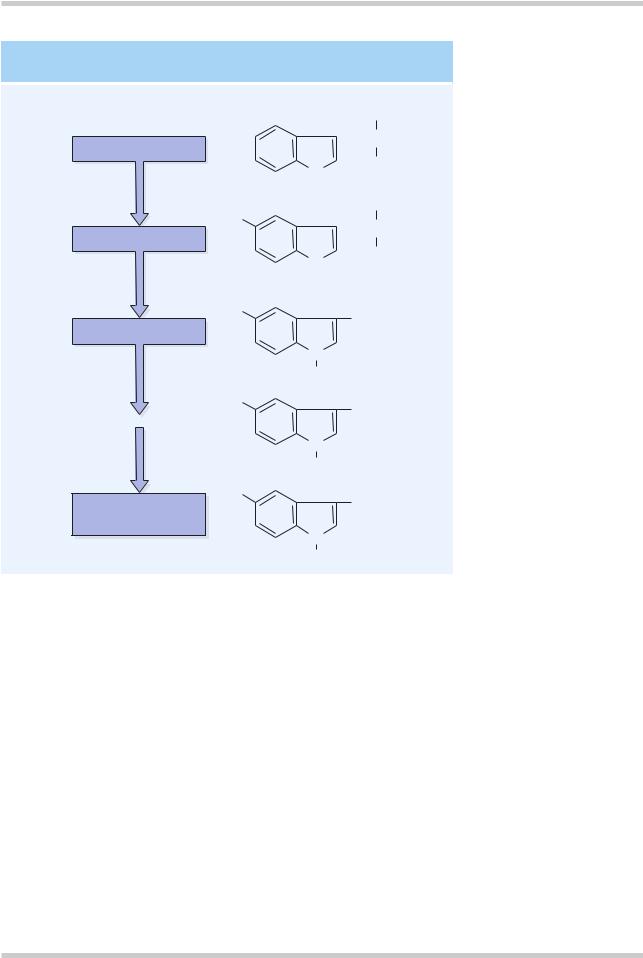
5-HT is synthesized from tryptophan by tryptophan hydroxylase, and the supply of tryptophan is the rate-limiting step in the
©2002 CRC Press LLC
SEROTONIN SYNTHESIS AND METABOLISM
|
|
|
|
COOH |
Tryptophan |
|
|
|
CH2CH |
|
|
|
||
|
|
|
|
|
Tryptophan |
N |
|
NH2 |
|
|
|
|
|
|
hydroxylase |
H |
|
COOH |
|
|
HO |
|
||
|
|
CH2CH |
||
5-Hydroxytryptophan |
|
|
|
|
|
|
|
||
|
|
|
|
|
L-aromatic |
N |
|
NH2 |
|
|
|
|
|
|
H |
|
|
||
acid decarboxylase |
|
|
||
HO
CH2CH2NH2
Serotonin (5-HT)
Monoamine |
N |
|
H |
||
oxidase |
||
|
||
|
HO |
|
|
CH2CHO |
|
Aldehyde |
N |
|
dehydrogenase |
H |
|
|
||
|
HO |
|
5-Hydroxyindoleacetic |
CH2COOH |
|
acid (5-HIAA) |
|
|
|
N |
|
|
H |
Figure 4.11 The rate-limiting step for serotonin synthesis is the availability of the precursor tryptophan. Tryptophan hydroxylase is the rate limiting enzyme. Serotonin in the CNS is primarily metabolized by monoamine oxidase. The primary metabolite is 5- hydroxyindoleacetic acid
synthesis of 5-HT (Figure 4.11). 5-HT is primarily broken down by monoamine oxidase and the primary metabolite is 5-HIAA.
Other neurotransmitters
Recent efforts have been directed towards finding an alternative neurochemical target in schizophrenia. The first of these that should be considered is gamma aminobutyric acid (GABA). GABA appears to have a regulatory role on dopaminergic function. The balance of evidence tends to suggest that GABA decreases dopaminergic firing. This links with human postmortem data indicating that GABAergic reductions correlate with increased dopamine concentrations19,20. Thus, it is possible that in schizophrenia there is a reduction in GABAergic function which leads to a
dysregulation of dopamine and the production of psychotic symptoms. A more likely candidate, however, appears to be the glutamatergic system. Glutamatergic dysfunction, particularly at the level of the N-methyl-D-aspartate (NMDA) receptor, has also been implicated in the pathophysiology of schizophrenia. Drugs which are antagonistic at the NMDA receptor, such as ketamine and phencyclidine, produce in healthy volunteers, both the positive, negative and neurocognitive symptoms that are characteristic of schizophrenia21. There is evidence that the propsychotic effects of these drugs may be mediated via an increase in the release of glutamate acting on non-NMDA receptors22.
If the function of NMDA receptors themselves is decreased this may remove the glutamatergic
©2002 CRC Press LLC