

Учебники / Auditory Trauma, Protection, and Repair Fay 2008
.pdf170 K.K. Ohlemiller and R.D. Frisina
mice showed a time course for age-linked MOC deterioration analogous to that in humans. Middle-aged animals showed a significant decline relative to the young adults, and further deficits in the efferent system were evident in old mice. As has been observed for humans (Varghese et al. 2005), wideband noise is more effective as a suppressing stimulus than narrowband signals such as pure tones.
8.3 Right Ear Advantage
Frisina’s group also examined the effects of age on the peripheral “right ear advantage” (Tadros et al. 2005a). In most young adult listeners, the right ear shows a lower audiometric threshold and higher amplitude DPOAEs. Tadros et al. compared these measures in “golden ear” old subjects (audiograms in the normal range) to those in subjects with typical, sloping high-frequency hearing loss characteristic of presbycusis. The golden ear subjects tended to have lower thresholds and higher otoacoustic emission amplitudes in the right ear, whereas this situation was reversed in the presbycusis subject group (Fig. 6.8). These findings suggest that the peripheral right ear advantage is not lost with age per se, but rather is lost as a part of presbycusis hearing loss.
Jerger and colleagues conducted an elegant series of dichotic listening experiments to shed light on hemispheric changes in central auditory processing with age. Young adult observers with normal hearing typically perceive auditory information more accurately when presented to the right ear (Jerger and Martin 2004). The opposite is true for nonlinguistic materials. Jerger and Jordan (1992) and Jerger et al. (1994) provided convincing evidence that asymmetric cortical processing of speech materials increases with age, i.e., there is an increased right ear advantage in subjects with presbycusis. This robust finding was apparent for
Figure 6.8. A significant difference in DPOAE amplitude decline, i.e., the normal hearing group relative to the presbycusis group. The right ear DPOAE decrement is more than the left ear decline especially in the f2 = 2–5 kHz region. (From Tadros et al. [2005a], Fig. 3, with permission.)
6. Age-Related Hearing Loss |
171 |
measures of correct responses using speech material, as well as for reaction-time measurements of auditory performance (Jerger et al. 1995).
9. Cellular Aging Mechanisms in ARHL
Interfacing with the environment exposes all sensory epithelia to injury risk, because sensory stimuli contain energy that cannot all be usefully transduced. Just as excessive light injures the retina, excessive sound injures the organ of Corti. Unlike the retina, the organ of Corti is subject to both biochemical and mechanical injury in the course of its normal function. A lifetime of operating as an intermediary between the acoustic world and the brain inevitably yields injury that cannot be distinguished from ostensibly “pure” aging processes, so that aging-as-injury is a theme of this chapter.
Aging research in humans and animals has sought “longevity genes” and longevity-promoting environments and practices (Karasik et al. 2005; Sinclair 2005; Geesaman 2006; Harper et al. 2006). Alleles and environments that promote healthy aging and longevity—or the opposite—will probably often impact the apparent rate of ARHL. As in all tissues, however, the uniqueness of the cochlea arises from the activity of a unique set of genes. Some mutations in genes that govern hearing-specific structures and processes may therefore emerge as candidate “ARHL genes,” actually meaning that certain alleles at particular loci can promote ARHL Most likely, all pro-ARHL alleles will be subject to strong environmental influences, as well as modulation by several other loci. Although no clear candidates have yet been identified in humans (Ates et al. 2005; Carlsson et al. 2005; Unal et al. 2005), possible ARHL-promoting genes and gene functions have been identified in mice. The following sections attempt to place ARHL into a wider context of cellular aging and the prevailing theories in this area.
9.1 Theories of Cellular Aging
Models of cellular aging can generally be placed into two categories: (1) Aging as a regulated “program” and (2) aging as dysregulation. The first category supposes that aging evolved as an adaptive process, and has largely fallen out of favor. It is far more likely that longevity is simply not selected for after peak reproductive age. Nevertheless, one type of “built-in” limitation, a limit on the total number of cell divisions in mitotically active cell populations, may yet be adaptive. Each time chromosomes are duplicated as part of mitosis, end segments called telomeres are shortened (von Zglinicki and Martin-Ruiz 2005). At some point, the telomeres become too short for duplication to occur. This mechanism of aging appears most relevant to tissues that emphasize cell replacement over repair, and may have evolved to help protect against cancer. While it may have some applicability to cochlear aging, its meaning for actual age-related hearing loss is less clear, as is considered further.
172 K.K. Ohlemiller and R.D. Frisina
The notion of aging as dysregulation and loss of homeostasis forms the basis of several aging hypotheses having relevance to ARHL. Cellular changes with aging typically include crippling modifications to DNA, important housekeeping proteins, and membrane lipids (e.g., Fenech 1998; Squier and Bigelow 2000; Leutner et al. 2001). Why should this occur given that cells have a host of DNA repair enzymes, and are constantly making or importing new proteins and lipids? The most significant and debilitating changes may permanently alter the DNA “blueprints” themselves. Proteins made from corrupted genes may have reduced function and be subject to improper folding and aggregation (Squier 2001). Proteins whose job it is to promote proper folding (chaperones) and to degrade damaged proteins (proteasomes) are also subject to modification, so that nonfunctional proteins may accumulate.
9.2 Progressive Cell Injury by Oxidative Stress
If much of aging can be equated with injury, the question arises regarding the kinds of injury to which the cells are subject. Most likely is oxidative stress (see also Wangemann, Chapter 3). The evolution of aerobic (oxygen-based) metabolism made it possible for cells to increase their energy output, and their range of activities. Oxygen is useful precisely because of its ability to break down carbon–carbon and carbon–hydrogen chemical bonds, the types of bonds that form biomolecules. This ability, however, might also present a problem. Cells use oxygen to synthesize metabolic intermediates and to fuel energy production, but must avoid being oxidized themselves. Inevitably, some oxidative attack on cellular DNA, proteins, and lipids does occur and is exacerbated by nearly any type of environmental stress. The observation that most injury to cells appears to be oxidative led to the Free Radical Theory of Aging, first proposed by Harman (1956), which asserts that aging is basically progressive oxidation. Aerobic cells have evolved to fend off oxidative attack in several ways. First, key reactions involving oxygen are quarantined to the mitochondrion. Inevitably, however, reactive oxygen-containing molecules (also known as reactive oxygen species or ROS) “escape” the intended reactions and boundaries. Cells then reduce oxidative injury by antioxidants, which either catalyze reactions that remove ROS or attenuate the activity of ROS. The Free Radical Theory has found much support, and currently provides the major framework for aging research (Fenech 1998; Squier and Bigelow 2000; Leutner et al. 2001; Barda 2002; Sinclair 2005; Hulbert et al. 2006). Oxidative modifications to cell constituents have detectable biochemical “signatures” for localization and semiquantitation, and studies have shown age-related increases in such changes in a host of tissues. Consequently, dietary antioxidants both decrease age-related infirmity and increase lifespan in animals. Moreover, treatments that increase lifespan, such as caloric restriction, also bolster antioxidant defenses and reduce oxidative tissue injury.
6. Age-Related Hearing Loss |
173 |
Experiments in humans and animals support the contention that the Free Radical Theory is applicable to ARHL. Cochlear injury caused by noise, ototoxins, and ischemia involves oxidative stress (see Henderson, Hu, and Bielefeld, Chapter 7 and Rybak, Talaska, and Schacht, Chapter 8 on noiseinduced and drug-induced hearing loss, respectively). Oxidative modification to DNA, proteins, and lipids of cochlear sensory cells is increased during aging (Jiang et al. 2006). The inactivation of genes encoding antioxidant enzymes SOD1 and glutathione peroxidase (GPx1) exacerbates apparent age-related cochlear pathology such as loss of hair cells and neurons, as well as thinning of stria vascularis (Ohlemiller et al. 1999, 2000; McFadden et al. 1999, 2001; Keithley et al. 2005). Interestingly, deficiency of SOD1 and GPx1 does not appear to shorten lifespan. Impairment of these critical and widely expressed antioxidant enzymes might be expected to promote broad pro-aging effects, and to decrease longevity. The narrower effects that are observed may testify to the special susceptibility of sensory epithelia to oxidative injury. Consistent with an involvement of oxidative stress in these pathologies, applications of antioxidants such as glutathione, d-methionine, and N-acetylcysteine reduce noise and ototoxic injury (reviews: Forge and Schacht 2000; Le Prell et al. 2006), and dietary application of vitamin E, vitamin C, or L-carnitine slow the progression of cochlear degeneration and hearing loss (Seidman 2000; Derin et al. 2004; Takumida and Anniko 2005; Le and Keithley 2006).
9.3 Mitochondrial Viability as a Key Factor in Aging
Mitochondria are both key targets of age-associated oxidative injury, and key mediators of aging effects on cells (Sastre et al. 2000; Kujothet al. 2005). Uniquely among intracellular organelles, mitochondria house their own DNA. This DNA codes for many essential components of energy production, but not for protective or repair components as nuclear DNA does. Damage to mitochondrial DNA also means that new mitochondria will carry the same errors, as new mitochondria come from replication of the old. Finally, as stated earlier, reactions carried out in mitochondria create most of the cell’s ROS, and ROS production may be exacerbated in compromised mitochondria. All of these factors may combine to promote the accumulation of DNA errors within individual mitochondria, so that overall energy production is reduced, and the function of the entire cell is impaired. A related view of aging, known as the Mitochondrial Clock Theory (see Seidman 2000), is perhaps a corollary to the Free Radical Theory. Accumulation of mitochondrial DNA mutations with age has been observed in essentially all tissues including the cochlea (Bai et al. 1997; Seidman et al. 2002; Pickles 2004;) and can be reduced both by antioxidants and by caloric restriction (Seidman 2000). Medical conditions that impact cochlear blood flow and possibly ARHL (see later), also promote mitochondrial DNA mutations (Dai et al. 2004).
174 K.K. Ohlemiller and R.D. Frisina
9.4 Calcium Dysregulation
Calcium is a critical regulator of cellular events (see Wangemann, Chapter 3). Its levels normally remain very low in cytoplasm and in extracellular fluids, so that minute changes can modulate specialized functions such as transduction in stereocilia and neurotransmitter release (e.g., Chan and Hudspeth 2005; Keen and Hudspeth 2006), as well as fundamental functions such as growth, division, and death (Lu and Means 1993; Krebs 1998). Major cytoplasmic proteins that buffer calcium or bind calcium for signaling include calmodulin, calbindin, parvalbumin, and calretinin (Lu and Means 1993; Schwaller et al. 2002). Because of the prominence of Ca2+ in many vital processes, it is not surprising that dysregulation of calcium may contribute to cellular aging (Squier and Bigelow 2000; Crompton 2004; Toescu 2005). Disruption of calcium homeostasis is part of the mechanism of excitotoxicity, and may be among the causes of neural ARHL (Lang et al. 2006a). In addition, the Cdh23 gene locus modifies other loci related to calcium, such as the one encoding plasma membrane Ca2+-ATPase 2 (Pmca2) (Davis et al. 2003). Thus, the sensory ARHL-like pathology associated with the Cdh23ahlallele may in part reflect calcium dysregulation.
Age-related changes in ACNS function also involve changes in calcium regulation. Zettel et al. (1997) examined changes in intracellular calcium binding proteins in the region of the IC shown by Walton’s group to undergo agerelated decline in temporal processing. In both CBA and B6 strains, calbindin levels declined with age. However, calretinin exhibited upregulation with age in CBA mice. To test whether this upregulation was strain-dependent or activitydependent, Zettel et al. (2001) deafened young adult CBA mice by cochlear ablation and examined changes in calretinin immunochemistry in the IC with aging. Control CBAs showed upregulation of calretinin with aging, but the deafened CBAs did not, supporting the idea that maintenance of neuronal activity in IC (through preservation of cochlear function) is important for calretinin regulation. Subsequent work in B6 mice by the same group (O’Neill et al. 1997) also revealed an age-related decline in calbindin-labeled neurons in the medial nucleus of the trapezoid body.
Canlon and colleagues (Idrizbegovic et al. 2001a,b) examined age-related changes in calcium-binding proteins in the cochlear nucleus of CBA mice. The percentage of neurons in the DCN staining for calbindin, calretinin, and parvalbumin was upregulated with age for ages up to 29 months. The age-related upregulation of calretinin and parvalbumin was correlated with the degree of peripheral hearing loss, as measured by inner and outer hair cell loss and spiral ganglion cell loss, suggesting peripherally induced central effects. Quantitative stereology using optical fractionation to obtain total neuron counts in PVCN and DCN revealed that the total number of PVCN neurons remained constant with age, whereas DCN cell numbers declined. Only parvalbumin showed an age-related upregulation in the PVCN.
Subsequent investigations by the same group contrasted the changes in the CBA cochlear nucleus with those in B6. Like CBA mice, B6 showed age-related upregulation of calbindin and parvalbumin in the DCN and PVCN for ages
6. Age-Related Hearing Loss |
175 |
up to 30 months (Idrizbegovic et al. 2003). Also as in CBA, calbindin and parvalbumin were correlated with peripheral hair cell and neurons loss in DCN and PVCN. The upregulation was accompanied by age-related declines in the absolute number of neurons in the DCN and PVCN (Idrizbegovic et al. 2004).
9.5 Limitations on Cell Repair and Replacement
Tissues that have sustained some degree of injury might compensate for cell loss by replacement of constituent cells by mitosis. Only a few cell types are normally replaced in the auditory system of adult mammals. Neurons are not replaced, nor are cochlear hair cells, nor most cells of the organ of Corti. This limitation places a premium on protective and repair capabilities. Clumping of hair cell stereocilia and deformation of the cuticular plate can be seen in aged humans (Scholtz et al. 2001), suggesting that normal bundle renewal is impaired in old hair cells, yet these nonfunctional hair cells may survive for some time. Supporting cells with pyknotic nuclei and dark cytoplasm are frequently observed in the organ of Corti of old mice (Ohlemiller and Gagnon 2004b). It is not clear whether cells showing these signs die and are replaced, or whether supporting cell pathology can also promote hair cell loss.
Within the cochlea, cell replacement appears limited to fibrocytes of the lateral wall and intermediate cells and marginal cells of the stria vascularis (Conlee et al. 1994; Yamashita et al. 1999; Dunaway et al. 2003; Hirose et al. 2005). Still these decrease in number with age (Wright and Schuknecht 1972; Ichimiya et al. 2000; Ohlemiller 2006), perhaps as a result of limits on proliferative capacity such as telomere status. Some new fibrocytes in ligament may derive from bone marrow (Lang et al. 2006b), so that it is not clear why there is net loss of fibrocytes with age. Both strial cells and fibrocytes in spiral ligament and limbus serve a distributed function, and are initially present in excess, as indicated by the fact that substantial numbers of these can be lost without any effect on hearing. While limits on strial cell replacement may play a role in strial ARHL (Ohlemiller 2006), it is at present not clear that ligament pathology is a significant factor in ARHL.
10. Risk Factors Affecting ARHL
The prevalence of clinically defined ARHL in the very old, while high, is not 100%. Although it appears in all societies, it occurs more frequently in industrial cultures than in nonindustrial cultures (Rosen et al. 1962). Such evidence argues that much of ARHL is influenced by the interplay between pro-ARHL alleles (or pro-aging alleles) and environment. No behavior, event, or environment carries a fixed degree of ARHL risk. Rather, the risk will depend on unknown alleles carried by the individual. Until such alleles are identified, the best strategy is to minimize environmental risks, which take many forms.
176 K.K. Ohlemiller and R.D. Frisina
10.1 Noise and Ototoxins
Environmental risk factors for apparent ARHL include acute or chronic exposure to noise, ototoxic medications, industrial solvents, or combinations of these (Gilad and Glorig 1979b; Rosenhall et al. 1993; Rosenhall and Pedersen 1995; Toppila et al. 2001; Fransen et al. 2003; Fechter 2004). Assaults by these agents appear to promote largely oxidative injury that primarily injures hair cells (see Henderson, Hu, and Bielefeld, Chapter 7; Rybak, Talaska, and Schacht, Chapter 8). Note that the intention here is not to equate cochlear noise and ototoxic injury with ARHL, or to suggest that the cellular pattern of injury is exactly the same in all three cases although there are intriguing similarities. Both noise and ototoxin exposure can, for example, cause permanent strial injury (e.g., Ulehlova, 1983; Garetz and Schacht 1996; Hirose and Liberman 2003). They mostly do not, however, cause permanent EP reduction, and thus would not be expected to draw a diagnosis of strial ARHL. There is likewise no compelling evidence that ototoxins promote primary neural loss sufficient to bring a diagnosis of neural ARHL. By contrast, early noise exposure may yield neural ARHL (Kujawa and Liberman 2006). However, because the principal targets of noise and ototoxins will be hair cells, the diagnosis can often be confused with sensory ARHL.
10.2 Lifestyle and Risk of Vascular Pathology
Proper function of the cochlea, particularly the lateral wall, is energy intensive, and likely to be vulnerable to any restriction of blood flow. Accordingly, the role of vascular insufficiency has long been a prominent topic in ARHL research (reviews: Gilad and Glorig 1979b; Nakashima et al. 2003). Obesity and conditions to which it may lead (hyperlipidemia, hypercholesterolemia, hypertension, hyperhomocysteinemia, hyperlipoproteinemia, and cardiovascular disease) have all been implicated in ARHL (Rosen et al. 1970; Spencer 1973; Drettner et al. 1975; Tachibana et al. 1984; Axellson and Lindgren 1985; Pillsbury 1986; Saito et al. 1986; Sikora et al. 1986; Suzuki et al. 2000; Satar et al. 2001; Fransen et al. 2003). Poor health habits with regard to exercise, smoking, and diet may also be risk factors for ARHL insofar as they impact vascular health, tissue oxygenation, and diabetes risk (see later) (Rosenhall et al. 1993; Cruickshanks et al. 1998; Torre et al. 2005; Uchida et al. 2005), although probably only as part of a spectrum of conditions of aging. While it has been suggested that the most immediate cochlear target of vascular pathology is likely to be the stria, limited observations of affected human and animal cochleae suggest broad tissue degeneration, and no special relationship to strial ARHL.
10.3 Early Exposure to Stress
Environment extends to the prenatal environment. It has been hypothesized that prenatal stress can “program” individuals to pathology that resembles accelerated
6. Age-Related Hearing Loss |
177 |
aging, or to possible risk factors for age-related pathology such as hypertension and cardiovascular disease (Barrenas et al. 2003). Suggested mechanisms involve “redeployment” of resources by the fetus to favor some tissues and organs, leaving others with fewer stem cells or poorly vascularized, and permanent alterations in endocrine function (Barker 1998). One result of such events may be sensorineural hearing loss associated with shortened stature in adulthood (Barrenäs et al. 2005). A potentially related finding is that exposure of prenatal rats to glucocorticoid stress hormones increases susceptibility in the young adult to NIHL possibly due to an overall increased vulnerability to oxidative stress (Canlon et al. 2003).
10.4 Mineralocorticoid Levels
Aging is often accompanied by medical comorbidities such as decreases in hormonal levels. A decline in levels of aldosterone, a mineralocorticoid produced by the adrenal cortex, may affect ionic balance, partly by its actions on Na+, K+-ATPase and the K+, Na+, Cl− cotransporter. Because these enzymes are highly expressed in the cochlear lateral wall and are critical to ion regulation in the cochlea, aldosterone may participate in the regulation of the EP. Alternatively, it may operate at a systemic level by averting hypertension or reducing inflammation. Blood aldosterone titer probably reflects both genetic and environmental influences. Frisina’s group examined the relation between aldosterone levels and age-related hearing loss in aged human subjects who had good hearing and showed good health overall (Tadros et al. 2005b). Based on standard audiometric criteria, the subjects were classified into three groups: golden ear (audiometric thresholds in the normal range), mild/moderate hearing loss, and severe loss. Serum aldosterone levels were significantly different between the groups, with the golden ears showing the highest aldosterone, the mild/moderate group next, and the subjects with severe hearing loss the least amount of aldosterone (Fig. 6.9). Interestingly, all aldosterone levels were within normal clinical limits. Regression analyses showed significant correlations between aldosterone levels, pure tone thresholds, and hearing-in-noise test (HINT) scores. These results suggest that aldosterone may be protective against presbycusis, as has been found for autoimmune hearing loss (Trune et al. 2006). At present there is no direct evidence to indicate which cochlear structures are preserved or affected.
10.5 Diabetes Mellitus
Non-insulin-dependent (type 2, adult onset) diabetes mellitus often appears as a condition of aging, frequently as a complication of obesity. In middle age, diabetes also produces multisystemic pathology that mimics aspects of aging (Geesaman 2006). Chronically elevated plasma glucose promotes malconformation and aggregation of proteins in all tissues, yet with particularly deleterious

178 K.K. Ohlemiller and R.D. Frisina
(a) |
(b) |
(c)
Figure 6.9. (A) A significant difference in serum aldosterone concentration was found between normal hearing and presbycusic groups, with a higher concentration in the normal hearing group. (B) A significant difference in serum aldosterone concentrations was found for the 58to 73-year-old groups of normal hearing subjects and presbycusic subjects, with higher concentrations in the normal hearing group. This analysis eliminated the age factor. (C) A significant difference in serum aldosterone concentrations was found between normal hearing and both mild/moderate and severe presbycusic groups.(From Tadros et al. [2005b], Fig. 1. Reprinted with permission.)
consequences for the microvasculature. The most wide-ranging pathologies of diabetes therefore appear mediated by microangiopathy. Both type 1 (juvenile) and type 2 diabetes promote hearing loss and cochlear pathology in humans (Wackym and Linthicum 1986; Fukushima et al. 2005) and animals (Rust et al. 1992; Raynor et al. 1995; Ishikawa et al. 1995). Type 2 diabetes has been proposed as a cause of ARHL, but the evidence for this is mixed (Malpas et al. 1989; Ma et al. 1998; Ologe et al. 2005; Vaughan et al. 2005). To clarify whether presbycusis is accelerated in aged type 2 diabetics, a group of type 2 diabetics older than the age of 60 years were compared with a group of ageand sex-matched controls (Frisina et al. 2006). Both groups were otherwise
6. Age-Related Hearing Loss |
179 |
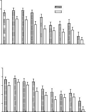
healthy and had no history of major health or hearing problems. Audiometric thresholds, otoacoustic emission levels, and speech thresholds revealed deficits in the diabetic group, with the right ear showing a more severe loss relative to the left (Fig. 6.10). Tests involving the ACNS, such as suprathreshold gap detection and HINT scores, also exposed relative deficits in the diabetic group. These findings support a causal link between type 2 diabetes and both peripheral and central aspects of ARHL.
Right Ear DPOAEs
DP amplitude (dB SPL)
DP amplitude (dB SPL)
5
Non-diabetics
0
Diabetics
–5 


–10 


–15 


–20
1.0 |
1.3 |
1.6 |
2.0 |
2.5 |
3.2 |
4.0 |
5.0 |
6.8 |
Frequency (kHz)
Left Ear DPOAEs
5 |
|
|
|
|
|
|
|
|
0 |
|
|
|
|
|
|
|
|
–5 |
|
|
|
|
|
|
|
|
–10 |
|
|
|
|
|
|
|
|
–15 |
|
|
|
|
|
|
|
|
–20 |
1.3 |
1.6 |
2.0 |
2.5 |
3.2 |
4.0 |
5.0 |
6.8 |
1.0 |
GM Frequency (kHz)
Figure 6.10. At all frequencies, DPOAEs were smaller for diabetics relative to nondiabetics. Like the threshold measures presented in the previous figures, the right ear was more affected than the left. ANOVA showed significant main effects of subject group (Right: p < 0.0001, F = 31.1, df= 1; Left: p < 0.0001, F = 15.2, df = 1). Interactions and subject group Bonferroni post-hoc analyses were not significant, except for the right ear at 2 kHz: p < 0.05, t = 2.82, df = 1. GM = geometric mean of f1 and f2. Error bars are SEM. (From Frisina et al. [2006], Fig. 3. Reprinted with permission.)