

GUIDELINES AND STANDARDS
Recommendations for Cardiac Chamber Quantification by Echocardiography in Adults: An Update from the American Society
of Echocardiography and the European Association
of Cardiovascular Imaging
Roberto M. Lang, MD, FASE, FESC, Luigi P. Badano, MD, PhD, FESC, Victor Mor-Avi, PhD, FASE, Jonathan Afilalo, MD, MSc, Anderson Armstrong, MD, MSc, Laura Ernande, MD, PhD,
Frank A. Flachskampf, MD, FESC, Elyse Foster, MD, FASE, Steven A. Goldstein, MD, Tatiana Kuznetsova, MD, PhD, Patrizio Lancellotti, MD, PhD, FESC, Denisa Muraru, MD, PhD,
Michael H. Picard, MD, FASE, Ernst R. Rietzschel, MD, PhD, Lawrence Rudski, MD, FASE, Kirk T. Spencer, MD, FASE, Wendy Tsang, MD, and Jens-Uwe Voigt, MD, PhD, FESC, Chicago, Illinois; Padua, Italy; Montreal, Quebec and Toronto, Ontario, Canada; Baltimore, Maryland; Creteil, France; Uppsala, Sweden; San Francisco, California; Washington, District of Columbia; Leuven, Liege, and Ghent, Belgium; Boston, Massachusetts
The rapid technological developments of the past decade and the changes in echocardiographic practice brought about by these developments have resulted in the need for updated recommendations to the previously published guidelines for cardiac chamber quantification, which was the goal of the joint writing group assembled by the American Society of Echocardiography and the European Association of Cardiovascular Imaging. This document provides updated normal values for all four cardiac chambers, including threedimensional echocardiography and myocardial deformation, when possible, on the basis of considerably larger numbers of normal subjects, compiled from multiple databases. In addition, this document attempts to eliminate several minor discrepancies that existed between previously published guidelines. (J Am Soc Echocardiogr 2015;28:1-39.)
Keywords: Adult echocardiography, Transthoracic echocardiography, Ventricular function, Normal values
From the University of Chicago Medical Center, Chicago, Illinois (R.M.L., V.M.-A., K.T.S.); the University of Padua, Padua, Italy (L.P.B., D.M.); Jewish General Hospital, McGill University, Montreal, Quebec, Canada (J.A., L.R.); Johns Hopkins University, Baltimore, Maryland (A.A.); INSERM U955 and Hopital^ Henri Mondor, Creteil, France (L.E.); Uppsala University, Uppsala, Sweden (F.A.F.); the University of California, San Francisco, San Francisco, California (E.F.); Medstar Washington Hospital Center, Washington, District of Columbia (S.A.G.); University Hospital Leuven, Leuven, Belgium (T.K., J.-U.V.); the University of Liege Hospital, Liege, Belgium (P.L.); Massachusetts General Hospital and Harvard Medical School, Boston, Massachusetts (M.H.P.); Ghent University Hospital, Ghent, Belgium (E.R.R.); and the University of Toronto, Toronto, Ontario, Canada (W.T.).
The following authors reported no actual or potential conflicts of interest in relation to this document: Jonathan Afilalo, MD, MSc, Anderson Armstrong, MD, MSc, Laura Ernande, MD, PhD, Frank A. Flachskampf, MD, FESC, Steven A. Goldstein, MD, Tatiana Kuznetsova, MD, PhD, Patrizio Lancellotti, MD, PhD, FESC, Victor Mor-Avi, PhD, FASE, Michael H. Picard, MD, FASE, Ernst R. Rietzschel, MD, PhD, Kirk T. Spencer, MD, FASE, Wendy Tsang, MD, and Jens-Uwe Voigt, MD, PhD, FESC. The following authors reported relationships with one or more commercial interests: Luigi P. Badano, MD, PhD, FESC, received grants from GE Healthcare, Siemens, and Esaote and serves on the speakers’ bureau for GE
Healthcare. Elyse Foster, MD, FASE, received grant support from Abbott Vascular Structural Heart. Roberto M. Lang, MD, FASE, FESC, received grants from and serves on the speakers’ bureau and advisory board for Philips Medical Systems. Denisa Muraru, MD, received research equipment from and served as a consultant for GE Healthcare. Lawrence Rudski, MD, FASE, holds stock in GE.
Attention ASE Members:
The ASE has gone green! Visit www.aseuniversity.org to earn free continuing medical education credit through an online activity related to this article. Certificates are available for immediate access upon successful completion of the activity. Nonmembers will need to join the ASE to access this great member benefit!
Drs Lang and Badano co-chaired the Writing Group.
Reprint requests: American Society of Echocardiography, 2100 Gateway Centre Boulevard, Suite 310, Morrisville, NC 27560 (E-mail: ase@asecho.org).
0894-7317/$36.00
Copyright 2015 by the American Society of Echocardiography.
http://dx.doi.org/10.1016/j.echo.2014.10.003
1

2 Lang et al
Abbreviations
AP = Anteroposterior
ASE = American Society of
Echocardiography
BSA = Body surface area
CMR = Cardiac magnetic resonance
DTI = Doppler tissue imaging
EACVI = European
Association of Cardiovascular
Imaging
EDV = End-diastolic volume
EF = Ejection fraction
ESV = End-systolic volume
FAC = Fractional area change
GLS = Global longitudinal strain
I-I = Inner edge–to–inner edge
IVC = Inferior vena cava
LA = Left atrial
L-L = Leading edge–to– leading edge
LV = Left ventricular
MDCT = Multidetector computed tomography
PW = Pulsed-wave
RA = Right atrial
RIMP = Right ventricular index of myocardial performance
RV = Right ventricular
RWT = Relative wall thickness
STE = Speckle-tracking echocardiography
TAPSE = Tricuspid annular plane systolic excursion
TAVI = Transcatheter aortic valve implantation
TAVR = Transcatheter aortic valve replacement
TEE = Transesophageal echocardiography
3D = Three-dimensional
3DE = Three-dimensional echocardiography
TTE = Transthoracic echocardiography
2D = Two-dimensional
2DE = Two-dimensional echocardiography
TABLE OF CONTENTS
I. The Left Ventricle |
3 |
|
1. Measurement |
of |
LV |
Size 3 |
|
|
1.1. Linear |
Measure- |
|
ments 3 |
|
|
1.2.Volumetric Measurements 3
1.3.Normal Reference
Values for 2DE |
6 |
|
1.4. Normal |
Reference |
|
Values for 3DE |
6 |
|
Recommendation |
6 |
|
2. LV Global Systolic Func- |
||
tion 6 |
|
|
2.1. Fractional |
Short- |
|
ening |
6 |
|
2.2.EF 7
2.3.Global Longitudinal
Strain (GLS) |
7 |
2.4.Normal Reference Values 7
Recommendations 10 |
|
|
3. LV |
Regional |
Func- |
tion |
10 |
|
3.1. Segmentation |
of the |
|
Left Ventricle |
10 |
|
3.2. Visual |
Assess- |
|
ment 11 |
|
|
3.3.Regional Wall Motion during Infarction and Ischemia 11
3.4.Regional Abnormalities in the Absence of Coronary Artery Dis-
ease 11
3.5.Quantification of
Regional Wall Motion
Using Doppler and
STE 11
Recommendations |
12 |
4. LV Mass |
13 |
Recommendations |
16 |
II. The Right Ventricle 16
5.General Recommendations for RV Quantifica-
|
tion 16 |
|
|
6. |
Essential |
Imaging |
Win- |
|
dows and Views |
16 |
|
7. |
RV Measurements |
17 |
|
7.1. Linear |
Measure- |
||
|
ments |
17 |
|
7.2.Volumetric Measurements 17
Recommendations 17 |
|
|
8. RV |
Systolic |
Func- |
tion |
19 |
|
8.1.RIMP 19
8.2.TAPSE 19
8.3. RV 2D FAC 19
8.4.DTI-Derived Tricuspid Lateral Annular Systolic Velocity 20
Journal of the American Society of Echocardiography
January 2015
8.5. RV Strain and Strain Rate |
20 |
|
||||
Recommendations |
20 |
|
|
|
|
|
8.6. RV 3D EF |
20 |
|
|
|
||
Recommendation |
20 |
|
|
|
|
|
III. The Left and Right Atria |
20 |
|
|
|||
9. LA Measurements |
|
24 |
|
|
||
9.1. General Considerations for LA Size |
24 |
|||||
9.2. Linear Dimensions and Area Measurements 25 |
||||||
9.3. Volume Measurements |
25 |
|
||||
9.4. Normal Values of LA Measurements |
25 |
|||||
Recommendations |
28 |
|
|
|
|
|
10. Right Atrial measurements |
28 |
|
||||
Recommendations |
28 |
|
|
|
|
|
IV. The Aortic Annulus and Aortic Root 28 |
|
|||||
11. The Aortic Annulus |
28 |
|
|
|||
12. The Aortic Root |
30 |
|
|
|||
13. Identification of Aortic Root Dilatation |
32 |
|||||
Recommendations |
32 |
|
|
|
|
|
V. The Inferior Vena Cava |
32 |
|
|
|||
Notice and Disclaimer |
33 |
|
|
|
||
References |
33 |
|
|
|
|
|
Appendix |
39.e1 |
|
|
|
|
|
Methods |
39.e1 |
|
|
|
|
|
Echocardiographic Measurements |
39.e1 |
|
||||
Statistical Analysis |
39.e1 |
|
|
|
||
The quantification of cardiac chamber size and function is the cornerstone of cardiac imaging, with echocardiography being the most commonly used noninvasive modality because of its unique ability to provide real-time images of the beating heart, combined with its availability and portability. Standardization of the methodology used to quantify cardiac chambers is maintained by creating and disseminating official recommendations, which when followed by practitioners provides uniformity and facilitates communication. Recommendations for echocardiographic chamber quantification were last published in 2005 by the American Society of Echocardiography (ASE) and the European Association of Echocardiography (renamed the European Association of Cardiovascular Imaging [EACVI]).1,2
Since then, echocardiographic technology has continued evolving, with two major developments being real-time threedimensional (3D) echocardiography (3DE) and myocardial deformation imaging. The goal of this document is to provide an update to the previously published guidelines, as well as recommendations and reference values, while eliminating the minor discrepancies that existed between previous guidelines. The normal values in this update include 3DE and myocardial deformation, when possible. Importantly, compared with the previous guidelines, this update is based on considerably larger numbers of normal subjects, compiled from multiple databases, to improve the reliability of the reference values.
Although most issues covered in this document reflect a broad consensus among the members of the writing group, one important issue the group debated was partition values for severity of abnormalities. Most often, in addition to describing a parameter as normal or abnormal (reference values), clinical echocardiographers qualify the degree of abnormality with terms such as mildly, moderately, and

Journal of the American Society of Echocardiography |
Lang et al 3 |
Volume 28 Number 1 |
|
severely abnormal, which reflect the degree to which measurements deviate from normal. In addition to providing normative data, it would be beneficial to standardize cutoffs for severity of abnormality for all parameters across echocardiography laboratories, such that the term moderately abnormal, for example, would have the same meaning universally. However, different approaches may be used for determining cutoff values for the different degrees of abnormality, all of which have significant limitations.
The first approach would be to empirically define cutoffs for mild, moderate, and severe abnormalities on the basis of SDs above or below the reference limit derived from a group of healthy people. The advantage of this method is that these data readily exist for most echocardiographic parameters. However, this approach is fundamentally flawed. First, not all echocardiographic parameters are normally distributed (or Gaussian), even in a normal population. Second, even if a particular parameter is normally distributed in normal subjects, most echocardiographic parameters, when measured in the general population, have a significant asymmetric distribution in one direction (abnormally large for size or abnormally low for function parameters). An alternative method would be to define abnormalities on the basis of percentile values (e.g., 95th, 99th) of measurements derived from a population that includes both healthy people and those with disease. Although these data would still not be normally distributed, they would account for the asymmetric distribution and the range of abnormality present within the general population. The major limitation of this approach is that such population data sets simply do not exist for most echocardiographic variables.
Ideally, an approach that predicts outcomes or prognosis would be preferred. That is, defining a variable as moderately deviated from normal would imply that there is a moderate risk for a particular adverse outcome for a patient. Although sufficient data linking risk and cardiac chamber sizes exist for several parameters (e.g., left ventricular [LV] size and ejection fraction [EF], left atrial [LA] volume), outcomes data are lacking for many other parameters. Unfortunately, this approach also has limitations. The first obstacle is how to best define risk. The cutoffs suggested for the same parameter vary broadly for different risks in different patient populations and disease states.
Last, cutoff values may be determined by experience-based consensus of expert opinions. An extensive debate arose among the members of the writing group, some of whom felt that providing partition values on the basis of this scientifically less- than-rigorous approach would be a disservice to the echocardiography community and that a disease-specific approach might be required to achieve meaningful clinical categorization of the severity of abnormality. Others felt that such cutoffs would provide a uniform reference for echocardiographic reporting, which would be easier to interpret by referring clinicians. The compromise was to provide experience-based partition values only for LV EF and LA volume, while suggested partition values for additional parameters of LV size and mass are listed in the Appendix. All partition values should interpreted with caution in this perspective.
For parameters other than LV size, function, and mass as well as LA volume, only the mean value and the SD of gender-, age-, and body surface area (BSA)–normalized cutoffs or upper and lower limits are reported in the appropriate sections of this document. For these parameters, measurements exceeding 61.96 SDs (i.e., the 95% confidence interval) should be classified as abnormal. Any description of the degree of deviation from normality in the
echocardiographic report should remain at the discretion of the individual laboratory, and the writing group does not recommend specific partition values.
Quantification using transesophageal echocardiography (TEE) has advantages and disadvantages compared with transthoracic echocardiography (TTE). Although visualization of many cardiac structures is improved with TEE, some differences in measurements have been found between TEE and TTE, particularly for chamber dimensions and thickness. These differences are primarily attributable to the inability to obtain from the transesophageal approach the standardized imaging planes and views used when quantifying chamber dimensions transthoracically. It is the recommendation of this writing group that the same range of normal values for LV and right ventricular (RV) chamber dimensions and volumes apply for both TEE and TTE. For details on specific views for optimal measurements, please refer to the recently published TEE guidelines.3
All measurements described in this document should be performed on more than one cardiac cycle to account for interbeat variability. The committee suggests the average of three beats for patients in normal sinus rhythm and a minimum of five beats in patients with atrial fibrillation. Because the committee acknowledges that the implementation of this recommendation is time consuming, the use of representative beats is acceptable in the clinical setting.
I. THE LEFT VENTRICLE
1. Measurement of LV Size
The most commonly used parameters to describe LV cavity size include linear internal dimensions and volumes. Measurements are commonly reported for end-diastole and end-systole, which are then used to derive parameters of global LV function. To allow comparison among individuals with different body sizes, chamber measurements should be reported indexed to BSA.
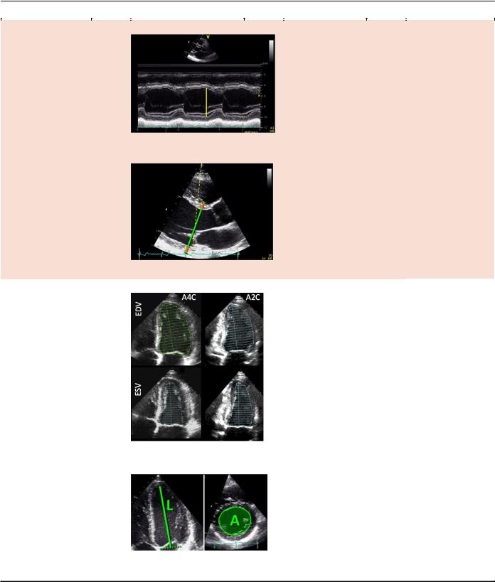
1.1.Linear Measurements. It is recommended that linear internal measurements of the left ventricle and its walls be performed in the parasternal long-axis view. Values should be carefully obtained perpendicular to the LV long axis and measured at or immediately below the level of the mitral valve leaflet tips. In this regard, the electronic calipers should be positioned on the interface between the myocardial wall and cavity and the interface between the wall and the pericardium. Internal dimensions can be obtained with a twodimensional (2D) echocardiography (2DE)–guided M-mode approach, although linear measurements obtained from 2D echocardiographic images are preferred to avoid oblique sections of the ventricle (Table 1).
1.2.Volumetric Measurements. LV volumes are measured using 2DE or 3DE. Volume calculations derived from linear measurements may be inaccurate, because they rely on the assumption of a fixed geometric LV shape such as a prolate ellipsoid, which does not apply in a variety of cardiac pathologies. Accordingly, the Teichholz and Quinones methods for calculating LV volumes from LV linear dimensions are no longer recommended for clinical use.
Volumetric measurements are usually based on tracings of the interface between the compacted myocardium and the LV cavity.
4 Lang et al |
|
Journal of the American Society of Echocardiography |
|
|
|
|
January 2015 |
|
|
||
Table 1 Recommendations for the echocardiographic assessment of LV size and function |
|
||
|
|
|
|
Parameter and method |
Technique |
Advantages |
Limitations |
|
|
|
|
Internal linear |
M-mode tracing |
Reproducible |
Beam orientation |
dimensions. |
|
High temporal |
frequently off axis |
Linear internal |
|
resolution |
Single dimension, i.e., |
measurements of the LV |
|
Wealth of published |
representative only in |
should be acquired in the |
|
data |
normally shaped |
parasternal long-axis |
|
|
ventricles |
view carefully obtained |
|
|
|
perpendicular to the LV |
|
|
|
long axis, and measured |
|
|
|
at the level of the mitral |
|
|
|
valve leaflet tips. |
|
|
|
Electronic calipers |
|
|
|
should be positioned on |
|
|
|
the interface between |
2D-guided linear measurements |
|
Lower frame rates |
myocardial wall and |
|
||
|
Facilitates orientation |
||
cavity and the interface |
|
than M-mode |
|
between wall and |
|
perpendicular to the |
Single dimension, i.e., |
pericardium (orange |
|
ventricular long axis |
representative only in |
|
|
|
|
arrows). |
normally shaped |
|
ventricles |
||
|
Volumes. |
Biplane disk summation |
Corrects for shape |
Apex frequently |
Volume measurements |
|
distortions |
foreshortened |
are usually based on |
|
Less geometrical |
Endocardial dropout |
tracings of the blood- |
|
assumptions |
Blind to shape distor- |
tissue interface in the |
|
compared with linear |
tions not visualized in |
apical fourand two- |
|
dimensions |
the apical twoand |
chamber views. At the |
|
|
four-chamber planes |
mitral valve level, the |
|
|
|
contour is closed by |
|
|
|
connecting the two |
|
|
|
opposite sections of the |
|
|
|
mitral ring with a straight |
|
|
|
line. LV length is defined |
|
|
|
as the distance between |
|
|
|
the middle of this line |
|
|
|
and the most distant |
|
|
|
point of the LV contour. |
|
|
|
|
Area-length |
Partial correction for |
Apex frequently |
|
|
||
|
|
shape distortion |
foreshortened |
|
|
|
Heavily based on |
|
|
|
geometrical |
|
|
|
assumptions |
|
|
|
Limited published |
|
|
|
data on normal |
|
|
|
population |
(Continued)
Journal of the American Society of Echocardiography |
|
Lang et al 5 |
|
||
Volume 28 Number 1 |
|
|
|
|
|
|
|
|
|
|
|
|
Table 1 (Continued) |
|
|
|
|
|
|
|
|
|
|
|
Parameter and method |
Technique |
Advantages |
Limitations |
|
|
|
|
|
|
|
|
|
Endocardial border enhancement |
Helpful in patients with |
Same limitations as |
|
|
|
|
suboptimal acoustic |
the above non- |
|
|
|
|
window |
contrast 2D |
|
|
|
|
Provides volumes that |
techniques |
|
|
|
|
are closer to those |
Acoustic shadowing in |
|
|
|
|
measured with cardiac |
LV basal segments |
|
|
|
|
magnetic resonance |
with excess contrast |
|
3D data sets |
No geometrical |
|
|
Lower temporal |
|
|
assumption |
|
|
Unaffected by |
resolution |
|
foreshortening |
Less published data |
|
More accurate and |
on normal values |
|
reproducible |
Image quality |
|
compared to other |
dependent |
|
imaging modalities |
|
Global Longitudinal |
Angle independent |
Vendor dependent |
Strain. |
Established |
|
Peak value of 2D |
prognostic value |
|
longitudinal speckle |
|
|
tracking derived strain |
|
|
(%). |
|
|
|
|
|
2D, two-dimensional; 3D, three-dimensional; A2C, apical 2-chamber view; A4C, apical 4-chamber view; EDV, end-diastolic volume; ESV, end-sys- tolic volume; LV, left ventricular.
At the mitral valve level, the contour is closed by connecting the two opposite sections of the mitral ring with a straight line. LV length is defined as the distance between the bisector of this line and the apical point of the LV contour, which is most distant to it. The use of the longer LV length between the apical twoand four-chamber views is recommended.
LV volumes should be measured from the apical fourand two-chamber views. Two-dimensional echocardiographic image acquisition should aim to maximize LV areas, while avoiding foreshortening of the left ventricle, which results in volume underestimation. Acquiring LV views at a reduced depth to focus on the LV cavity will reduce the likelihood of foreshortening and
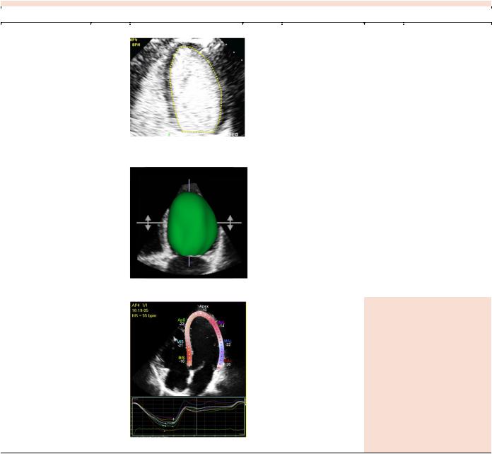
minimize errors in endocardial border tracings (Table 1). Because the issue of foreshortening is less relevant in 3D data sets, 3D image acquisition should focus primarily on including the entire left ventricle within the pyramidal data set. To ensure reasonably accurate identification of end-systole, the temporal resolution of 3D imaging should be maximized without compromising spatial resolution.
Contrast agents should be used when needed to improve endocardial delineation when two or more contiguous LV endocardial segments are poorly visualized in apical views, as per published guidelines.4 Contrast-enhanced images may provide larger volumes than unenhanced images that are closer to those obtained with cardiac