The Science of Health by Cassandra Willyard
Cassandra Willyard is a New York– based science writer with a passion for medicine. Her articles have appeared in
Nature, ScienceNow and The Scientist.
How Do Tumors Grow?
An alternative explanation for cancer’s origins could lead to better therapies
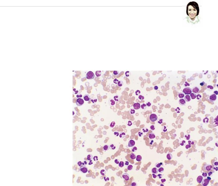
On a sweltering August evening in 2009 Pat Elliott noticed that her feet seemed swollen. Because she had been standing for hours while teaching a workshop in Phoenix, she was not surprised. “I thought it was the heat,” she says. But her feet hurt, too, so Elliott decided to play it safe and called her doctor, who suggested she come in for some tests. Days later the marketing professional learned that she had developed an uncommon form of blood cancer called chronic myelogenous leukemia (CML).
Elliott’s cancer is the result of a genetic change that arose in one or more stem cells in her bone marrow. (Normally these stem cells give rise to various blood cells in the body.) The defect caused the stem cells and their progeny to produce an abnormal enzyme known as Bcr-Abl. This enzyme signals the marrow to produce too many immature white blood cells and allows them to persist longer than they should. The proliferating blood cells then crowd out healthy cells, damage the bone marrow and allow infections to take hold. Once these abnormal cells enter the bloodstream, they can also cause the spleen to swell and damage other organs. The pain and swelling in Elliott’s feet most likely resulted from kid-
ney problems caused by her disease. CML usually starts off being fairly innocuous, especially if treated. It can, however, become aggressive and lethal if left untreated.
To keep her disease in check, Elliott takes a bright yellow pill daily. The medication, called imatinib (sold under the brand name Gleevec), binds to the abnormal enzyme and shuts off the proliferation signal. Without this enzyme, the extra white blood cells mature normally and die as they should. Indeed, a recent study suggests that CML patients who live at least two years after starting imatinib treatment can look forward to a normal life span. But the pill is not a cure. A small number of long-lived cancer cells persist inside Elliott’s body. If she stops taking her medication, the cancer will return.
Although imatinib keeps Elliott—and some 22,000 other CML patients in the U.S.—alive and healthy, the drug’s inability to eradicate the cancer suggests that perhaps the standard model that most researchers use to visualize how tumors grow—
32 Scientific American, August 2011
and the treatments that have arisen from that model—is flawed. An alternative hypothesis has recently gained traction; if it proves to be more accurate, physicians may need to adjust their therapeutic approaches, targeting their treatments to destroy particular subsets of cells within the tumor.
TWO MODELS
Oncologists have long worked under the assumption that most tumors develop from a single cell. After a series of genetic mutations, which occur as a result of exposure to radiation, cigarette smoke, dietary choices or a genetic predisposition, this single cell begins to divide uncontrollably into more cells. Each succeeding generation of cells accumulates more genetic mistakes that make the tumor grow bigger, invade local tissues and eventually spread (metastasize) to other parts of the body. Under the standard model of cancer growth, once a tumor has gained the ability to spread, any one of the founding cell’s descendants can
JOAQUIN CARRILLO-FARGA Photo Researchers, Inc.
© 2011 Scientific American

Creative Commons presents The Power of Open, a book that highlights the role of copyright, content sharing and collaboration in driving innovation in the digital age.
The Power of Open includes case studies of a wide variety of scientists, educators, artists and entrepreneurs around the globe who successfully use Creative Commons’ tools to advance their research, art, teaching and businesses.
GO TO HTTP://THEPOWEROFOPEN.ORG TO DOWNLOAD A FREE COPY TODAY!
ABOUT CREATIVE COMMONS: Creative Commons is the global non-pro t organization behind a copyright framework and global standard for licensing that meets the needs of individual creators, companies and Internet users. It makes creative, educational and scienti c content more accessible and usable on the Internet, helping to unlock the creative power of today’s connected world.

The Science of Health
break off and form a new mass—which is why health authorities emphasize early diagnosis and the need to destroy all tumor cells to prevent a recurrence.
The alternative view proposes that only a handful of the cells in a tumor—known as cancer stem cells—have the ability to grow uncontrollably and spread. These cells renew themselves indefinitely (essentially making close duplicates of themselves) and also give rise to a mix of cells having different properties and a finite life span. In this way, cancer stem cells resemble the normal stem cells sprinkled throughout the body that replace old or damaged tissues, such as skin or the lining of the intestine. Unlike normal stem cells, however, cancer stem cells ignore any and all chemical signals that tell them to stop dividing. According to this alternative conception, most cells in the tumor will eventually die and so should be less dangerous. The few stem cells in the tumor, however, would be particularly deadly: if even a single cancer stem cell survived the initial therapy, it could give rise to a whole new tumor weeks, months or even years later.
At least four decades of research support the idea that cancer stem cells play a major role in the origin of the so-called liquid tumors of the blood and lymphatic systems. But scientists have only recently begun exploring the possibility that cancer stem cells may be responsible for the development of solid tumors—such as cancers of the lung, breast and liver—as well. The presence of cancer stem cells might also clarify why imatinib does not cure CML. If stem cells uniquely did not need the Bcr-Abl enzyme to survive, no amount of the enzyme-blocking drug would eliminate them. “Those cells can be swimming in a sea of imatinib, but they’re not being killed,” says John E. Dick of the University of Toronto, a pioneer in cancer stem cell research. Investigators also suspect that cancer stem cells may be the reason relapses can occur years after an apparently successful treatment. Some cancer stem cells seemingly enter a dormant state, allowing them to survive the initial treatment relatively unscathed.
EVIDENCE
The first strong evidence that cancer stem cells might play a role in solid as well as liquid tumors came in 2003 from the study of malignant breast tissue. The study kicked off an era of what Dick calls “breathless excitement.” The cancer stem cell idea appealed to many researchers because it seemed to explain so much, not only the variety of cells within tumors but also why traditional cancer therapies often fail. A second study, also of breast tumors, by Jenny C. Chang, who is now an oncologist at Methodist Hospital in Houston, provided compelling evidence that cancer stem cells might be unusually resistant to standard treament.
At the time, Chang was working with patients at Ben Taub General Hospital in Houston, which serves the region’s 1.5 million uninsured residents. Because so many Ben Taub patients have limited access to health care, they tend to delay going to the doctor, so their cancers are more advanced when they receive a diagnosis. In fact, the tumors are often so big that they require a dose
of chemotherapy to shrink the growths before surgery can even be attempted. These circumstances afforded Chang a unique opportunity to look at drug resistance.
Chang began taking biopsies of the tumors before
and after chemotherapy. When she, along with Jeffrey M. Rosen of Baylor College of Medicine, and colleagues compared these biopsies, they found that the samples taken after treatment contained a greater proportion of what appeared to be cancer stem cells, as determined by the presence of certain proteins on their surface. Before the treatment, the putative cancer stem cells accounted for about 4.7 percent of the tumor, on average. After 12 weeks of chemotherapy, the ratio had risen to 13.6 percent, suggesting that the cancer stem cells were better able to survive chemotherapy than other cells in the tumor. In addition, when the cells from the postchemotherapy biopsies were grown in suspension, they formed more multicellular balls than the prechemotherapy ones, something only stem cells typically do.
CONTROVERSY
To date, researchers have reported finding evidence of cancer stem cells in tumors of the breast, brain, skin, colon, prostate, pancreas and liver, among others. But as more and more scientists have joined the field—and tried to replicate one another’s work—the picture has gotten a lot more complicated. In 2008, for example, Sean J. Morrison, director of the University of Michigan Center for Stem Cell Biology, found that if he tweaked a test, or assay, in mice that is used to detect cancer stem cells, he could dramatically change the results. A previous study had suggested that only one cell out of every million was a melanoma stem cell. Morrison found one in four cells could form a tumor. “If you make changes in the assay and you get huge changes in the spectrum of human cancer cells that can form a tumor, then that makes you really worry about drawing conclusions,” he says.
Today the field is divided. Although it seems clear that at least some cancers follow the stem cell model, researchers disagree over which tumors belong in that category. And no one yet knows whether therapies that specifically target cancer stem cells will save lives or how best to identify cancer stem cells. Nor do researchers understand how cancer stem cells originate, how often they derive from normal stem cells or whether each tumor has just a few or many of the cells. Some of the latest study results suggest that a complex mix of both the standard model and the cancer stem cell idea comes closest to elucidating how cancers form.
Despite the controversy, the research is prompting scientists to pay closer attention to the diverse subsets of cells found within all tumors. Ravindra Majeti, a cancer researcher at Stanford University, is working to develop diagnostic tools that indicate how aggressive a particular cancer will be—and therefore what type of treatment is needed—based on which genes are most active in the tumor cells. The hope is that this approach would be a more accurate way to gauge a tumor’s degree of malignancy than the standard method, which involves examining the cells under a microscope and grading them based on their how abnormal they appear.
For now Elliott will continue to take her yellow pills and try to stay positive. “We appreciate everything we do have,” she says, speaking for herself and her fellow patients. “But what we really want is a cure for this and to live a normal life.” 
34 Scientific American, August 2011
© 2011 Scientific American