

ECHO 2013 / Contrast for Diagnosis and Therapy
.pdfOPTIMIZE Methods
•108 pts with intermediate to high pretest probability of CAD
•Underwent two DSE studies, one with, and one without contrast.
•The order of the studies was randomized and performed at least 4 hours apart, within a period of 24 hours.
•Patients were not screened for image quality to allow evaluation of the impact of contrast on accuracy in the whole spectrum of image quality.
•Cath performed within one month and interpreted with QCA
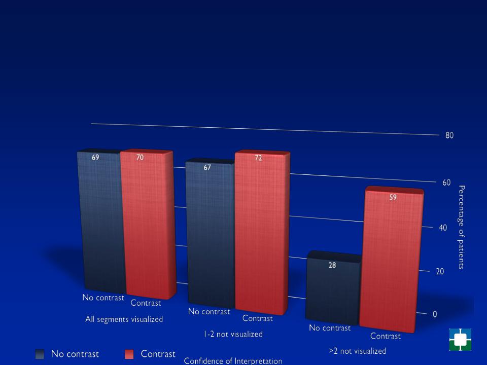
Plana et al. JACC Img 2008; 1: 145-52 |
Contrast12:21 |

Visualization of Segments
Contrast vs. Non-contrast enhancement
*p<0.001
* |
* |
|
Rest |
|
Plana et al. JACC Img 2008; 1: 145-52 |
Contrast12:22 |

Agreement on presence or absence of CAD
with Coronary Angiography
Contrast vs Non-contrast Enhancement
p<0.03
Plana et al. JACC Img 2008; 1: 145-52 |
Contrast12:23 |
Agreement of Detection of CAD with angiography
Effect of contrast and number of segments not visualized
p=NS p=NS p=0.005
Plana et al. JACC Img 2008; 1: 145-52
Contrast12:24

Contrast12:25

Fellow Echo
Middle of the Night
What‟s going on????
Contrast12:26

Contrast Administered
Clear extravasation beyond LV cavity
Contrast12:27

CT Scan
“multiple serpiginous wall defects |
|
coalescing into 2 distinct adjacent |
|
pseudoaneurysms…resulting in a large |
|
loculated hemopericardium … |
|
compressing the left atrium” |
Contrast12:28 |

Contrast Perfusion Echocardiography
Contrast Is Seen in the Myocardium
|
|
|
|
|
|
Baseline |
|
LV Phase Myocardial Phase |
|||
Contrast12:29

Bubble Destruction and Wash-In
Quantification of Myocardial Perfusion
Wei et al., Circulation 1998; 97: 473-483
Contrast12:30