

ECHO 2013 / Mitral Stenosis Quantitation It’s Not All About the Gradient
.pdfState-of-the-Art Echo 2013
Mitral Stenosis Quantitation: It’s
not all about the gradient!
Rebecca T. Hahn, MD
Director of Interventional Echocardiography
Columbia University
No Disclosures

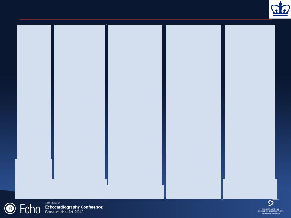
Severity of Mitral Stenosis
|
MVA (cm2) |
Mean (mm Hg) |
Pap (mm Hg) |
Normal |
4.0 – 5.0 |
|
|
|
|
|
|
Mild |
> 1.5(<2.5) |
<5 |
< 30 |
|
|
|
|
Moderate |
1.0 – 1.5 |
5-10 |
30-50 |
|
|
|
|
Severe |
< 1.0 |
> 10 |
> 50 |
|
|
|
|
Note: Above criteria applicable when HR = 60-90 bpm No single value defines severity in mitral stenosis

Mitral Stenosis: Pathophysiology
•MVA <2.5 cm2 before development of symptoms
•MVA >1.5 cm2 will not typically produce symptoms at rest
Increase in transmitral flow or decrease in diastolic filling period (ie: tachycardia) may increase LAP
Dyspnea may occur with exercise, emotional stress, infection, pregnancy, AFib with rapid VR
Pulmonary vascular disease
•Increased pulmonary arteriolar resistance
•Reversible pulmonary venous obstruction
Bonow et al, JACC 2006, 48(3) e1-148

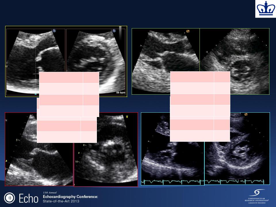
Pathology
Rheumatic |
Degenerative (mitral |
|
annular and leaflet |
||
(commissural fusion) |
||
calcification) |
||
|
Pk/Mn = 17/11 mmHg MVOA = 1.0 cm2
Pk/Mn = 16/6 mmHg MVOA = 1.5 cm2

Pathology
|
Degenerative (mitral |
Rheumatic |
annular and leaflet |
(commissural fusion) |
calcification) |
Commissural Fusion
Baumgartner H et al. JASE 2009:22(1):1-23

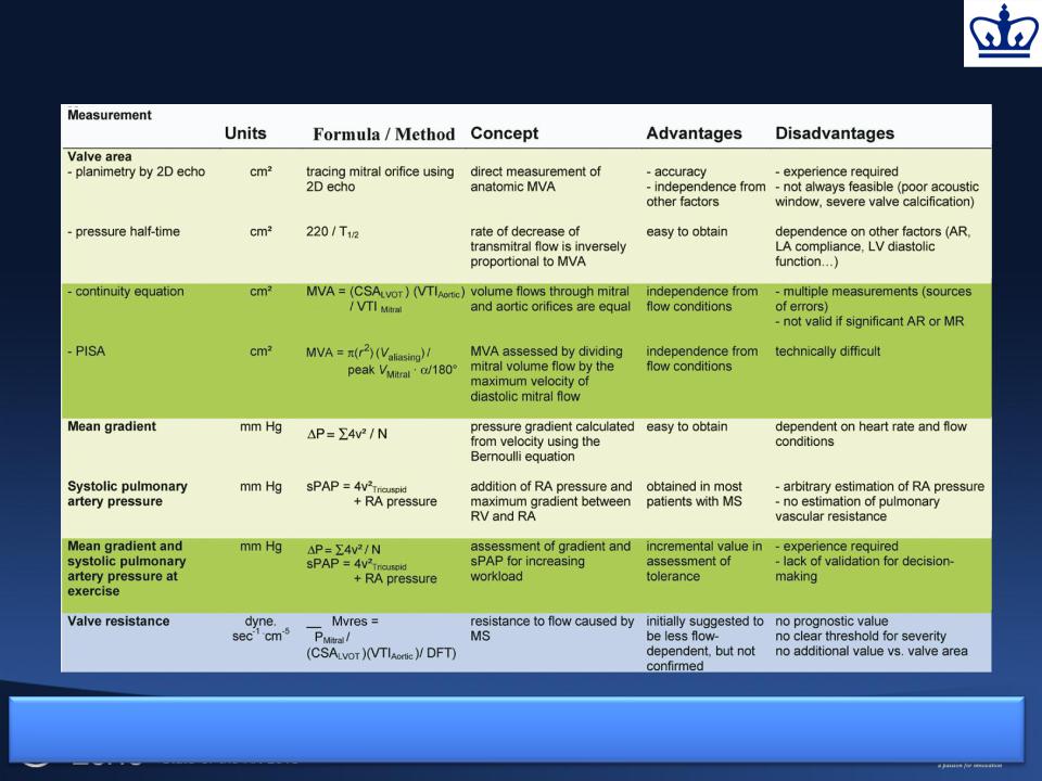
Imaging Views
Data Element |
Recording |
Measurement |
||
Planimetry |
• |
2D parasternal SAX |
• Contour of the inner mitral |
|
|
• Scan valve for smallest orifice |
|
orifice |
|
|
• Position of measurement can be |
• |
Include commissures when |
|
|
|
oriented by 3D |
|
opened |
|
• Lowest gain to visualize whole |
• In mid-diastole (use cine) |
||
|
|
orifice |
• |
Average measurements in atrial |
|
|
|
|
fibrillation |
Mitral Flow |
• |
CW Doppler |
• |
Mean gradient |
|
• |
Apical windows often suitable |
• |
PHT from descending (mid- |
|
|
(optimize intercept angle |
|
diastolic) slope of E-wave |
|
• Adjust gain to obtain well-defined |
• |
Average measurements in atrial |
|
|
|
contour |
|
fibrillation |
Systolic PAP |
• |
CW Doppler |
• |
Maximum velocity of TR |
|
• Multiple acoustic windows to |
• |
Estimate RAP |
|
|
|
optimize intercept angle |
|
Mitral Valve Score |
Valve anatomy |
• |
Parasternal SAX |
• |
Valve thickness |
|
• |
Parasternal LAX |
• |
Commissural fusion |
|
• |
Apical 2Ch view |
• Extension and location of bright |
|
|
|
|
|
zones |
|
|
|
• |
Valve pliability |
Baumgartner H et al. JASE |
• |
Subvalvular apparatus |
||
2009:22(1):1-23 |
|
|
|
(thickening/fusion/shortening |
Echo Score Index for Mitral Stenosis
|
Grade |
|
|
Mobility |
|
|
Subvalvular |
|
|
Leaflet Thickening |
|
|
Calcification |
|
|
|
|
|
|
|
|
thickening |
|
|
|
|
|
|
|
|
1 |
|
|
Highly mobile |
|
|
Minimal thickening |
|
|
Leaflets near normal |
|
|
A single area of |
|
|
|
|
|
valve with only |
|
|
just below the mitral |
|
|
in thickness (4 – 5 |
|
|
echo brightness |
|
|
|
|
|
leaflet tips |
|
|
leaflets |
|
|
mm) |
|
|
|
|
|
|
|
|
restricted |
|
|
|
|
|
|
|
|
|
|
|
2 |
|
|
Leaflet mid and |
|
|
Thickening of |
|
|
Mid-leaflets normal, |
|
|
Scattered areas of |
|
|
|
|
|
base portions have |
|
|
chordal structures |
|
|
considerable |
|
|
brightness |
|
|
|
|
|
normal mobility |
|
|
extending up to 1/3 |
|
|
thickening of margins |
|
|
confined to leaflet |
|
|
|
|
|
|
|
|
of the chordal length |
|
|
(5 – 8 mm) |
|
|
margins |
|
|
3 |
|
|
Valve continues to |
|
|
Thickening |
|
|
Thickening |
|
|
Brightness |
|
|
|
|
|
move forward in |
|
|
extending to the |
|
|
extending the entire |
|
|
extending into the |
|
|
|
|
|
diastole, mainly |
|
|
distal 1/3 of the |
|
|
length of the leaflet |
|
|
midportion of the |
|
|
|
|
|
from the base |
|
|
chords |
|
|
(5-8mm)t |
|
|
leaflets |
|
|
4 |
|
|
No or minimal |
|
|
Extensive thickening |
|
|
Considerable |
|
|
Extensive |
|
|
|
|
|
forward movement |
|
|
and shortening of all |
|
|
thickening of al |
|
|
brightness |
|
|
|
|
|
of the leaflets in |
|
|
chordal structures |
|
|
leaflet tissue (> 8 – |
|
|
throughout much |
|
|
|
|
|
diastole |
|
|
extending down the |
|
|
10mm) |
|
|
of the leaflet tissue |
|
|
|
|
|
|
|
|
papillary muscles |
|
|
|
|
|
|
|
|
|
|
|
|
|
|
|
|
|
|
|
|
|
|
|
|
|
|
|
|
|
|
|
|
|
|
|
|
|
Abascal VM et al, JACC 1988;12:606-615
Mitral Valve Score
Mobility 2
Subvalvular 2
Leaflet 2
Mobility 1
Calcification 2
Subvalvular 1
Leaflet 2
Calcification 1
Mobility 3
Subvalvular 3
Leaflet 3
Mobility 1
Calcification 2
Subvalvular 2
Leaflet 2
Calcification 1
Approaches to Evaluating MS
Level of recommendations: (1) appropriate in all patients (yellow); (2) reasonable in selected patients (green);and (3) not recommended (blue).