

ECHO 2013 / Interventional Echocardiography
.pdfState-of-the-Art Echo 2013
Interventional Echocardiography
Rebecca T. Hahn, MD
Associate Professor of Clinical Medicine
Director of Interventional Echocardiography
Columbia University
No Disclosures

Spectrum of structural heart disease.
„Structural heart disease‟ is a term first introduced by Martin Leon at the 1999
Transcatheter Cardiovascular Therapeutics meeting to provide an over-reaching term encompassing non-coronary cardiac disease processes and developing interventional techniques
Steinberg D H et al. Eur Heart J Suppl 2010;12:E2-E9

The Heart Team
Ideally, such a team would be comprised of the patient’s primary cardiologist, cardiac surgeon, interventional cardiologist,
echocardiographer, imaging specialists…, heart failure and valve |
|
disease specialist, cardiac anesthesiologist, nurse practitioner, |
|
and cardiac rehabilitation specialists. |
3 |
|
|

Echo (2D and 3D) in Structural 
Heart Disease
Improved structural imaging
Understanding disease morphology
Patient selection
-- Flail Gap = 4 mm
Echo (2D and 3D) in Structural 
Heart Disease
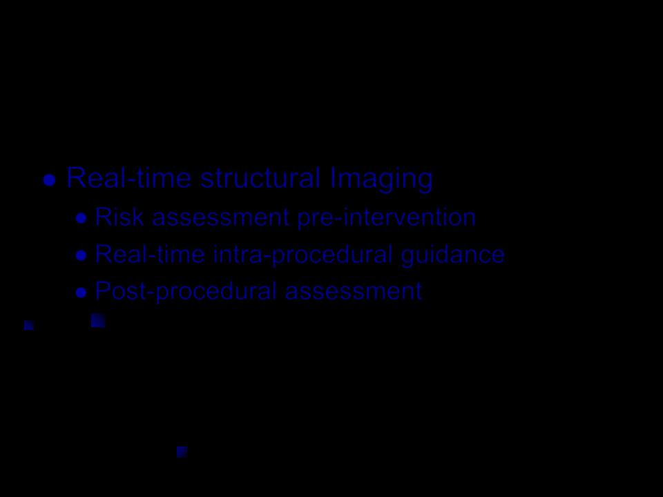
Real-time structural Imaging
Risk assessment pre-intervention
Real-time intra-procedural guidance
Post-procedural assessment

Echo (2D and 3D) in Structural 
Heart Disease
Value of Echocardiographic Imaging
Improve procedural safety
Reduction in complications
Reduce radiation and contrast exposure
Improved outcomes
Assessment of procedural success
Predictors of long-term outcome

Consensus Statement
J Am Soc Echocardiogr 2011;24:937-65.)
Patient Selection: TTE and TEE (2D and 3D)
Peri-procedural echocardiography during transcatheter procedures
Post-implant

Structural Heart Disease in
the Cath Lab
Septal Closure
LAA occlusion
Alcohol septal ablation
Transcatheter AVR
Percutaneous MV repair
Prosthetic Valve Dysfunction
Paravalvular Regurgitation
Prosthetic Valve Stenosis

Atrial Septal Defect
Is this suitable for percutaneous closure?
To be suitable for closure, a defect should have sufficient rims (1 cm) or margins to the mitral valve, the base of the aorta, and the orifices of the venae cavae and coronary sinus
3D TEE provides views of the ASD allowing its measurement and identifying its spatial relation with neighboring structures
Abdel-Massih T et al. Echocardiography 2005;22(2):121–127

Associated Anomalies: PAPVR