

ECHO 2013 / Mitral Stenosis Quantitation It’s Not All About the Gradient
.pdfCase Study
2D
1.thickened leaflets (tips) and subvalvular apparatus
2.Diastolic doming AMV
3.Restricted motion of PMV
4.Enlarged atrium
Ejection fraction by biplane
Simpson’s method = 55%

Case: Assess LA, LV, MV
•Peak/mean gradient = 16/6 mmHg
•MV VTI = 41.5 cm
• Pressure Halftime = 133 msec

Is this severe MS?
• Yes |
• No |
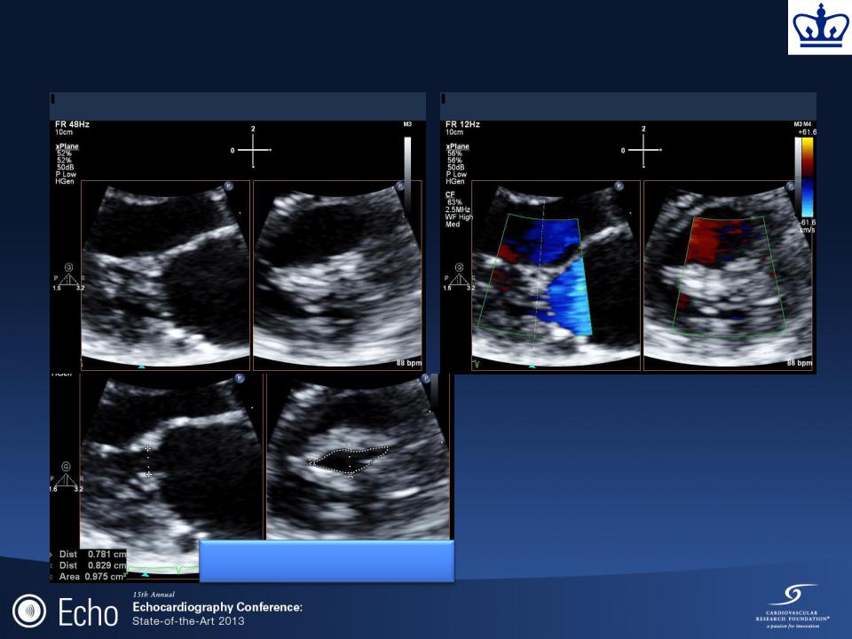
Case: 2D Mitral Assessment
• Look at orientation of opening
|
• Will you get a good SAX |
|
view from this window (Use |
|
3D!) |
|
• Measure the maximum tip |
MVOA = 0.98 cm2 |
separation |
• Match to parasternal |
|
|
dimension in mid-diastole |

Mitral Stenosis: Planimetry
•
•
•
•Advantage: relatively hemodynamicindependent assessment MVOA
•Limitation:
Difficulty measuring the smallest area of a funnel-shaped orifice
2D: Planimetry of area in parasternal short-axis view
Feasible in 95% of patients Accuracy:
Underestimates compared to surgical valve area (Henry et al, Circ 1975;51:827)
Overestimates compared to hemodynamic valve area (Weyman et al, AJC 1979;43:386)
Best correlation with direct anatomic measurement (Faletra F et al. J Am Coll Cardiol 1996;28:1190-7)

Case: 3D

Case: Multiplanar
Reconstruction
MVOA = 1.03 cm2
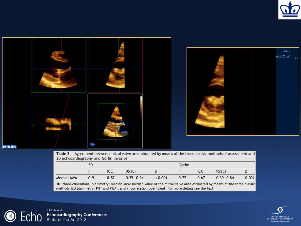
Use of Three-D Echo
Planimetered MVA = 1.05 cm2
Median MVA of 3 classic measures: 2D planimetry, PHT and PISA
Pérez de Isla, L et al. Eur J Echocardiography (2007) 8, 470-473

Case: LV Stroke Volume
LVOT Diam = 18.5 cm
LVOT VTI = 17.5 cm
LVOT Stroke Volume = 47 cc

Conservation of Mass: Continuity
Equation
LVOT Diam = 18.5 cm |
|
LVOT VTI = 17.5 cm |
|
|
|
|
LV (or RV) |
|
|
|
|
|
X |
|
Stroke Volume |
|
|
|
MVOA = 1.1 cm2
MV VTI = 42.5
?
X
=
Mitral Valve Area
Mitral Valve Stroke Volume