ECHO 2013 / Management Decisions in the ICU and ER The Role of the FOCUS Echo
.pdf
FOCUS Echo of the Pericardial
Effusion
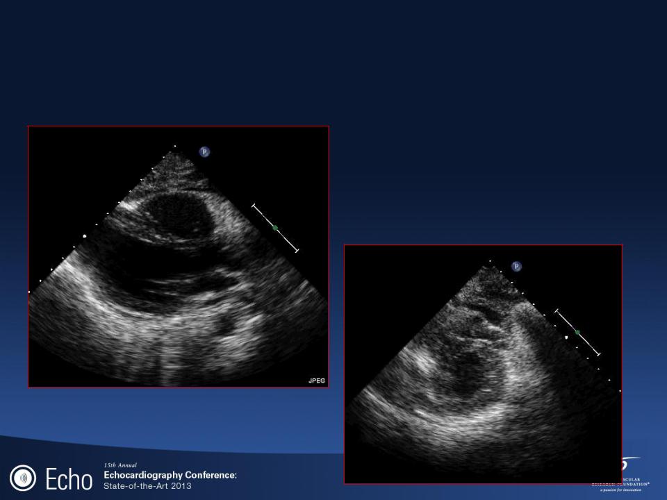
LOCALIZED
PERICARDIAL
EFFUSION
Patient with lymphoma

FOCUS Echo of the Pericardial Effusion
|
|
|
Subcostal |
|
Apical |
|
|||
|
|
|
|
|
|
|
|
|
|
|
|
|
|
|
LOCALIZED
Patient with lymphoma PERICARDIAL
EFFUSION

Differential Diagnosis PE
• Epicardial fat
• Pleural effusion
•Tumor
•Thrombus
•Ascites

Pleural Effusions
Pericardium reflects at the posterior atrioventricular groove.
Pleural effusion continues under the left atrium, posterior to the descendng aorta.

Pleural Effusions
Right
Pleural
Effusion
Left
Pleural
Effusion

Pericardial Tamponade
Hemodynamics: Comprehensive Echo
•Equilibration intracardiac diastolic pressures
usually between 10 and 30 mmHg
Within 44 mmHg
•Inspiratory increase in right-sided pressures and reduction in leftsided pressures (pulsus paradoxus)
•X - descent
descent of the base in systole
•Y - descent
occurs as the tricuspid valve opens and ventricular filling begins from the high-pressure right atrium
in constrictive pericarditis, filling is truncated in early to mid diastole
in tamponade, filling is restricted throughout diastole
•Kussmaul‟s Sign
in constriction, venous return increases with inspiration and a high right atrial pressure resists filling resulting in an increased JVP

Pericardial Tamponade
Comprehensive Echo
•Doppler
Mitral valve opening is delayed
Trans-mitral E velocity
is decreased > 25-30% (normally 10%)
Respiratory variation
tricuspid valve > 40-50 % (normally 17%)
MV
TV

Comprehensive Echo Pericardial Tamponade
Tricuspid |
|
Mitral |
|
|
|
Normal mitral E respiratory variability = 10% Normal tricuspid E respiratory variability = 17-25%
|
E wave |
A wave |
|
MV |
43 |
9% |
28 12% |
TV |
83 |
53% |
58 25% |
Appleton C et al, JACC. 1988;11:1020-1030

Comprehensive Echo Pericardial Tamponade
|
|
|
|
|
|
|
|
|
|
|
|
|
|
|
|
|
|
|
|
|
|
|
Respiratory variation of |
|
Respiratory variation of |
||||
pulmonary outflow |
aortic outflow |
|||
> 30% |
|
|
> 20% |
|
Appleton C et al, JACC. 1988;11:1020-1030

Comprehensive Echo Imaging the
Superior Vena Cava
Subcostal View
