

New Products and New Areas of Bioprocess Engineering
.pdfMultistage Magnetic and Electrophoretic Extraction of Cells, Particles and Macromolecules |
141 |
|
CZE |
capillary zone electrophoresis °C |
|
ddiameter of the solute or droplet sphere (m)
dB/dz |
magnetic field gradient in the direction of motion |
dy/dt |
velocity of the bioparticle in electric field (m s–1) |
dz/dt |
one-dimensional particle motion |
Ddiameter of the chamber or distance (perpendicular to g) from the high temperature to the closest lateral boundary (m)
DX dextran
eelectrical charge (C)
Eelectric field strength (V m–1)
Ep |
parameter in Eq. 27 (Ep = KR) |
ECCD |
electrophoretic counter current distribution |
ELECSEP |
electrophoretic separator |
Ftotal force on a particle (N)
Fb |
buoyancy force (N) |
Fd |
drag force (N) |
Fg |
gravitational force (N) |
Fm |
magnetic force (N) |
gacceleration due to gravity (m s–2)
Gr |
Grasshof number |
G-6-PDH |
glucose-6-phosphate dehydrogenase |
htotal depth of chamber (m)
hT |
heat transfer coefficient (kW m–2 °C–1) |
Hmagnetic field strength (A m–1)
Hc |
chamber height (m) |
HP |
volume of the heavy phase (m–3) |
HGMSs |
high gradient magnetic separations |
iunit vector in the z direction
Icurrent (A)
Jflux of cells in magnetic field (cell m–2 s–1)
kE |
electrical conductivity of the medium (S m–1) |
kT |
thermal conductivity of the medium (kW m–1 °C–1) |
Ksolute partition coefficient (K = xs /ys)
llength of element (in Eq. 1)
LP |
volumes of the light phase (m–3) |
LDH |
lactate dehydrogenase |
mnumber of bioparticles that would migrate during a single step
mp |
mass of the particle (kg) |
(m1)n, r |
number of type-1 particles with mobility m1E |
Minduced polarization (Eq. 3)
Mc |
flow rate of the coolant that is to be circulated to remove the re- |
|
quired heat (kg s–1) |
MAGSEP |
magnetic separator |
MDX |
maltodextrin |
nstage or chamber number
Ntotal number of bioparticles initially (t = 0) present in the first bottom chamber (N = N1 + N2)
142 |
K.S.M.S. Raghavarao et al. |
[N1]r–1 |
number of particles of mobility mE1 in stage ‘n’ at transfer or step |
|
(r – 1) |
N1 |
number of bioparticles with mobility m1E [N1 = x1(N)] |
N2 |
number of bioparticles with mobility m2E [N2 = x2 (N)] |
Nu |
Nusselt number |
ORSEP |
organic separator |
p |
parameter in Eq. (27) [p = E/(E + 1)] |
Pd |
palladium |
6-PGDH |
6-phosphogluconate dehydrogenase |
PEG |
poly(ethylene glycol) |
PPE |
phase partition experiment |
PTK |
phophofructokinase |
Pr |
Prandtl number |
Qheat to be removed from the system (kW)
rtransfer or step number
rc |
radius of chamber (m) |
Rphase volume ratio (R = L/H)
Ra |
Rayleigh number |
Rac |
critical Rayleigh number |
Re |
Reynolds number |
RME |
reverse micellar extraction |
ttime of application of electric field(s)
um |
magnetic energy density |
Um |
magnetostatic potential energy |
vdrop velocity (m s–1)
v |
velocity of the cell (m s–1) |
cell |
drop velocity due to electric field (m s–1) |
vE |
|
vmedium |
velocity (circulation) of the medium (m s–1) |
vp |
velocity and can be assumed to be equal to Dz/Dt (m s–1) |
V |
volume of the lower cavity half (m3) |
b |
volume of the cell (m3) |
V |
|
cell |
particle volume (m3) |
V |
|
p |
|
Vvolume of the chamber (m3)
V* |
mis-alignment volume (m3) |
Wpower density (kW m–3)
xs |
mass concentration of the solute in the light phase (kg m–3) |
x1 |
fraction of type 1 particles |
ys |
mass concentration of the solute in the heavy phase (kg m–3) |
ydistance of electrophoretic migration by bioparticles during the electric field application of time t
zdistance of unidimensional motion (m)
bcoefficient of thermal expansion (°C–1)
lfunction of the conductivities of the dispersed or droplet phase and continuous phase (Eq. 26)
mchemical potential
m0 |
magnetic permeability of free space |
mE |
electrophoretic mobility of bioparticle (m2 V–1 s–1) |
Multistage Magnetic and Electrophoretic Extraction of Cells, Particles and Macromolecules |
143 |
|
m1E |
electrophoretic mobility of type 1 particles (m2 V–1 s–1) |
|
m2E |
electrophoretic mobility of type 2 particles (m2 V–1 s–1) |
|
Dcm |
magnetic susceptibility difference |
|
Dcc |
bulk magnetic susceptibility between the particle and the sur- |
|
ccell |
rounding medium |
|
magnetic susceptibility of cell |
|
|
cm |
magnetic susceptibility |
|
cmedium |
magnetic susceptibility of medium |
|
t |
time of application of the electric field(s) |
|
∂B/∂r |
rate of change of the magnetic field along the vertical axis r |
|
∂B/∂z |
rate of change of the magnetic field along the vertical axis z |
|
Çdensity of the system (kg m–3)
Çcell |
density of the cell (kg m–3) |
Çmedium |
density of the medium (kg m–3) |
ÇC |
density of solvent or the continuous phase (kg m–3) |
ÇD |
dispersed or droplet phase density (kg m–3) |
Ço |
medium density (kg m–3) |
Çp |
particle density (kg m–3) |
ÇS |
density of solute sphere or dispersed phase droplet (kg m–3) |
DÇ |
density difference (kg m–3) |
sE |
surface charge density (C m–2) |
Dt |
time interval required for particle separation (s) |
DT |
rise in temperature (°C) |
Dz |
vertical height of particle migration (m) |
hviscosity (kg m s–1)
hC |
viscosity of the solvent or continuous phase (kg m s–1) |
hD |
viscosity of the dispersed phase (kg m s–1) |
1 Introduction
Separation science and technology is one of the most complex and important area of biotechnology and biochemical engineering. New separation technologies, capable of treating dilute solutions in both small and large-scale processes, even in the presence of particulate matter, are necessary. Electrophoretic and magnetic separation techniques appear to have potential in this area with a wide range of applications. Manufacturers of new enzymes and pharmaceutical products require improved methods for recovering intact cells and intracellular products. There is an increasing need for efficient methods to recover cells selectively from other bioparticles to generate a feedstock for product separation processes involving chromatography and membrane separations. Similarly, isolation, purification, and concentration of many biomolecules produced in fermentation processes are extremely expensive. Often such downstream processing contributes a large portion of the product cost [1, 2]. Many methods of cell separation have been reviewed in the literature [3, 4]. In the present review we briefly discuss the suitability of various methods for different applications.
144 |
K.S.M.S. Raghavarao et al. |
Magnetic separation technologies, though conventionally being used for ore and chemical industries, have gained importance for biotechnological applications quite recently [5, 6]. Similarly, aqueous two-phase extraction and reverse micellar extraction have recently gained prominence for the downstream processing of macromolecules. In the present review these methods are analyzed especially in terms of the reasons for not realizing their full market potential. We present mainly our own research on the development of unified multistage extraction processes that are versatile enough to handle cells as well as macromolecules as given below.
We describe multistage methods, namely ADSEP (Advanced Separator), MAGSEP (Magnetic Separator), and ELECSEP (Electrophoretic Separator), for quantitatively separating cells, particles, and other macromolecules by using magnetically and electrophoretically assisted extraction processes. These methods are expected to cost much less in comparison with the competing technologies at similar scale and precision. These multistage methods build on the counter-current distribution technology of classical chemical engineering separations, stemming from the seminal work of Craig and Craig [7] and later applied to aqueous two-phase extraction (ATPE). Earlier exploitation of this sliding chamber concept has been attempted with only partial success due to some shortcomings related to convenience and practicability. In the present review we indicate how these shortcomings can be alleviated by combining concepts, so that more efficient and convenient automated magnetic and electrophoretic bioextraction processes can be developed. We also presented a few suggestions for future work while analyzing the scale-up and economic aspects of these extraction processes. Commercial uses of the magnetic and electrophoretic processes, having both ground and space based research elements, also are presented in this review.
To the best of our knowledge, multistage magnetic and electophoretic separations have not been reported in the earlier literature. While other processes involve some element of trial and error (e.g., identifying an appropriate solution), the theory behind magnetic and electric field assisted separations provide the ability to predict how unknown cells, particles, and macromolecules will react to magnetic and electric fields. Hence mathematical modeling is stressed in the proposed review, discussing our own models while reviewing the models reported in the literature.
2
Cell and Particle Separations
2.1
Magnetic Extraction
Magnetic extraction has been used in various biological processes such as biochemistry, biotechnology, and environmental technologies. Magnetic extraction, based on magnetic sorbents, carriers, and modifiers, has been used for isolation, modification, detection, immobilization, and removal of a variety of biologically active compounds, xenobiotics, cellular organelles, and cells.
Multistage Magnetic and Electrophoretic Extraction of Cells, Particles and Macromolecules |
145 |
The developments that aroused fresh technical interest in magnetic separations are:
1.New methods for generating high magnetic field gradients.
2.The introduction of reasonably priced efficient and superconducting magnets [8].
3.New developments in magnetic labeling techniques for cells and microspheres that have extended the useful range of High Gradient Magnetic Separations (HGMSs) into many important areas of biotechnology [1].
No widely-used magnetic method has yet been developed to separate cells on the basis of the amount of ligand bound. By combining magnetic extraction, used as a rate process, with countercurrent extraction, it is now possible to use magnetic separation of cells as a quantitative technique, separating on the basis of the number of ligands bound per cell. This could be qualitative, based on the amount of ligand bound to each kind of cell, or quantitative, based on the amount of ligand bound to cells of the same type, some with high receptor content and some with low.
There are numerous examples of potential uses of magnetic extraction methods. Separation of high CD4 from low CD4 binding cells from all other cells in whole blood is a very significant procedure for the study of immune function in leukemia, AIDS, and immunomodulation caused by environmental stresses. Research laboratories have recently used receptor number as a dependent variable in a variety of scientific applications, such as endocrinology [9], growth regulation [10, 11], virology [12, 13], carcinogenesis [14, 15], infectious diseases [16], neurology [17], and nutrition [18].
2.1.1
Existing Methods – Brief Summary
Magnetic separation methods can be classified into two main types. In the first type, separand is intrinsically magnetic so that magnetic separation can take place without any modification. There are only a few examples of such materials in biotechnology, such as red blood cells containing high concentrations of paramagnetic hemoglobin, magnetotactic bacteria containing small magnetite particles within their cells, and magnetic particles used in waste water purification systems. In the second type, one or more non-magnetic components of a mixture have to be rendered magnetic by the attachment of a magnetically responsive entity. The newly formed complexes have magnetic properties and can be manipulated using an external magnetic field [5].
Magnetic properties include ferromagnetism, paramagnetism, and diamagnetism, and specific materials including cells and biomolecules can exhibit these properties [8]. The most naturally labeled cells that are separated by HGMS are probably the magnetoactive aquatic bacteria [19]. These organisms contain small single-domain crystals of magnetite, and the ability to be oriented in a magnetic field remains even when the organism dies. Several patents have already been granted in the USA and Japan describing potential uses of these rare organisms in medical applications [1].
146 |
K.S.M.S. Raghavarao et al. |
The simplest technique for adding ferromagnetic properties to biological matter is to supplement the solution with small magnetite particles and using the natural surface and colloidal properties of both the magnetite and the biological material to obtain the desired separation from the solution [1]. Although this technique is not very selective, it works very well in cases where total removal of cell mass is required, for example, from fermentation broth or waste water. The technique can be further improved by adjusting surface and/or colloidal properties and/or by adding a flocculating agent [6]. Ferrofluids (surfactant stabilized magnetite colloids) had also been extensively used for ferromagnetic labeling as reviewed by Rosenberg [20] and their application in affinity chromatography was discussed by Mosbach and Anderson [21].
Most of the applications of HGMS of paramagnetic material pertain to red blood cells (erythrocytes) [6, 22]. The technique relies on the paramagnetic properties of hemoglobin groups in erythrocytes when they are in the reduced state (Fe2+). When hemoglobin combines with oxygen it becomes diamagnetic. No evidence of damage to erythrocytes was found when they were separated from whole blood by HGMS [22]. This opened up a wide range of clinical and research uses of HGMS of red blood cells as this method has advantages over centrifugation in that it is completely selective and can be made a continuous process. The low magnetic susceptibility of erythrocytes [22] is compensated by using lower flow rates than in the other (magnetically labeled cells) applications of HGMS to maintain the level of adsorption [1]. There are other ways to induce paramagnetism. For instance, large positive susceptibilities can be introduced into biological particles by their adsorption of magnetic cations. The trivalent cations of the lanthanide series are especially effective at relatively low magnetizing fields compared to hemoglobin. It was demonstrated that cells from whole blood, yeast cells, bacteria and Vistna virus can be magnetically recovered from Er3+-containing solutions with an efficiency greater than 80% [23]. The binding of the ion is affected by the pKa of the binding group, the pH, and the ionic strength and hence these parameters could be varied to magnetize a specific material preferentially [1].
Diamagnetic materials can be separated by using a strongly paramagnetic background fluid which increases the contrast. White blood cells and platelets have been extracted by this method though in general the range of concentration of the carrier medium is limited by the osmotic pressure effects on the cell membrane [6].
Two possible modes – direct and indirect – are available for the isolation of target cells or compounds. The former is often employed, in which magnetic particles with immobilized affinity ligands are directly suspended in the solution or suspension. During the course of incubation the target cells bind with the immobilized ligand and the whole complex can then be separated using a magnet. In the indirect mode, the target cells interact with the affinity ligand, usually a primary antibody, that has first been added to the solution or suspension in the free form, to form a complex during incubation. Magnetic particles with immobilized secondary antibodies are then added. Finally the magnetic complex formed is separated in a magnetic separator.
Multistage Magnetic and Electrophoretic Extraction of Cells, Particles and Macromolecules |
147 |
Magnetic particles with immobilized lectins can be used for the isolation of polysaccharides or structure containing sugar moities. This technique has been used for the isolation of various microbial cells from culture media [24]. Immunomagnetic particles – magnetic particles bearing immobilized specific antibodies against the target structures – can be used for the selective isolation of specific cells such as prokaryotic and eukaryotic [25, 26].
Magnetic modifications of standard immunoassays can be successfully used for the determination of microbial cells [26], parasites [27], and viruses [28]. Cells to which magnetic carriers are attached can be removed from the system simply by using an external magnetic field, or can also be targeted to the desired place. Non-porous magnetic supports with a diameter of 1 mm or smaller are advantageous as they are more resistant to fouling, diffusional limitations, and attrition than porous supports [1, 29]. There are many ways to immobilize the cells or compounds of interest and practically all the standard procedures used in affinity chromatography can be used for this purpose. Again, similar to the chromatography methods, a spacer must often be inserted between the magnetic particle and the ligand to overcome steric hindrance at the cell surface. Several microorganisms have been immobilized on magnetic carriers or have been entrapped in biopolymers containing magnetic material. These methods are useful in applications where suspended particles and viscous substances are present in the fermentation medium. The magnetic material usually does not interfere with the catalytic function of the cells, as shown for Saccharomyces cerevisiae immobilized in calcium alginate together with magnetite [30]. HGMS of yeast cells by means of irreversible adsorption of very fine particles of maghemite (gamma-Fe2O3) on the cell wall was reported [31]. Binding of paramagnetic erbium ions on various surface structures of microbial cells also leads to the formation of magnetized microorganisms [32]. Many other applications in different biorelated areas, apart from the specific ones given in this section, are discussed by Safarik and Safarika [33], in an excellent review article on magnetic separations.
Gravity-dependent phenomena associated with separation processes have been studied in the low-gravity environment of space flight [34]. Magnetic field-based separations, on the other hand, have not been pursued in microgravity research. In general, gravity assists the separation of magnetized from unmagnetized separands [35], or magnetic stabilization is designed to avert effects of gravity [36, 37]. Flowing systems [38] can be considered relatively gravity independent. However, certain gradient-dependent separations have a meaningful gravitational component, and Owen’s work suggests that enhanced separation could be achieved in a ferrofluid relieved of the gravity vector [39].
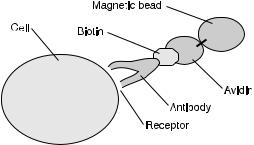
The method of cell separation using a magnetic field has been implemented as a binary separation between cells that have and have not bound magnetic microspheres on the basis of a cell receptor (a specific type of surface ligand), as shown in Fig. 1.
Apart from the above-mentioned interests in low-gravity research applied to separation science and technology, there is an interest in performing separations in support of low-gravity biotechnology, immunology, cytology, serology, microbiology, and chemical procedures on the International Space Station
148 |
K.S.M.S. Raghavarao et al. |
Fig. 1. Magnetic bead attached to cell receptor
Alpha [40]. Addressing this concern is likely to require innovative research on separation processes that require a body force but will function independently of the gravitational vector, yet possibly be enhanced by gravitational unloading.
Continuous collection and processing of blood samples from animals and crew members is best done by analyzing samples for cell types when the samples are fresh. Methods available for separating cells for characterization are sedimentation, flow cytometry, electrophoresis, differential adsorption, and magnetic separation [41]. Sedimentation can provide only a binary separation unless elutriation or a density gradient is used. Elutriation is complicated and requires fraction collecting capability. Density gradients are difficult to form in low gravity and on a centrifuge while it is operating at moderate speed for cell separation, and, in any case, differential sedimentation would be a multistep process. Flow sorting produces small sample sizes and requires unnecessarily complex and expensive equipment ($200,000 on the ground). Electrophoresis, like sedimentation, separates cells on the basis of an intrinsic physical property. Adsorption and magnetic separations are usually based on affinity ligands immobilized to adsorbents in the former case and magnetic microspheres in the latter.
It is easy to argue that magnetic cell separation methods are the easiest to implement in low gravity, and that they can be made quantitative; that is, cells with low amounts of microspheres bound by ligand can be separated from cells with high amounts of microspheres bound by ligand.
2.1.2
Multistage Magnetic Method
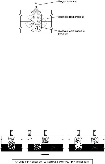
Magnetic field gradients can be set up as shown in Fig. 2, but many configurations of magnetic poles can be used to create the gradient; for example, N and S poles could be placed on opposite sides of the upper cavity, and a metal hairpin could hang into the cavity to create a gradient around the hairpin [38]. Additional configurations are considered below.
The development of user-friendly devices that are capable of separating particles according to quantity of ligand on their surfaces appears to be the area of greatest need in improving magnetically-assisted separation devices. The
Multistage Magnetic and Electrophoretic Extraction of Cells, Particles and Macromolecules |
149 |
|||||
|
|
|
|
|
|
|
|
|
|
|
|
|
|
|
|
|
|
|
|
|
|
|
|
|
|
|
|
|
|
|
|
|
|
|
|
|
|
|
|
|
|
Fig. 2. Single stage of the magnetic separation process
magnetic separation industry has made considerable progress in this regard, but the commercial technology to date has been limited to binary separation methods. The innovation presented here represents progress by finally providing a reliable method for differential magnetic separation on the basis of small differences in surface composition.
The model equations (discussed in Sect. 2.1.3) are used as the basis for the design of a multistage separation system where the separation driving force is electromagnetic (Fig. 3). In staged magnetic separation, the final distribution of separands can be calculated from a simple relationship involving the number of transfers and the equivalent of a partition coefficient, K, defined as the ratio of upper and lower compartment concentrations.
Capture could be isocratic (magnets in all stages having equal strength) or gradient (magnets at increasing stage numbers having increasing field strength). In the latter case, in a typical application the first stage has no magnet and no upper cavity and serves the purpose of homogenizing the cell mixture by stirring just before the beginning of transfers. The second stage also has no magnet and serves the purpose of adding magnetic particles to the cell suspension from a low-volume upper cavity, mixing them together, and allowing them to react. The third stage has a very weak magnet in the upper cavity, and attracts only the most highly magnetized cells, namely those with the most receptors for bonding with the magnetic microspheres. The fourth stage has a
Fig. 3. One transfer in the multistage electromagnetic separator process
150 |
K.S.M.S. Raghavarao et al. |
stronger magnet than the third in its upper compartment and attracts more weakly magnetized cells, etc., until, at the last-but-one stage the strongest magnet of all captures the cells with the fewest receptors. The final stage also has no magnet and will contain any remaining completely unmagnetized cells after the final transfer. In the presence of gravity, uncaptured cells settle into the lower cavities by gravitational sedimentation if the transfer times are made sufficiently long. In the absence of gravity, uncaptured cells would remain in both the upper and lower cavities at each transfer. However, continued mixing with each transfer would have the effect of removing the uncaptured cells in each cavity.
This method can separate both particulate (cells) and soluble (proteins) separands. The electromagnetic method for separating solute molecules resembles the magnetically stabilized fluidized adsorption bed developed by Noble and co-workers [36, 37]. The separand will bind to magnetized chromatography beads,and these will be drawn to the upper chamber by the electromagnetic field. If the binding is due to specific affinity, then K will be very high, as non-binding solutes will be quickly diluted away by subsequent transfers.
A comprehensive mass-balance model of multistage separation has been developed [42]. Figure 4 graphically represents how the multistage separator is equivalent to a tall separation column.
The electromagnetically-assisted separation process was employed by modifying a multistage technology previously developed ADSEP. The ultimate objective is to design and fabricate a prototype of a multistage electromagnetic separator for purifying cells and proteins. The combination of these two innovative technologies promises to provide a unique new method for performing cell and particle separations and meeting a growing commercial demand.
In order to establish the feasibility of multistage electromagnetic separation, the following technical objectives were addressed:
1.Determination of how closely a theoretical model can predict the outcome of an electromagnetically-assisted separation.
2.Optimization of magnetic field, stirring procedure, and magnet design to capture magnetized cells and particles.
3.Resolve if (and how) the existing multistage configuration may need to be altered to accommodate the electromagnetic separation process.
4.Establish the effectiveness of separating and classifying model particles and model cells in a multistage process, which has magnets of gradually increasing strength at each successive stage.
5.Determine optimal number of cavities to accommodate various degrees of separation in the electromagnetic separation process.
6.Evaluate user requirements, applications, and commercial potential for a multistage electromagnetic separator.
A preliminary concept for magnetic separation (MAGSEP) has been developed and the feasibility of the concept was established.
MAGSEP is designed to operate based on separation governed by particle migration rates rather than static binary separations. Due to the non-static nature of MAGSEP, there is no equilibrium point constant or partition coef-